Protease-antiprotease imbalance in patients with severe COVID-19 - De Gruyter
←
→
Page content transcription
If your browser does not render page correctly, please read the page content below
Clin Chem Lab Med 2021; aop

Letter to the Editor

Farid Zerimech, Merce Jourdain, Brigitte Onraed, Marion Bouchecareilh, Boualem Sendid,
Alain Duhamel, Malika Balduyck and Pascal Pigny*, on behalf of the LICORNE Study Groupa

Protease-antiprotease imbalance in patients with
severe COVID-19
https://doi.org/10.1515/cclm-2021-0137                                   characteristic of acute respiratory distress syndrome (ARDS),
Received December 14, 2020; accepted February 20, 2021;                  requires an admission in an intensive care unit (ICU) for
published online March 5, 2021
                                                                         ventilatory support [1]. Mortality in these patients is high,
                                                                         around 20–35% depending on series [1]. Post-mortem histo-
Keywords: alpha-1 antitrypsin; ARDS; COVID-19; elastase;                 pathological analysis of lungs from patients who died from
matrix metalloprotease-12 (MMP-12).                                      COVID-19 showed bilateral diffuse alveolar damage consis-
                                                                         tent with an ARDS [2]. Other associated features frequently
                                                                         reported are intra-alveolar fibrin deposition and a strong
To the Editor,
                                                                         fibrotic reaction [3], protein-enriched interstitial edema and
Patients infected with SARS-CoV-2 may develop either mild or             inflammatory infiltrates dominated by lymphocytes [3] or by
suddenly severe symptoms of acute respiratory infection for              pro-inflammatory macrophages [2]. In several animal models
yet unknown reasons. However, risk factors of severe Coro-               of SARS-CoV-2 infection histological analysis of the lungs also
navirus Disease-19 (COVID-19) such as age, cardiomyopathy,               demonstrated a progressive infiltration of alveolar cavities by
diabetes, hypertension, obesity and male sex have been                   inflammatory cells mainly T lymphocytes, macrophages but
identified. In severe cases, the worsening of hypoxemia,                 also neutrophils [4]. Regarding pathogenesis, we hypothesize
                                                                         that the release by neutrophils and/or alveolar macrophages
                                                                         of stored proteases such as neutrophil elastase (NE), matrix
Malika Balduyck and Pascal Pigny share joint senior authorship.          metalloprotease-12 (MMP-12) or both, contributes to the
                                                                         diffuse alveolar damage. Indeed these proteases are able to
a
    See Appendix for details.                                            degrade elastin fibers [5] which are core components of the
                                                                         interstitial matrix in the airways and fundamental for their
*Corresponding author: Prof. Pascal Pigny, PharmD, PhD, CHU Lille,
                                                                         viscoelastic properties. The aim of our study was to determine
Laboratoire de Biochimie « Hormonologie, Metabolisme, Nutrition-
Oncologie », 59037 Lille, France, Phone : +33 3 204 44246,               whether severe COVID-19 patients with different outcomes
E-mail: pascal.pigny@chru-lille.fr. https://orcid.org/0000-0003-         have different pattern of circulating proteases (NE, MMP-12)
3926-4487                                                                and of the major antiprotease alpha-1 antitrypsin (A1AT)
Farid Zerimech, CHU Lille, Laboratoire de Biochimie « Hormonologie,
                                                                         during their stay in ICU.
Metabolisme, Nutrition-Oncologie », Lille, France; Univ. Lille, ULR
4483 – IMPECS – IMPact de l’Environnement Chimique sur la Santé
                                                                              Thirty-nine patients admitted into the ICU of CHU Lille
humaine, Lille, France                                                   for intubation and mechanical ventilation following
Merce Jourdain, CHU Lille, Univ-Lille, INSERM UMR 1190, Pôle de          COVID-19 between 18 March and 5 May 2020 were included.
Réanimation, Lille, France                                              This study is nested within the LICORNE clinical and bio-
Brigitte Onraed, CHU Lille, Laboratoire de Biochimie, Biologie           logical database which received an Ethics Committee
Prédictive, Lille, France
                                                                         approval (CPP Nord-Ouest 4, ECH20/09). SARS-CoV-2
Marion Bouchecareilh, Univ. Bordeaux, CNRS, INSERM, BaRITOn,
U1053, Bordeaux, France                                                  infection was confirmed by RT-qPCR analysis of naso-
Boualem Sendid, CHU Lille, Univ Lille, INSERM UMR 1285, Institut de      pharyngeal swabs. Patients were followed up longitudi-
Microbiologie, Lille, France                                             nally during the ICU stay. Through reviewing of the
Alain Duhamel, Univ. Lille, CHU Lille, ULR 2694 – METRICS: Évaluation   electronic medical records patients were classified into two
des Technologies de Santé et des Pratiques Médicales, Lille, France
                                                                         groups: patients who died during (n=10) vs. patients alive
Malika Balduyck, CHU Lille, Laboratoire de Biochimie « Hormonologie,
Metabolisme, Nutrition-Oncologie », Lille, France; and Faculté de
                                                                         at the end of ICU stay (n=29). Initial blood samples for
Pharmacie et EA 7364 RADEME, Laboratoire de Biochimie et Biologie        routine inflammatory markers testing (C-reactive protein
Moléculaire, Université de Lille, Lille, France                        [CRP], haptoglobin, transthyretin) were drawn at
  Open Access. © 2021 Farid Zerimech et al., published by De Gruyter.      This work is licensed under the Creative Commons Attribution 4.0
International License.
2 Zerimech et al.: Protease-antiprotease imbalance in severe COVID-19 patients
intubation (T1) and then periodically while in ICU. For the were compared by Mann Whitney U test. Linear mixed
current study, we measured proteases and antiproteases regression model (covariance pattern model) was used to
in the remaining serum samples collected at five time evaluate the association of outcome with repeated
points of the follow-up i.e. T1 (baseline), T2 (days 2–4), T3 biomarker measures before the first occurrence of death.
(days 5–7), T4 (days 8–10) and T5 (days 11–18). The first Statistical analyses were performed with SPSS.
death occurred at day nine. At the time of sample acqui- At the time of admission in the ICU (T1), COVID-19
sition and processing, investigators were unaware of the patients with a positive outcome did not differ from
patients’ outcome. Broncho-alveolar lavage fluids (BALF) patients who deceased in terms of age, Body Mass Index,
collected in 23 COVID-19 patients at any time of their ICU sex-ratio, co-morbidities, or circulating levels of routine
stay for ARDS were analysed for MMP-12, NE and total biochemical markers (Table 1). All patients displayed
protein levels (one sample per patient). To eliminate any increased levels of acute phase proteins (CRP, hapto-
bias linked to difference in sampling time between alive globin, fibrinogen [Table 1]) testifying an intense inflam-
and dead patients, statistical analysis was performed on matory response. Serum A1AT, another acute phase
BALF samples taken between day one and day 11 of the ICU protein, and, consequently, EIC were increased in both
stay (n=15). Among these 15 patients, 10 were new with no groups of patients, but weakly (median≅1.5fold the upper
available matched serum samples. normal value (UPN) Figure 1A). By contrast, MMP-12 and
NE and MMP-12 serum and BALF levels were measured NE serum levels were strongly increased (median≅10 or
by ELISA using commercial kits (Abcam, Cambridge, UK). 4–7 fold the UPN, respectively) in both groups with no
A1AT serum level was measured by an immunoturbidi- significant differences between groups (Figure 1A). Dur-
metric automated method [6]. The reference values ranged ing the whole course of ICU stay, the peak value of MMP-12
from 0.9 to 2 g/L as commonly accepted [7]. Elastase was significantly higher in COVID-19 patients who will
inhibitory capacity (EIC) was determined by a kinetic decease later on than in those with a positive outcome; a
spectrophotometric method based on the inhibition by similar non-significant increase was observed for NE
A1AT of the hydrolytic activity of porcine pancreatic elas- (Figure 1B). Peak value of EIC did not differ between the
tase on a chromogenic substrate as described [8]. The two groups. Analysis of the longitudinal evolution of NE,
reference values of EIC determined in healthy subjects MMP-12, A1AT and EIC levels before first death by mixed
ranged from 17,500 to 31,500 inhibitory units (IU)/L [8]. model showed that only NE tended to be influenced by
A1AT immunophenotyping was performed by iso- outcome (p for time interaction=0.075; Figure 1C). More
electric focusing as described [9]. Total protein amount was precisely, NE levels peaked at T2 in patients who deceased
measured by Pierce BCA kit. Categorical variables were (p=0.001). The longitudinal evolution of the protease/
compared by Fisher’s exact test. Quantitative variables antiprotease ratios (NE/EIC and MMP-12/EIC) depicted in
Table : Baseline characteristics of COVID- patients.
Clinical findings Patients alive Patients deceased p-Value
n= n=
Age, years . [.–.] . [.–.] . ns
BMI . [.–.] . [.–.] . ns
Gender M/ F M/ F . ns
Hypertension (.%) (.%) . ns
Obesity (BMI≥) (.%) (.%) . ns
NIDDM (.%) (.%) . ns
Cardiovascular disease (.%) (.%) . ns
COPD (.%) (.%) . ns
Dyslipidemia (.%) (.%) . ns
Apnea (.%) (.%) . ns
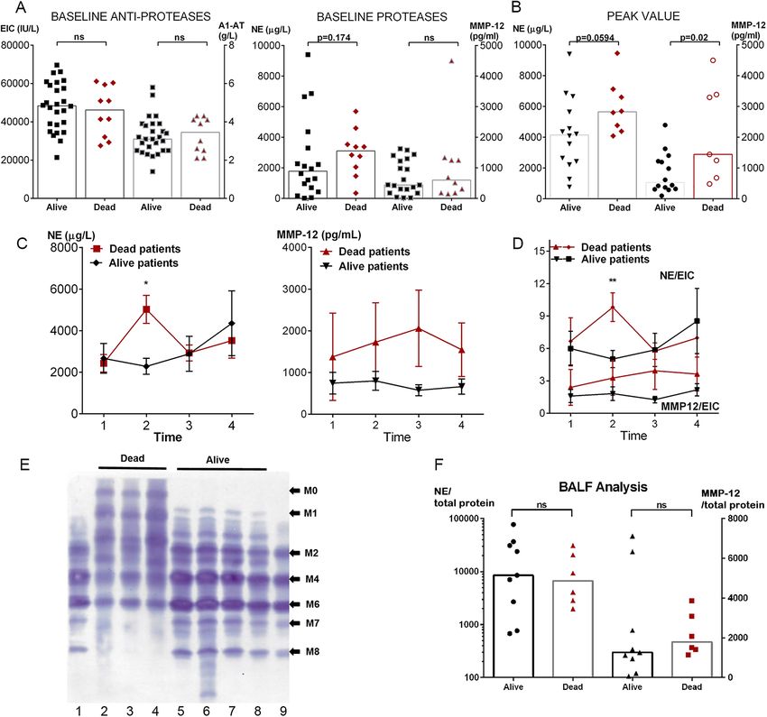
CRP, mg/L (Zerimech et al.: Protease-antiprotease imbalance in severe COVID-19 patients 3 Figure 1D is very similar to those of the corresponding M1 A1AT glycoforms [10] was observed in 100% of patients protease i.e. NE or MMP-12 (Figure 1C). Indeed, the NE/EIC with a negative outcome and in 59% patients with a ratio also peaked at T2 in patients with a fatal outcome positive one (Figure 1E) (p=0.057, Fisher’s exact test). (Figure 1D). Moreover, A1AT immunophenotyping was Regarding BALF, samples taken from nine alive and six conducted in 17 alive and eight deceased patients of our deceased patients were analyzed. Sampling time was not cohort. An unusual isoelectric focusing profile with statistically different between the two groups (day median anodic bands corresponding to highly sialylated M0 and (alive vs. deceased): 3 vs. 4.5). The ratios NE/total protein Figure 1: Proteases and antiproteases in severe COVID-19 patients. (A) Serum levels of EIC (elastase inhibitory capacity), A1AT, NE and MMP-12 at admission into ICU (T1) of COVID-19 patients with ARDS according to their final outcome. The height of the box represents the median. ns, not significant. Normal range for A1AT: 0.9–2 g/L, for EIC: 17,500– 31,500 IU/L. (B) Peak value of NE and MMP-12 at the serum level during the whole ICU stay (T1–T5) according to patient’s outcome. The height of the box represents the median. (C–D) Longitudinal follow-up of serum levels of NE, MMP-12, and protease/antiprotease ratios (NE/EIC or MMP12/EIC) in COVID-19 patients admitted into ICU before first death (median ± SEM). The first death occurred at day 9 (n=1), the others at day 13 (n=2) or later on (n=7). The number of samples analyzed per subject during the follow up was: 2 (1–4), median (range); **, p
4        Zerimech et al.: Protease-antiprotease imbalance in severe COVID-19 patients

and MMP-12/total protein did not differ between deceased             Acknowledgments: We thank Julien Labreuche for his help
and alive patients (Figure 1F).                                      with statistical analysis, Catherine Lelorne, Jordan Soutif
     Our preliminary data showed that COVID-19 patients              and Nadine François for their expert technical assistance,
with fatal ARDS exhibited during their ICU stay signifi-             and Virginie Deprez for her help with protocol mana-
cantly higher peak values of serum MMP-12 than those with            dgment. The Lille COVID Research Network (LICORNE)
a positive outcome. A similar trend was observed for NE.             wishes to acknowledge the contribution of residents,
Moreover, NE levels evolution before first death tended              medical students, nursing teams, lab technicians, and
to be associated with outcome with higher NE levels and              clinical research associates in the midst of the SARS-CoV-2
NE/EIC ratio at T2 in deceased patients. This attests that a         pandemic.
higher elastase activity is released from macrophages and            Research funding: This work was funded by the Institut de
neutrophils of COVID-19 patients with fatal ARDS.                    Biochimie & Biologie Moleculaire, CHU Lille (grant 8860).
Concomitantly, they shifted their A1AT biosynthesis to-              Author contributions: All authors have accepted
wards more anodic, more sialylated glycoforms in an                  responsibility for the entire content of this manuscript
attempt to boost its anti-inflammatory properties, as                and approved its submission.
reported in patients with community-acquired pneumonia               Competing interests: Authors state no conflict of interest.
[10] or severe COVID-19 [11]. However, here the presence of          Informed consent: Informed consent was obtained from all
the highly sialylated M0 and M1 glycoforms in COVID-19               individuals included in this study.
patients is not correlated to the AAT serum levels or to the         Ethical approval: This study is nested within the LICORNE
intensity of the inflammatory response (Sup Table 1), but to          clinical and biological database which received an Ethics
a higher NE/EIC ratio (Supplemental Table 1). Therefore,             Committee approval (CPP Nord-Ouest 4, ECH20/09).
we postulate that the qualitative shift in A1AT glycoforms
represents an attempt to trigger its anti-elastase activity.
Unfortunately, this attempt was unsuccessful since the               Appendix
qualitative shift tends to be associated with a worse clinical
outcome in COVID-19 patients.                                        LICORNE Study Group Scientific Committee: Dominique
     Despite our cross-sectional analysis of BALF was                Deplanque, Karine Faure, Guillaume Lefevre, Kazali
negative, we postulate that an elastase-antielastase                 Alidjinou, Régis Bordet, Ilka Engelmann, Delphine
imbalance also exists locally during ICU stay leading to a           Garrigue, Anne Goffard, Eric Kipnis, Myriam Labalette,
net proteolytic activity in the lungs. Such an imbalance has         Marc Lambert, David Launay, Daniel Mathieu, Claude
been proposed as a pathogenic mechanism in emphysema                 Alain Maurage, Julien Poissy, Martine Remy, Boualem
[12]. Interestingly some COVID-19 patients developed pul-            Sendid, Sophie Susen, Maxime Caillier, Laurent Schwarb
monary lesions complicated by a mediastinal emphysema                Laurent, Michael Hisbergues.
as observed in patients with an A1AT genetic deficiency.
Indirect arguments further support this similarity. In Italy,
a geographical overlap between COVID-19 cases and A1AT              References
deficient-patients was reported [13]. Obese male patients
with a higher risk of severe COVID-19 had reduced serum               1. Bhatraju PK, Ghassemieh BJ, Nichols M, Kim R, Jerome KR, Nalla AK,
A1AT levels and increased NE activity [14] suggestive of                 et al. COVID-19 in critically ill patients in the Seattle region-case
an elastase-antielastase imbalance. By contrast, female                  series. N Engl J Med 2020;382:2012–22.
                                                                      2. Carsana L, Sonzogni A, Nasr A, Rossi RS, Pellegrinelli A, Zerbi P,
gender is positively correlated with serum A1AT levels [15].
                                                                         et al. Pulmonary post-mortem findings in a series of COVID-19
In conclusion, this preliminary study is the first charac-                cases form northern Italy: a two-centre descriptive study. Lancet
terization of a protease-antiprotease imbalance in blood of              Infect Dis 2020;20:1135–40.
COVID-19 patients with severe ARDS leading to death. We               3. Copin MC, Parmentier E, Duburcq T, Poissy J, Mathieu D, The Lille
believe that our research could open new therapeutic                     COVID-19 ICU and Anatomopathology Group. Time to consider
                                                                         histological pattern of lung injury to treat critically ill patients with
perspectives in this setting with the introduction of phar-
                                                                         COVID-19 infection. Intensive Care Med 2020;46:1124–6.
macological (as Sivelestat® or Alvelestat®) or physiological          4. Bao LL, Deng W, Huang B, Gao H, Liu J, Ren L, et al. The
inhibitors of NE such as A1AT prepared by plasma frac-                   pathogenicity of SARSCoV-1 in hACE2 transgenic mice. Nature
tionation (Alfalastin®, Prolastin® or Respreeza®).                       2020;583:830–3.
Zerimech et al.: Protease-antiprotease imbalance in severe COVID-19 patients 5
5. Heinz A. Elastases and elastokines: elastin degradation and its for resolution of community-acquired pnemonia. Am J Respir Crit
significance in health and disease. Crit Rev Biochem Mol Biol Care Med 2018;197:1346–9.
2020;55:252–73. 11. McElvaney OJ, McEvoy NL, McElvaney OF, Carroll TP, Murphy MP,
6. Balduyck M, Odou MF, Zerimech F, Porchet N, Lafitte JJ, Maitre B. Dunlea DM, et al. Characterization of the inflammatory response
Diagnosis of alpha-1 antitrypsin deficiency: modalities, indications to severe COVID-19 Illness. Am J Respir Crit Care Med 2020;202:
and diagnosis strategy. Rev Mal Resp 2014;31:729–45. 812–21.
7. Dati F, Schumann G, Thomas L, Aguzzi F, Baudner S, Bienvenu J, et al. 12. McGarry Houghton A. Matrix metalloproteinases in destructive
Consensus of a group of professional societies and diagnostic lung diseases. Matrix Biol 2015;44–46:167–74.
companies on guidelines for interim reference ranges for 14 proteins 13. Vianello A, Braccioni F. Geographical overlap between alpha-1
in serum based on the standardization against the IFCC/BCR/CAP antitrypsin deficiency and COVID-19 infection in Italy: casual or
Reference Material (CRM 470). International Federation of Clinical causal ? Arch Bronconeumol 2020;56:609–10.
Chemistry. Community Bureau of Reference of the Commission of 14. Mansuy-Aubert V, Zhou QL, Xie X, Gong Z, Huang JY, Khan AR,
the European Communities. College of American Pathologists. Eur J et al. Imbalance between neutrophil elastase and its inhibitor
Clin Chem Clin Biochem 1996;34:517–20. α1-antitrypsin in obesity alters insulin sensitivity,
8. Klumpp T, Bieth JG. Automated measurement of the elastase- inflammation and energy expenditure. Cell Metabol 2013;17:
inhibitory capacity of plasma with a centrifugal analyzer. Clin 534–48.
Chem 1979;25:269–72. 15. Senn O, Russi EW, Schindler C, Imboden M, von Eckardstein A,
9. Zerimech F, Hennache G, Bellon F, Barouh G, Jacques Lafitte J, Brandli O, et al. Circulating Alpha-1 antitrypsin in the general
Porchet N, et al. Evaluation of a new Sebia isoelectrofocusing kit population: determinants and association with lung function.
for alpha-1 antitrypsin phenotyping with the Hydrasis system. Resp Res 2008;9:35.
Clin Chem Lab Med 2008;46:260–3.
10. McCarthy C, Dunlea DM, Saldova R, Henry M, Meleady P, Supplementary Material: The online version of this article offers
McElvaney OJ, et al. Glycosylation repurposes Alpha-1 antitrypsin supplementary material (https://doi.org/10.1515/cclm-2021-0137).You can also read