Application of optical coherence tomography and high-frequency ultrasound imaging during noninvasive laser vasectomy
←
→
Page content transcription
If your browser does not render page correctly, please read the page content below
Application of optical coherence
                 tomography and high-frequency
                 ultrasound imaging during noninvasive
                 laser vasectomy

                 Christopher M. Cilip
                 Mohamad E. Allaf
                 Nathaniel M. Fried

Downloaded From: https://www.spiedigitallibrary.org/journals/Journal-of-Biomedical-Optics on 20 Sep 2021
Terms of Use: https://www.spiedigitallibrary.org/terms-of-use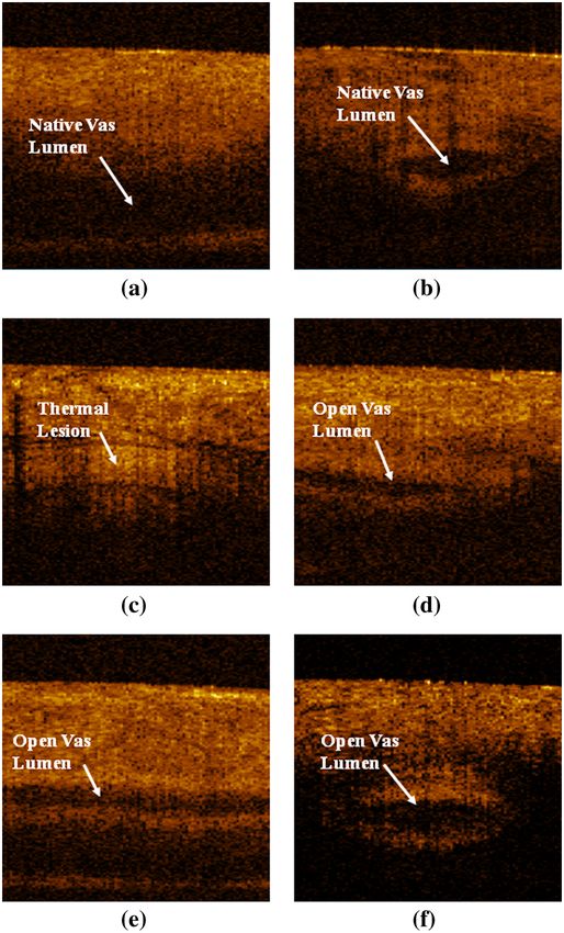
Journal of Biomedical Optics 17(4), 046006 (April 2012)

                Application of optical coherence tomography and
                high-frequency ultrasound imaging during noninvasive
                laser vasectomy
                Christopher M. Cilip,a Mohamad E. Allaf,b and Nathaniel M. Frieda,b
                a
                    University of North Carolina at Charlotte, Department of Physics and Optical Science, North Carolina
                b
                    Johns Hopkins Medical Institutions, Department of Urology, Baltimore, Maryland

                               Abstract. A noninvasive approach to vasectomy may eliminate male fear of complications related to surgery and
                               increase its acceptance. Noninvasive laser thermal occlusion of the canine vas deferens has recently been reported.
                               Optical coherence tomography (OCT) and high-frequency ultrasound (HFUS) are compared for monitoring laser
                               thermal coagulation of the vas in an acute canine model. Bilateral noninvasive laser coagulation of the vas was
                               performed in six dogs (n ¼ 12 vasa) using a Ytterbium fiber laser wavelength of 1075 nm, incident power of 9.0 W,
                               pulse duration of 500 ms, pulse rate of 1 Hz, and 3-mm-diameter spot. Cryogen spray cooling was used to prevent
                               skin burns during the procedure. An OCT system with endoscopic probe and a HFUS system with 20-MHz trans-
                               ducer were used to image the vas immediately before and after the procedure. Vasa were then excised and pro-
                               cessed for gross and histologic analysis for comparison with OCT and HFUS images. OCT provided high-resolution,
                               superficial imaging of the compressed vas within the vas ring clamp, while HFUS provided deeper imaging of the
                               vas held manually in the scrotal fold. Both OCT and high HFUS are promising imaging modalities for real-time
                               confirmation of vas occlusion during noninvasive laser vasectomy. © 2012 Society of Photo-Optical Instrumentation Engineers
                               (SPIE). [DOI: 10.1117/1.JBO.17.4.046006]

                               Keywords: coagulation; laser; male sterilization; noninvasive; optical coherence tomography; ultrasound; vasectomy.
                               Paper 11773P received Dec. 20, 2011; revised manuscript received Feb. 13, 2012; accepted for publication Feb. 20, 2012; published
                               online Apr. 11, 2012.

                1       Introduction                                                               measure the anatomy of the normal human vas deferens.11–13
                Approximately 500,000 vasectomies are performed in the                             Our laboratory has also used HFUS (with a 13-MHz probe)
                United States per year, making it the most common urological pro-                  for imaging the canine vas before and after laser thermal coa-
                cedure in the U.S.1 Although vasectomy is more effective and less                  gulation.14 This study aims to improve on these previous studies
                likely to have complications than tubal ligation, the number of men                by using a higher-frequency (20 MHz) probe capable of
                undergoing surgical sterilization is approximately three times less                improved resolution and to compare HFUS with another
                than women.2–4 Fear of complications related to surgical vasect-                   high-resolution imaging modality, optical coherence tomogra-
                omy (e.g. hematoma, infection, acute and chronic pain, and ster-                   phy (OCT), for imaging the canine vas before and after laser
                ilization failure) is a major factor in a couples’ choice of surgical              thermal coagulation. Both imaging modalities are safe, compact,
                                                                                                   and inexpensive and are therefore evaluated as possible choices
                sterilization.5,6 A noninvasive technique for male sterilization may
                                                                                                   for potential use in laser vasectomy during this preclinical study.
                improve acceptance of vasectomy.
                                                                                                   However, each imaging modality serves a slightly different pur-
                    Our laboratory is currently developing a noninvasive vasect-
                                                                                                   pose: HFUS provides deep imaging with intermediate scale
                omy technique utilizing near-infrared laser irradiation in con-
                                                                                                   resolution while OCT provides high resolution but is limited
                junction with cryogen spray cooling of the scrotal skin
                                                                                                   in its superficial imaging depth.
                surface for thermal occlusion of the vas. During previous pre-
                liminary noninvasive laser vasectomy studies, we reported suc-
                cessful thermal occlusion and scarring of the vas in both ex vivo                  2       Methods
                and in vivo canine models,7–9 with the thermally coagulated vas
                segment withstanding burst pressures over twice that of normal                     All procedures were conducted under an animal protocol
                canine ejaculation pressures.10 However, this completely nonin-                    approved by the Johns Hopkins Animal Review Committee.
                vasive therapeutic procedure may still benefit from the use of a                   Noninvasive thermal occlusion of the vas was performed bilat-
                compact and inexpensive imaging modality to confirm, in real                       erally in a total of six dogs (n ¼ 12 vasa). After completion of
                time, accurate targeting, thermal coagulation, and occlusion of                    the procedure, all vasa were harvested and processed for histol-
                the vas.                                                                           ogy with H&E staining. The animals were neutered, monitored
                    Optical coherence tomography (OCT) and high-frequency                          for a few days during recovery, and then adopted out to car-
                diagnostic ultrasound (HFUS) have recently been used to                            ing homes.
                                                                                                       A compact, tabletop, 50-watt Ytterbium fiber laser (Model
                                                                                                   TLR1075-50, IPG Photonics, Oxford, MA) emitted near-
                Address all correspondence to: Nathaniel Fried, Ph.D., Department of Physics       infrared laser radiation with a wavelength of 1075 nm, which
                and Optical Science, University of North Carolina at Charlotte 9201 University
                City Boulevard, Charlotte, NC 28223. Tel: 704-687-8149; Fax: 704-687-8197;
                E-mail: nmfried@uncc.edu.                                                          0091-3286/2012/$25.00 © 2012 SPIE

                Journal of Biomedical Optics                                                046006-1                                           April 2012   •   Vol. 17(4)

Downloaded From: https://www.spiedigitallibrary.org/journals/Journal-of-Biomedical-Optics on 20 Sep 2021
Terms of Use: https://www.spiedigitallibrary.org/terms-of-use
Cilip, Allaf, and Fried: Application of optical coherence tomography and high-frequency ultrasound : : :

                                                                                                   4-mm-OD, vasectomy ring clamp was used to manually isolate
                                                                                                   the vas beneath the scrotal skin surface for co-location with the
                                                                                                   cryogen and laser spots (Fig. 1). It should be noted that although
                                                                                                   a modified vasectomy ring clamp design was used for these pro-
                                                                                                   cedures, the base of the clamp (hemostat) is essentially the same
                                                                                                   as the instrument currently used during a standard no scalpel
                                                                                                   vasectomy procedure.
                                                                                                       A compact, tabletop, inexpensive, FDA-approved, endo-
                                                                                                   scopic, optical coherence tomography system (Niris II, Imalux,
                                                                                                   Cleveland, OH) with a handheld 8-Fr (2.7-mm-OD) probe was
                                                                                                   used [Fig. 2(a)]. The OCT system acquired real-time images at
                                                                                                   eight frames/s with ∼11 μm axial resolution and ∼25 μm lateral
                                                                                                   resolution in tissue with a lateral scan length of 2 mm and an
                                                                                                   imaging depth of 1.6 mm. The OCT system was used for super-
                                                                                                   ficial imaging of the canine vas once it was compressed inside
                                                                                                   the vas ring clamp, immediately before and after laser irradia-
                                                                                                   tion. This approach to OCT imaging of the vas is entirely fea-
                Fig. 1 A conventional no-scalpel vasectomy ring clamp was used to
                isolate the vas deferens beneath the scrotal skin surface, and then a              sible during a clinical procedure.
                detachable customized clamp extension was used to safely co-locate                     A compact, tabletop, inexpensive, FDA-approved high-
                the cryogen spray spot and laser spot on the tissue surface inside the             frequency ultrasound system (Episcan-I-200, Longport Interna-
                ring.                                                                              tional, Silchester, United Kingdom) with 20-MHz linear
                                                                                                   scanning transducer was used for comparison with OCT
                                                                                                   [Fig. 2(b)]. This transducer provided a scan length of 15 mm,
                was focused with a lens into a 400-μm-core fiber optic patch-                      an axial image resolution of ∼100 μm, and an imaging depth
                cord (Thorlabs, Newton, NJ). Another lens at the end of the                        of ∼1 cm. The HFUS transducer was used to provide deeper
                patch-cord delivered a collimated laser beam to the tissue sur-                    imaging of the canine vas when it was held manually within
                face. The laser was externally triggered by a function generator                   a scrotal fold immediately before and after application of the
                (DS345, Stanford Research Systems, Sunnyvale, CA) to operate                       vas ring clamp.
                in long-pulse mode, with an average incident power of 9.0 W,                           Several indicators were used to determine successful laser
                500-ms pulse duration, 1-Hz pulse rate, and 3-mm-diameter                          thermal coagulation and occlusion of the canine vas, including
                (1∕e2 ) spot at the scrotal skin surface.                                          OCT, HFUS, gross analysis, and histology.
                    A dynamic cooling device (DCD, Candela Laser Corpora-
                tion, Wayland, MA) was used to deliver cryogen (1,1,1,2-                           3       Results
                tetrafluoroethane, boiling point ¼ − 26 °C) to the scrotal skin                    Longitudinal and cross-sectional OCT images of the vas were
                surface through a solenoid valve to prevent overheating and for-                   taken, in vivo, before and after the procedure to confirm success-
                mation of skin burns during the procedure. Three cryogen pulses                    ful thermal coagulation of the vas [Fig. 3(a), 3(b), and 3(c)].
                were applied to pre-cool the skin prior to laser irradiation over a                OCT images of the excised vas were also taken, ex vivo, imme-
                period of eight seconds. For all of the procedures, laser irradia-                 diately after the procedure for comparison [Fig. 3(d), 3(e), and
                tion was performed for a period of 60 sec for each vas. Cryogen                    3(f)]. The native vas appeared with a dark, fluid-filled lumen
                spray was delivered intermittently between laser pulses with a                     (absent of reflected signal intensity), while the thermal lesion
                60-ms pulse duration, pulse rate of 0.25 Hz, and a 2-cm-                           was represented by a lighter region (indicative of enhanced
                diameter spot concentric with the laser spot. A cryogen mask                       reflected signal intensity). This is consistent with a significant
                was used to thermally insulate surrounding scrotal skin from                       increase in the scattering coefficient of soft tissue once it has
                cryogen spray to avoid superficial freeze burns. Details of the                    been thermally denatured. It should also be noted that since
                experimental setup have been previously reported.9 A modified,                     the thermal lesion length along the vas (3.58  0.36 mm)

                Fig. 2 (a) Photograph of the optical coherence tomography system and probe used in these studies; (b) photograph of the high-frequency ultrasound
                system and probe used in these studies.

                Journal of Biomedical Optics                                              046006-2                                            April 2012   •   Vol. 17(4)

Downloaded From: https://www.spiedigitallibrary.org/journals/Journal-of-Biomedical-Optics on 20 Sep 2021
Terms of Use: https://www.spiedigitallibrary.org/terms-of-use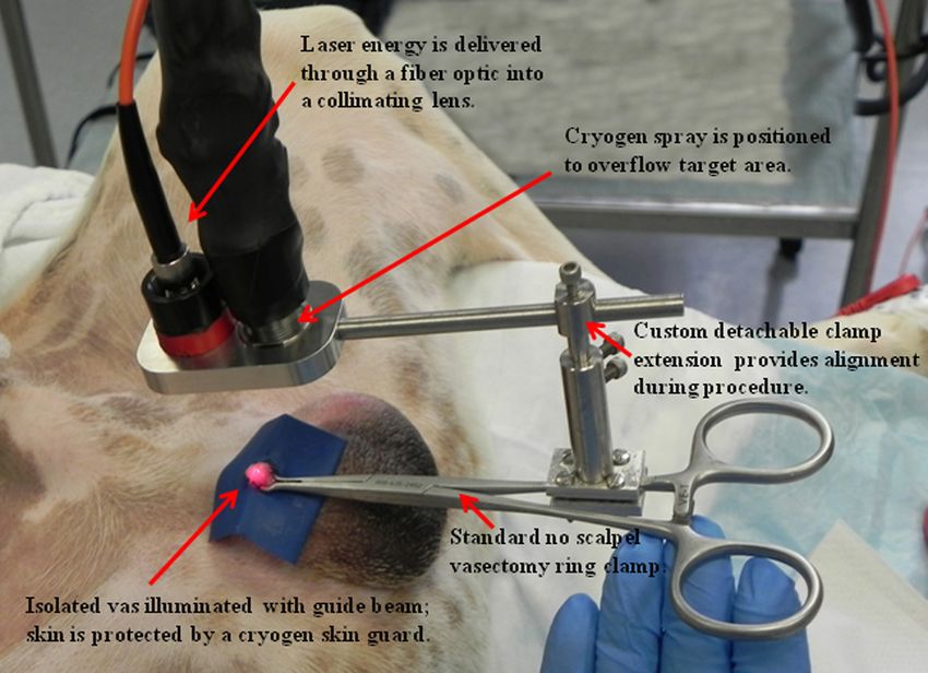
Cilip, Allaf, and Fried: Application of optical coherence tomography and high-frequency ultrasound : : :
similar range to that previously reported for the native human
vas deferens.12,13 The canine vas deferens in general is
known to have similar dimensions to that of the human vas defe-
rens.15 The thermal lesion length is strongly correlated with the
3-mm-diameter laser spot used in these studies and is also simi-
lar to measurements reported in previous studies.7–9,14
The thermally coagulated vas was excised for examination to
verify the OCT and HFUS images. Figure 5 shows a represen-
tative image of the thermally coagulated vas segment, which can
be identified by several indicators including blanching and
shrinkage of the vas wall.
The vas samples were then processed for histology using
standard techniques. For each dog, serial sectioning of the
vas was performed in both the longitudinal and cross-sectional
directions (one orientation for each vas—left and right sides)
through the entire vas sample at 100 μm intervals. Figure 6 pro-
vides representative longitudinal and cross-sectional histologic
sections of the canine vas with open lumen sections (taken
beyond the thermally coagulated segment) also provided for
comparison.
4 Discussion
Vasectomy is a safe, simple, effective, and inexpensive surgical
procedure for male sterilization. Despite the low morbidity of
this procedure, societal pressures, psychological factors such
as the perception of the loss of “manhood,” and male fear of
surgery are reasons frequently cited by couples choosing
other forms of contraception. A noninvasive laser vasectomy
technique may eliminate many of these concerns and increase
male acceptance of vasectomy. While some of our earlier non-
invasive laser vasectomy studies have noted the formation of
minor scrotal skin burns with the use of higher laser powers,8
there was no evidence of skin burns in this study. Only tempor-
ary skin irritation and reddening was observed due to the blunt
trauma of the vasectomy ring clamp tips on the skin. Such
trauma caused by the vas ring clamp is normal for the conven-
Fig. 3 (a) and (b) Longitudinal and cross-sectional OCT images of the tional vasectomy technique as well. This irritation disappeared
native canine vas acquired while the vas and scrotal skin was com- approximately 15 min after the clamp was removed and the
pressed inside the vas ring clamp prior to the procedure. (c) Longitudinal
OCT image of the thermally coagulated canine vas acquired while the
procedure completed.
vas and scrotal skin were compressed inside the vas ring clamp after the The development of any completely noninvasive therapeutic
procedure. (d) Longitudinal OCT image of the thermally coagulated procedure may benefit from the introduction of a diagnostic
canine vas acquired ex vivo after the vas tissue was harvested; modality to confirm success since no tissue is removed for
(e) and (f) longitudinal and cross-sectional OCT images of the native analysis during the noninvasive procedure. OCT and HFUS
canine vas acquired ex vivo after the tissue was harvested. All OCT are obvious choices for use in vas imaging during noninvasive
images measure 1.6 × 2.0 mm (depth × width).
laser vasectomy because they are both relatively compact,
inexpensive imaging modalities that provide sufficient image
resolution and depth to view the vas deferens, which has a
was longer than the OCT lateral scanning distance of 2 mm, it lumen and wall thickness of ∼300 μm and ∼1 mm, respectively.
was impossible to capture the entire lesion in a single image. OCT has an order of magnitude better resolution and an order
Furthermore, the vas was not a straight tube, but rather traveled of magnitude worse imaging depth than HFUS. Therefore, OCT
in and out of the OCT (and HFUS) image plane. was limited in these studies to imaging the vas once it was fixed
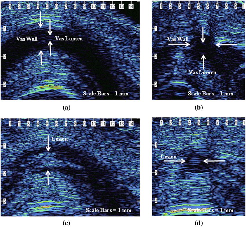
Longitudinal and cross-sectional HFUS images of the vas and compressed beneath the scrotal skin inside the 4-mm-
were also taken, in vivo, before and after the procedure to con- diameter vas ring clamp. However, while the native vas was
firm successful thermal coagulation of the vas (Fig. 4). In the easily visible during OCT imaging before the procedure, it
native vas images, the fluid-filled lumen also appears dark was more difficult to identify the thermally coagulated vas seg-
(with an absence of reflected signal intensity), while the ther- ment after the procedure. Only longitudinal sections were
mally denatured vas appears lighter (again indicative of obtained, and cross-sectional images of the thermally coagu-
increased reflected signal intensity). Using HFUS, the average lated vas could not easily be confirmed. It may be that the
vas lumen diameter, vas wall thickness, and vas thermal lesion need to perform OCT imaging of the compressed vas and scrotal
length were measured to be 0.27 0.07 mm (n ¼ 6), skin within the clamp resulted in a distorted view of the anat-
1.09 0.06 mm (n ¼ 6), and 3.58 0.36 mm (n ¼ 9), respec- omy, which in turn made interpretation of the OCT images more
tively. These lumen diameter and wall dimensions are within a difficult. HFUS imaging of the vas was more successful, with
Journal of Biomedical Optics 046006-3 April 2012 • Vol. 17(4)
Downloaded From: https://www.spiedigitallibrary.org/journals/Journal-of-Biomedical-Optics on 20 Sep 2021
Terms of Use: https://www.spiedigitallibrary.org/terms-of-use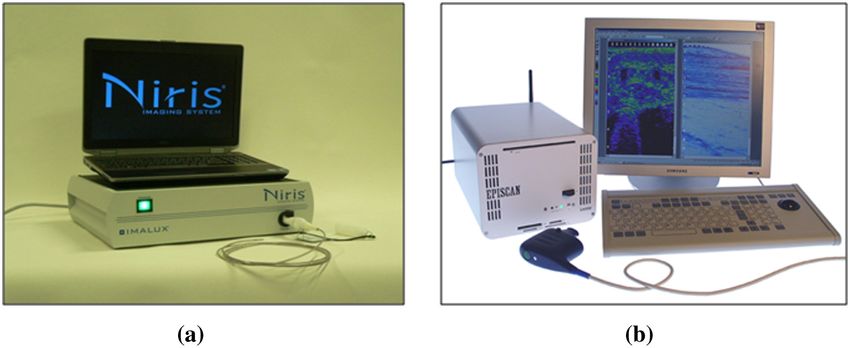
Cilip, Allaf, and Fried: Application of optical coherence tomography and high-frequency ultrasound : : :

                Fig. 4 (a) and (b) Longitudinal and cross-sectional HFUS images of the native canine vas, acquired by manually isolating the vas within the scrotal skin
                fold prior to placement of the vas ring clamp. The vas wall and lumen can be identified. (c) and (d) Longitudinal and cross-sectional HFUS images of the
                thermally coagulated canine vas acquired by manually isolating the vas within the scrotal skin fold after removal of the vas ring clamp. A thermal lesion
                encompassing both the vas wall and lumen is observed.

                                                                                                   postoperative imaging of the thermal lesion more difficult and
                                                                                                   resulted in a slightly lower vas sample set for evaluation.
                                                                                                   In some vas, it was also difficult to differentiate the lumen
                                                                                                   from the wall before the procedure and provide two distinct
                                                                                                   measurements for these two structures. This may have been
                                                                                                   due to suboptimal probe placement or the method of manual
                                                                                                   isolation causing excessive pressure and flattening of the vas.
                                                                                                   It may be possible in future studies to design the imaging
                                                                                                   probe tip and vas clamp to minimize these limitations.
                                                                                                      From a clinical perspective, it is also worth briefly mention-
                                                                                                   ing the costs associated with noninvasive laser vasectomy, since
                                                                                                   surgical vasectomy is currently a low-cost procedure (∼500 to
                                                                                                   $1,000). For noninvasive laser vasectomy, there are capital costs
                                                                                                   associated with purchase of the laser (∼20; 000) and cryogen
                                                                                                   (∼5; 000) systems. The compact, portable, tabletop, time-
                Fig. 5 Gross image of the thermally coagulated vas segment showing                 domain OCT system and probe used in this study is relatively
                significant blanching and shrinkage of the vas wall.                               inexpensive (∼40; 000), in comparison with other OCT systems.
                                                                                                   The compact, tabletop HFUS system and probe used in this
                                                                                                   study is also relative inexpensive (∼20; 000) in comparison
                longitudinal and cross-sectional images of the native and ther-                    with conventional clinical HFUS consoles. However, a patient
                mally coagulated vas obtained while the vas was held manually                      may be willing to pay a slightly higher fee for nonsurgical
                within a scrotal fold, both before and after removal of the vas                    vasectomy.
                from the ring clamp.                                                                  Finally, more definitive and longer-term chronic canine stu-
                   However, it should be noted that the limited imaging depth of                   dies are currently being performed to examine vas recanalization
                the HFUS system required manual isolation of the vas prior to                      and azospermia rates, with comparison of surgical vasectomy
                measurement. This made locating the region of interest during                      and noninvasive laser vasectomy, prior to clinical studies.

                Journal of Biomedical Optics                                              046006-4                                              April 2012   •   Vol. 17(4)

Downloaded From: https://www.spiedigitallibrary.org/journals/Journal-of-Biomedical-Optics on 20 Sep 2021
Terms of Use: https://www.spiedigitallibrary.org/terms-of-use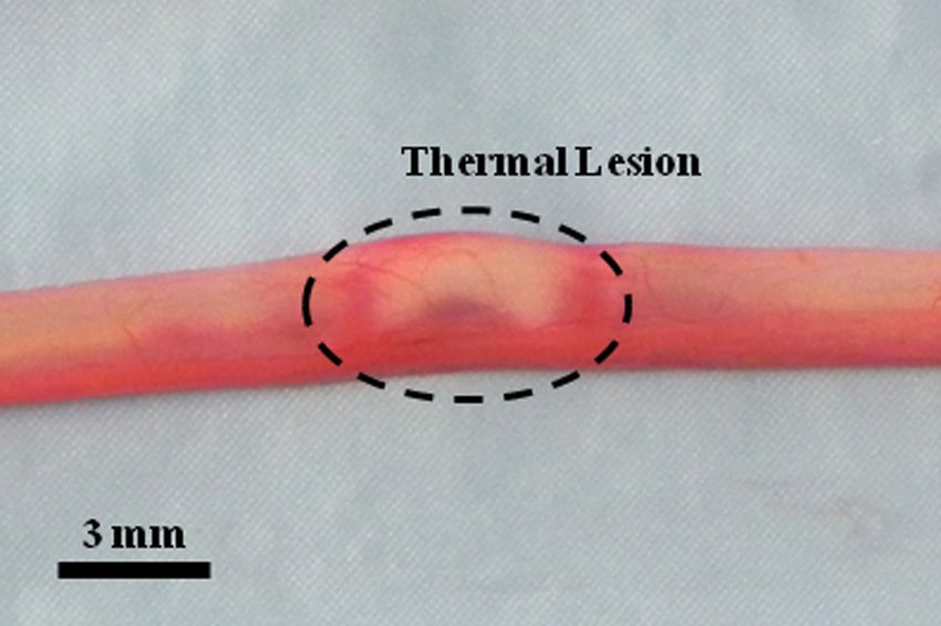
Cilip, Allaf, and Fried: Application of optical coherence tomography and high-frequency ultrasound : : :

                                                                                                   and the United States Agency for International Development,
                                                                                                   through a subcontract from Family Health International
                                                                                                   (Durham, NC). The authors thank Dawn Ruben and Laurie
                                                                                                   Pipitone for assisting with the animal studies, James Hsia of
                                                                                                   the Candela Corporation (Wayland, MA) for providing the cryo-
                                                                                                   gen cooling system, Paul Wilson of Longport (Chadd’s Ford,
                                                                                                   PA) for providing the high-frequency ultrasound system, and
                                                                                                   Nancy Tresser of Imalux (Cleveland, OH) for providing the opti-
                                                                                                   cal coherence tomography system used in these studies.

                                                                                                   References
                                                                                                    1. M. A. Barone et al., “Vasectomy in the United States, 2002,” J. Urol.
                                                                                                       176(1), 232–236 (2006).
                                                                                                    2. W. D. Mosher et al., “Use of contraception and use of family planning
                                                                                                       services in the United States: 1982–2002,” Adv. Data 350, 1–36
                                                                                                       (Dec.10, 2004).
                                                                                                    3. A. Chandra et al., “Fertility, family planning, and reproductive health of
                                                                                                       U.S. women: data from the 2002 National Survey of Family Growth,”
                                                                                                       Vital Health Stat. 25, 1–160 (Dec.2005).
                                                                                                    4. G. M. Martinez et al., “Fertility, contraception, and fatherhood: data on
                                                                                                       men and women from cycle 6 (2002) of the 2002 National Survey of
                                                                                                       Family Growth,” Vital Health Stat. 26, 1–142 (May2006).
                                                                                                    5. W. B. Miller et al., “Tubal sterilization or vasectomy: how do married
                                                                                                       couples make the choice,” Fertil. Steril. 56(2), 278–284
                                                                                                       (1991).
                                                                                                    6. R. N. Shain et al., “Factors associated with married women’s selection
                                                                                                       of tubal sterilization and vasectomy,” Fertil. Steril. 43(2), 234–244
                                                                                                       (1985).
                                                                                                    7. C. M. Cilip et al., “Noninvasive laser vasectomy: preliminary ex vivo
                Fig. 6 H&E-stained histologic sections of the vas: (a) cross-section of                tissue studies,” Lasers Surg. Med. 41(3), 203–207 (2009).
                open lumen; (b) cross-section of thermally occluded lumen; (c) longitu-             8. C. M. Cilip et al., “Noninvasive laser coagulation of the canine vas defe-
                dinal section of open lumen; (d) longitudinal section of thermally                     rens, in vivo,” Proc. SPIE 7548, 75481D (2010).
                occluded lumen.                                                                     9. C. M. Cilip et al., “Application of an optical clearing agent during non-
                                                                                                       invasive laser coagulation of the canine vas deferens,” J. Biomed. Opt.
                                                                                                       15(4), 048001 (2010).
                Both OCT and HFUS may be used in these chronic studies for                         10. A. Shafik, “Electrovasogram: a canine study of the electromechanical
                imaging of the scarred vas.                                                            activity of the vas deferens,” Urology 46(5), 692–696 (1995).
                                                                                                   11. C. Davis and W. Kuang, “Optical coherence tomography: a novel mod-
                5    Conclusions                                                                       ality for scrotal imaging,” Can. Urol. Assoc. J. 3(4), 319–322
                High-frequency ultrasound may be used as a diagnostic tool                             (2009).
                                                                                                   12. T. Puttemans, A. Delvigne, and D. Murillo, “Normal and variant appear-
                to assist in determining successful laser thermal coagulation                          ances of the adult epididymis and vas deferens on high-resolution sono-
                and scarring of the vas during noninvasive laser vasectomy.                            graphy,” J. Clin. Ultrasound 34(8), 385–392 (2006).
                While optical coherence tomography also shows promise,                             13. W. D. Middleton et al., “High-resolution sonography of the normal
                further improvements are necessary before this imaging modal-                          extrapelvic vas deferens,” J. Ultrasound Med. 28(7), 839–846
                ity can be reliably used for evaluation of the thermally coagu-                        (2009).
                lated vas.                                                                         14. C. M. Cilip et al., “High-frequency ultrasound imaging of noninvasive
                                                                                                       laser coagulation of the canine vas deferens,” Lasers Surg. Med. 43(8),
                Acknowledgments                                                                        838–842 (2011).
                                                                                                   15. D. E. Leocadio et al., “Anatomical and histological equivalence of the
                This project is supported by a research grant from the NIH,                            human, canine, and bull vas deferens,” Can. J. Urol. 18(3), 5699–5704
                National Institute of Child Health and Human Development,                              (2011).

                Journal of Biomedical Optics                                              046006-5                                                    April 2012    •   Vol. 17(4)

Downloaded From: https://www.spiedigitallibrary.org/journals/Journal-of-Biomedical-Optics on 20 Sep 2021
Terms of Use: https://www.spiedigitallibrary.org/terms-of-use
You can also read