Recurrent hydronephrosis and spontaneous renal rupture caused by lymphoplasmacytic inflammation in a cat
←
→
Page content transcription
If your browser does not render page correctly, please read the page content below
Case Report                                                       Veterinarni Medicina, 66, 2021 (02): 80–86

                                                                  https://doi.org/10.17221/153/2020-VETMED

Recurrent hydronephrosis and spontaneous
renal rupture caused by lymphoplasmacytic
inflammation in a cat
Junyoung Kim1,2, Dongkeun Oh1, Junho Cho1, Seongjun Kim1,
Junghee Yoon2*
1
    BOM Animal Hospital, Seoul, Republic of Korea
2
    College of Veterinary Medicine and the Research Institute for Veterinary Science,
    Seoul National University, Seoul, Republic of Korea
*Corresponding author: heeyoon@snu.ac.kr

Citation: Kim J, Oh D, Cho J, Kim S, Yoon J (2021): Recurrent hydronephrosis and spontaneous renal rupture by lym-
phoplasmacytic inflammation in a cat. Vet Med-Czech 66, 80–86.

Abstract: A seven-year-old male cat that was previously diagnosed with – and treated for – left hydronephrosis
due to suspected idiopathic proteinaceous materials in the renal pelvis, presented with a short history of anorexia
and vomiting. The abdominal ultrasound revealed bilateral hydronephrosis, and the intravenous pyelography
showed a moderate amount of free fluid in the retroperitoneal space at 48 hours. After the nephrectomy, the gross
examination of the right kidney revealed a very thin capsule with urine leakage, and the right renal pelvis showed
small, black deposits. The histopathology of the right kidney revealed hydronephrosis with compression atrophy,
necrosis of the renal cortex/medulla, and a moderate lymphoplasmacytic inflammation. The crystallographic
analysis revealed that the black deposits were composed of 100% protein and no minerals. The cat was diagnosed
with hydronephrosis and spontaneous renal rupture caused by proteinaceous pelvic materials, secondary to the idi-
opathic renal lymphoplasmacytic inflammation. In addition to revealing the possibility that immune-mediated
renal disease can induce spontaneous renal rupture in cats, this case report demonstrates the utility of imaging
for diagnosing and monitoring hydronephrosis, detecting urine leakage, and planning surgery.

Keywords: computed tomography; crystallography; feline; intravenous pyelography; renal; ultrasonography

  Hydronephrosis refers to the dilatation of the renal     solidified blood (DSB) calculi, and retroperitoneal
pelvis and calyces due to a renal or postrenal uri-        fibrosis after a renal transplantation (Vanden et al.
nary obstruction that induces the progressive atro-        2005; D’lppolito et al. 2006; Ragni and Fews 2008;
phy of the renal parenchyma (Rawlings et al. 2003).        D’Anjou et al. 2011; Zaid et al. 2011; Cohen et al.
Hydronephrosis in cats is caused by a ureteral or ure-     2012; Foster and Pinkerton 2012; Lee et al. 2014;
thral blockage by urinary tract calculi, inflammation,     Selgas et al. 2014). Blood clots in the urinary tract
neoplasia, or retroperitoneal masses resulting in ex-      are rare and may result from urinary tract calculi,
traluminal ureteral compression (D’lppolito et al.         infections, inflammation, neoplasia, trauma, clotting
2006; Ragni and Fews 2008; D’Anjou et al. 2011; Zaid       disorders, or idiopathic causes (Rawlings et al. 2003;
et al. 2011; Cohen et al. 2012; Foster and Pinkerton       DiBartola 2005; Vanden et al. 2005).
2012; Selgas et al. 2014; Evans and Fowlkes 2016).           A spontaneous renal rupture is defined as the
Less common causes of hydronephrosis include ob-           rupture of the renal parenchyma due to the renal
structions induced by blood clots in the renal pelvis      pathology (Zhang et al. 2017). Possibly reflecting
or ureters after a renal biopsy, ectopic ureters, dried    the dysregulation of the immune system, a lympho-

80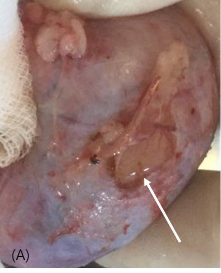
Case Report                                                             Veterinarni Medicina, 66, 2021 (02): 80–86

                                                                        https://doi.org/10.17221/153/2020-VETMED

cytic-plasmacytic inflammation is characterised                 titis and bilateral renal diverticular calculi. After
by the infiltration of lymphocytes and plasma cells             the cystitis was resolved, the cat’s bilateral renal
into certain tissues, particularly in the gastrointes-          calculi were monitored with an abdominal radiog-
tinal tract and nasal or oral cavities (Willard 2003;           raphy, abdominal ultrasound, and laboratory tests
Hall and German 2005; Simpson 2005). However,                   every month thereafter at a hospital. At the nine-
hydronephrosis and a spontaneous renal rupture                  month follow-up, the laboratory findings showed
due to lymphoplasmacytic inflammation in the kid-               increased creatinine [274.3 μmol/l; reference range
ney have not been reported in veterinary medicine.              (RR): 70.8–159.3 μmol/l]. The total protein (8.3 g/l;
  We document the rare case of a seven-year-old                 RR: 5.7–7.8 g/l), albumin (3.6 g/l; RR: 2.3–3.5 g/l),
cat with recurrent hydronephrosis and a spontane-               and calcium (1 079.6 μmol/l; RR: 778.7–1 053.1
ous renal rupture with a suspected blood clot for-              μmol/l) concentrations were mildly elevated. On the
mation in the renal pelvis caused by an idiopathic              abdominal radiography, the location, size, and num-
lymphoplasmacytic inflammation in the kidney.                   ber of the renal calculi remained unchanged; how-
                                                                ever, the left kidney was slightly enlarged (2.64 times
                                                                the length of the second lumbar vertebral body)
Case presentation                                               relative to the right kidney (2.48 times the length
                                                                of the second lumbar vertebral body). The abdominal
  A six-year-old castrated male Korean Shorthair                ultrasound revealed a moderate-to-severe dilation
cat weighing 6.0 kg first presented with haematu-               of the left renal pelvis and a mild dilation of the
ria in January 2019 and was diagnosed with cys-                 left proximal ureter, with amorphous and echo-

           Figure                     1                               Figure 1B

     (A)                                                             (B)

     Figure 1C                                                       Figure 1D

     (C)                                                             (D)
Figure 1. Abdominal ultrasound findings in a cat with recurrent hydronephrosis and spontaneous renal rupture
Longitudinal (A) and transverse (B) images of the left hydronephrotic kidney with amorphous and echogenic materials
in the left renal pelvis at the nine-month follow-up. Dorsal (C) and transverse (D) images of the right kidney at five months
after the first surgery showing hydronephrosis due to the amorphous, echogenic materials in the right renal pelvis along
which it was asymmetrically dilated (white arrow) and a renal calculus (D)

81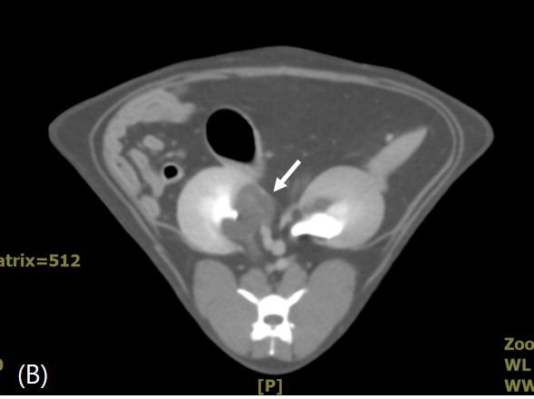
Case Report                                                      Veterinarni Medicina, 66, 2021 (02): 80–86

                                                                 https://doi.org/10.17221/153/2020-VETMED

genic material in the pelvis (Figure 1A,B). The cat       sample showed an increased urinary protein/creati-
was diagnosed with left hydronephrosis with radio-        nine ratio (UPC) (0.8; RR: < 0.2) and predominantly
lucent, amorphous, and echogenic material in the          red blood cells (RBCs; > 50/high power field) on the
renal pelvis. The differential diagnoses included         microscopic examination of the sediment. The cysto-
blood clots, haemorrhage, inf lammatory debris,           centesis sample showed a normal UPC (0.1; RR: < 0.2)
DSB calculi, or mucus plugs in the left renal pelvis.     and no mucus, bacteria, or casts. Both samples were
The cat was hospitalised for observation and treat-       devoid of crystals or minerals. The cat recovered well
ment, including intravenous (i.v.) fluids (22.5 ml/h,     and was discharged after five days.
5% dextrose and sodium chloride; CJ Healthcare,             The hydronephrosis did not recur for approxi-
Eumseong, Republic of Korea), amoxicillin-cla-            mately four months. The cat presented at seven
vulanic acid (13.75 mg/kg, q12h, Amocla; Kuhnil           years of age with a one-day history of anorexia and
Pharmaceutical Co., Cheonan, Republic of Korea),          vomiting.
metronidazole (15 mg/kg, q12h, Metrynal; Dai Han            The laboratory testing revealed a mildly increased
Pharmaceutical Co., Ansan, Republic of Korea), and        globulin (5.3 g/l; RR: 2.7–5.2 g/l), severe azotae-
famotidine (0.5 mg/kg, q12h, Gaster; Dong-A ST,           mia, including an increased blood urea nitrogen
Seoul, Republic of Korea). After four days of treat-      (6 911.5 μmol/l), creatinine (929.2 μmol/l) and phos-
ment, the hydronephrosis was resolved and the cat         phorus (9.4 mg/l; RR: 2.6–6.0 mg/l), and metabolic
was discharged. However, the cat was readmitted           acidosis (pH = 7.248). However, the right and left kid-
seven days later due to a 2-day history of anorexia,      neys were enlarged to 2.74 and 2.65 times the length
vomiting, and diarrhoea. The subsequent abdominal         of the second lumbar vertebral body, respectively. The
ultrasound revealed recurrent, moderate-to-severe         abdominal ultrasound showed moderate right hydro-
left hydronephrosis with amorphous and echogenic          nephrosis and recurrent left hydronephrosis second-
pelvic material, a small amount of free fluid in the      ary to the amorphous and echogenic pelvic material
retroperitoneal space around the left kidney, and         in the renal pelvis (Figure 1C). The right hydrone-
a mild dilation with corrugation of the intestinal        phrosis was accompanied by a fluid-dilated, tubular
loops. The laboratory tests showed leukocytosis           structure connected to the renal pelvis toward the
(25.7 × 10 9/l; RR: 5.5–19.5 × 10 9/l), increased cre-    periphery of the right renal parenchyma (Figure 1D).
atinine (247.8 μmol/l; RR 70.8–159.3 μmol/l), and         Moreover, a small amount of free fluid with increased
metabolic acidosis (pH = 7.242; RR: 7.32–7.44). The       fat echogenicity was identified around both kidneys,
cat was hospitalised and administered fluids and          suggesting pyelonephritis. The cat was hospitalised
an antibiotic therapy. Over next five days, the white     for a fluid and antibiotic therapy. Within six days,
blood cell (WBC) count and creatinine levels re-          all the laboratory findings had returned to normal
turned to normal, and the vomiting ceased. Despite        and the cat had stopped vomiting and regained its
improvement in the pelvic dilation, the hydrone-          appetite. However, despite the ongoing treatment,
phrosis on the abdominal ultrasound and anorexia          the cat became anorexic again and developed an in-
persisted. An intravenous pyelography (IVP) showed        creased creatinine concentration (247.8 μg/l) on day
a normal bilateral renal excretory function. An ex-       seven. The abdominal ultrasound revealed persistent
ploratory laparotomy was performed to directly            moderate bilateral hydronephrosis. Computed to-
identify the cause of the left hydronephrosis and         mography (CT) imaging with pre- and post-contrast
revealed a slightly dilated left renal pelvis, a normal   was performed to investigate the hydronephrosis and
right kidney and bilateral ureters, and no obstructive    the dilated tubular structure connected to the re-
material. On the pyelocentesis, approximately 0.8 ml      nal pelvis of the right kidney (Figure 2A). The post-
of a mildly hazy urine was obtained using a 26-gauge      contrast images revealed a very thin capsule on the
needle. The abdominal ultrasound revealed allevia-        right kidney, and the dilated tubular structure was
tion of the hydronephrosis and a reduction in the         identified as an asymmetric, severely dilated renal
amorphous material in the renal pelvis. An ultra-         pelvis (Figure 2B). No other obstructions were noted.
sound-guided cystocentesis was performed using            An IVP was then performed for 48 h showing good
a 23-gauge needle, and a urinalysis was performed         initial nephrographic opacification of both kidneys.
on both samples. The specific gravities of the pyelo-     However, the right kidney showed no evidence of py-
centesis and cystocentesis samples were 1.022 and         elographic opacification during the procedure, and
1.021 (RR: > 1.035), respectively. The pyelocentesis      the left kidney showed a delayed pyelogram with hy-

82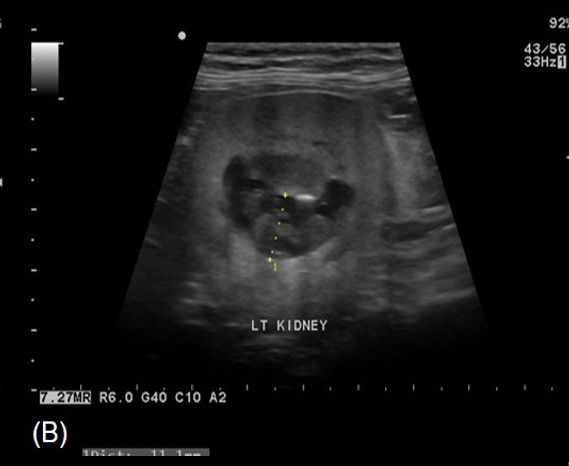
Case Report                                                            Veterinarni Medicina, 66, 2021 (02): 80–86

                                                                       https://doi.org/10.17221/153/2020-VETMED

      Figure 2A                                                  Figure 2B

     (A)                                                        (B)
Figure 2. Pre-contrast (A) and post-contrast (B) transverse computed tomography (CT) images of the right renal
pelvis in a cat with recurrent hydronephrosis and spontaneous renal rupture
The pre-contrast image shows a right renal diverticular calculus (A). The post-contrast CT image shows hydronephrosis
of the right kidney with a thin capsule (white arrow), accompanied by an asymmetric and severe dilation of the pelvis (B)

dronephrosis and hydroureter (Figure 3A,B). A mod-            space around the right kidney was newly identified
erate amount of free fluid in the retroperitoneal             (Figure 3C,D). This fluid was also seen on the abdom-

                                                                    Figure 3B
           Figure 3A

           (A)                                                        (B)

           Figure 3C                                               Figure 3D

           (C)
             (C)                                                    (D)

Figure 3. Intravenous pyelography at 24 h (A, B) and 48 h (C, D) in a cat with recurrent hydronephrosis and sponta-
neous renal rupture
Bilaterally, good initial nephrographic opacification is shown; the right kidney shows no pyelographic opacification
until 48 hours. The left kidney shows a delayed pyelogram with hydronephrosis and hydroureter during the procedure.
A moderate amount of free fluid in the retroperitoneal and peritoneal spaces is newly identified at 48 hours (C, asterisk)

83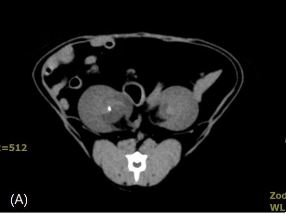
Case Report Veterinarni Medicina, 66, 2021 (02): 80–86
https://doi.org/10.17221/153/2020-VETMED
inal ultrasound. The ultrasound-guided abdominal DISCUSSION
fluid analysis revealed that the creatinine concentra-
tion in the abdominal fluid was higher (1 681.4 μg/l) Our case involved a seven-year-old cat with
than that in the serum (16 mg/l suggesting the pres- persistent and marked bilateral hydronephro-
ence of a uroabdomen. An exploratory laparotomy sis associated with amorphous, echogenic pelvic
was performed to detect the urine leakage from the materials with no shadowing or mineralisation
thin right renal capsule. During surgery, a moderate- identified on the diagnostic imaging. In such cas-
to-severe amount of abdominal fluid was suctioned; es, an exploratory laparotomy can help directly
a thin capsule with vascularisation, congestion, and identify the pelvic material. Although there was
an irregularly coarse parenchymal surface adjacent a 5-month interval between the first and second
to the right renal pelvis was identified. Urine leak- surgeries for the left and right kidneys, respectively,
age was present (Figure 4A), and the proximal lu- the hydronephrosis on both sides was attributed
men of the right ureter showed a mild dilation with to the same cause because of their similar imag-
palpable, small, and soft deposits. A right nephrec- ing characteristics. The predominance of RBCs
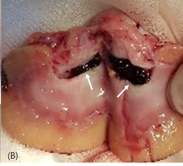
tomy was performed. The gross examination of the on the cytology at the time of the first surgery and
right kidney revealed several small, black deposits the protein composition of the deposits in the re-
in the right renal pelvis and proximal ureteral lu- nal pelvis affirmed our presumption that the de-
men (Figure 4B). The histopathology of the right posits were renal blood clots with necrosis. The
kidney revealed hydronephrosis with compression lack of haematuria or cloudy urine on the gross
atrophy, necrosis of the renal cortex/medulla, and examination prompted our consideration of other
a moderate lymphoplasmacytic inflammation. The aetiologies. Most mucus plugs observed in cats
crystallographic analysis showed that the deposits are composed of large quantities of matrices with
were composed of 100% protein and no minerals. varying amounts of minerals, and fewer than
The cat was hospitalised after the surgery, and ab- 10% of plugs lack crystals (Houston et al. 2003).
dominal fluid was again detected on the abdominal Hence, we ruled out mucus plugs. DSB calculi are
ultrasound on the third day, with no definitive cause radiolucent and do not appear as discrete calculi
identified. The owner decided to discontinue treat- on an abdominal ultrasound (Westropp et al. 2006;
ment and requested to take the cat home, and it suc- Novak and Craig 2011). However, unlike gelatinous
cumbed to the disease after 10 days. blood clots, these calculi are very firm and stone-
Figure 4A Figure 4B
(A) (B)
Figure 4. The right kidney and renal pelvis of a cat with recurrent hydronephrosis and spontaneous renal rupture after
the right nephrectomy
This was accompanied by urine leakage (A, white arrow). A substantial amount of small, black deposits (100% protein
composition) could be seen in the right renal pelvis (B, white arrows)
84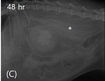
Case Report                                                    Veterinarni Medicina, 66, 2021 (02): 80–86

                                                               https://doi.org/10.17221/153/2020-VETMED

like (Westropp et al. 2006). Therefore, DSB calculi    dronephrosis and renal rupture. If a specific cause
were less likely to be present in our case, because    cannot be identified from test results, an immune-
the pelvic material was soft when palpated during      mediated disease should be tested by biopsy, when
surgery. In addition, the hydronephrosis showed        possible. If a biopsy is infeasible, treatment with
a spontaneous resolution with fluid and antibiotic     corticosteroids and a dietary change should be in-
therapy during hospitalisation, and the left hydro-    stituted, and the cat’s response should be closely
nephrosis improved after the pyelocentesis during      monitored.
the first exploratory laparotomy. We presumed the
material to be renal blood clots caused by an idi-
opathic lymphoplasmacytic inflammation because         Conflict of interest
there was no history of abdominal surgery or trau-
ma; moreover, chronic renal disease, blood clotting      The authors declare no conflict of interest.
disorders, and other inflammations or infections
in the urinary tract were excluded by the imaging
and laboratory examinations, including the sym-        REFERENCES
metric dimethylarginine test and urinalysis.
  Our case shows that progressive hydronephrosis       Cohen L, Shipov A, Ranen E, Bruchim Y, Segev G. Bilateral
can result in spontaneous renal rupture and identi-      ureteral obstruction in a cat due to a ureteral transitional
fies the unique characteristics of hydronephrosis        cell carcinoma. Can Vet J. 2012 May;53(5):535-8.
with radiolucent, amorphous, non-shadowing, and        D’Anjou MA, Bedard A, Dunn ME. Clinical significance of
echogenic materials on diagnostic tests. We fur-         renal pelvic dilatation on ultrasound in dogs and cats.
ther demonstrated that the right renal rupture re-       Vet Radiol Ultrasound. 2011 Jan-Feb;52(1):88-94.
sulted from a lymphoplasmacytic inflammation,          D’lppolito P, Nicoli S, Zatelli A. Proximal ureteral ectopia
atrophy, and necrosis of the renal parenchyma due        causing hydronephrosis in a kitten. J Feline Med Surg.
to progressive hydronephrosis. In addition, while        2006 Dec;8(6):420-3.
the moderate ascites that recurred 3 days after the    DiBartola SP. Renal disease: Clinical approach and labora-
right nephrectomy could be attributed to the spon-       tory evaluation. In: Ettinger SJ, Feldman EC, editors.
taneous rupture of the left kidney; this could not       Textbook of veterinary internal medicine. 6th ed. St. Louis:
be confirmed because the owner refused further           Elsevier Saunders; 2005. p. 1722-3.
examinations of the cat. If the presence of a lym-     Evans D, Fowlkes N. Renal leiomyosarcoma in a cat. J Vet
phoplasmacytic inflammation in the kidney had            Diagn Invest. 2016 May;28(3):315-8.
been confirmed, we could have improved the             Foster JD, Pinkerton ME. Bilateral ureteropelvic junction
cat’s condition or prolonged its life with dietary       stenosis causing hydronephrosis and renal failure in an
modification (e.g., hypoallergenic diet) and the ad-     adult cat. J Feline Med Surg. 2012 Dec;14(12):938-41.
ministration of high-dose corticosteroids or im-       Hall EJ, German AJ. Diseases of the small intestine. In: Et-
munosuppressive drugs (Willard 2003; Zaid et al.         tinger SJ, Feldman EC, editors. Textbook of veterinary
2011; Zhang et al. 2017).                                internal medicine. 6 th ed. St. Louis: Elsevier Saunders;
  Although the diagnostic imaging modalities             2005. p. 1367-72.
could not definitively confirm the causative mate-     Houston DM, Moore AEP, Favrin MG, Hoff B. Feline ure-
rial for the hydronephrosis, they helped to diagnose     thral plugs and bladder uroliths: A review of 5484 submis-
and monitor the hydronephrosis, detect the urine         sions 1998–2003. Can Vet J. 2003 Dec;44(12):974-7.
leakage, and plan the surgery. Our findings suggest    Lee N, Choi M, Keh S, Oh Y, Seo J, Choi H, Kim H, Yoon J.
that blood clots or a proteinaceous material should      Bilateral congenital ureteral strictures in a young cat. Can
be considered as differential diagnoses in cats pre-     Vet J. 2014 Sep;55(9):841-4.
senting with an upper urinary tract obstruction due    Novak JM, Craig LE. Pathology in practice. Hydronephro-
to radiolucent, amorphous, and echogenic mate-           sis and intrapelvic blood nephroliths with acute tubular
rial with no shadowing. The continuous monitor-          degeneration and end-stage nephropathy. J Am Vet Med
ing with an ultrasound and CT is important in such       Assoc. 2011 Apr 1;238(7):863-5.
cases. The rapid surgical removal of clots or the      Ragni RA, Fews D. Ureteral obstruction and hydronephrosis
injection of thrombolytic agents such as strepto-        in a cat associated with retroperitoneal infarction. J Feline
kinase into the clots may prevent progressive hy-        Med Surg. 2008 Jul;10(3):259-63.

85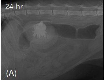
Case Report                                                             Veterinarni Medicina, 66, 2021 (02): 80–86

                                                                         https://doi.org/10.17221/153/2020-VETMED

Rawlings CA, Bjorling DE, Christie BA. Kidneys. In: Slat-        Westropp JL, Ruby AL, Bailiff NL, Kyles AE, Ling GV. Dried
  ter D, editor. Textbook of small animal surgery. 3rd ed.         solidified blood calculi in the urinary tract of cats. J Vet
  Philadelphia: W.B. Saunders; 2003. p. 1607-16.                   Intern Med. 2006 Jul-Aug;20(4):828-34.
Selgas AG, Scase TJ, Foale RD. Unilateral squamous cell          Willard MD. Digestive system disorders. In: Nelson RW,
  carcinoma of the renal pelvis with hydronephrosis in a cat.      Couto CG, editors. Small animal internal medicine. 3rd ed.
  J Feline Med Surg. 2014 Feb;16(2):183-8.                         St. Louis: Mosby; 2003. p. 408-48.
Simpson KW. Diseases of the stomach. In: Ettinger SJ, Feld-      Zaid MS, Berent AC, Weisse C, Caceres A. Feline ureteral
  man EC, editors. Textbook of veterinary internal medi-           strictures: 10 cases (2007–2009). J Vet Intern Med. 2011
  cine. 6th ed. St. Louis: Elsevier Saunders; 2005. p. 1321-5.     Mar-Apr;25(2):222-9.
Vanden SL, Levine JF, Lees GE, Groman RP, Grauer GF,             Zhang H, Zhuang G, Sun D, Deng T, Zhang J. Spontaneous
  Forrester SD. Renal biopsy: A retrospective study of meth-       rupture of the renal pelvis caused by upper urinary tract
  ods and complications in 283 dogs and 65 cats. J Vet In-         obstruction: A case report and review of the literature.
  tern Med. 2005 Nov-Dec;19(6):794-801.                            Medicine (Baltimore). 2017 Dec;96(50):e9190.

                                                                                                     Received: July 26, 2020
                                                                                                  Accepted: October 7, 2020

86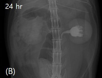
You can also read