Case Report: Combining 18F PET with MR for Detection of Bone Metastases in One Simultaneous Examination
←
→
Page content transcription
If your browser does not render page correctly, please read the page content below
Body Imaging Clinical

Case Report: Combining [18F] PET
with MR for Detection of Bone Metastases
in One Simultaneous Examination
Markus Lentschig, M.D.; Christiane Franzius, M.D.

ZEMODI, Zentrum für Moderne Diagnostik, Bremen, Germany

Background
In nuclear medicine, bone scanning is           scintigraphy are eminent; [18F] PET adds            importance of [18F] PET has increased
based on the principle of scintigraphy          diagnostic information mainly by its                recently because of its importance
using bone-seeking radiopharmaceuti-            superior resolution compared to scinti-             as a substitute for conventional skeletal
cals. [99mTc] (or [99Mo]) -labelled poly-       graphy and nowadays PET is routinely                scintigraphy in a time with limited
phosphonates are used as tracers for this       acquired as 3D data. In addition, CT used           availability of [99Mo]/[99mTc]. To ensure
purpose. They accumulate in sites of            as input for attenuation correction helps           healthcare, [18F] PET has now become
increased bone formation; metastases            to characterize suspicious bone model-              part of common outpatient care [1–5].
are detected either by increased uptake         ling. Also it should be kept in mind that           Within the last decade, MRI has also
of the lesion itself (osteoplastic), as         from a patient perspective, [18F] PET/CT            increasingly challenged the clinical value
reaction of the surrounding healthy bone        is considered to be the more convenient             of bone scintigraphy with superior diag-
matrix or as defect (osteolytic).               procedure (with special focus on prepa-             nostic performance. The potential to
Bone scintigraphy has found its way into        ration time and scan duration). Indepen-            assess not only changes of the bone but
several clinical guidelines over the last       dent of its diagnostic advantages, the              especially of the bone marrow and soft
decades and is a standard procedure in
the evaluation of bone metastases.              1A                                                  1B
However, degenerative changes of bones
are challenging to diagnose accurately
especially in elderly patients. The low
sensitivity of scintigraphy for (small)
osteolytic lesions often requires compli-
mentary imaging, either X-ray, computed
tomography (CT) or – especially in case
of bone tumors and bone marrow involve-
ment – magnetic resonance imaging
(MRI). It has to be stated that bone scin-
tigraphy is also associated with poor spa-
tial resolution and as a consequence of
the imaging mechanism itself this method
has limited diagnostic specificity for lesion
characterization and an insufficient
sensitivity for bone marrow diseases.
Positron emission tomography (PET)
using [18F]-fluoride has already demon-
strated to be a clinically useful alterna-
tive to traditional bone scintigraphy.
Interestingly, [18F] was initially replaced
by [99mTc]-labelled polyphosphonates as
osteotropic tracer. But with the develop-            1 Maximum intensity projection (MIP) of the [18F] PET data (1A) uncorrected and (1B)
ment of modern PET/CT technology,                 after attenuation correction (MR based).
the advantages over traditional bone

                                                                              MAGNETOM Flash · 1/2012 · www.siemens.com/magnetom-world      13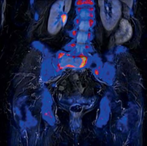
Clinical Body Imaging

tissue in general at highest sensitivity       A very obvious aspect is that it clearly       Case report
can add important information and have         improves spatial registration between          Patient history and sequence details
a clear impact on patient care. This is        metabolic and morphological information        A 91-year-old female with severe sacral
already proven for dedicated patient           by reducing the time gap between the           pain was referred to our institution for
cohorts. In combination with the advent        acquisition of MR and PET. This is a           a bone scan with [18F] as substitute to
of multi-regional MRI and further              clear advantage not only in imaging the        bone scintigraphy. The patient was diag-
advances in MR technology, diffusion-          pelvis, bowel, lung and liver, but also        nosed in 1986 with beast cancer and
weighted imaging (DWI) is used more            in patients with limited capability for        in 2004 a malignoma of the uterus was
and more routinely to add functional           holding still in one position over a longer    treated. Application of [18F] was per-
information to MRI. The images derived         time period. Also it should be mentioned       formed according to guidelines. MR/PET
from such an exam show PET-like appear-        that there are no limitations in the per-      was conducted as a multi-step exam
ance; however, the underlying mecha-           formance of the individual imaging             covering the whole body. During simul-
nism is restriction of water motion. How       methods of such a combined simultane-          taneous PET acquisition, a coronal T1w
DWI will add further diagnostic accuracy       ous MR/PET system. Hybrid MR/PET sys-          TSE (512 matrix, 450 mm FOV, 5 mm SL)
in the detection of bone metastases and        tems rely on segmentation algorithms           and T2w STIR (384 matrix, 450 FOV,
especially therapy follow-up is still          for providing the input function of the        5 mm SL) was acquired. In addition, a
subject of debate but its potential is         attenuation correction. At this point in       transversal DWI was measured (b-values
more than evident [6–11].                      time, bone segmentation is available           50, 400, 800 s/mm2, spectral fat satura-
Combining [18F] PET and MRI for evalua-        only for dedicated areas like the skull base   tion, 192 matrix, 5 mm SL; inline ADC
tion of bone processes is therefore            and not yet integrated in whole-body           calculation). All images shown were
appealing but was only available in a          scanning. Nevertheless, based on exist-        acquired using Biograph mMR (Siemens
small number of very selected cases up         ing data and experience, the need for          Healthcare, Erlangen, Germany) and a
to now. One practical reason is the asso-      bone segmentation can be negated in a          combination of the head/neck, spine
ciated effort for conducting, synchroniz-      clinical setting especially when a qualita-    and body coils.
ing (time and indication wise) and             tive reading of PET is performed. The
reading two complex exams (this is espe-       need for quantification of PET is unques-      Imaging findings
cially true for MRI, where a standard          tioned for follow-up exams and, so far,        A large osteolytic lesion is shown within
whole-body scan produces more than             the introduced error as compared to a          the sacral bone (massa lateralis). A clear
1000 images which have to be read). In         standard PET/CT (which has also a certain      mismatch between lesion size and corre-
addition, fusion techniques, which are         level of confidence only) seems to be          sponding bone formation is obvious. In
often used to assist in this task, are of      negligible even for longitudinal studies –     addition, tumor-suspicious bone forma-
limited value especially for scans cover-      if performed with an MR/PET system.            tion with corresponding lytic aspect in
ing a larger volume simply because of          Nevertheless, the advent of this technol-      MRI is demonstrated for the 10th and 7th
different positions of the bones between       ogy has reminded us that the discussion        right rib. Based on DWI, these lesions are
the two examinations. The limitation of        about the accuracy of PET quantification       characterized by high signal on the origi-
two separate exams can only partially          is of high importance and far from con-        nal b-value images and restriction of
be overcome by positioning aids (with          cluded (which is also true for the compa-      water diffusion. In addition, multiple
all their associated disadvantages). It can,   rability of results acquired with different    degenerative bone formations without
however, be overcome by using hybrid           PET/CT systems).                               corresponding oedema in MRI are visual-
MR/PET systems (with the advantage to          The diagnostic capabilities of MR and PET      ized: spondylosis of the thoracic spine
perform only one scan). Much has been          alone and in combination are of course         and coxarthrosis of the right hip are the
written about the need and the technol-        dependent on the underlying pathology          most obvious ones. Focal uptake is also
ogy behind this new hybrid imaging             and the applied MR imaging methods             seen in the dorsal processus of the 6th
modality (see also the most recent issues      and tracers. [12–15]                           and 7th cervical vertebra. Based on T2w
of MAGNETOM Flash). It should be                                                              STIR images at least for the bone forma-
pointed out that simultaneous MR and                                                          tion of the 7th vertebra a corresponding
PET imaging also has advantages in                                                            hyperintense lesion with space occupy-
clinical routine over a sequential approach                                                   ing aspects at least on the coronal original
– not only from a workflow aspect.                                                            orientation can be shown. Often reac-
                                                                                              tive oedema can be seen also in degen-
                                                                                              erative findings, however, based on the
                                                                                              space occupying appearance further
                                                                                              manifestation of the bone metastases

14 MAGNETOM Flash · 1/2012 · www.siemens.com/magnetom-world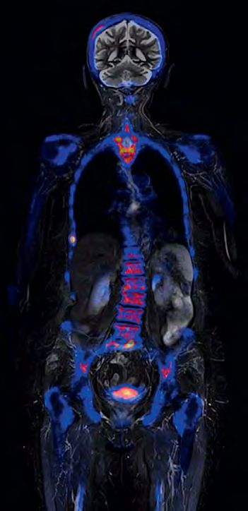
Body Imaging Clinical
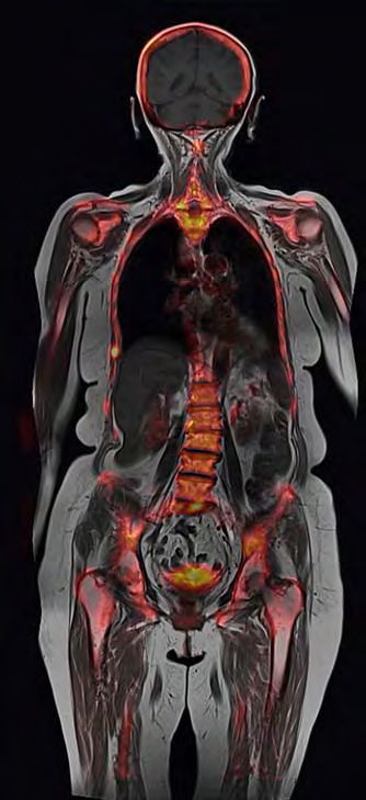
2A 2B 2C
*
*
2 Sagittal MIP (2A), coronal (2B), thick-slice MIP (2C) showing pathologic bone formation within the os sacrum and the 10th and 7th right rib
as well as the 7th / 6th cervical vertebra (dorsal processus) (arrows). In addition, multiple degenerative bone formation can be seen (e.g. spondy-
losis of the thoracic spine; asterisk).
3A 3B 3C
* * *
3 Clear mismatch between bone formation (arrows) and true extend of the metastasis (asterisk) in the massa lateralis of the os sacrum is shown.
Coronal multiplanar reconstruction (MPR) of the [18F] PET (3A), overlay of metabolic information on MRI (3B), corresponding coronal T1w TIRM (3C).
MAGNETOM Flash · 1/2012 · www.siemens.com/magnetom-world 15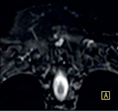
Clinical Body Imaging

4A                                              4B
                                                                    4 Thick-slice MPR
                                                                   based on the
                                                                   b = 800 s/mm2 DWI
                                                                   images. By suppres-
                                                                   sion of the back-
                                                                   ground the tumor
                                                                   tissue is well delin-
                                                                   eated. ADC mapping
                                                                   (not shown) did
                                                                   proof restriction of
                                                                   water diffusion.
                                                                   Coronal (4A) and
                                                                   transversal (4B)
                                                                   reformation.

5A                                              5B            5C

16 MAGNETOM Flash · 1/2012 · www.siemens.com/magnetom-world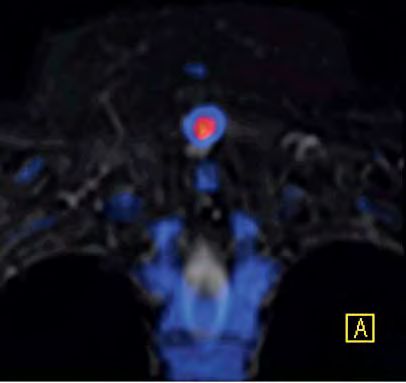
Body Imaging Clinical

must be concluded. No evidence for           numbers of successful treatment of the      Conclusion
further metastases within the long bones     primary tumor and also in concordance       Combining [18F] PET and MRI in one
of the upper and lower (not shown)           with latest epidemiological data, tumor     simultaneous exam is appropriate when
extremities, no fractures or soft tissue     recurrence of mamma carcinoma after         it comes to providing best patient care.
involvement, no spinal cord compres-         the 5-years follow-up interval has to be    Based on the knowledge with PET and
sion. The used protocol was mainly           taken into account. However, a third        MRI alone, it is more than justified in our
focused on the skeletal system, how-         tumor manifestation cannot be ruled out     opinion to state that this imaging
ever, further tumor manifestations out-      based only on imaging findings and          method can be applied to a large cohort
side the bone (including lymph nodes)        missing presence of a potential primary     of patients. While the presented case
can be ruled out with sufficient diagnos-    tumor. Therefore the final conclusion       may be a not so common clinical sce-
tic accuracy.                                of this exam has to be bone metastases      nario for the future application of MR/
                                             of a cancer of unknown primary (CUP).       PET, it clearly demonstrates the potential
Diagnosis                                    Because of clinical presentation (severe    of this method as the most accurate
Multifocal metastatic disease of the skel-   pain), a therapy relevance is obvious       method for evaluation of osseous and
etal system has to be concluded. Based       but further diagnosis and therapy will be   bone marrow processes. Especially in
on imaging findings and patient history,     to be discussed in detail and based on      cases with suspicion of bone marrow
a late metastatic manifestation of the       a very individual decision as a conse-      involvement and for younger patients,
mamma carcinoma seems to be the most         quence of the patients age and general      simultaneous MR/PET will play an
plausible explanation. With increased        condition.                                  important role in the future. How far

5D                                             5E
                                                                                             5 Excellent spatial regis-
                                                                                            tration between MRI and
                                                                                            PET data is shown exem-
                                                                                            plary with the small osteo-
                                                                                            lytic metastasis of the right
                                                                                            lateral thoracic wall (10 th
                                                                                            rib). Coronal MPR of [18F]
                                                                                            PET (5A), corresponding
                                                                                            T2w TIMR (5B), overlay of
                                                                                            PET data on T2w TIRM (5C),
                                                                                            corresponding T1w TSE
                                                                                            without (5D) and with (5E)
                                                                                            overlay of PET information.

                                                                     MAGNETOM Flash · 1/2012 · www.siemens.com/magnetom-world    17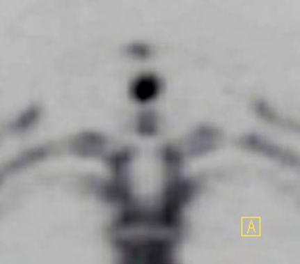
Clinical Body Imaging

6A                                                        6B                                                     6C

  6 The pathologic bone formation of the 6th and especially 7th (coronal MPR of the PET is shown in (6A)) dorsal processus of the cervical vertebra
  has also to be rated as potentially metastastic. A space occupying lesion with similar imaging features as shown for the other metastases can be
  demonstrated on the coronal T2w TIRM MRI (6B); the overly of the PET data (6C) shows a slight spatial mismatch of the two imaging modalities as
  a consequence of the different imaging mechanisms of [18F] PET (bone formation metabolism) and MRI (soft tissue characteristics).

this method will be added to, or will                    5 Yen RF, Chen CY, Cheng MF, Wu YW, Shiau YC,          12 Schlemmer HP, Pichler BJ, Krieg R, Heiss WD.
                                                           Wu K, Hong RL, Yu CJ, Wang KL, Yang RS. The             An integrated MR/PET system: prospective appli-
even replace, conventional imaging will
                                                           diagnostic and prognostic effectiveness of F-18         cations. Abdom Imaging. 2009 Nov;34(6):668-74.
of course be also a question of upcom-                     sodium fluoride PET-CT in detecting bone me-         13 Schwenzer NF, Schmidt H, Claussen CD. Whole-
ing therapy options and tracers e.g. for                   tastases for hepatocellular carcinoma patients.         body MR/PET: applications in abdominal imag-
evaluation of hormone receptor status.                     Nucl Med Commun. 2010 Jul;31(7):637-45.                 ing. Abdom Imaging. 2012 Feb;37(1):20-8.
But certainly the presented combination                  6 Pfannenberg C, Aschoff P, Schanz S, Eschmann         14 Delso G, Martinez-Möller A, Bundschuh RA,
                                                           SM, Plathow C, Eigentler TK, Garbe C, Brechtel          Ladebeck R, Candidus Y, Faul D, Ziegler SI. Eval-
of [18F] PET and MRI is already a further
                                                           K, Vonthein R, Bares R, Claussen CD, Schlemmer          uation of the attenuation properties of MR
step towards a more accurate and patient-                  HP. Prospective comparison of 18F-fluorodeoxy-          equipment for its use in a whole-body PET/MR
specific diagnoses and therapy selection                   glucose positron emission tomography/com-               scanner. Phys Med Biol. 2010 Aug 7;55(15):
– and all within one exam.                                 puted tomography and whole-body magnetic                4361-74. Epub 2010 Jul 20.
                                                           resonance imaging in staging of advanced             15 Delso G, Fürst S, Jakoby B, Ladebeck R, Ganter
                                                           malignant melanoma. Eur J Cancer. 2007                  C, Nekolla SG, Schwaiger M, Ziegler SI. Perfor-
                                                           Feb;43(3):557-64. Epub 2007 Jan 16.                     mance measurements of the Siemens mMR
  References
                                                         7 Müller-Horvat C, Radny P, Eigentler TK, Schäfer J,      integrated whole-body PET/MR scanner. J Nucl
1 Cook GJ. PET and PET/CT imaging of skeletal
                                                           Pfannenberg C, Horger M, Khorchidi S, Nägele            Med. 2011 Dec;52(12):1914-22. Epub 2011
  metastases. Cancer Imaging. 2010 Jul 19;10:1-8.
                                                           T, Garbe C, Claussen CD, Schlemmer HP. Pro-             Nov 11.
2 Hellwig D, Krause BJ, Schirrmeister H, Freesmeyer
                                                           spective comparison of the impact on treatment
  M. [Bone scanning with sodium 18F-fluoride PET
                                                           decisions of whole-body magnetic resonance
  and PET/CT. German guideline Version 1.0.].                                                                      Contact
                                                           imaging and computed tomography in patients
  Nuklearmedizin. 2010;49(5):195-201. Epub                                                                         Markus G. Lentschig, M.D.
                                                           with metastatic malignant melanoma. Eur J Can-
  2010 Sep 13.                                                                                                     ZEMODI
                                                           cer. 2006 Feb;42(3):342-50. Epub 2005 Dec 20.
3 Hahn S, Heusner T, Kümmel S, Köninger A, Naga-                                                                   Zentrum für Moderne Diagnostik
                                                         8 Schmidt GP, Reiser MF, Baur-Melnyk A. Whole-
  rajah J, Müller S, Boy C, Forsting M, Bockisch A,                                                                Schwachhauser Heerstr. 63a
                                                           body MRI for the staging and follow-up of
  Antoch G, Stahl A. Comparison of FDG-PET/CT                                                                      28211 Bremen
                                                           patients with metastasis. Eur J Radiol. 2009
  and bone scintigraphy for detection of bone                                                                      Germany
                                                           Jun;70(3):393-400. Epub 2009 May 19. Review.
  metastases in breast cancer. Acta Radiol. 2011                                                                   Phone +49 421 69641-600
                                                         9 Ketelsen D, Röthke M, Aschoff P, Merseburger AS,
  Nov 1;52(9):1009-14. Epub 2011 Oct 3.                                                                            Fax +49 421 69641-649
                                                           Lichy MP, Reimold M, Claussen CD, Schlemmer
4 Withofs N, Grayet B, Tancredi T, Rorive A, Mella C,                                                              www.zemodi.de
                                                           HP. [Detection of bone metastasis of prostate
  Giacomelli F, Mievis F, Aerts J, Waltregny D,
                                                           cancer – comparison of whole-body MRI and
  Jerusalem G, Hustinx R. ¹⁸F-fluoride PET/CT for
                                                           bone scintigraphy]. Rofo. 2008 Aug;180(8):
  assessing bone involvement in prostate and
                                                           746-52. Epub 2008 May 29. German.
  breast cancers. Nucl Med Commun. 2011
                                                        10 Wu LM, Gu HY, Zheng J, Xu X, Lin LH, Deng X,
  Mar;32(3):168-76.
                                                           Zhang W, Xu JR. Diagnostic value of whole-body
                                                           magnetic resonance imaging for bone metasta-
                                                           ses: a systematic review and meta-analysis.
                                                           J Magn Reson Imaging. 2011 Jul;34(1):128-35.
                                                           doi: 10.1002/jmri.22608. Epub 2011 May 25.
                                                        11 Padhani AR, Koh DM, Collins DJ. Whole-body
                                                           diffusion-weighted MR imaging in cancer: cur-
                                                           rent status and research directions. Radiology.
                                                           2011 Dec;261(3):700-18. Review.

18 MAGNETOM Flash · 1/2012 · www.siemens.com/magnetom-world
You can also read