Closed spinal dysraphism in a 6-month-old mixed breed dog
←
→
Page content transcription
If your browser does not render page correctly, please read the page content below
Case Report                                                                    Veterinarni Medicina, 66, 2021

                                                                   https://doi.org/10.17221/68/2020-VETMED

Closed spinal dysraphism in a 6-month-old
mixed breed dog
Guilherme Galhardo Franco1*, Emerson Goncalves Martins
de Siqueira2, Joao Augusto Leonel de Souza2, Leonardo
Oliveira de Castro Prado2, Sheila Canevese Rahal2,
Maria Jaqueline Mamprim3, Bruno Watanabe Minto4,
Claudia Valeria Seullner Brandao2, Jose Sergio Costa Junior4
1
  Department of Surgery, Federal University of Espirito Santo, Alegre,
  Espírito Santo, Brazil
2
  Department of Veterinary Surgery and Anesthesiology, College of Veterinary
  and Animal Science, São Paulo State University “Júlio de Mesquita Filho”,
  Botucatu, São Paulo, Brazil
3
  Department of Animal Reproduction and Veterinary Radiology, College
  of Veterinary and Animal Science, São Paulo State University “Júlio de
  Mesquita Filho”, Botucatu, São Paulo, Brazil
4
  Department of Clinical and Veterinary Surgery, College of Agricultural
  and Veterinary Sciences, São Paulo State University “Júlio de Mesquita Filho”,
  Jaboticabal, São Paulo, Brasil
*Corresponding author: guilherme.franco.vet@gmail.com

Citation: Franco GG, Siqueira EGM, Souza JAL, Prado LOC, Rahal SC, Mamprim MJ, Minto BW, Brandao CVS, Costa
Jr JS (2021): Closed spinal dysraphism in a 6-month-old mixed breed dog. Vet Med-Czech 66.

Abstract: The term spinal dysraphism defines an incomplete fusion or a bone defect that affects the neural struc-
tures of the spinal cord due to a neural tube malformation. A 6-month-old, male, mixed-breed dog, was evaluated
for paraparesis, pelvic limb proprioceptive ataxia, faecal and urinary incontinence. A neurological examination
indicated an L4-S3 spinal cord segment lesion. A thoracolumbar and lumbosacral spine magnetic resonance imag-
ing was performed and a closed spinal dysraphism, with the presence of a lipomyelomeningocele, was detected.
The magnetic resonance imaging showed an entire narrowing passage and a fibrous mass stemming from a wedge-
shaped gap in the caudal region of the dorsal lamina of the L4 vertebra, leaving the spinal canal towards the skin
surface in the region where the skin stigma was observed. A dorsal laminectomy was performed, the spinal cord
was exposed, and the entire fibrous mass was excised. Three months postoperatively, the patient had a complete
resolution of the urinary and faecal incontinence showing only a residual mild paraparesis, which remained six
months after surgery. The surgical procedure resulted in the satisfactory recovery of the neurological signs.

Keywords: congenital malformation; lipomyelomeningocele; spina bifida

  Spinal dysraphism defined as an incomplete              formation represents a spectrum of congenital
fusion or defect of the bone and other neural             anomalies classified into two subtypes: open and
structures of the spine due to a neural tube mal-         closed (Westworth and Sturges 2010).

1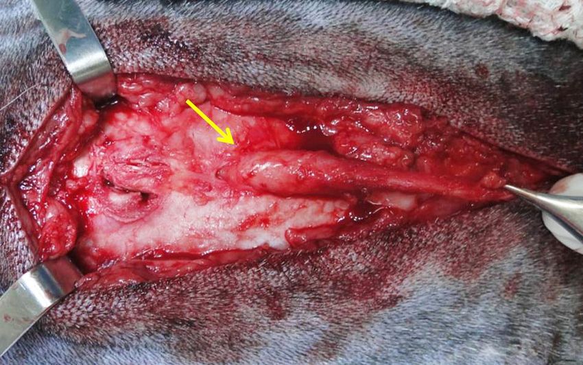
Case Report Veterinarni Medicina, 66, 2021
https://doi.org/10.17221/68/2020-VETMED
Open spinal dysraphism, characterised by nerve decreased. The anal sphincter reflex and sensitivity
tissue exposition through a defect in the meninges were also diminished. The other segmental reflexes
and/or vertebral column, is the most often diag- of the thoracic and pelvic limbs were normal. These
nosed in the neonatal period. In closed spinal dysra- findings indicated an L4-S3 spinal cord lesion.
phism (CSD), the resulting malformation is covered Upon palpation, a mass of soft consistency was
by the skin without exposure of the neural tissue observed in the L4 spinous process region. The dor-
(Rossi et al. 2006), and was reported in English sal midline region also had skin and hair changes,
Bulldogs, French Bulldogs, Collies, and Manx cats characterised by a 0.5-cm diameter, circular hypo-
(Wilson et al. 1979; Plummer et al. 1993; Kopke pigmentation lesion with a changing hair direction
et al. 2019). CSD can occur anywhere in the spine, (Figure 1).
but it is most commonly diagnosed in the lumbosa- Palpation of the head revealed an open fronto-
cral area (Westworth and Sturges 2010). parietal fontanelle. The orthopaedic examination
Lipomyelomeningocele is a type of CSD and detected a grade IV medial patellar luxation in the
consists of a subcutaneous fibroadipose mass that right pelvic limb. The complete cell blood count
intersects the lumbar fascia, causing a laminar spi- (CBC) and biochemistry profile were unremarkable.
nal defect, displacing the dura and, in some cases, Radiographs of the thoracolumbar, lumbosacral
anchoring and infiltrating the spinal cord (Rossi spine and pelvic limbs were obtained. They showed
et al. 2006). a radiolucent wedge in the caudal region of the dor-
sal lamina of the L4 vertebra and a medial patellar
luxation in the right pelvic limb with varus devia-
Case description tion and an external rotation of the distal femur.
The ultrasound of the caudal lumbar region showed
A 6-month-old, male, mixed-breed dog, 8.8 kg, a hyperechoic structure in the subcutaneous region
was presented with a 3-month history of progres- suggestive of a fat deposition due to its echogenicity.
sive weakness in the pelvic limbs, ataxia and uri- Magnetic resonance imaging (MRI) (Vet-MR,
nary and faecal incontinence. There was no history 0.25 Tesla; ESAOTE, Génova, Italy) of the brain,
of trauma. thoracolumbar and lumbosacral spine were per-
The neurological examination showed ambu- formed. The MRI showed the entire narrowing
latory paraparesis with decreased muscle tone, spinal canal and a fibrous mass stemming from
proprioceptive ataxia and proprioceptive deficits a wedge-shaped gap in the caudal region of the
in the pelvic limbs. The nociception of both pelvic dorsal lamina of the L4 vertebra, leaving the spi-
limbs was normal and the patellar reflexes were nal canal towards the skin surface in the region
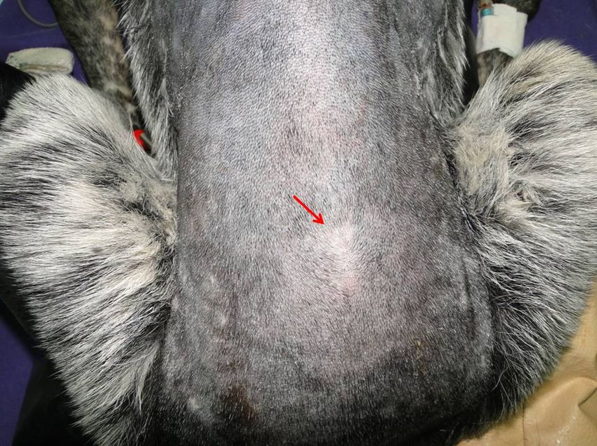
Figure 1
Figure 1. Photograph of the dog’s
lumbosacral region. Note the skin
changes (arrow) present in the
dorsal midline
2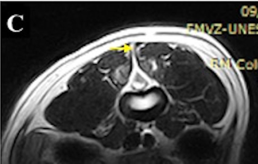
Case Report                                                                     Veterinarni Medicina, 66, 2021

                                                                    https://doi.org/10.17221/68/2020-VETMED

      Figure 2
    (A)                                                     (D)

    (B)                                                      (E)

    (C)                                                      (F)

Figure 2. Sagittal T2-weighted MRI (A) and transversal T2-weighted MRI (C) of the thoracolumbar spine showing
a defective closure of the spinal canal at the L4 vertebrae, through which a fibroadipose mass protrudes (yellow
arrow) toward the epidermis. Sagittal STIR-weighted MRI (B) of the thoracolumbar spine showing a defective clo-
sure of the spinal canal at the L4 vertebrae with the suppressed adipose tissue (green arrow) compatible with lipo-
myelomeningocele. Follow up sagittal T2-weighted MRI of the thoracolumbar spine showing a complete resection
of the lipomyelomeningocele. Follow up sagittal (D) and transversal (E) T2-weighted MRI (F) and sagittal STIR-
weighted MRI of the thoracolumbar spine showing a complete resection of the lipomyelomeningocele

where the skin stigma was observed (Figure 2).             by the radiographs. Similarly, a pelvic limb CT was
These findings were suggestive of lipomyelomenin-          performed in order to plan the surgical correction
gocele. The brain MRI ruled out other congeni-             of medial patellar luxation (Figure 3).
tal anomalies. Computed tomography (CT) scans                 The patient underwent inhalational anaesthe-
(SCT-7800CT; Shimadzu, Kyoto, Japan) of the                sia after 8 h of fasting and was positioned in ster-
thoracolumbar, lumbosacral spine and pelvic limbs          nal recumbency. Ceftriaxone (Ceftrion; Redson
were also conducted. The lumbosacral spine CT              Pharmaceuticals, Ahmedabad, India), 30 mg/kg,
allowed a three-dimensional reconstruction of the          i.v., was given before induction. The thoracolum-
L4 dorsal lamina deformation, previously detected          bar and lumbar regions were aseptically prepared.

3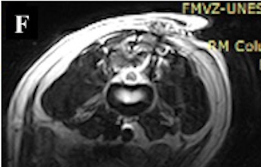
Case Report                                                                       Veterinarni Medicina, 66, 2021

                                                                        https://doi.org/10.17221/68/2020-VETMED

                                                               (B)
     Figure 3
    (A)

                                                               (C)

Figure 3. Dorsal (A), transversal (B) and sagittal (C) plane of the lumbosacral spine CT showing the incomplete fusion
of the caudal L4 dorsal lamina

A blunt dissection through the deep layers of the           ataxia and deficits in the pelvic limbs remained, and
skin revealed a subcutaneous fibroadipose mass              the owner reported improvement in both the faecal
protruding from the lumbodorsal fascia through
the laminar defect of the L4 vertebra. This mass                     Figure 4
adhered to the dura while infiltrating and anchor-
ing the spinal cord (Figure 4).
  After performing the dorsal laminectomy, the
spinal cord was exposed. A durotomy was per-
formed, and the mass along the entire path was
excised, fixed in formalin and sent for a histo-
pathological examination. The durotomy inci-
sion was sutured with a 6-0 polypropylene suture
(Prolene; Ethicon Inc, Somerville, NJ, USA) using
a simple interrupted pattern. The histopathologi-
cal analysis revealed a lipoma associated with the          Figure 4. Intraoperative photograph. Observe the sub-
fibrocartilaginous tissue.                                  cutaneous fibroadipose mass that crosses the lumbodor-
  Ten days postoperatively, the patient was in good         sal fascia through the laminar defect of the L4 vertebra
general condition. However, the proprioceptive              (arrow)

4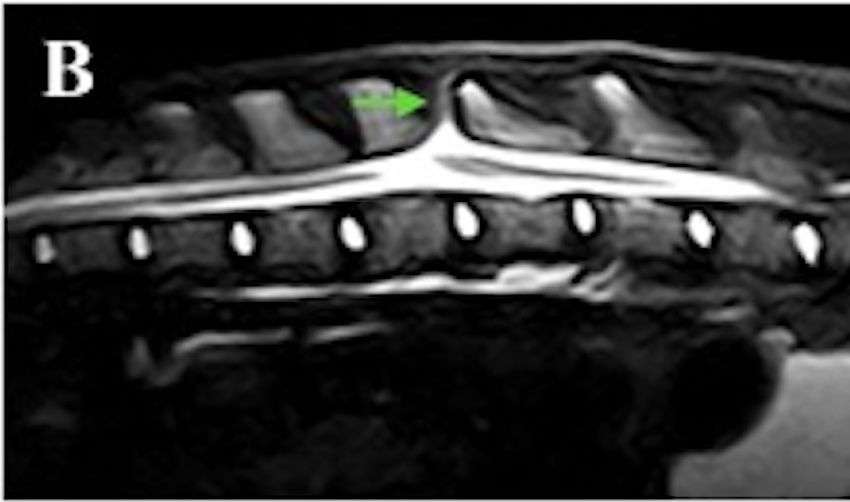
Case Report                                                                    Veterinarni Medicina, 66, 2021

                                                                   https://doi.org/10.17221/68/2020-VETMED

and urinary incontinence. Thirty days after surgery,       MRI correlated well, but an ultrasound is only
partial improvement of the pelvic limb ataxia oc-          a screening test that requires an MRI confirma-
curred, while the faecal incontinence was entirely         tion (Sewell et al. 2015).
resolved, with only sporadic episodes of urinary             Although the radiographs showed a fusion failure
incontinence. A follow-up MRI of the thoracolum-           in the caudal portion of the L4 dorsal lamina, it was
bar and lumbosacral spine was performed for the            not possible to visualise the lipomyelomeningocele.
postoperative evaluation, and the complete resec-          The CT scan enabled a three-dimensional recon-
tion of the lipomyelomeningocele was confirmed             struction of the lumbar segment of the spine and
(Figure 2).                                                showed the spinal malformation more accurately.
  The patient was monitored monthly for six                The MRI was essential to support the diagnosis,
months. At three months postoperatively, the dog           due to the parenchyma and spinal cord lesion site
had a complete resolution of the urinary and faecal        evaluation, and the continuity of the meninges pro-
incontinence while only a residual mild paraparesis        truding through the bone defect of the L4 dorsal
remained at six months.                                    lamina heading towards the epidermis. A postop-
                                                           erative MRI is advised to rule out any immediate
                                                           surgical complications in the first few weeks and,
DISCUSSION                                                 in some cases, it is also recommended after one
                                                           year, based on the clinical symptoms, to exclude
   In humans, axial lumbosacral cutaneous symp-            the re-anchoring of the bone marrow by the scar
toms are present in 50% to 80% of the cases, which         (Valentini et al. 2013). In this case report, the dog
may include sacrococcygeal dimples, dermoid si-            underwent an MRI 30 days after the surgery.
nus, haemangioma, lipoma, areas of hyper- or hy-             The clinical evolution of a CSD is variable and
popigmentation, hair tufts, and caudal appendages          often unpredictable (Westworth and Sturges 2010).
(Rossi et al. 2006; Sewell et al. 2015; Blount et al.      In this case, the clinical signs and their evolution
2019). This dog had both skin and hair alterations         from birth to 6 months of age were compatible with
in the dorsal midline, characterised by a circular         the tethered cord syndrome. These signs appear
hypopigmentation lesion with a 0.5 cm diameter             while the animal is growing, as the pathological fixa-
and changing hair direction, as shown in Figure 1.         tion of the medullary cone causes traction, resulting
   Congenital dermoid sinuses are cutaneous paths          in the distortion of the spinal anatomy and ischemic
lined by stratified squamous epithelium that estab-        lesions (Ricci et al. 2011; De Decker et al. 2015).
lish communication between the skin and deeper               The ideal time to perform the procedure is con-
structures (Kiviranta et al. 2011). In this case report,   troversial, but several studies have shown improve-
the biopsy sample sent for the histopathological           ment in motor function and urological signs with
analysis revealed no stratified squamous epithe-           an early surgical intervention (Valentini et al. 2013;
lium inside the mass, unlike a dermoid sinus, and          Song et al. 2014). In this case, a dorsal laminectomy,
a lipoma associated with a fibrocartilaginous tis-         an intradural exploration and a lipomyelomenin-
sue was observed, confirming the lipomyelomenin-           gocele resection have been shown to be effective
gocele type lesion.                                        to decompress and release the spinal cord.
   In humans, CSD is commonly associated with hy-            The post-operatory evolution of this dog was
drocephalus and a Chiari II malformation (Rossi            satisfactory and corroborated with Shamir et al.
et al. 2006; Blount et al. 2019), although this dog        (2001) who reported that an intradural surgical ex-
did not have any of these conditions. There are            ploration might be essential to improve or preserve
few reports of hydrocephalus associated with               the neurological signals of patients with lipomy-
myelomeningocele in dogs (Wilson et al. 1979;              elomeningocele.
Westworth and Sturges 2010).                                 In conclusion, spinal radiography, an ultrasound,
   Spinal radiographs are of little use to investigate     and a CT scan are essential tools to identify and
the CSD due to its low specificity (Sewell et al. 2015).   characterise the condition, as well as an MRI
In humans, an ultrasound is the frontline exam             to support the lesion type, providing the appro-
used to investigate the disease, because it is fast,       priate information for the surgical plan. In this
inexpensive, innocuous, and does not require an-           dog, the surgery resulted in a satisfactory clinical
aesthesia (Chern et al. 2012). The ultrasound and          recovery.

5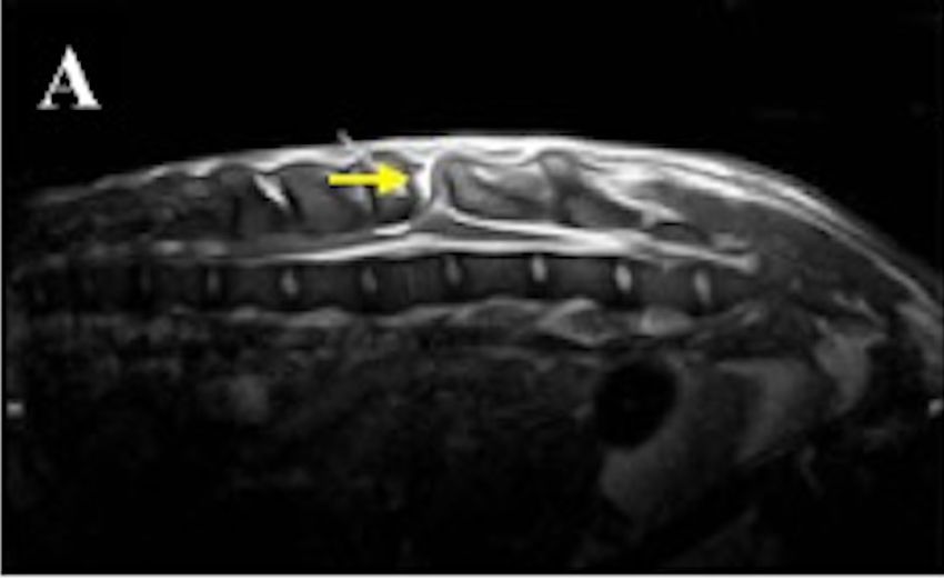
Case Report                                                                         Veterinarni Medicina, 66, 2021

                                                                       https://doi.org/10.17221/68/2020-VETMED

Conflict of interest                                          Ricci E, Cherubini GB, Jakovljevic S, Aprea F, Cantile C.
                                                                MRI findings, surgical treatment and follow-up of a my-
    The authors declare no conflict of interest.                elomeningocele with tethered spinal cord syndrome
                                                                in a cat. J Feline Med Surg. 2011 Jun;13(6):467-72.
                                                              Rossi A, Gandolfo C, Morana G, Piatelli G, Ravegnani M,
REFERENCES                                                      Consales A, Pavanello M, Cama A, Tortori-Donati P. Cur-
                                                                rent classification and imaging of congenital spinal ab-
Blount JP, George TM, Koueik J, Iskandar BJ. Concepts           normalities. Semin Roentgenol. 2006 Oct;41(4):250-73.
  in the neurosurgical care of patients with spinal neural    Sewell MJ, Chiu YE, Drolet BA. Neural tube dysraphism:
  tube defects: An embryologic approach. Birth Defects          Review of cutaneous markers and imaging. Pediatr Der-
  Res. 2019 Nov 15;111(19):1564-76.                             matol. 2015 Mar-Apr;32(2):161-70.
Chern JJ, Kirkman JL, Shannon CN, Tubbs RS, Stone JD,         Shamir M, Rochkind S, Johnston D. Surgical treatment
  Royal SA, Oakes WJ, Rozzelle CJ, Wellons JC. Use of lum-      of tethered spinal cord syndrome in a dog with mye-
  bar ultrasonography to detect occult spinal dysraphism.       lomeningocele. Vet Rec. 2001 Jun 16;148(24):755-6.
  J Neurosurg Pediatr. 2012 Mar;9(3):274-9.                   Song RB, Glass EN, Kent M, Sanchez MD, Smith DM, de La-
De Decker S, Gregori T, Kenny PJ, Hoy C, Erles K, Volk HA.      hunta A. Surgical correction of a sacral meningomye-
  Tethered cord syndrome associated with a thickened fi-        locele in a dog. J Am Anim Hosp Assoc. 2014 Nov-Dec;
  lum terminale in a dog. J Vet Intern Med. 2015 Jan;29(1):     50(6):436-43.
  405-9.                                                      Valentini LG, Selvaggio G, Erbetta A, Cordella R, Pecoraro
Kiviranta AM, Lappalainen AK, Hagner K, Jokinen T. Der-         MG, Bova S, Boni E, Beretta E, Furlanetto M. Occult spi-
  moid sinus and spina bifida in three dogs and a cat.          nal dysraphism: Lessons learned by retrospective analy-
  J Small Anim Pract. 2011 Jun;52(6):319-24.                    sis of 149 surgical cases about natural history, surgical
Kopke MA, Jack MW, Baltzer WI, Wightman PF, Gal A.              indications, urodynamic testing, and intraoperative neu-
  Dermoid sinus type VI associated with spina bifida and        rophysiological monitoring. Childs Nerv Syst. 2013 Sep;
  tethered cord syndrome in a French Bulldog. J Vet Diagn       29(9):1657-69.
  Invest. 2019 Mar;31(2):294-7.                               Westworth DR, Sturges BK. Congenital spinal malforma-
Plummer SB, Bunch SE, Khoo LH, Spaulding KA, Kornegay           tions in small animals. Vet Clin North Am Small Anim
  JN. Tethered spinal cord and an intradural lipoma associ-     Pract. 2010 Sep;40(5):951-81.
  ated with a meningocele in a Manx-type cat. J Am Vet        Wilson JW, Kurtz HJ, Leipold HW, Lees GE. Spina bifida
  Med Assoc. 1993 Oct 15;203(8):1159-61.                        in the dog. Vet Pathol. 1979 Mar;16(2):165-79.

                                                                                              Received: March 25, 2020
                                                                                          Accepted: November 24, 2020

6
You can also read