Fusobacterium genomics using MinION and Illumina sequencing enables genome completion and correction - bioRxiv
←
→
Page content transcription
If your browser does not render page correctly, please read the page content below
bioRxiv preprint first posted online Apr. 20, 2018; doi: http://dx.doi.org/10.1101/305573. The copyright holder for this preprint (which was not
                         peer-reviewed) is the author/funder. It is made available under a CC-BY-NC-ND 4.0 International license.

Fusobacterium genomics using MinION
and Illumina sequencing enables genome
completion and correction
   S. Michelle Todd1,                    Robert E. Settlage2,                   Kevin K. Lahmers1,                   Daniel J. Slade3#

1Department of Biomedical Sciences and Pathology, Virginia-Maryland College of Veterinary Medicine, Blacksburg,
VA, USA.
2Advanced Research Computing, Virginia Polytechnic Institute and State University, Blacksburg, VA, USA.
3Department of Biochemistry, Virginia Polytechnic Institute and State University, Blacksburg, VA, USA.

#To whom correspondence should be addressed: Dr. Daniel J. Slade, Department of Biochemistry, Virginia
Polytechnic Institute and State University, Blacksburg, VA 24061. Telephone: +1 (540) 231-2842. Email:
dslade@vt.edu

Design Type sequence assembly objective
Measurement Type whole genome sequencing
Technology Type DNA sequencing
Factor Type
Sample Characteristics Fusobacterium nucleatum, Fusobacterium periodonticum, Fusobacterium necrophorum,
Fusobacterium gonidiaformans, Fusobacterium ulcerans, Fusobacterium varium, Fusobacterium mortiferum

Understanding the virulence mechanisms of human pathogens from the genus Fusobacterium has been hindered by
a lack of properly assembled and annotated genomes. Here we report the first complete genomes for seven
Fusobacterium strains, as well as resequencing of the reference strain F. nucleatum subsp. nucleatum ATCC 25586
(seven total species, eight total genomes). A highly efficient and cost-effective sequencing pipeline was achieved
using sample multiplexing for short-read Illumina (150 bp) and long-read Oxford Nanopore MinION (>80 kbp)
platforms, coupled with genome assembly using the open-source software Unicycler. When compared to currently
available draft assemblies (previously 24-67 contigs), these genomes are highly accurate and consist of only one
complete chromosome. We present the complete genome sequence of F. nucleatum 23726, a genetically tractable
and biomedically important strain, and in addition, reveal that the previous F. nucleatum 25586 genome assembly
contains a 452 kb genomic inversion that has been corrected using our sequencing and assembly pipeline. To enable
the scientific community, we concurrently use these genomes to launch FusoPortal, a repository of interactive and
downloadable genomic data, genome maps, gene annotations, and protein functional analysis and classification. In
summary, this study provides detailed methods for accurately sequencing, assembling, and annotating
Fusobacterium genomes, which will enhance efforts to properly identify virulence proteins that may contribute to a
repertoire of diseases including periodontitis, pre-term birth, and colorectal cancer.

Summary of Visuals: Table 1 | Table 2 | Table 3 | Table 4 | Figure 1 | Figure 2 | Figure 3 | Figure 4
bioRxiv preprint first posted online Apr. 20, 2018; doi: http://dx.doi.org/10.1101/305573. The copyright holder for this preprint (which was not
                       peer-reviewed) is the author/funder. It is made available under a CC-BY-NC-ND 4.0 International license.

Background & Summary
Multiple Fusobacterium species are oral pathogens that can infect a broad range of human organ and tissue
niches1,2. Fusobacterium nucleatum has recently been connected with colorectal cancer (CRC) 3,4, with studies
showing this bacterium induces a pro-inflammatory tumor microenvironment5,6 and chemoresistance against drugs
used to treat CRC7. Despite the importance of this bacterium in human diseases, there is a lack of completed
genomes of biomedically relevant isolates to allow for virulence factor identification. Further motivation for complete
sequencing and assembly of a library of Fusobacterium genomes came from the observation that our bioinformatic
analysis frequently uncovered a high percentage of large, predicted secreted proteins (~3,000-11,000 bp) in the F.
nucleatum 23726 genome that were missing critical protein domains at either the N- or C-terminus (e.g. N-terminal
Sec signal sequences).

The genome of F. nucleatum subsp. nucleatum ATCC 25586, which is the standard F. nucleatum reference strain,
was completed in 2002 using cosmid and λ phage technologies to achieve long reads (10-35 kb) and facilitate
genome assembly8. More recently, several Fusobacterium draft genomes have been sequenced using short-read
technologies (454 Life Sciences), presumably making complete genome assembly difficult due to repeat regions
(e.g. CRISPR arrays, transposons). Similarly, Illumina sequencing is highly accurate and widely available, but is not
optimal for assembling whole genomes because of read length limitations (~ 150 bp). With the emergence of next
generation long-read sequencing (Pacific Biosciences, Oxford Nanopore Technologies MinION), assembling whole
genomes is now becoming standard and affordable for academic research settings. The recent combination of
MinION long-read and Illumina short-read technologies to scaffold and polish DNA sequencing data, respectively,
has created a robust pipeline for microbial genome completion and subsequent gene identification and
characterization9. A follow up study by these scientists detailed their methods for concurrently sequencing twelve
Klebsiella genomes through multiplex sampling 10. Following this experimental road map, we outline our experimental
methods for the first completely sequenced, assembled, and annotated Fusobacterium genomes using MinION
technology. In addition, these inaugural genomes are used to launch the FusoPortal genome and bioinformatic
analysis repository. In summary, this study provides key resources to further determine how multiple Fusobacterium
species contribute to a variety of human infections and diseases.

Methods
Bacterial growth and genomic DNA preparation
All strains of Fusobacterium were grown overnight in CBHK (Columbia Broth, hemin (5 μg/ml), and menadione (0.5 μ
g/ml) at 37 °C in an anaerobic chamber (90% N 2, 5% CO2, 5% H2). Genomic DNA from stationary phase bacteria
was isolated in diH2O from each strain using a Wizard isolation kit (Promega), and quantitated using a Qubit
fluorimeter (Life Technologies).

Short-read Illumina sequencing
Short-read DNA sequencing was carried out at the Genomic Sequence Center at the Virginia Tech Biocomplexity
Institute and Novogene (strain F. nucleatum 25586). For sequencing at Virginia Tech, DNA-seq libraries were
constructed using PrepX ILM 32i DNA Library Reagent Kit on an Apollo 324 NGS library prep system. Briefly 150 ng
of genomic DNA was fragmented using a Covaris M220 Focused-ultrasonicator to 400 bp. The ends were repaired
and an ‘A’ base added to the 3’ end for ligation to the adapters which have a single ‘T’ base overhang at their 3’ end.
Following ligation, the libraries were amplified by 7 cycles of PCR and barcoded. The library generated was validated
by Agilent TapeStation and quantitated using Quant-iT dsDNA HS Kit (Invitrogen) and qPCR. The libraries were then
pooled and sequenced using a NextSeq 500/550 Mid Output kit V2 (300 cycles) (P/N FC-404-2003) to 2 x 150
cycles. BCL files were generated using Illumina NextSeq Control Software v2.1.0.32 with Real Time Analysis RTA
v2.4.11.0. BCL files were converted to FASTQ files, adapters trimmed and demultiplexed using bcl2fastq Conversion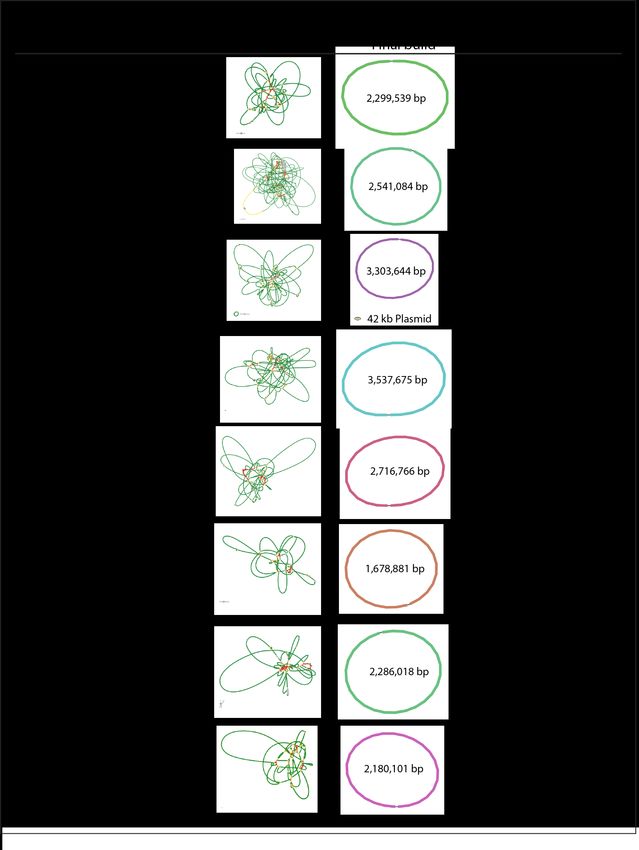
bioRxiv preprint first posted online Apr. 20, 2018; doi: http://dx.doi.org/10.1101/305573. The copyright holder for this preprint (which was not
                      peer-reviewed) is the author/funder. It is made available under a CC-BY-NC-ND 4.0 International license.

Software v2.20. Illumina sequencing statistics and genome coverage are detailed in Table 1, and the public
availability of the data at NCBI is detailed in Table 4.

Table 1. Statistics for short read Illumina sequencing.

                                      # Illumina Base                  Mean                                  Genome
Species              Strain           Sequences Pairs                  Length           Max Length           Size                Mean Depth

F. nucleatum         23726            5,163,442        774.5 mb 150.2                   151                  2,299,539           336 X

F. nucleatum         25586            1,250,000        187.5 mb 150.0                   150                  2,180,101           86.0 X

F. varium            27725            1,250,000        187.5 mb 150.2                   151                  3,303,644           56.8 X
                                                                                                             Plasmid:
                                                                                                             42,814

F. ulcerans          49185            1,250,000        187.5 mb 150.2                   151                  3,537,675           53.1 X

F. mortiferum        9817             1,250,000        187.5 mb 150.2                   151                  2,716,766           69.2 X

F.             25563                  1,250,000        187.5 mb 150.2                   151                  1,678,881           111 X
gonidiaformans

F.                   2_1_31           1,250,000        187.5 mb 150.2                   151                  2,541,084           73.8 X
periodonticum

F.                   1_1_36S          1,250,000        187.5 mb 150.2                   151                  2,286,018           82.2 X
necrophorum

Long-read MinION sequencing
Purified Fusobacterium genomic DNA was sequenced on a MinION sequencing device (Oxford Nanopore
Technologies) using the 1D Genomic DNA sequencing kit SQK-LSK108 according to Oxford Nanopore Technologies
instructions. Multiplexed samples were barcoded using the 1D Native Barcoding Kit (EXP-NBD103) according to
instructions. Briefly, purified genomic DNA was repaired with NEBNext FFPE Repair Mix (New England Biolabs). The
NEBNext Ultra II End-Repair/dA-tailing Module was utilized to phosphorylate 5’ ends and add a deoxyadenosine
monophosphate (dAMP) to the 3’ ends of the repaired DNA. For multiplexed samples, barcodes were ligated to the
end-prepped DNA using the NEB Blunt/TA Master Mix (New England Biolabs). Barcoded samples were pooled into a
single reaction and an adapter (Oxford Nanopore Technologies) was ligated to the DNA using the NEBNext Quick T4
DNA Ligase (New England Biolabs). For single reactions, an adapter (Oxford Nanopore Technologies) was ligated to
the end-prepped DNA using the NEB Blunt/TA Master Mix (New England Biolabs). The DNA was purified with
AMPureXP beads (Beckman Coulter, Danvers, MA) following each enzymatic reaction. Purified, adapted DNA was
sequenced on a MK1B (MIN-101B) MinION with a FLO-MIN106 (SpotON) R9.4 or FLO-MIN107 (SpotON) 9.5 flow cell
using MinKNOW software version 1.7.10 or 1.7.14 (Oxford Nanopore Technologies). After sequencing, Fast5 files
were basecalled using Albacore version 2.1.7 (Oxford Nanopore) on a Macbook Pro with a 3.3 GHz Intel Core i7
processor. For multiplexed samples, basecalled fastq files were demultiplexed based on the ligated barcode using
Porechop and adaptors were trimmed. Sample preparation and sequencing details are described in Table 2, and the
MinION sequencing statistics and genome coverage are detailed in Table 3. As an example of data quality, Figure 1
shows the long read coverage obtained using MinION sequences for the F. necrophorum funduliforme 1_1_36S
genome.
bioRxiv preprint first posted online Apr. 20, 2018; doi: http://dx.doi.org/10.1101/305573. The copyright holder for this preprint (which was not
peer-reviewed) is the author/funder. It is made available under a CC-BY-NC-ND 4.0 International license.
Table 2. Experimental details for MinION sequencing.
Sample IDs Sequencing Kit Flow Cell MinKNOW Version Input DNA (μg)
23726 SQK-LSK108 FLO-MIN106 1.7.10 2.38
(SpotON) R9.4
25586 SQK-LSK108 FLO-MIN107 1.7.14 3.96
(SpotON) R9.5
49185, 9817, SQK-LSK108, EXP- FLO-MIN107 1.7.14 3.72, 3.12, 2.94, 0.6,
25563, 2_1_31, NBD103 (SpotON) R9.5 2.31, 1.83
1_1_36S, 27725
#
# Multi-plexed on one flow cell.
Table 3. Results for MinION sequencing.
# MinION Mean Genome
Species Strain Sequences Base Pairs Length Max Length Size Mean Depth
F. nucleatum 23726 13,904 81,303,330 5,847.5 55,886 2,299,539 35.4 X
F. nucleatum 25586 25,240 111,179,924 4,404.9 84,482 2,180,101 60.0 X
F. varium 27725 31,066 89,376,121 2,877.0 62,500 3,303,644 27.0 X
Plasmid:
42,814
F. ulcerans 49185 20,313 42,439,006 2,089.3 74,841 3.537,675 12.0 X
F. mortiferum 9817 37,658 118,090,402 3,135.9 83,201 2,716,766 43.5 X
F. 25563 58,596 105,220,409 1,784.7 87,683 1,678,881 62.7 X
gonidiaformans
F. 2_1_31 52,316 178,049,514 3,403.3 68,386 2,541,084 70.0 X
periodonticum
F. 1_1_36S 66,335 129,875,928 1957.9 81,384 2,286,018 56.8 X
necrophorumbioRxiv preprint first posted online Apr. 20, 2018; doi: http://dx.doi.org/10.1101/305573. The copyright holder for this preprint (which was not
peer-reviewed) is the author/funder. It is made available under a CC-BY-NC-ND 4.0 International license.
Figure 1: Statistics and Mapping of F. necrophorum funduliforme 1_1_36S MinION long-reads. Post complete
genome assembly, MinION reads were mapped to the F. necrophorum funduliforme 1_1_36S genome using
Geneious version 9.1.4 software.
Genome assembly
Genome assemblies were carried out using the open-source software Unicycler9, resulting in complete
chromosomes for each of the eight sequenced genomes. While both the Illumina and MinION sequencing runs
produced far more data than necessary, data sets were split to utilize ample yet reasonable mean depth of coverage
for 1.6 mb to 3.5 mb genomes. Using the mean depths of coverage for each genome described in Table 1 and Table
2, each genome can be constructed in 2-3 hours using a standard Macbook Pro laptop (2.8 GHz Intel Core i7). The
utility of Unicycler therefore opens up a robust method for researchers without the need for a super computer tobioRxiv preprint first posted online Apr. 20, 2018; doi: http://dx.doi.org/10.1101/305573. The copyright holder for this preprint (which was not
peer-reviewed) is the author/funder. It is made available under a CC-BY-NC-ND 4.0 International license.
handle data processing. The details of all final assemblies are shown in Figure 2, and the public availability of the
data at NCBI is detailed in Table 4. For consistent starts to the circular chromosome, each genome was rotated to
have Gene 1, which encodes for the rod-shape determining protein MreC, in the reverse orientation as is seen for the
beginning of the F. nucleatum subsp. nucleatum ATCC 25586 reference genome8.
Figure 2: Genome assembly and Annotation of eight Fusobacterium genomes from seven species. Short-read
only and complete genome assembly representations were created using Bandage11.
In addition, we believe that we have identified a previously undocumented 42 kb plasmid in F. varium ATCC 27725.
To show the effectiveness of our genome assembly pipeline, Figure 3a shows the alignment of 67 contigs from the
previous F. nucleatum subsp. nucleatum ATCC 23726 draft genome on our completed circular genome. We show
that all contigs map, with our genome completing previous gaps. The accuracy of our genome when compared to
mapped base pairs from the draft genome assembly at NCBI shows 99.99% base identification as determined bybioRxiv preprint first posted online Apr. 20, 2018; doi: http://dx.doi.org/10.1101/305573. The copyright holder for this preprint (which was not
peer-reviewed) is the author/funder. It is made available under a CC-BY-NC-ND 4.0 International license.
Geneious version 9.1.4. Strikingly, upon Geneious alignment of our F. nucleatum subsp. nucleatum ATCC 25586
genome with the previously complete genome deposited at NCBI (GCA_000007325.1), we discovered a ~ 452 kb
genomic inversion (Figure 3b). This region is flanked on both ends by ~ 8 kb repeats that are likely the reason for the
previous inability to discover this genomic feature. To validate this inversion, we aligned eight MinION reads (30-68
kb) that spanned this region, and show that these sequences confirm this genomic correction.
Figure 3: Analysis of Fusobacterium nucleatum. (A) Alignment of the complete F. nucleatum subps. nucleatum
ATCC 23726 genome with the 67 contig draft assembly (Genbank: ADVK01000000). (B) Confirmation of a 452 kb
genomeic inversion in the previous F. nucleatum subps. nucleatum ATCC 25586 genome assembly (Genbank:
GCA_000007325.1).
Open reading frame predictions
Gene predictions for protein encoding open reading frames were carried out using the bacterial specific program
Prodigal via command line on a Mac 12. Genes for tRNA encoding were predicted with Prokka13 using the KBase
server14. rRNA were identified using Barrnap (Bacterial ribosomal RNA predictor). In addition, we used thebioRxiv preprint first posted online Apr. 20, 2018; doi: http://dx.doi.org/10.1101/305573. The copyright holder for this preprint (which was not
peer-reviewed) is the author/funder. It is made available under a CC-BY-NC-ND 4.0 International license.
CRISPRone web server to identify all CRISPR associated proteins and arrays, which consist of spacer and repeat
regions. Details of each of these components are found on the FusoPortal repository. In each genome, protein
encoding gene predictions by Prodigal and Prokka were in nearly complete agreement (data not reported). In
addition, genome annotation for each genome was requested at NCBI upon data deposition into Genbank (Table 4).
Software and code availability
All software and scripts used in this study have been described and properly referenced in previous methods
sections.
Data Records
Raw data and completed genomes for each of the eight Fusobacterium strains have been deposited at NCBI under
the BioProject, BioSamples, Sequence Read Archives (SRA), and Genbank accession numbers detailed in Table 4.
Table 4. Data deposited at NCBI for all sequenced Fusobacterium strains.
Genbank SRA
Species Strain Genome BioProject BioSample Illumina1 SRA MinION1
F. nucleatum 23726 GCA_003019785.1 PRJNA433545 SAMN08501025 SRX3740879 SRX3740878
F. nucleatum 25586 GCA_003019295.1 PRJNA433545 SAMN08706662 SRX3786193 SRX3786192
F. varium 27725 GCA_003019655.1 PRJNA433545 SAMN08501142 SRX3740889 SRX3740888
F. ulcerans 49185 GCA_003019675.1 PRJNA433545 SAMN08501141 SRX3740885 SRX3740884
F. mortiferum 9817 GCA_003019315.1 PRJNA433545 SAMN08501148 SRX3740887 SRX3740886
F. 25563 GCA_003019695.1 PRJNA433545 SAMN08501140 SRX3740881 SRX3740880
gonidiaformans
F. 2_1_31 GCA_003019755.1 PRJNA433545 SAMN08501101 SRX3740877 SRX3740876
periodonticum
F. 1_1_36S GCA_003019715.1 PRJNA433545 SAMN08501105 SRX3740883 SRX3740882
necrophorum
1 Sequence Read Archive at NCBI
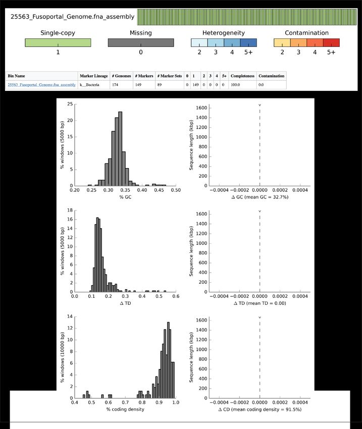
Technical Validation
CheckM15 on the Kbase14 server was used to check the quality of each genome using the reduced tree data set
setting. An example of this data analysis is shown for strain F. gonidiaformans ATCC 25563 is shown in figure Figure
4, and data analysis for all genomes are available on the FusoPortal repository.bioRxiv preprint first posted online Apr. 20, 2018; doi: http://dx.doi.org/10.1101/305573. The copyright holder for this preprint (which was not
peer-reviewed) is the author/funder. It is made available under a CC-BY-NC-ND 4.0 International license.
Figure 4: CheckM genome analysis of F. gonidiaformans ATCC 25563. CheckM analysis on the Kbase server
shows that the F. gonidiaformans ATCC 25563 is of high quality and contains all core bacterial genes tested. Data
analysis by CheckM for all eight Fusobacterium genomes described in this study are detailed on the FusoPortal
repository.
Usage Notes
The raw data, genome assemblies, and annotations can be accessed via the NCBI BioProject PRJNA433545, and
further details of these files can be found in Table 4. In addition, all of this data is easily accessible in our newly
implemented FusoPortal data repository or on our Open Science Framework database.
Data Citations
1. NCBI BioProject PRJNA433545 http://www.ncbi.nlm.nih.gov/bioproject/433545
2. FusoPortal http://fusoportal.org
3. Open Science Framework http://osf.io/2c8pv
Acknowledgements
We would like to thank the laboratory of Emma Allen-Vercoe (University of Guelph) for providing many of the strainsbioRxiv preprint first posted online Apr. 20, 2018; doi: http://dx.doi.org/10.1101/305573. The copyright holder for this preprint (which was not
peer-reviewed) is the author/funder. It is made available under a CC-BY-NC-ND 4.0 International license.
sequenced in this study. This work is supported by the USDA National Institute of Food and Agriculture.
Author Contributions
S.M.T. performed all MinION sequencing, and wrote and edited the manuscript. K.K.L. prepared raw MinION
sequences for genome assembly and wrote and edited the manuscript. R.E.S. assembled genomes and edited the
manuscript. D.J.S. conceived and designed the experiments, assembled genomes, analyzed the data, and wrote and
edited the manuscript.
Competing Interests The authors declare no competing financial interests.
References
1. Dahya, V., Patel, J., Wheeler, M. & Ketsela, G. Fusobacterium nucleatum endocarditis presenting as liver and brain
abscesses in an immunocompetent patient. Am. J. Med. Sci. 349, 284–285 (2015).
2. Signat, B., Roques, C., Poulet, P. & Duffaut, D. Fusobacterium nucleatum in periodontal health and disease. Curr.
Issues Mol. Biol. 13, 25–36 (2011).
3. Castellarin, M. et al. Fusobacterium nucleatum infection is prevalent in human colorectal carcinoma. Genome Res.
22, 299–306 (2012).
4. Kostic, A. D. et al. Genomic analysis identifies association of fusobacterium with colorectal carcinoma. Genome
Res. 22, 292–298 (2012).
5. Kostic, A. D. et al. Fusobacterium nucleatum potentiates intestinal tumorigenesis and modulates the tumor-
immune microenvironment. Cell Host Microbe 14, 207–215 (2013).
6. Rubinstein, M. R. et al. Fusobacterium nucleatum promotes colorectal carcinogenesis by modulating E-cadherin/
β-catenin signaling via its FadA adhesin. Cell Host Microbe 14, 195–206 (2013).
7. Yu, T. et al. Fusobacterium nucleatum promotes chemoresistance to colorectal cancer by modulating autophagy.
Cell 170, 548–563.e16 (2017).
8. Kapatral, V. et al. Genome sequence and analysis of the oral bacterium fusobacterium nucleatum strain ATCC
25586. J. Bacteriol. 184, 2005–2018 (2002).
9. Wick, R. R., Judd, L. M., Gorrie, C. L. & Holt, K. E. Unicycler: Resolving bacterial genome assemblies from short
and long sequencing reads. PLoS Comput. Biol. 13, e1005595 (2017).
10. Wick, R. R., Judd, L. M., Gorrie, C. L. & Holt, K. E. Completing bacterial genome assemblies with multiplex
MinION sequencing. Microb Genom 3, e000132 (2017).
11. Wick, R. R., Schultz, M. B., Zobel, J. & Holt, K. E. Bandage: Interactive visualization of de novo genome
assemblies. Bioinformatics 31, 3350–3352 (2015).
12. Hyatt, D. et al. Prodigal: Prokaryotic gene recognition and translation initiation site identification. BMC
Bioinformatics 11, 119 (2010).
13. Seemann, T. Prokka: Rapid prokaryotic genome annotation. Bioinformatics 30, 2068–2069 (2014).
14. Arkin, A. P. et al. The DOE systems biology knowledgebase (KBase). bioRxiv 096354 (2016).
15. Parks, D. H., Imelfort, M., Skennerton, C. T., Hugenholtz, P. & Tyson, G. W. CheckM: Assessing the quality of
microbial genomes recovered from isolates, single cells, and metagenomes. Genome Res. (2015).You can also read