Periarticular histiocytic sarcoma of a thoracic limb in a Rottweiler
←
→
Page content transcription
If your browser does not render page correctly, please read the page content below
pISSN 2466-1384 eISSN 2466-1392
大韓獸醫學會誌 (2018) 第 58 卷 第 1 號
Korean J Vet Res(2018) 58(1) : 57~60
https://doi.org/10.14405/kjvr.2018.58.1.57

        Periarticular histiocytic sarcoma of a thoracic limb in a Rottweiler
                                      Hyeok-Soo Shin1, Ye-In Oh2, Byung-Jae Kang1,*
              1
             Department of Veterinary Surgery, College of Veterinary Medicine and Institute of Veterinary Science,
                                    Kangwon National University, Chuncheon 24341, Korea
   2
    Department of Veterinary Internal Medicine, College of Veterinary Medicine, Seoul National University, Seoul 08826, Korea

                     (Received: January 2, 2018; Revised: February 21, 2018; Accepted: February 26, 2018)

       Abstract: An 8-year-old, castrated, male Rottweiler was referred for evaluation of chronic right thoracic limb lameness
       and a progressively growing mass surrounding the right elbow joint. On admission, the dog’s general health was good,
       without abnormalities detected on physical examination. The dog was diagnosed with periarticular histiocytic sarcoma.
       Although draining lymph nodes and lung metastases were suspected, palliative amputation was performed. Localized
       histiocytic sarcomas, with destructive lesions involving multiple bones of a joint and periarticular soft-tissue masses,
       are uncommon in dogs. This case report presents clinical findings, imaging characteristics, and histopathologic and
       immunohistochemical features of a periarticular joint histiocytic sarcoma.
       Keywords: bone, dogs, immunohistochemistry, periarticular histiocytic sarcoma

  Histiocytes are derived from bone marrow stem cells and              nated HS, which is an invasive multiorgan disorder with a
can be either macrophages or dendritic cells. Dendritic cells          grave prognosis [2]. The etiology of HS is unknown, but a
can be further subdivided into Langerhans cells found in the           clear genetic link has been observed. Rottweiler, Golden
skin, interstitial dendritic cells found in many organ systems,        Retriever, Labrador Retriever, Doberman Pincher, and Bernese
and interdigitating dendritic cells found in the T cell zone.          Mountain Dog are predisposed breeds, and middle- to old-
Because of these various differentiation potentials of histio-         aged dogs are often affected [1, 5, 13].
cytes, canine histiocytic proliferative disorders represent a             Dogs with localized HS, which occurs at a focal limb,
diagnostic and therapeutic challenge [11, 12, 14].                     often present with lameness associated with one joint because
  Various canine histiocytic proliferative diseases have been          the tumor can arise from the dendritic cells of the synovial
reported in dogs, including reactive diseases such as cutane-          lining. Moreover, the tumor can affect the surrounding bone
ous and systemic histiocytosis. Neoplastic histiocytic dis-            and soft tissue [4]. The Rottweiler, described herein, devel-
eases are classified into histiocytomas (benign forms) or              oped chronic lameness and pain due to periarticular histio-
histiocytic sarcomas (HSs; malignant forms). HSs are further           cytic sarcoma (PAHS).
classified into the disseminated or localized form. Localized             An 8-year-old, castrated, male Rottweiler, weighing 38 kg,
HS involves a single primary lesion, whereas disseminated              was referred for the evaluation of chronic right thoracic limb
HS involves multiple organ systems [2] and presents multi-             lameness and pain. Three months previously, mild soft-tis-
ple tumor masses in several organ systems. Primary lesions             sue swelling and pain in the region of the elbow joint was
are usually seen in the bone marrow, spleen, and lung,                 detected by the referring veterinarian and treated with non-
whereas secondary lesions are observed in the liver, lymph             steroidal anti-inflammatory drugs, antibiotics, and restricted
nodes, and sometimes other organs. Dogs with disseminated              physical activity. However, the dog showed only temporary
HS often show anorexia, weight loss, and lethargy [2, 15].             improvement. Over the previous month, the mass rapidly
Localized HSs arise from a single organ. They are locally              increased in size and the dog showed intermittent lameness
invasive and can metastasize to regional lymph nodes. The              in the right thoracic limb.
subcutis is a major invasion site, but other primary sites have           On admission, the dog showed good general health but had
been observed in the stomach, lungs, liver, spleen, pancreas,          been showing mild anorexia for 1 week. Right axillary lymph
and central nervous system. A more favorable prognosis can             node enlargement was identified on a physical examination.
be expected for dogs with localized HS than with dissemi-              An orthopedic examination revealed intermittent lameness in

*Corresponding author
Tel: +82-33-250-8207, Fax: +82-33-259-5625
E-mail: bjkang@kangwon.ac.kr
                                                                  57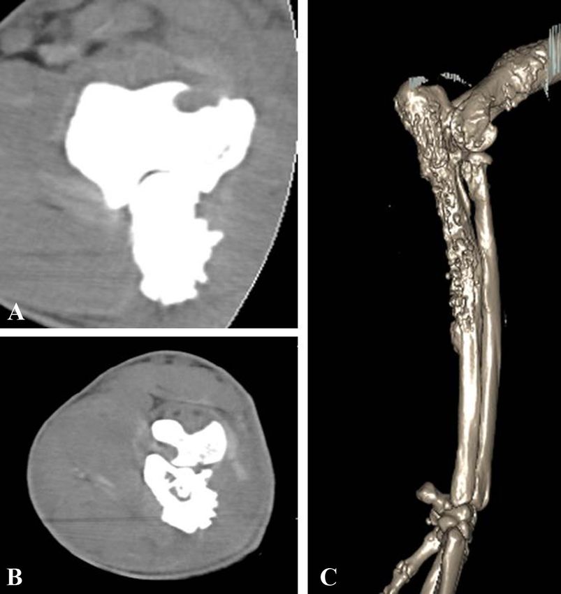
58                                        Hyeok-Soo Shin, Ye-In Oh, Byung-Jae Kang

Fig. 1. Lateral (A) and dorsoventral (B) radiographs demon-
strating a large soft-tissue mass surrounding the right elbow
joint and irregular bone proliferation at the distal humerus,
proximal radius, and ulna.
                                                                 Fig. 2. Computed tomography images of the humeral condyle
                                                                 (A) and proximal radius and ulna (B) demonstrate the combi-
the right forelimb. The range of motion of the elbow joint
                                                                 nation of irregular bone lysis and periosteal reaction. (C) Three-
was restricted to about 60% of that of the normal leg, and the
                                                                 dimensionally reconstructed image of the distal humerus, radius,
dog showed pain when the affected elbow joint was manipu-        and ulna in the lateral aspect.
lated. Radiography revealed a large soft-tissue mass around
the elbow joint. Moreover, new bone production, cortical
destruction, and perceptive lysis were present within the dis-   thoracic limb was amputated through the removal of the
tal humerus and proximal ulna (Fig. 1). Neoplastic and infec-    scapula. Axillary lymph nodes and other regional lymph
tious diseases were listed as the main causes. Fine-needle       nodes were also removed during the surgery. The dog recov-
aspiration of the mass was performed under ultrasound guid-      ered without any complication and showed good general
ance. Microscopic examination revealed marked cellular and       health. However, the owner refused the administration of
nuclear pleomorphism and giant multinucleated cells, charac-     adjuvant chemotherapy. The limb and removed lymph nodes
teristic of a malignant tumor. In addition, the microscopic      were submitted for histopathologic evaluation. On gross
examination revealed the dominance of spindle cells, which       examination, the bone marrow and bony structure of the
are features of mesenchymal cell origin tumors. Microorgan-      proximal ulna and distal humerus were effaced by an unen-
isms were not observed on synovial fluid examination, and a      capsulated, poorly circumscribed, infiltrative neoplasm. The
synovial culture showed no bacterial growth. A tentative         large mass surrounding the elbow joint was due to the neo-
diagnosis of a neoplastic disorder was made, and osteosar-       plastic proliferation of neighboring muscles and joint cap-
coma, fibrosarcoma, chondrosarcoma, osteochondrosarcoma,         sule. Furthermore, proliferated soft tissues were filled with a
synovial cell sarcoma, and HS were included in the differen-     hemopurulent discharge. The bone neoplasm was composed
tial diagnosis list. Computed tomography (CT) was per-           of spindle cells and occasional round cells, sometimes
formed to evaluate the metastasis and neoplastic change in       arranged in pleomorphic sheets supported by existing stroma.
the right periarticular region. A complete blood count, serum    Bone and axillary lymph node had the same features on
biochemical analysis, and thoracic radiography were per-         microscopic observation. Neoplastic cells had distinct cell
formed prior to anesthesia, and these revealed only mild ane-    borders and moderate amount of amphophilic cytoplasm.
mia. The iso- to hypo-attenuating mass was found near the        Nuclei were round to oval and had finely to coarsely stip-
right elbow joint on pre-contrast CT. In addition to the sur-    pled chromatin and 1–3 indistinct nucleoli. Marked anisocy-
rounding bone structure, the synovial membrane showed            tosis and anisokaryosis were seen. Marked osteolysis and
neoplastic changes (Fig. 2). The right axillary lymph node       mesenchymal proliferation were also seen (Fig. 3A). More
was enlarged (6 × 3 cm2). Chest CT revealed multiple small       than 90% of the local lymph nodes showed necrotic changes,
metastatic pulmonary nodules. The prognosis was expected         and multiple aggregates of lymphocytes, plasma cells, and
to be negative because of the suspicion of metastasis, the       macrophages with fibrous tissue were seen at the periphery.
degree of pain worsened, and the size of the tumor increased;    To obtain a definitive diagnosis, immunohistochemical eval-
therefore, palliative amputation was considered. The right       uation was performed, which revealed a large number of
Periarticular histiocytic sarcoma in a dog 59
affected the humerus. Therefore, amputation was a reason-
able treatment choice.
A more favorable prognosis may be expected for dogs with
localized HS than with disseminated HS, which is an aggres-
sive multisystem disease with a grave prognosis. The dog in
this case survived only 5 months after the amputation because
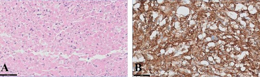
Fig. 3. (A) Histological appearance of the histiocytic sarcoma although the original tumor site in the elbow was completely
characterized by marked anisocytosis and anisokaryosis. H&E excised, the tumor had already metastasized to the draining
stain. (B) Positive immunohistochemical staining of neoplastic lymph nodes and lungs. Many multimodal therapies, includ-
cells for CD18. Scale bars = 50 µm. ing surgical resection, chemotherapy, and radiation therapy,
have been reported and may be helpful in improving the
prognosis [10].
CD18-positive cells and low to moderate number of vimen- Localized HS with destructive lesions involving multiple
tin-positive cells (Fig. 3B). On the basis of the histological bones of a joint and a periarticular soft-tissue mass is uncom-
criteria and immunohistochemical features, a final diagnosis mon in dogs and is of unknown etiology. PAHS should be
of PAHS was made. The dog showed good general health considered an additional differential diagnosis in middle-aged
during 4 months after the surgery, but showed weight loss to older predisposed dog breeds, including Rottweiler, pre-
and intermittent vomiting and dyspnea for 1 month. There- senting with lameness when radiography reveals an aggres-
fore, the owner elected euthanasia. sive joint lesion. Additional diagnostic evaluations described
Localized HSs that develop from a single region are in this case report can be performed to make a definitive
locally invasive and tend to metastasize to draining lymph diagnosis.
nodes. Most localized HSs are reported in the subcutaneous
tissues of the extremities [2]. The tumor masses often invade Acknowledgments
the deep dermis and underlying skeletal muscles and fascia.
Furthermore, localized HS can develop from a periarticular This study was supported by the Basic Science Research
region. Previous studies have suggested that the association Program through the National Research Foundation of Korea
with a joint is because the tumor can develop from the den- (NRF) funded by the Ministry of Science, ICT & Future
dritic cells of the synovial lining [3, 7]. Planning (NRF-2015R1C1A1A01051759) and 2016 Research
In a review of joint tumors in dogs, PAHS was three times Grant from Kangwon National University (No. 520160533).
more common than was synovial cell sarcoma, especially in
the area of the stifle joint [4]. Furthermore, an association References
between PAHS in the stifle joint and previous cranial cruci-
ate ligament disease or previous joint disease has been reported 1. Abadie J, Hédan B, Cadieu E, De Brito C, Devauchelle P,
[4]. Only few reports have documented PAHS in the elbow Bourgain C, Parker HG, Vaysse A, Margaritte-Jeannin P,
joint; this could be because of the characteristics of the elbow Galibert F, Ostrander EA, André C. Epidemiology, pathology,
joints that show relatively few ligamentous diseases [4]. In and genetics of histiocytic sarcoma in the Bernese mountain
dog breed. J Hered 2009, 100 (Suppl 1), S19-27.
this case, the tumor site was the elbow joint, which was an
2. Affolter VK, Moore PF. Localized and disseminated histiocytic
uncommon site, and the only clinical sign was that of fore-
sarcoma of dendritic cell origin in dogs. Vet Pathol 2002, 39,
limb lameness and pain, without previous elbow joint disease. 74-83.
HS is usually observed in middle-aged Bernese Mountain 3. Argyle DJ, Brearley MJ, Turek MM. Decision Making in
Dogs, Rottweilers, Golden Retrievers, and Flat-Coated Retriev- Small Animal Oncology. 1st ed. pp. 161-170, Ames, Wiley-
ers. Although the patient’s signs can lead to a suspicion of Blackwell, 2008.
HS, diagnostic evaluation is necessary for confirming the 4. Craig LE, Julian ME, Ferracone JD. The diagnosis and
diagnosis. Determining the exact sublineages of dendritic prognosis of synovial tumors in dogs: 35 cases. Vet Pathol
cells from which HSs arise is not yet possible. However, 2002, 39, 66-73.
immunohistochemical evaluation using CD18 and vimentin 5. Fidel J, Schiller I, Hauser B, Jausi Y, Rohrer-Bley C,
revealed the origin of the tumor cells, and was very helpful Roos M, Kaser-Hotz B. Histiocytic sarcomas in flat-coated
retrievers: a summary of 37 cases (November 1998–March
in differentiating these tumors from osteosarcoma, synovial
2005). Vet Comp Oncol 2006, 4, 63-74.
cell sarcoma, and malignant fibrous histiocytomas [6, 8].
6. Fulmer AK, Mauldin GE. Canine histiocytic neoplasia: an
With the recent development of limb-sparing techniques overview. Can Vet J 2007, 48, 1041-1043, 1046-1050.
using allografts and xenografts, many successful cases of 7. Harasen GL, Simko E. Histiocytic sarcoma of the stifle in
treatment of distal limb neoplastic disease have been reported a dog with cranial cruciate ligament failure and TPLO
[9, 12]. In the present case, although the dog showed good treatment. Vet Comp Orthop Traumatol 2008, 21, 375-377.
general health and an intact shoulder joint, the region with 8. Hasegawa T, Hasegawa F, Hirose T, Sano T, Matsuno Y.
the tumor surrounded the elbow joint and the neoplasm Expression of smooth muscle markers in so called malignant60 Hyeok-Soo Shin, Ye-In Oh, Byung-Jae Kang
fibrous histiocytomas. J Clin Pathol 2003, 56, 666-671. 12. Straw RC, Withrow SJ. Limb-sparing surgery versus
9. Liptak JM, Dernell WS, Ehrhart N, Lafferty MH, amputation for dogs with bone tumors. Vet Clin North Am
Monteith GJ, Withrow SJ. Cortical allograft and endopros- Small Anim Pract 1996, 26, 135-143.
thesis for limb-sparing surgery in dogs with distal radial 13. Voegeli E, Welle M, Hauser B, Dolf G, Flückiger M.
osteosarcoma: a prospective clinical comparison of two [Histiocytic sarcoma in the Swiss population of Bernese
different limb-sparing techniques. Vet Surg 2006, 35, 518- mountain dogs: a retrospective study of its genetic predisposi-
533. tion]. Schweiz Arch Tierheilkd 2006, 148, 281-288. German.
10. Rassnick KM, Moore AS, Russell DS, Northrup NC, 14. Wellman ML, Davenport DJ, Morton D, Jacobs RM.
Kristal O, Bailey DB, Flory AB, Kiselow MA, Intile JL. Malignant histiocytosis in four dogs. J Am Vet Med Assoc
Phase II, open-label trial of single-agent CCNU in dogs with 1985, 187, 919-921.
previously untreated histiocytic sarcoma. J Vet Intern Med 15. Withrow SJ, Page R, Vail DM. Withrow and MacEwen’s
2010, 24, 1528-1531. Small Animal Clinical Oncology. 4th ed. pp. 818-821,
11. Shortman K, Liu YJ. Mouse and human dendritic cell Philadelphia, WB Saunders, 2007.
subtypes. Nat Rev Immunol 2002, 2, 151-161.You can also read