Breast Cancer Tumor Stroma: Cellular Components, Phenotypic Heterogeneity, Intercellular Communication, Prognostic Implications and Therapeutic ...
←
→
Page content transcription
If your browser does not render page correctly, please read the page content below
Review

Breast Cancer Tumor Stroma: Cellular Components,
Phenotypic Heterogeneity, Intercellular
Communication, Prognostic Implications and
Therapeutic Opportunities
Noemi Eiro 1,*, Luis O. Gonzalez 2, María Fraile 1, Sandra Cid 1, Jose Schneider 3 and
Francisco J. Vizoso 1,4,*
 1 Research Unit, Fundación Hospital de Jove, Avda. Eduardo Castro, 161, 33290 Gijón, Spain;
   investigacion@hospitaldejove.com (M.F. and S.C.)
 2 Department of Anatomical Pathology, Fundación Hospital de Jove, Avda. Eduardo Castro, 161,

   33290 Gijón, Spain; a.patologica2@hospitaldejove.com
 3 Department of Obstetrics and Gynecology, Universidad Rey Juan Carlos, Avda. de Atenas s/n, 28922,

   Alcorcón, Madrid, Spain; jose.schneider@urjc.es
 4 Department of Surgery, Fundación Hospital de Jove, Avda. Eduardo Castro, 161, 33290 Gijón, Spain

 * Correspondence: noemi.eiro@gmail.com (N.E.); investigacion@hospitaldejove.com (F.J.V.);
   Tel.: +34-9853-200-50 (N.E. and F.J.V.)

 Received: 12 April 2019; Accepted: 8 May 2019; Published: 13 May 2019

 Abstract: Although the mechanisms underlying the genesis and progression of breast cancer are
 better understood than ever, it is still the most frequent malignant tumor in women and one of the
 leading causes of cancer death. Therefore, we need to establish new approaches that lead us to
 better understand the prognosis of this heterogeneous systemic disease and to propose new
 therapeutic strategies. Cancer is not only a malignant transformation of the epithelial cells merely
 based on their autonomous or acquired proliferative capacity. Today, data support the concept of
 cancer as an ecosystem based on a cellular sociology, with diverse components and complex
 interactions between them. Among the different cell types that make up the stroma, which have a
 relevant role in the dynamics of tumor/stromal cell interactions, the main ones are cancer
 associated fibroblasts, endothelial cells, immune cells and mesenchymal stromal cells. Several
 factors expressed by the stroma of breast carcinomas are associated with the development of
 metastasis, such as matrix metalloproteases, their tissular inhibitors or some of their regulators like
 integrins, cytokines or toll-like receptors. Based on the expression of these factors, two types of
 breast cancer stroma can be proposed with significantly different influence on the prognosis of
 patients. In addition, there is evidence about the existence of bi-directional signals between cancer
 cells and tumor stroma cells with prognostic implications, suggesting new therapeutic strategies in
 breast cancer.

 Keywords: CAFs; MMPs; TIMPs; cytokines; TLRs; mesenchymal stromal cells; exosomes; integrins

1. Introduction
     Breast cancer is the most frequent malignant tumor in women and the leading cause of cancer
death, since 30% of breast cancers develop distant metastases after the initial treatment of the
apparently localized tumors [1]. Nowadays, the mechanisms underlying the genesis and
progression of breast cancer are better understood [2], but despite an improvement of the survival
rates for some molecular subtypes of breast cancer, we are still far from a cure for all patients [3].
Cancers 2019, 11, 664; doi:10.3390/cancers11050664                            www.mdpi.com/journal/cancers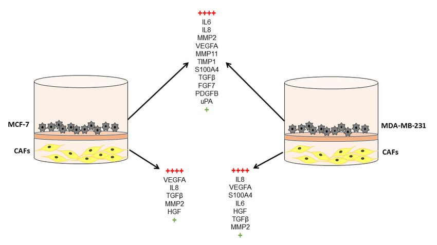
Cancers 2019, 11, 664                                                                           2 of 26

Furthermore, classical (chemotherapy and radiation therapy) or new therapeutic strategies (selective
targeting of oncogenes, immune toxicity or oncolytic virotherapy), are far from satisfactory and
often associated with significant side effects, including collateral damage, drug resistance, immune
toxicity or virus mutability and unexpected toxicity. It seems increasingly clear that the old dogma
of cancer based only on a malignant transformation of the epithelial cells is too simplistic, and a new
concept considering cancer as an ecosystem based on a cell sociology and the tumor-stroma
crosstalk, is gaining strength. A better knowledge of the role of tumor stroma and its interaction with
cancer cells can lead us to implement new prognostic tools or new therapeutic strategies aiming at a
disruption of the dynamics of tumor-stroma interactions.
     In the present work, we reviewed several key pathophysiological aspects related to tumor stroma
and breast cancer progression, their clinical implications and possible therapeutic opportunities.

2. Tumor Stroma Components
      The tumor stroma consists of the non-malignant cells of the tumor mass. Among the various
cell types that make up the tumor stroma, and play a key role in tumor-stroma interactions, we
mainly find resident cells such as cancer-associated fibroblasts (CAFs), endothelial cells and
perycites, blood derived cells such as immune cells, and mesenchymal stroma cells which may be
resident or attracted by the tumor itself and sometimes, by adipocytes surrounding it [1].

2.1. Cancer-Associated Fibroblasts (CAFs)
      Cancer-asssociated-fibroblasts (CAFs) are one of the most abundant cell types in breast cancer
stroma with a well recognized role in the initiation and progression of tumor progression. The CAF
population derives from resident fibroblasts, but CAFs can also stem from other origins, including
mesenchymal stromal cells (MSCs), epithelial cells, pericytes, adipocytes and endothelial cells [2].
CAFs differ from normal fibroblasts by showing a different gene expression profile and promoting
cancer cell aggressiveness [3–5].
      However, although it has been proposed that contractile forces exerted by CAFs can alter the
organization and the physical properties of the basement membrane (interface of epithelium and
stroma), making it permissive for cancer cell invasion [6], the role of CAFs in tumor progression is
more complex. CAFs show a high proliferation rate and can induce the degradation and remodeling
of the extracellular matrix (ECM), epithelial mesenchymal transition (EMT) activation, angiogenic
shift, metabolic reprogramming toward a reverse Warburg phenotype, or promote stem cell trait
achievement, as compared with normal fibroblasts [7–9]. The influence of CAFs is effected through a
paracrine function by means of the secretion of growth factors and cytokines [10–13], such as IL-1β,
IL-6, IL-8, SDF-1, and NFκB, in order to induce immune cell recruitment that may contribute to
tumor progression [14,15]. CAFs are not only able to promote cancer progression but also cancer
survival, by means of the creation of a “protective niche” that maintains residual tumor cell survival,
such as through the induction of resistance to cancer therapy. In this sense, secretion of hepatocyte
growth factor (HGF) and IL-6 by CAFs has been related to lapatinib resistance in HER2+ breast
cancer [16] and tamoxifen resistance [17], respectively.

2.2. Immune Cells
     The immune system plays a complex role in tumorigenesis [18] and immune cells, along with
CAFs, are one of the main cell populations making up the tumor mass in invasive breast carcinomas.
Tumor-infiltrating leukocytes have been historically considered to be manifestations of an intrinsic
defense mechanism against developing tumors [19] and also, subsequently, interpreted as an
aborted attempt of the immune system to reject the tumor. However, nowadays, it is well known
that leukocyte infiltration can promote tumor growth, angiogenesis and tumor cell invasion [20,21],
due to the secretion of cytokines, growth factors, chemokines and proteases [22–24].
     The immune cell infiltrate includes a variable representation of leukocytes, including
macrophages, neutrophils, mast cells, T- and B-lymphocytes [20]. Breast carcinomas may have
Cancers 2019, 11, 664 3 of 26
different types of immune cell infiltrate able to control tumor growth. In this sense, the infiltration of
macrophages, also named tumor-associated macrophages (TAMs) and known to have pro-tumoral
functions, has been associated with a worse prognosis [20,25,26]. Macrophages can be polarized into
two phenotypes: classically activated (M1) macrophages and alternatively activated (M2)
macrophages driven by a cytokine repertoire of T helper cells (Th1 or Th2). M1 has been established
as a tumor-suppressive phenotype and M2 as a tumor-promoting phenotype, considering that
TAMs are primarily M2 polarized. On the other hand, the presence of both T- and B-cells in the
microenvironment has been related to an immunological response, inhibiting cancer development
and progression [27–35]. In this sense, a subgroup of CD4+ T helper cells, known as regulatory T cells
(Tregs), has been associated with the suppression of T-cell immunity. Forkhead box protein P3
(FoxP3) is a transcription factor able to induce the immunosuppressive function of Tregs, being the
most specific marker for Tregs in tumors. Due to the ability to inhibit anti-tumor immunity,
tumor-infiltrating Foxp3+ Tregs were associated with poor prognosis [36], but recently, it has been
shown that Foxp3+Tregs could improve survival in some tumors [37,38]. In a previous study, we
described the clinical relevance of the relative amount of macrophages (CD68+), T-cells (CD3+) and
B-cells (CD20+) at the invasive front of breast carcinomas. Thus, an increased CD68 count and CD68 /
(CD3 + CD20) ratio were both directly associated with a higher probability of shortened relapse-free
survival [39]. Nevertheless, the prognostic significance of the immune cell infiltrate in the tumor
microenvironment remains controversial, perhaps due to non-standardized evaluation. Therefore, it
is important, and possible as described below, to identify new factors as markers of immune stromal
cells associated with tumor aggressiveness. Due to the important role of the host immune system in
cancer, immune checkpoint inhibitors have garnered attention during the last years, especially
against cytotoxic T-lymphocyte antigen-4 (CTLA-4) and programmed cell death protein 1 (PD-1) or
its ligands (PDL-1) [40].
2.3. Endothelial Cells
Endothelial cells (ECs) are ubiquitous within tumors and necessary for the development and
functionality of vessels, especially blood vessels, essential to supply oxygen and nutrients for tumor
progression. The endothelial barrier maintains vascular and tissue homeostasis but its alteration
leads to vascular permeability and drives tumor-induced angiogenesis, blood flow disturbances,
inflammatory cell infiltration and tumor cell extravasation. In addition, regardless of perfusion, ECs
can regulate tumor growth through the secretion of paracrine factors, thereby increasing
tumorigenicity, stemness and invasiveness [41–44]. All of these effects are due to the crosstalk
between tumor and ECs, leading to the initiation of angiogenesis [45] and also to the development of
an abnormal phenotype of ECs, which can be different depending on tumors. Thus, for example, it
has been reported that ECs from highly metastatic tumors have a more proangiogenic phenotype
than those from low metastatic tumors. [46].
2.4. Mesenchymal Stromal Cells
Mesenchymal stromal cells (MSCs) are adult multipotent stromal cells, which exhibit certain
common properties, ranging from the expression of surface markers (CD73, CD90, CD105), to
self-renewal capability and differentiation into osteoblasts, chondrocytes and adipocytes [47,48].
MSCs preferentially reside in perivascular niches of nearly all neonatal human tissues and organs.
MSCs are recruited to sites of injury where their functions are extremely diverse and depend on the
tissue-specific origins and the microenvironment in which MSCs are embedded. Migration of MSCs
towards the inflammation site leads to cellular interactions that occur both directly via gap junctions,
membrane receptors and nanotubes and indirectly via soluble structures and factors. In
physiological situations, MSCs contribute to support tissue repair, stem cell homeostasis and
immunomodulation. MSCs interact with the surrounding cells by means of the secretion of soluble
factors, such as cytokines or growth factors, with activities that can dramatically alter the key cellular
functions of neighboring cells, such as survival, apoptosis, maturation and differentiation [49].Cancers 2019, 11, 664 4 of 26
However, MSCs and their paracrine-based activity also have a relevant role in the
tumor-stroma crosstalk [50,51], since MSCs themselves can be stimulated by tumor cells to develop
an aberrant tumor-associated phenotype [52]. Tumor cells can recruit MSCs to the tumor site using
chemotactic factors such as MMPs, growth factors and inflammatory cytokines [53]. In turn, MSCs
can release paracrine signals, including cytokines, chemokines and growth factors [51], to stimulate
neighboring cells with pro- and/or anti-tumorigenic activities. Thereby, MSCs, having the capability
to migrate to the tumor site [54,55], display several pro-tumor functions, such as the promotion of
tumor growth [56] and angiogenesis [57–59], epithelial-to-mesenchymal transition (EMT) induction
[60,61], as well as alteration of the extracellular matrix [62,63] to promote the migration and
implantation of tumor cells to metastatic sites [54,64]. All of these effects, in their turn, are attained
mainly by means of the secretion of pro-inflammatory cytokines [65], the suppression of immune
effector cells [66–71], the expansion of immune regulatory cells [66,70,72] and increasing resistance to
cancer therapies [73,74].
Additionally, although as previously mentioned, MSCs may be a source of CAFs, MSCs from
the tumor microenvironment can also transdifferentiate into M2-type macrophages, myeloid-derived
suppressor cells (MDSC) or M2-type macrophages under the influence of cytokines or chemokines
[75–77].
3. Stroma Phenotype Associated with Tumor Metastasis
During the past 10 years, our group has investigated the relationship of different factors
expressed by stromal cells associated with breast cancer metastasis. These factors include matrix
metalloproteases (MMPs) and their tissue inhibitors (TIMPs), cytokines and Toll-like receptors (TLRs).
Based on these data, we can propose two types of breast carcinoma stroma with significantly
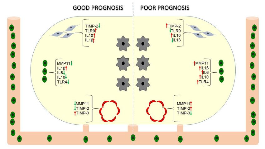
different influences on prognosis (Figure 1).
Figure 1. Stroma phenotype and prognosis. Prognostic significance of factors expressed by
cancer-associated fibroblasts (CAFs, ), mononuclear inflammatory cells (MICs, ) and
endothelial cells ( ) in breast cancer (tumor cells, ).
3.1. Metalloproteases and Their Inhibitors
The human MMP family is composed by 26 members divided into six groups based on
substrate specificity and homology (Table 1). MMPs play an essential role in the degradation of the
stromal connective tissue and basement membrane components, which are key elements in tumorCancers 2019, 11, 664 5 of 26
invasion and metastasis. MMPs are also able to influence in vivo tumor cell behavior due to their
capacity to cleave growth factors, cell surface receptors, cell adhesion molecules and
chemokines/cytokines [78,79]. Furthermore, by cleaving proapoptotic factors, MMPs produce a more
aggressive phenotype through the generation of cells resistant to apoptosis [78]. MMPs also
positively regulate cancer-related angiogenesis, through their ability to mobilize or activate
proangiogenic factors (bFGF, VEGF, TGFβ and integrin αvβ3) after the degradation of basement
membrane or ECM components and negatively via generation of angiogenesis inhibitors, such as
angiostatin and endostatin [80]. MMP activity is specifically inhibited by TIMPs, but it is now
accepted that TIMPs are multifactorial proteins also involved in the induction of tumor cell
proliferation and the inhibition of apoptosis [81,82]. The role of MMPs in ECM remodeling is mainly
due to their capacity to degrade the ECM, thus allowing tumor progression. In this sense, MMP-7
(matrilysin 1), with the capacity to degrade type IV collagen, fibronectin and laminin, is highly
expressed in human breast tumors, inducing tumor growth and invasiveness [83]. MMP-9
(gelatinase B) is related to tumor invasion and metastasis by its special capacity to degrade type IV
collagen [84] and to induce angiogenesis [78]. MMP-11 (stromelysin-3) is preferentially expressed by
peritumoral stromal cells and is associated with tumor progression and poor prognosis [85–87].
MMP-13 (collagenase-3), due to its wide substrate specificity compared with other MMPs, plays a
central role in the MMP activation cascade [88]. MMP-14 (membrane type 1 MMP or MT1-MMP) is
involved in ECM degradation, activation of MMP-13 and MMP-2 zymogen, and in molecular
carcinogenesis, tumor cell growth, invasion and angiogenesis.
Breast carcinomas containing mononuclear inflammatory cells (MICs) or CAFs with a high
expression profile of MMPs and TIMPs have a higher rate of distant metastasis development
compared with tumors with a low expression profile [86,87,89,90], independently of luminal or
basal-like phenotype of breast carcinomas [91]. However, variations in MMP/TIMP expression
among the different histological subtypes of breast carcinomas (ductal, lobular, mucinous, tubular,
papillary and medullary invasive carcinomas) have been found [92]. In a more recent study, we
found that MMP-11 (also known as stromelysin 3) expression by MICs, and TIMP-2 expression by
CAFs, either at the tumor center or at the invasive front, were the most potent independent
prognostic factors for predicting the clinical outcome of patients [87]. Considering that the
expression of MMP-11 may constitute a useful biological marker for pro-metastatic MICs, we
investigated its relationship with the gene expression of 65 factors associated with inflammation and
tumor progression in a population of breast cancer patients stratified into two groups according to
MMP-11 expression by intratumoral MICs (positive or negative). Among all analyzed factors,
interleukin 1β (IL-1β), IL-6, IL-17, interferon β (IFNβ) and nuclear factor kappa B (NFκB) were
expressed at high levels in tumors with MMP-11 positive MICs [93,94]. It has been evidenced that
MMPs can either promote or repress inflammation by the direct proteolytic processing of cytokines
to activate, inactivate, or antagonize their function. For example, IL-1β requires proteolytic
processing for activation. Indeed, the IL-1β-converting enzyme (ICE, nowadays known as caspase-1)
needs the activity of MMPs, such as MMP2, -3, and -9, to activate the IL-1β precursor to the active
form [95].
We also found that MMP/TIMP expression by endothelial cells (ECs) from the adjacent
non-neoplastic tissue was absent or very low compared with ECs from the tumor itself. In addition,
MMP-11 expression by ECs was related to distant metastasis development and shorter relapse-free
survival, whereas, conversely, TIMP-3 expression was related to low occurrence of distant
metastasis [96]. These results support our previous data indicating that the expression of MMP-11 by
stromal cells is associated with distant metastasis development in breast cancer. Regarding TIMP-3,
there are data in agreement with our own findings indicating that TIMP-3 is a naturally occurring
inhibitor of angiogenesis that limits vessel density in the vascular bed of tumors and curtails tumor
growth [97,98]. In addition, it has been reported that TIMP-3 may induce apoptosis in ECs by
triggering a caspase-independent cell death pathway [99].Cancers 2019, 11, 664 6 of 26
Table 1. Human matrix metalloproteases.
Name of Class MMP Enzyme Name Substrates
Collagens (I, II, III, VII and X), proteoglycans,
MMP-1 Collagenase-1
entactin, ovostatin, MMP-2, MMP-9
Collagenase-2/neutrophil Collagens (I, II, III, VII, VIII and X), fibronectin,
MMP-8
collagenase proteoglycans
Collagenases
Collagens (I, II, III, VII, VIII and X), tenascin,
MMP-13 Collagenase-3 plasminogen, aggrecan, fibronectin,
osteonectin, MMP-9
MMP-18 Collagenase-4 Type I collagen
Gelatin, collagen (IV, V, VII VI, IX and X),
MMP-2 Gelatinase-A
elastin, fibronectin
Gelatinases Collagens (IV, V, VII, X and XIV), gelatin,
MMP-9 Gelatinase-A entactin, elastin, fibronectin, osteonectin,
plasminogen, proteoglycans
Collagens (IV, V and IX), gelatin, aggrecan,
MMP-3 Stromelysin-1 laminin, elastin, casein, osteonectin, fibronectin,
ovostatin, entactin, plasminogen
Collagens (I, II, IV and V), gelatin, casein,
Stromelysins MMP-10 Stromelysin2
elastin, fibronectin
Collagens (IV, V, IX and X), laminin, elastin,
MMP-11 Stromelysin2
fibronectin, casein, proteoglycans
MMP-17 Homology tostromelysin-2 Pro-MMP2, fibrin/fibrinogen, gelatin
Collagens IV, gelatin, fibronectin, laminin,
MMP-7 Matrilysin
elastin, casein, transferrin
Matrilysins
Collagen IV, fibronectin, fibrinogen, gelatin,
MMP-26 Matrilysin-2
pro-MMP9
Collagens (I, II, III), gelatin, fibronectin, laminin,
MMP-14 MT1-MMP
vitronectin, entactin, pro-MMP2
Fibronectin, gelatine, vitronectin, entactin,
MMP-15 MT2-MMP
laminin, pro-MMP-2
MT-MMP
Collagen III, gelatin, casein, fibronectin, pro-
(membrane MMP-16 MT3-MMP
MMP-2
type-MMP)
MMP-17 MT4-MMP Pro-MMP2, fibrinogen, gelatin
MMP-24 MT5-MMP Fibronectin, pro-MMP2, proteoglycans, gelatin
Pro-MMP2, pro-MMP9, collagen IV, gelatine,
MMP-25 MT6-MMP
fibronectin, Proteinase A
Collagen IV, gelatin, elastin, casein, fibronectin,
MMP-12 Macrophage metalloelastase
vitronectin, laminin, entactin, fibrin/fibrinogen
MMP-19 RASI-1 Collagen (I, IV) gelatin, fibronectin, laminin
MMP-20 Enamelysin Amelogenin, aggrecan
Other enzymes MMP-21
MMP-22
MMP-23 Gelatin
MMP-28 Epilysin
MMP-29 Unnamed
3.2. Cytokines
Cytokines are low-molecular-weight proteins that mediate cell-to-cell communication. Immune
and stromal cells, such as fibroblasts and endothelial cells, synthesize cytokines and regulate
through them several processes, such as proliferation, cell survival, differentiation, immune cell
activation, cell migration and death. Besides the central role of cytokines in the inflammatory
process, they have also been recognized as powerful players in tumor progression through several
pathways, including the generation of free radicals that can damage DNA, potentially causingCancers 2019, 11, 664 7 of 26
mutations that lead to tumor initiation, stimulating cell proliferation and reducing apoptosis,
stimulating EMT and angiogenesis or allowing tumor cell evasion of immune surveillance. On the
other hand, cytokines can modulate an anti-tumoral response that seems conditional on the balance
of pro- and anti-inflammatory cytokines, their relative concentrations, cytokine receptor expression,
the activation state of surrounding cells [100] and the stage of tumor development [101].
Although previous reports have shown several positive associations between high cytokine
levels and tumor aggressiveness, most of them were based on serum levels and few of them
evaluated the impact on tumor recurrence [102–104]. We have reported high cytokine expression by
cancer cells, supporting the recognized fact that cancer cells secrete cytokines that can act as
autocrine factors contributing to their malignant phenotype. We also found different profiles of
cytokine expression by stromal cells related with patient outcome [105]. Indeed, IL-1β expression by
stromal cells (CAFs and MICs) was significantly associated with both low metastasis rate and longer
relapse-free survival and overall survival, whereas IL-10 expression by stromal cells was
significantly associated with higher metastasis occurrence and both shorter relapse-free survival and
overall survival. In addition, the combination of IL-1β, IL-6 and IL-10 expression by MICs showed an
important association with prognosis and improved the prognostic significance of MMP-11 status by
MICs [105]. All these data seem to indicate that the non-expression of pro-inflammatory cytokines
(such as IL-1β and IL-6), together with the expression of an anti-inflammatory cytokine (such as
IL-10) could contribute to tumor immune escape. IL-10 is an important anti-inflammatory cytokine
and due to its immunosuppressive effect on dendritic cells and macrophages, IL-10 can dampen
antigen presentation, cell maturation and differentiation, allowing tumor cells to evade immune
surveillance mechanisms [106]. These findings are in agreement with some studies suggesting that
interleukins can inhibit tumor growth [107,108] and that their expression can be correlated with
good prognosis [109,110]. All of these data suggest the complexity of the tumor stroma and the
importance to consider the particular cell type expressing cytokines in the context of the tumor
environment.
3.3. Toll-Like Receptors (TLRs)
TLRs play an essential role in the activation of innate and adaptive immunity, contributing to
the capacity of the immune system to combat pathogens [111]. Cancer cells activated by TLR signals
may also release cytokines and chemokines that in turn may recruit immune cells and stimulate
them to release further cytokines and chemokines, resulting in a cytokine profile associated with
immune tolerance, tumor cell proliferation and resistance to apoptosis; but it also enhances tumor
cell invasion and metastasis by regulating metalloproteases and integrins [112–114]. However, there
are data supporting the importance of TLR expression by stromal cells in tumor behavior. In this
sense, we have shown that TLR4 expression by MICs was associated with an increased incidence of
metastasis, whereas TLR9 expression by CAFs was associated with a low metastasis-rate [115],
pointing towards a protective role of TLR9 against tumor progression. Indeed, it has been shown
that stimulation of TLR9 activates human plasmacytoid dendritic cells and B cells, inducing a potent
innate immune response, in preclinical tumor models as well as in patients [116]. TLR9 ligands
induce the expression of OX40 on CD4+ T cells in the tumor microenvironment. OX40, a
co-stimulatory molecule belonging to the TNFR superfamily expressed on activated effector T cells
and regulatory T cells, can promote effector T cell activation and inhibit regulatory T cell function,
which may contribute to the eradication of spontaneous malignancy by local immunotherapy [117].
3.4 Integrins
Cell adhesion to the ECM is fundamental for tissue integrity and basic behavioral responses of
cells under physiological conditions. Integrins are the main cell adhesion receptors for components
of the ECM. The name integrin refers to the function of family members to integrate cell exteriors
(e.g., ECM) to the cell interiors (e.g., the cytoskeleton) [118]. They are a family of 24 transmembrane
heterodimers generated from a combination of 18α integrin (with a size of 120–170 kDa) and 8β integrin
(with a size of 90–100 kDa) chains, connected by monovalent bonds. Both α and β subunits possess aCancers 2019, 11, 664 8 of 26
large extracellular domain, a transmembrane domain and usually a small cytoplasmic tail [119]. Thus,
both integrin subunits are required for interactions with the cytoskeleton and the ECM [120]. Integrins
markedly differ from each other in ligand specificity and expression levels in mammalian tissues.
The most common and vital for mammalian cells are fibronectin-binding integrin α5β1, the
collagen-binding receptor α2β1, and integrin αvβ3, having a diverse ligand specificity.
The role of integrins, primary receptors for ECM and bi-directional signaling molecules, is
complex because they bind to some matrix proteins and to other receptors at the same time.
Integrins act as sensors of the epithelial microenvironment by affecting cells in two key ways:
regulating the actin cytoskeleton of cells through binding directly with proteins (filamin, talin and
vinculin) [121], or by phosphorylating the relative kinases (focal adhesion kinases (FAKs),
proto-oncogene tyrosine-protein kinase (Src)-family kinases (SFKs) and integrin-linked kinase (ILK), to
activate or cooperate with the other cell signaling pathways including the PI3K/Akt and MAPK/Erk
pathways [122,123]. Thus, they transduce information (“outside-in”) from the extracellular
environment to modulate cell responses, including survival signaling, growth signaling, adhesion,
spreading, migration, secretion of proteases and invasion [124,125]. For all of these reasons, integrins
do not just bind a cell to its environment, but they have also been shown to regulate several
intracellular signaling pathways, rendering their physiological role far more complex [118]. Indeed,
dysregulated integrin-mediated adhesion and signaling is a precursor in the pathogenesis of many
human diseases, including cancer [126].
4. Intercellular Communication System in the Tumor Microenvironment
The bi-directional signaling between cancer cells and stroma, mainly composed by CAFs and
MICs, induces the expression of pro-tumoral factors by both tumor compartments [127–129]. These
signals can be mediated by soluble factors, exosomes or via integrins.
4.1. Soluble Factors
Cancer cells secrete cytokines and chemokines, such as TGF-β, involved in the recruitment and
activation of CAFs [128,130]. Furthermore, cytokines and chemokines secreted by cancer cells and
CAFs, such as CCL2, contribute to the recruitment of macrophages and the induction of their
transformation into tumor-associated macrophages (TAMs) [131–133]. In this sense, it has been
reported that the oncogenic dysregulation of the RAS, MYC and the MAPK pathways in cancer cells
contributes to the secretion of growth factors and cytokines such as VEGF, IL-6, IL-10, and IL-1β,
leading to the recruitment and the tumorigenic transformation of macrophages [134,135].
Nevertheless, after the recruitment of stromal cells in the tumor microenvironment, a complex
dynamic interaction takes place, which can be evidenced by transwell-type experiments. Thus, it
was demonstrated that after co-culture of CAFs and breast cancer cell lines (both MCF-7 and
MDA-MB-231 cell lines), gene expression of factors related to tumor progression was significantly
upregulated in both cell types (Figure 2) and invasion and angiogenic capacities of breast cancer
cells were increased. Among all analyzed factors, S100A4, FGF7, PDGFB, VEGFA, TGFβ, IL6, IL8,
uPA, MMP2, MMP11 and TIMP1 are the more differentially expressed (Table 2) [13,136].
Additionally, the expression of some of these factors [13,136] was highly increased after co-culture of
breast cancer cell lines with CAFs from the primary tumor and MIC-MMP11+ cells [136], which
suggests a strong pro-tumor influence of the immune original microenvironment of the CAFs.Cancers 2019, 11, 664 9 of 26
Figure 2. Gene expression of factors after co-culture between CAFs and breast cancer cell lines
(MCF-7 or MDA-MB-231).
Table 2. Main role of factors implicated in the crosstalk between cancer cells and CAFs.
Gene Symbol Gene Name Main Role
S100A4 S100 calcium binding protein A4 Invasion
FGF7 Fibroblast growth factor 7 Cell growth/Invasion
PDGFB Platelet-derived growth factor beta Angiogenesis
VEGFA Vascular endothelial growth factor A Angiogenesis
TGFβ Transforming growth factor beta Inflammation/Invasion
IL6 Interleukin 6 Inflammation
IL8 Interleukin 8 Inflammation
uPA Urokinase-type plasminogen activator ECM remodeling
MMP2 Matrix metalloproteases 2 ECM remodeling
MMP11 Matrix metalloproteases 11 ECM remodeling
TIMP1 Tissue inhibitor of metalloproteases 1 ECM remodeling
4.2. Exosomes
Although soluble factors, chemokines and cytokines produced within the tumor are mainly
responsible of modifications in cancer cells and stromal cells, new evidence indicates the
contribution of another mode of cell communication involving extracellular vesicles (EVs). EVs,
resulting in a particulate nano-communication system that may be responsible for dissemination of
messages among the different cell types of the tumor, are divided into different categories
depending on their size: apoptotic bodies (1000–5000 nm), microvesicles (500–1000 nm in diameter)
and exosomes (30–150 nm) [137]. Exosomes, the smallest subset of EVs, originate in the endocytic
compartment of the parent cell via a series of intraluminal invaginations taking place in the
multivesicular bodies (MVBs). Consequently, their molecular content recapitulates the content of the
parent cell, at least partially [138]. Exosomes are enclosed by a protein-phospholipid bilayer
membrane interspersed by cell type-specific proteins, lipids and glycans. The exosome lumen is
filled with various cellular proteins, nucleic acids, mRNA, miRNA and DNA, soluble factors,
including cytokines and chemokines, enzymes and cofactors [139]. Exosomes produced by different
cell types carry distinct molecular and genetic components, and they may be “addressed” by the
parent cell to reach a specific molecular address of the recipient cell. Upon contacting a local or
distantly-located recipient cell, exosomes deliver signals that culminate in cellular re-programming
[140,141]. The mechanisms responsible for delivery and processing of the exosome cargo in recipient
cells are not entirely understood, but may include the initial ligand-receptor type of binding on the
cell surface followed by endocytosis or phagocytosis of exosomes [142].Cancers 2019, 11, 664 10 of 26
4.3. Tumor-Derived Exosomes (T-D-EXs)
Tumor cells are important producers of exosomes with molecular signatures depending on the
type of tumor cell, which are different from those of exosomes derived from normal cells [143,144].
Tumor-derived exosomes (T-D-EXs), ubiquitously present in the tumor environment and in body
fluids of patients with cancer [58,143], circulate and disseminate information to tissues and cells
distant from the tumor. T-D-EXs carry messages from the parent tumor cell to other normal or
malignant cells in the tumor microenvironment, including MSCs [145]. Thus, T-D-EXs can mediate
autocrine, juxtacrine and paracrine signaling that the tumor cells establish and that is necessary for
their survival in the tumor microenvironment [146] (Figure 3).
Figure 3. Paracrine communication between cancer cells and tumor microenvironment through
exosomes.
4.4. MSC-Derived Exosomes (MSC-D-EXs)
Under normal physiological conditions, MSCs are a rich source of exosomes [64], which seem
responsible for many functions generally attributed to MSCs [147], such as their contribution to the
modulation of physiological functions of neighboring stromal cells [148]. It is known that
MSC-derived exosomes (MSC-D-EXs), through spontaneous or organelle-mediated release,
significantly contribute to the increase in extracellular ATP levels [149], which induces a favorable
effect in migration and invasion of breast cancer cells [150].
Importantly, MSC-D-EXs have the capacity to interact with multiple cell types in the tumor
microenvironment and to ensure they adequately support tumor growth. MSC-D-EXs carry a
complex cargo of molecules and genes comprising more than 850 unique gene products and more
than 150 different miRNAs [151,152] and thus have the potential to elicit different cellular responses
in a broad variety of cells [153]. Nevertheless, MSCs are recognized as recipients of signals
emanating from the tumor, but also as efficient producers of their own and abundant exosomes
[66,153]. Thus, exosomes horizontally transfer information to neighboring cells and transform the
cellular milieu to one supporting tumor survival [153]. (Figure 3). It is known that T-D-EXs induceCancers 2019, 11, 664 11 of 26
phenotypic and functional changes in MSCs which may exert profound effects on tumor growth
[154], and that epigenetic modifications mediated by genetic elements introduced by exosomes to
recipient MSCs appear to be involved [155]. At present, the cellular, molecular and genetic
mechanisms responsible for re-programming of MSCs by T-D-EXs are under intense scrutiny. The
initial contact may be due to T-D-EXs carrying numerous cell adhesion molecules (CAMs) whereby
they can readily fuse with adhesion receptors on MSCs allowing for the protein/gene transfer to the
MSCs cytosol. It remains unclear whether the protein transfer alone is sufficient for the
re-programming of MSCs by T-D-EXs or whether the transfer of transcription factors and nucleic
acids is mandatory. These changes in MSCs include the overexpression of genes involved in cell
migration (CXCR4 and CXCR7), in the matrix remodeling (collagen type IV alpha 3 chain) and in
angiogenesis or tumor growth (IL-8, OPN and myeloperoxidase) [156]. T-D-EXs from prostate
cancer, breast cancer or chronic lymphocytic leukaemia can promote MSC migration to the tumor
site [55] and induce MSC differentiation into myelofibroblasts overexpressing alpha smooth muscle
actin (αSMA) [65].
4.5. Integrins and Cancer
Alterations of integrin expression and function, linked to many types of cancer [157,158], lead to
modifications which are the basis of tumor progression, such as dedifferentiation, proliferation,
apoptosis or disorganization of the ECM and promotion of metastasis [122,157,159–163]. Therefore,
integrins have been implicated in different steps of cancer progression, such as cancer initiation and
proliferation, local invasion and intravasation, survival of circulating tumor cells (CTCs),
extravasation and metastatic colonization [163].
Regarding breast cancer, experimental data implicate integrins such as β1 integrins, and more
specifically α3β1 integrin, in mammary tumorigenesis [164,165]. Likewise, aberrant expression of β1
integrin in human breast carcinoma has been linked to cell adhesion, angiogenesis, tumor
progression and metastasis [166–168]. In agreement with these findings, there are clinical data
demonstrating that integrin overexpression, such as that of β1 integrin [167–169] or integrin α6 [170],
are associated with a poor prognosis and reduced survival, or with radiation resistance [171].
Nevertheless, the functions of individual integrins in invasion and metastasis processes are
controversial. Thus, for example, it has been reported that inhibition of β1 integrin significantly
reduces the formation of metastatic foci of several cancer types, including breast cancer [172]; or that
α2β1 suppressed metastatic dissemination in a mouse model of spontaneous breast cancer [173]. In
addition, some studies have reported that decreased β1 integrin protein expression is associated
with more aggressive breast cancer types [174,175], whereas other studies could not verify a
significant correlation between integrin subunit β1 (ITGB1) protein expression and survival of
patients with breast carcinoma [176,177]. These contradictory data may be due to the diversity of the
integrin family and the variety of signal pathways that a particular receptor can induce in different
cells, as well as to differences between the different tumor histotypes in signaling pathways initiated
by integrins. Therefore, it may be of key importance to consider the role of integrins in the context of
tumor stroma heterogeneity, representing an exciting new way to explore the complexity of
biological systems.
4.6. Possible Role of Integrins to Better Characterize the Tumor Stroma Phenotype in Breast Cancer
Integrins are widely expressed by many types of cells, including tumor cells, endothelial cells
(ECs), pericytes, fibroblasts and immune cells. Therefore, integrins play key roles in other
cancer-relevant processes, such as white blood cell trafficking and activation, chronic inflammation
and angiogenesis, which are strongly related to cancer progression [163]. The contribution of CAFs
to cancer progression and tumor invasion is mediated via several integrin-linked mechanisms
[178,179]. CAFs align fibronectin fibres within the tumor ECM and, through application of traction
forces mediated by α5β1 integrin, promote directional cancer cell migration [180]. Additionally,
CAFs are able to induce tamoxifen resistance by secreting fibronectin, which stimulates its ligandCancers 2019, 11, 664 12 of 26
integrin β1 to activate signaling pathways, such as the PI3K/AKT pathway [181] and long-term
exposure to CAFs makes breast cancer cells addictive to integrin β1 [182].
Although integrin-mediated cell-to-cell and cell-to-matrix interactions during the T-cell lifespan
still represent an open field of research, there are data indicating that integrins in lymphocytes or
macrophages also play a key role to promote tumor progression. Thus, several mechanisms
implicate integrins in the infiltration of lymphocytes and macrophages into tumors. This may be by a
dynamic integration of integrins with several extracellular factors, such as the ECM protein periostin
[183], osteopontin-rich matrix [184], VCAM-1 [185,186] or fibronectin [185]. In addition, it has been
reported that αLβ2 affinity down-modulation is crucial in promoting the intravascular crawling and
diapedesis of T-cells during homing to peripheral lymph nodes [187]. Integrins also generate a signal
that interacts with chemokines and antigens to modulate T-cell motility, proliferation and
differentiation [188]. Endothelial cells use integrins to interact with their underlying basement
membrane, which combined with tyrosine kinase-induced signaling are important regulators of
vessel integrity and tumor progression, since integrin expression both on cancer cells and on
endothelial cells is implicated in extravasation [189].
Interestingly, integrins also regulate proteins, such as MMPs and fibronectin, which play key
roles in the dynamics of the proteolytic activity from tumor stroma and the metastasic cascade.
Integrins contribute to the latter by upregulating the expression of MMP genes and facilitating
protease activation and function at the ECM interface [190]. In addition, integrins, such as αVβ3
cooperate with MMPs, such as MMP-2 [191], MMP-9 [192] and MMP-14 [193], in regulating
migration of metastatic breast cancer cells toward specific substrates in an activation-dependent
pathway. Indeed, it has been demonstrated that integrins control MMP-2 and MMP-9 expression
regulating angiogenesis in breast tumor cells and endothelial cells [194]. In fact, the inhibition of
MMP-9 and αVβ5-integrin interaction results in a reduced angiogenesis and tumor invasion [195].
The arginine (R)-glycine (G)-aspartic acid (D) (RGD) sequence is included in the adhesion
molecule fibronectin, which is the ligand for several types of integrins, such as α5β1, αvβ3 and αvβ5
[124]. Integrins, after binding to fibronectin, activate focal adhesion kinase (FAK), which further
activates multiple signaling proteins, promoting directional cancer cell migration through the
activation of cytoskeletal contractility [178,180]. It is also worth mentioning that integrin-fibronectin
interactions are also implicated in induced exit of cancer cells from dormancy [196,197], as well as in
radiotherapy resistance [198,199]. As was mentioned above, there are new data which implicate
exosomes in playing a vital role in the development of organ-specific metastasis [200], integrins
being the most highly expressed receptors on their surface. These associations might contribute to
support the “seed and soil” hypothesis proposed by Paget more than 100 years ago [201]. This is
because integrins facilitate the binding and fusion of extracellular vesicles to the plasma membrane
of their cell targets [202]. Thus, exosomal integrins can contribute to initiate organ colonization of
specific tissues by preparing favorable pre-metastatic niches to metastatic niche formation
[200,203–205]. With regard to this, it has been shown that integrins αvβ5, α6β4 and α6β1 on
tumor-derived exosomes could drive tumor cells to metastasize to specific organs like lung, liver or
brain [203].
4.7. Tumor Stroma and Therapeutic Opportunities
A consolidated example of targeting stroma in breast cancer are the different strategies
available to target angiogenic cells in clinical trials of advanced breast cancer [206]. In addition,
cancer therapies should aim for a progressive disruption of the dynamics of interactions between
cancer cells and the tumor microenvironment by targeting metabolic dysregulation and
inflammation to partially restore tissue homeostasis and turn on the immune cancer kill switch. The
translation of this therapeutic approach to established treatments would, however, require more
understanding of the adaptive complexity of cancer resulting from the interactions of cancer cells
with the tumor microenvironment and the immune system.
Studies investigating the role of CAFs have reported that the therapeutic targeting of cancer
cells alone is not enough for the treatment of cancer [207]. CAFs are essential components of theCancers 2019, 11, 664 13 of 26
tumor microenvironment and therefore, represent a molecular target for the treatment of cancer
[208]. In addition, compared with cancer cells, CAFs are genetically more stable with a reduced
probability of developing drug-resistance, thus representing a potential therapeutic target with a
lower probability of long-term chemoresistance development [209,210]. Different strategies
implicating CAFs could be developed, such as targeting the ability of CAFs to exert mechanical
forces on the basal membrane [6], or induce the reduction of lactate and steer the tumor
microenvironment to a state of reduced inflammation so as to enable an effective intervention of the
immune system. This is because the probability of dysregulation of the RTK, PI3K and MAPK
signaling pathways is significantly high for most types of cancer. Driven by growth factors from the
stroma, these pathways may, with high probability, be the first drivers of an upregulated glycolysis
in cancer cells. The consequent increase of lactate secretion into the tumor microenvironment will
thereafter lead to its acidification and the activation of TGF-β [211], leading to the recruitment and
transformation of CAFs. On the other hand, several new agents blocking CAF pro-tumor activity
have undergone pre-clinical and clinical evaluation [12,212,213]. Several clinical trials have also been
implemented in order to evaluate inhibitors of cytokine receptors or neutralizing antibodies that
prevent the sustained exposure to these inflammatory mediators that promote tumor progression
[214,215]. Among the most implemented clinical trials for breast cancer in the metastatic setting are
those involving drugs that target immune-cell-intrinsic checkpoints. Blockade of one of these
checkpoints, cytotoxic T-lymphocyte-associated antigen-4 (CTLA-4) or the programmed cell death 1
(PD-1) receptor, may provide proof of concept for the activity of an immune-modulation approach
in the treatment of breast cancer [216]. As it was previously reported, MSC biology is of great
interest for cancer progression and could lead to new therapeutic strategies. Thus, for example, it has
recently been reported that the trophic effect of MSCs on breast cancer cell growth is exerted via
ionotropic purinergic signaling, suggesting the inhibition of the purinergic signaling system as a
potential target for therapeutic intervention [217]. Nevertheless, contradictory results have been
described regarding induced pro- or anti-tumor effects, both in vitro and in vivo [218]. These
discrepancies in MSC functionality may also be due to their heterogeneity and to the fact that no
specific surface markers currently exist to isolate a more homogeneous population. Some data point
towards the importance of the source of MSCs with regard to their tumorigenic properties. Thus, for
example, bone marrow-derived MSCs (BM-MSC-CM) have both anti-tumor effects on non-small cell
lung cancer cells [219] and stimulatory effects on myeloma cells [220], whereas adipose
tissue-derived MSCs conditioned medium (ADSC-CM) has no effect on human glioblastoma cancer
stem cell subpopulations [221]. Therefore, a possible alternative would be to find an MSC-tissue
source providing a unique anti-tumor activity. Regarding this aspect, we recently identified the
human cervix as a new source of MSCs, named human uterine cervical stem cells (hUCESCs), which
are obtained from the transformation zone of the uterine cervix of healthy women by means of Pap
cervical smears [222]. Our results show specific anti-tumor effects of conditioned medium from
hUCESCs (hUCESCs-CM) on proliferation, apoptosis, tumor-cell invasiveness and aggressive
behavior of breast cancer cells in vitro (MDA-MB-231 breast cancer cell lines and primary tumors)
and reduced tumor growth in vivo in a mouse xenograft tumor model, which differ from those
reported for other types of MSC-CM [223–225]. We also explored the effect of hUCESCs-CM
treatment on CAFs and on monocyte to macrophage differentiation [222]. In this sense, we
established that co-culture with hUCESCs as well as treatment with hUCESCs-CM significantly
reduced CAF cell proliferation and invasion, and increased early apoptosis. We also observed that
treatment with hUCESCs-CM significantly inhibited and reversed macrophage differentiation.
These are interesting findings, because the use of secretome and conditioned medium from MSCs is
considered a new therapeutic strategy [226].
The secretome, emerging as a novel type of biological regulation involving the communication
between cells, is defined as the set of factors/molecules secreted to the extracellular space. These
factors include, among others, soluble proteins, free nucleic acids, lipids and extracellular vesicles
including exosomes. The use of cell-free therapies such as MSC-sourced secretome in regenerative
medicine provides key advantages over stem-cell based applications [226]: its application resolvesCancers 2019, 11, 664 14 of 26
several safety considerations potentially associated with the transplantation of living and
proliferating cell populations (immune compatibility, tumorigenicity, emboli formation and the
transmission of infections); it may be evaluated for safety, dosage and potency in a manner
analogous to conventional pharmaceutical agents; its storage can be performed without application
of potentially toxic cryopreservative agents for a long period without loss of product potency; it is
more economical and more practical for clinical application since it avoids invasive cell collection
procedures; its mass-production is possible through tailor-made cell lines under controlled
laboratory conditions; etc. Our study revealed that hUCESCs-CM had a higher concentration than
control media or ASCs-CM of factors such as LIGHT (or TNFSF14), Fms-related tyrosine kinase 3 ligand
(FLT-3 ligand), interferon-gamma-inducible protein-10 (IP-10) and latency-associated protein [222].
These factors have been associated with induction of apoptosis, inhibition of cell growth, reduced
cell invasion and tumor inhibition. It is also noteworthy that, compared to ASCs-CM, hUCESCs-CM
contains lower levels of factors known to participate in cancer progression, such as epidermal
growth factor receptor (EGFR), fibroblast growth factor (FGF) 4 and 9, Intercellular adhesion
molecule 3 (ICAM3), IL6, IL6R, monocyte chemotactic protein-3 MCP3 (also named CCL7),
macrophage migration inhibitory factor (MIF), sgp130 and vascular endothelial growth factor D
(VEGFD) [222]. This, again, indicates that the properties of MSCs and their secretomes vary
depending on their source. All of these findings related to the anatomical source of hUCESCs [227],
suggest that a complex paracrine signaling network is implicated in the anti-tumor potential of this
MSC type. In addition, recent in vitro data of our group demonstrate that exosomes derived from
hUCESCs show an anti-tumor effect (unpublished data), which is interesting because of the
potential for future therapeutic interventions for cancer control via MSCs-derived exosomes.
On the other hand, integrins may be interesting therapeutic targets because they are expressed
by stromal cells from primary tumors and from metastatic niches (CAFs, immune-cells or
endothelial cells) and on tumor-derived exosomes [163]. In fact, previous studies have demonstrated
that blocking integrins with synthetic peptides, antibodies, or disintegrins, interferes with tumor cell
invasion and metastasis in vitro and in vivo [228,229]. Nevertheless, there is still need for reliable
predictive biomarkers for patient stratification, as well as the correspondent detection tools, such as
antibodies which reliably function in immunohistochemistry performed on formalin-fixed,
paraffin-embedded material [229].
Their development would be a major step forward in the integrin field to translate preclinical
efficacy into a beneficial treatment for patients.
5. Conclusions and Future Perspectives
Studies on the cellular components of breast cancer stroma have identified new biological
markers, such as MMPs, TIMPs, cytokines or TLRs leading to the definition of main phenotypes
with different prognoses. These and other factors, such as exosomes derived from cancer cells,
MSCs and integrins, could allow us in the near future to identify pathways of intercellular
communication leading to new therapeutic strategies in breast cancer.
Studies investigating the role of tumor stromal cells have reported that the therapeutic
targeting of cancer cells alone is not enough for the treatment of cancer. There is evidence indicating
that the tumor microenvironment is a fertile ground for the development of novel therapies with the
potential to augment existing treatment and prevention options. In addition, compared to cancer
cells, stromal cells are relatively stable from the genetic point of view, with a reduced probability of
developing drug-resistance, thus representing a potential therapeutic target with lower chances for
the development of long-term chemoresistance. Several clinical trials have been implemented in
order to evaluate blocking CAFs or inhibitors of cytokine receptors or neutralizing antibodies that
prevent the sustained exposure to inflammatory mediators that promote tumor progression. MSC
biology is also of great interest in cancer progression and could lead to new therapeutic strategies. A
possible alternative would be to find an MSC-tissue source providing a unique anti-tumor activity.
With regard to it, hUCESCs have shown a wide anti-tumor effect against aggressive breast cancer in
both in vitro and in vivo studies, against CAFs and on monocyte to macrophage differentiation inCancers 2019, 11, 664 15 of 26
breast cancer. CM or secretome from hUCESCs emerges as an interesting and potential anticancer
cell-free therapy in regenerative medicine, providing key advantages over stem-cell based
applications, resolving several safety considerations potentially associated with the transplantation
of living and proliferative cell populations, and carrying several technical advantages for its
application. Finally, considering that integrins are key players in metastasis and are expressed by
stromal cells from primary tumors and from metastatic niches and on tumor-derived exosomes,
targeting integrins may be an important innovative therapy to explore when considering future
therapeutic options based on tumor stroma activity blockage.
Author Contributions: N.E., L.O.G., M.F. and S.C. analyzed the bibliography and prepared the tables and
figures. N.E., J.S. and F.J.V. designed the project and wrote the manuscript. All authors reviewed the
manuscript.
Funding: This study was supported by Instituto de Salud Carlos III (PI17/02236) to F.J.V., and by Fundación
para la Investigación en Células Madre Uterinas (FICEMU).
Conflicts of Interest: The authors declare the following competing interests: F.J.V. and N.E. are co-inventors of
a patent (“Human uterine cervical stem cell population and uses thereof”) owned by GiStem Research, of which
N.E., L.O.G., S.C., J.S. and F.J.V. are shareholders. The founding sponsors had no role in the design of this
review, in the collection, analyses, or interpretation of data, in the writing of the manuscript, or in the decision
to publish the results.
References
1. Balkwill, F.R.; Capasso, M.; Hagemann, T. The tumor microenvironment at a glance. J. Cell Sci. 2012, 125,
5591–5596, doi:10.1242/jcs.116392.
2. Anderberg, C.; Pietras, K. On the origin of cancer-associated fibroblasts. Cell Cycle 2009, 8, 1461–1462,
doi:10.4161/cc.8.10.8560.
3. Zhu, C.Q.; Popova, S.N.; Brown, E.R.; Barsyte-Lovejoy, D.; Navab, R.; Shih, W.; Li, M.; Lu, M.; Jurisica, I.;
Penn, L.Z.; et al. Integrin alpha 11 regulates IGF2 expression in fibroblasts to enhance tumorigenicity of
human non-small-cell lung cancer cells. Proc. Natl. Acad. Sci. USA 2007, 104, 11754–11759,
doi:10.1073/pnas.0703040104.
4. Nakagawa, H.; Liyanarachchi, S.; Davuluri, R.V.; Auer, H.; Martin, E.W., Jr.; de la Chapelle, A.; Frankel,
W.L. Role of cancer-associated stromal fibroblasts in metastatic colon cancer to the liver and their
expression profiles. Oncogene 2004, 23, 7366–7377, doi:10.1038/sj.onc.1208013.
5. Walter, K.; Omura, N.; Hong, S.M.; Griffith, M.; Vincent, A.; Borges, M.; Goggins, M. Overexpression of
smoothened activates the sonic hedgehog signaling pathway in pancreatic cancer-associated fibroblasts.
Clin. Cancer Res. 2010, 16, 1781–1789, doi:10.1158/1078-0432.CCR-09-1913.
6. Glentis, A.; Oertle, P.; Mariani, P.; Chikina, A.; El Marjou, F.; Attieh, Y.; Zaccarini, F.; Lae, M.; Loew, D.;
Dingli, F.; et al. Cancer-associated fibroblasts induce metalloprotease-independent cancer cell invasion of
the basement membrane. Nat. Commun. 2017, 8, 924, doi:10.1038/s41467-017-00985-8.
7. Koontongkaew, S. The tumor microenvironment contribution to development, growth, invasion and
metastasis of head and neck squamous cell carcinomas. J. Cancer 2013, 4, 66–83, doi:10.7150/jca.5112.
8. Madar, S.; Goldstein, I.; Rotter, V. ‘Cancer associated fibroblasts’—More than meets the eye. Trends Mol.
Med. 2013, 19, 447–453, doi:10.1016/j.molmed.2013.05.004.
9. Erez, N.; Truitt, M.; Olson, P.; Arron, S.T.; Hanahan, D. Cancer-Associated Fibroblasts Are Activated in
Incipient Neoplasia to Orchestrate Tumor-Promoting Inflammation in an NF-kappaB-Dependent Manner.
Cancer Cell 2010, 17, 135–147, doi:10.1016/j.ccr.2009.12.041.
10. Kalluri, R.; Zeisberg, M. Fibroblasts in cancer. Nat. Rev. Cancer 2006, 6, 392–401, doi:10.1038/nrc1877.
11. Allen, M.; Louise Jones, J. Jekyll and Hyde: The role of the microenvironment on the progression of cancer.
J. Pathol. 2011, 223, 162–176, doi:10.1002/path.2803.
12. Mao, Y.; Keller, E.T.; Garfield, D.H.; Shen, K.; Wang, J. Stromal cells in tumor microenvironment and
breast cancer. Cancer Metastasis Rev. 2013, 32, 303–315, doi:10.1007/s10555-012-9415-3.
13. Gonzalez, L.; Eiro, N.; Fernandez-Garcia, B.; Gonzalez, L.O.; Dominguez, F.; Vizoso, F.J. Gene expression
profile of normal and cancer-associated fibroblasts according to intratumoral inflammatory cells
phenotype from breast cancer tissue. Mol. Carcinog. 2016, 55, 1489–1502, doi:10.1002/mc.22403.You can also read