Decoding cancer's camouflage: epithelial-mesenchymal plasticity in resistance to immune checkpoint blockade
←
→
Page content transcription
If your browser does not render page correctly, please read the page content below
Lotsberg et al. Cancer Drug Resist 2020;3:[Online First]
DOI: 10.20517/cdr.2020.41
                                                                                                                       Cancer
                                                                                                               Drug Resistance

Review                                                                                                                         Open Access

Decoding cancer's camouflage: epithelial-
mesenchymal plasticity in resistance to immune
checkpoint blockade
Maria L. Lotsberg1,*, Austin Rayford1,2,*, Jean Paul Thiery1,3,4,5,6,7, Giuliana Belleggia8, Stacey D’Mello
Peters1, James B. Lorens1,2, Salem Chouaib3,9, Stephane Terry3,10, Agnete S. T. Engelsen1,3
1
 Centre for Cancer Biomarkers and Department of Biomedicine, University of Bergen, Bergen 5009, Norway.
2
  BerGenBio ASA, Jonas Lies vei 91, Bergen 5009, Norway.
3
  INSERM UMR 1186, Integrative Tumour Immunology and Immunotherapy, Gustave Roussy, Fac. de Médecine - Univ. Paris-Sud,
Université Paris-Saclay, Villejuif 94805, France.
4
  Cancer Science Institute of Singapore, National University of Singapore, Singapore, Singapore 117599, Singapore.
5
  Department of Biochemistry, Yong Loo Lin School of Medicine, National University of Singapore, Singapore, Singapore 119228,
Singapore.
6
  Institute of Molecular and Cell Biology, Agency for Science, Technology and Research, A-STAR, Singapore, Singapore 138673,
Singapore.
7
  Guangzhou Regenerative Medicine and Health, Guangdong Laboratory, Guangzhou 510005, China.
8
  School of Medicine, Clinical Skills Assessment Program, University of Connecticut, Farmington, CT 06030, USA.
9
  Thumbay Research Institute of Precision Medicine, Gulf Medical University, Ajman 4184, United Arab Emirates.
10
   Université Paris-Saclay, INRAE, AgroParisTech, GABI, Jouy-en-Josas 78350, France.
*Equal contribution.

Correspondence to: Dr. Agnete S. T. Engelsen, Centre for Cancer Biomarkers and Department of Biomedicine, University of
Bergen, Jonas Lies vei 91, Bergen 5009, Norway. E-mail: agnete.engelsen@uib.no

How to cite this article: Lotsberg ML, Rayford A, Thiery JP, Belleggia G, Peters SDM, Lorens JB, Chouaib S, Terry S, Engelsen AST.
Decoding cancer’s camouflage: epithelial-mesenchymal plasticity in resistance to immune checkpoint blockade. Cancer Drug
Resist 2020;3:[Online First]. http://dx.doi.org/10.20517/cdr.2020.41

Received: 12 Jun 2020 First Decision: 14 Jul 2020 Revised: 11 Sep 2020 Accepted: 17 Sep 2020 Available online: 12 Oct 2020

Academic Editor: Gerhard Hamilton Copy Editor: Cai-Hong Wang Production Editor: Jing Yu

Abstract
Epithelial-mesenchymal plasticity (EMP) of cancer cells contributes to cancer cell heterogeneity, and it is well
established that EMP is a critical determinant of acquired resistance to cancer treatment modalities including
radiation therapy, chemotherapy, and targeted therapies. Here, we aimed to explore how EMP contributes to
cancer cell camouflage, allowing an ever-changing population of cancer cells to pass under the radar of our
immune system and consequently compromise the effect of immune checkpoint blockade therapies. The ultimate
clinical benefit of any combination regimen is evidenced by the sum of the drug-induced alterations observed in
the variety of cellular populations composing the tumor immune microenvironment. The finely-tuned molecular
crosstalk between cancer and immune cells remains to be fully elucidated, particularly for the spectrum of

                       © The Author(s) 2020. Open Access This article is licensed under a Creative Commons Attribution 4.0
                       International License (https://creativecommons.org/licenses/by/4.0/), which permits unrestricted use,
    sharing, adaptation, distribution and reproduction in any medium or format, for any purpose, even commercially, as long
    as you give appropriate credit to the original author(s) and the source, provide a link to the Creative Commons license,
    and indicate if changes were made.

                                                                                                                          www.cdrjournal.com
Page 2 Lotsberg et al . Cancer Drug Resist 2020;3:[Online First] I http://dx.doi.org/10.20517/cdr.2020.41 malignant cells along the epithelial to mesenchymal axis. High-dimensional single cell analyses of specimens collected in ongoing clinical studies is becoming a key contributor to our understanding of these interactions. This review will explore to what extent targeting EMP in combination with immune checkpoint inhibition represents a promising therapeutic avenue within the overarching strategy to reactivate a halting cancer-immunity cycle and establish a robust host immune response against cancer cells. Therapeutic strategies currently in clinical development will be discussed. Keywords: Epithelial-to-mesenchymal transition, epithelial-mesenchymal plasticity, immune evasion, tumor immune microenvironment, intrinsic and extrinsic mechanisms of resistance to immune checkpoint blockade, therapeutic opportunity INTRODUCTION The early history of cancer immunotherapy The history of cancer immunology research dates all the way back to 1863 when Rudolf Virchow observed immune cell infiltration in tumors and hypothesized that sites of chronic inflammation served as a hot bed for cancer development[1,2]. Since then, numerous studies have explored the mechanistic link between chronic inflammation and cancer incidence, and tumor-promoting inflammation has been established as a hallmark of cancer[3-6]. However, in 1909, Paul Ehrlich postulated that the immune system could also have a protective role against tumorigenesis[7], and this concept was further developed and presented as the “theory of immunosurveillance” by Burnet and Thomas in the 1950’s[8-11]. Despite the fact that these early studies established a link between cancer and the immune system, the importance of the immune system in cancer progression and the therapeutic possibilities for cancer treatment remained largely neglected for nearly half a century. In fact, the theory of immunosurveillance was considered controversial until an important scientific discovery was published in Nature by Shankaran et al.[12] in 2001. The experimental evidence presented in this paper unambiguously showed that the immune system can, and often does, prevent tumors from developing, and that the immune status of mice is a critical determinant of their susceptibility to tumor development induced by chemical carcinogens[12,13]. A malignant cell may reveal its identity and alert the immune system in various ways. During malignant progression, cancer cells accumulate genetic mutations, which in turn may result in the expression of tumor-specific antigens, also known as neo-antigens. In addition, epigenetic alterations may also deregulate the expression of tumor-associated antigens such as cancer embryonic antigens or cancer testis antigens. Both tumor-specific and tumor-associated antigens can be recognized as foreign by the immune system and initiate immune cell-mediated clearing of the malignant cell population [14,15]. However, the strong selection pressure towards cancer cells that continuously expose tumor antigens and markers of cellular distress enforces a continuous interplay between cancer cells and immune cells, referred to as the process of “immunoediting”[9,13]. As a consequence of this constant selection pressure, malignant cells evolve to escape the immune system, and thus the concept of immunoediting could in part explain what was previously considered a controversy: the immune system may contribute to tumor suppression as well as tumor promotion[13]. Conceptually, the process of immunoediting consists of three sequential phases[9]. In the first phase, the innate and adaptive arms of the immune system work in concert to attack and eliminate the malignant cells (elimination phase). In the second phase, the adaptive arm of the immune system keeps the cancer under control by eliminating some cancer cells while others survive and proliferate, resulting in an equilibrium between dying and proliferating cells in the malignant tissue (equilibrium phase). In this phase, the cells that are still actively dividing will give rise to a population of more or less immunogenic cells, and clonal
Lotsberg et al . Cancer Drug Resist 2020;3:[Online First] I http://dx.doi.org/10.20517/cdr.2020.41      Page 3

expansion of less immunogenic cancer cells results in the third phase of the immunoediting process,
wherein the tumor escapes from the immune response (escape phase). It is believed that malignancies can
remain dormant and asymptomatic in the equilibrium phase of immunoediting for a prolonged period of
time, in some cases even an entire lifetime, unless they can eventually “escape” the immune pressure and
manifest as symptomatic and clinically detectable cancers. This notion is supported by autopsy studies
that have revealed the prevalence of well-differentiated indolent malignancies or “pseudo cancers” of the
thyroid, prostate and mammary glands[16]. The onset of the escape phase may be a result of alterations
in the cancer cells themselves or through alterations in the tumor immune microenvironment (TIME),
leading to an immunosuppressive state[9,13]. As all clinically detectable cancers have successfully escaped
the immune response at least to some degree, the process of immunoediting represents a non-linear model
where the evolution of the malignant cells and the TIME are mutually dependent on each other. The goal of
cancer immunotherapy is to shift the equilibrium of cancer immunoediting from tumor tolerance towards
eradication by modulating the crosstalk between malignant cells and the TIME.
The immune-oncology revolution
William Coley was the first to demonstrate that immune cells can kill cancer cells in patients. Already in
1893, he treated cancer patients with a mixture of bacterial toxins (Coley’s toxins) in a successful attempt to
activate the immune system to attack cancers[17,18]. Another approach to boost the immune system to fight
cancer has been used for decades in the clinical management of early stage high-risk non-muscle-invasive
bladder cancer, where Bacillus Calmette-Guerin, a vaccine originally intended for immunization against
M. tuberculosis, is applied as an intravesical immunotherapy to induce a localized immune response[19,20].
However, immunotherapy did not emerge as a revolutionary cancer therapy for multiple cancer types until
the recent development of immune checkpoint blockade (ICB) therapy. The concept of ICB is to utilize
antibodies to block the receptor-ligand interactions of molecules that serve as negative regulators of the
immune system, such as cytotoxic T lymphocyte-associated protein 4 (CTLA-4) and programmed cell
death protein 1 (PD-1). Work by Allison and colleagues showed that CTLA-4 functions as an immune
checkpoint or “off-switch” by binding CD80/CD86 with greater affinity than the stimulatory “on-switch”
CD28, thereby inhibiting activation and proliferation of T cells[21,22]. Ipilimumab (Yervoy®, Bristol-Myers
Squibb, New York City, NY, US) is a humanized antibody that blocks CTLA-4, thereby reactivating the
immune system. The clinical trials leading to US Food and Drug Administration (FDA) approval of
ipilimumab in 2011 received enormous attention, as it was the first cancer treatment ever to demonstrate an
increased overall survival in metastatic melanoma patients[23].

The interaction between the PD-1 receptor and its inhibitory ligand programmed death-ligand 1 (PD-L1)
is another example of an inhibitory checkpoint modulating the duration and amplitude of the immune
response against cancer cells[24-26]. Interactions between PD-1 and PD-L1 cause inhibition of T cell receptor
(TCR)-mediated lymphocyte proliferation and cytokine secretion[26]. In a physiological context, PD-L1
plays an important role in protecting host cells from immune-mediated tissue damage post-infection and
preventing autoimmune disorders. PD-L1 is frequently overexpressed by malignant cells, allowing cancers
to escape the adaptive immune system[27]. Numerous studies have reported that increased expression
of PD-L1 in tumors is associated with poor prognosis[28-31]. Antibodies blocking the PD-1/PD-L1 axis
have been shown to induce durable clinical responses in numerous cancer types including malignant
melanoma[22,23,32], non-small cell lung cancer (NSCLC) [33,34], urothelial carcinoma[35,36], head and neck
squamous cell carcinoma, Hodgkin’s lymphoma and renal cell carcinoma (RCC)[32-34,37-39]. This has led to
FDA approval of several immune checkpoint inhibitors including the fully humanized monoclonal PD-
L1-targeting antibodies atezolizumab (Tecentriq®, Genentech/Roche, South San Francisco, CA, US) and
durvalumab (Imfinzi®, AstraZeneca, Cambridge, UK), as well as monoclonal antibodies targeting PD-1;
namely pembrolizumab (Keytruda®, Merck Sharp & Dohme, Kenilworth, New Jersey, US) and nivolumab
(Opdivo®, Bristol-Myers Squibb, New York City, NY, US)[35,36,40,41].
Page 4                     Lotsberg et al . Cancer Drug Resist 2020;3:[Online First] I http://dx.doi.org/10.20517/cdr.2020.41

The remaining challenge
ICB truly represents a paradigm shift in cancer treatment[42]; not only do some patients experience
profound clinical benefit from the treatment, but also it has been shown to induce durable responses for a
subset of patients with aggressive malignancies[42,43]. Unfortunately, some of the most common cancers such
as breast, prostate and colon cancer have shown poor responses to ICB[37,44]. Furthermore, even within the
subtypes of cancers for which ICB therapy is indicated, heterogeneous responses have been shown, and a
majority of the patients do not benefit from ICB therapy[42]. Furthermore, heterogeneous responses have
been observed between multiple cancer lesions within the same patient[44]. The European Organisation
for Research and Treatment of Cancer 18071 trial reported that more than half of high-risk patients with
stage III melanoma who received adjuvant ipilimumab relapsed with a median recurrence-free survival of
26.1 months[45]. The KEYNOTE-001 trial reported that one in four patients with metastatic melanoma who
achieved an initial objective response to pembrolizumab subsequently experienced disease progression[45].
However, the reported outcomes of the efficacy of pembrolizumab treatment either alone or in combination
with other treatments for advanced/metastatic melanoma varies considerably in the multiple studies
performed, and a recent meta-analysis including 25 research articles with a total of 2,909 patients reported
an overall response rate of only 34% (progression-free survival: 5.7 months, overall survival 20.3 months)[46].

Multiple mechanisms have been proposed to explain why only a subset of tumors respond to ICB, yet
an urgent lack of reliable predictive biomarkers remains[37,42,47]. High cancer cell expression of immune
checkpoint ligands, high tumor mutational burden (TMB) and a favorable immune cell infiltration in the
TIME, i.e., immune cell inflamed “hot” as opposed to immune cell excluded “cold” tumors, have all been
associated with a more favorable response to ICB[37,48,49]. The best predictive biomarkers identified so far
include the TMB and neoantigen landscape of the tumor; however the adoption and harmonization of
these methods in the clinic remain in their infancy[15,50,51]. For lack of better alternatives, PD-L1 expression
is currently being used as a predictive biomarker for immune checkpoint inhibitors targeting the PD-1/PD-L1
axis in malignant melanoma, NSCLC and RCC, but the usefulness of PD-L1 expression as a predictor of
response to ICB remains a continuous matter of debate[42,52]. Thus, extensive efforts are currently being
made to identify better predictive biomarkers and explore rational combinational therapies, with the
ultimate aim to increase initial ICB response rates while also preventing or reversing the emergence of
resistance[49,53].

In this review, we aim to explore how epithelial-mesenchymal plasticity (EMP), i.e., the acquired ability of
epithelial cells to transition between epithelial and mesenchymal phenotypes, might contribute to cancer
cell camouflage, enabling an ever-changing population of cancer cells to escape the immune system.
Targeting EMP represents a promising therapeutic opportunity in combination with immune checkpoint
inhibitors. Here, we aim to elucidate how EMP affects the various steps of the cancer-immunity cycle
and to highlight some of the clinical evidence and molecular mechanisms supporting the hypothesis that
targeting EMP represents a promising therapeutic avenue within the overarching strategy to reactivate a
halting cancer-immunity cycle and establish a robust host immune response against cancer cells.

MAIN TEXT
Epithelial-mesenchymal transition and plasticity
The majority of human cancers are of epithelial origin[54]. As opposed to mesenchymal cells, which are
characterized by their spindle-shaped fibroblast-like morphology and loose connection to surrounding
cells, epithelial cells exhibit highly organized apicobasal polarity and junctional complexes ensuring
stable intercellular adhesion and controlling transepithelial permeability. The process of epithelial-to-
mesenchymal transition (EMT) was first described in embryonic development[55-57], but was later associated
with physiological and pathological processes occurring in adults such as wound healing, in the progression
of carcinoma and indirectly in tissue fibrosis[58-63]. EMT is a cellular process where epithelial cells lose their
Lotsberg et al . Cancer Drug Resist 2020;3:[Online First] I http://dx.doi.org/10.20517/cdr.2020.41     Page 5

cell polarity and tight cell-cell adhesions, acquiring a fibroblast-like morphology and cytoarchitecture as
well as the ability to remodel the extracellular matrix (ECM) and alter their cellular behavior to gain more
migratory and invasive properties[55,62,64]. Considerable remodeling of the cytoskeleton accompanies the
loss of epithelial cell polarity and confers new mechanobiological properties favoring migration and tissue
invasion[62,63]. At the molecular level, EMT is characterized by downregulation of epithelial markers (e.g.,
E-cadherin and EpCAM), and upregulation of mesenchymal markers (e.g., N-cadherin and vimentin).
Mesenchymal cells also exhibit increased expression of matrix metalloproteinases which are required for
their invasive properties[65,66]. The most commonly recognized EMT transcription factors (TFs) include
Snail (SNAI1), Slug (SNAI2), zinc finger E-box binding homeobox 1 and 2 (ZEB1 and ZEB2), and Twist
family BHLH transcription factor 1[62,67]. Since multiple TFs may activate EMT, different combinations of
TFs may be responsible, at least in part, for the heterogeneity that stems from different versions of the EMT
program[60]. Of note, the EMT International Association suggests that EMT should not be assessed solely
on the basis of EMT-TF expression or that of a small number of molecular markers, but rather the primary
criteria for defining EMT should consist of measurable alterations in cellular characteristics together with a
set of molecular markers[62].

Importantly, EMT is not an irreversible linear process, and the reverse process, mesenchymal-to-epithelial
transition (MET), describes the transition from the mesenchymal (M) to the epithelial (E) state [68].
Although EMT was initially believed to be a binary process, it is now well documented that this is indeed a
dynamic process and intermediate-state cells also exist[61,69]. Cells can thus undergo partial EMT or partial
MET to obtain intermediate E/M states with a cellular phenotype that is neither completely epithelial nor
completely mesenchymal and harbors attributes characteristic of both states[62,69]. Rather than acting as
an “on/off” switch between the E and M states, the process of EMT is therefore considered to generate a
continuum of cell states along an “epithelial-mesenchymal axis”, also frequently referred to as an “EMT
scale” or “EMT spectrum”[60,70]. Tan and colleagues have developed a universal and quantitative gene
expression-based EMT scoring system used to establish and evaluate an EMT spectrum across different
cancer types[69]. Recently, the term “epithelial-mesenchymal plasticity” (EMP) has been increasingly
recognized, referring to the ability of cells to adopt mixed E/M features and to switch between various
states along the epithelial-mesenchymal spectrum[62]. Although carcinoma cells may hijack this conserved
developmental program, its physiological role in maintaining homeostasis is highly regulated both spatially
and temporally, while the pathological EMT program detected in cancers, on the other hand, appears to be
a mainly stochastic and time-independent process[65,66,71]. Of note, non-epithelial cancers such as melanoma
have also been shown to undergo EMT-like programs leading to similar phenotypic changes induced
by EMT-related genes, and serve as a model system for studying metastasis and therapy resistance[72-74].
Phenotypic switching in melanoma involves re-organization of the cytoskeleton, cell membrane and cellular
adhesions, as well as alterations in common EMP-related phenotypic markers including gain of E-cadherin
and loss of N-cadherin expression[72].

Under pathological conditions, epithelial cells are believed to undergo EMT, but they rarely or perhaps
never undergo the full EMT transition to obtain a completely mesenchymal phenotype, suggesting
that partial EMT represents the norm rather than the exception[60,62]. Evidence also indicates that these
intermediate E/M phenotypes have the greatest malignant and metastatic potential[75-77]. Carcinoma cells
can also temporarily transition to a more plastic state to metastasize or overcome selection pressures from
therapy and subsequently revert to an epithelial-like state[75]. Expression of EMT-TFs is also frequently
accompanied by features of stemness. Retention of stemness has been linked to EMT and in particular to a
stemness window around intermediate E/M states[61]. Like EMP, the acquisition of stem cell traits in cancer
cells is not considered a fixed trait; rather it represents a hallmark of cancer within a spatial and temporal
window in cancer progression[61]. Accumulating evidence suggests that morphological phenotype and
stemness can be regulated independently in malignant cells, but that a stemness window along the EMT
axis may predispose malignant cells for acquisition of stem cell traits[61].
Page 6                     Lotsberg et al . Cancer Drug Resist 2020;3:[Online First] I http://dx.doi.org/10.20517/cdr.2020.41

EMP as a mediator of resistance to cancer therapies
Even for the most effective cancer therapies, be it chemotherapy, targeted therapies, radiotherapy or
immunotherapy, persisting cancer cells that survive treatment are believed to constitute a reservoir of slow-
cycling cells that eventually may acquire irreversible genetic mutations or epigenetic alterations causing
therapy resistance and relapse[78-80]. Acquired therapy resistance is one of the major obstacles to achieving
durable remission, and thus major efforts have been made to understand the mechanisms of drug resistance
against targeted therapies as well as more conventional cancer treatment regimens. Historically, the focus
of this research has been to uncover genetic mutations responsible for acquired drug resistance, such as
secondary mutations in the target gene, causing impaired drug binding, or mutations causing activation
of downstream signaling or alternative survival pathways[78,81]. However, recent attempts to characterize
resistance mechanisms to targeted therapies, such as that of third-generation anaplastic lymphoma kinase
(ALK) inhibitor loratinib resistance by next-generation sequencing and phenotypic analysis of longitudinal
tumor samples, have highlighted EMT as a mediator of resistance in cases where a specific mutation
affecting drug binding could not be detected[82]. Furthermore, the EMT-mediated loratinib resistance found
in these patient-derived cell lines could be overcome by combined SRC and ALK inhibition[82]. EMP has
been shown to be widely associated with therapy resistance against both cytotoxic and targeted therapy in
multiple cancer types, and increasing evidence also shows that EMP contributes to a multidrug resistance
phenotype[58,60,71,83]. However, the exact mechanism by which EMP contributes to drug resistance in these
various contexts remains to be fully elucidated. Proposed mechanisms include reduced levels of pro-
apoptotic proteins or increased drug efflux, which may be part of a broader association between EMT-
mediated cancer cell dedifferentiation, acquisition of stem cell traits, and drug resistance[78,83-87]. Although it
has been acknowledged for quite some time that dynamic chromatin modifications may be an independent
route to drug resistance in cancer cells and thus embodies a promising drug target in combination with
various drugs including ICB, it was not until the recent FDA approval of tazemetostat (Trade name:
Tazverik, developer: Epizyme), a first-in-class small molecule inhibitor of enhancer of zeste homolog 2
(EZH2), that this hypothesis has been feasible for clinical testing. Tazemetostat was FDA approved as a
monotherapy in January 2020 for patients with histologically confirmed, metastatic or locally advanced
epithelioid sarcoma with INI1 loss, and was thus the first “epigenetic” drug approved for solid cancers.
EZH2 is considered a master regulator of EMT through orchestrating the regulation of the H3K27me3
epigenetic mark, and in pancreatic cancer it has been shown to regulate EMT through miRNA 139-5p.
Tazemetostat is currently in phase 2 clinical trials in combination with the PD-1 inhibitor TecentriqTM
(atezolizumab) in patients with relapsed or refractory diffuse large B-cell lymphoma (DLBCL), and thus
future combination studies will need to address whether EZH2 inhibitors or other epigenetic drugs can
reduce EMT and increase response to ICB in pancreatic cancer or other solid cancers.

Novel technologies leveraging single cell analysis have only just begun to provide a better mechanistic
understanding of the processes at play. In several recent studies in vitro, drug-tolerant persister cells,
sometimes referred to as “jackpot” cells, have been observed in cancer cell cultures from a variety of tumor
types upon treatment[78,80,88-91]. From single-cell sequencing endeavors, these studies have shown that many
cancers, including melanoma, display a profound transcriptional variety at the single cell level. Through
single-cell gene expression analyses of melanoma cultures, Shaffer and colleagues were able to demonstrate
that non-heritable resistance to the V600E mutated BRAF inhibitor vemurafenib (Zelboraf, Plexxikon and
Genentech) was established from a very rare (1 per 50-500 cells) subpopulation of pre-existing pre-resistant
cells. These cells transiently express a semi-coordinated set of well-known plasticity and resistance markers,
including epidermal growth factor receptor (EGFR), platelet derived growth factor receptor beta, nerve
growth factor receptor, fibroblast growth factor receptor 1, Wnt family member 5A (WNT5A), JUN and
the receptor tyrosine kinase AXL[91]. Treatment with vemurafenib induced a stepwise reprogramming of the
pre-resistant cells into a stable resistant phenotype, which was not affected by drug holidays[91].
Lotsberg et al . Cancer Drug Resist 2020;3:[Online First] I http://dx.doi.org/10.20517/cdr.2020.41      Page 7

AXL expression in melanoma has been associated with resistance mechanisms mediated by mitogen-
activated protein kinase (MAPK)/extracellular signal-regulated kinase (ERK) signaling[92-94]. Tirosh and
colleagues provided strong evidence for the involvement of AXL as well as AXL-related genes (AXL
signature) in acquired melanoma resistance to both vemurafenib and the MEK inhibitor trametinib
(Mekinist, Novartis, Basel, Switzerland), and also found that the proportion of a similar population
of “jackpot” cells as well as the abundance of plasticity gene transcripts significantly increased after
resistance was acquired in vitro, while being selected against more differentiated populations expressing
melanocyte-inducing transcription factor (MITF) and SRY-box transcription factor 10[95]. Taken together,
the accumulating evidence for the role of rare “jackpot” cells in acquired therapy resistance supports the
hypothesis that intervention with cytotoxic or targeted therapies and immunotherapies displaying initial
efficacy in tumor eradication may also act as selection pressures, predisposing tumors to immune evasion
through the acquisition of plasticity phenotypes and priming cells for immune resistance. The adoption of
distinct phenotypes through the process of EMT may provide cells with properties adaptive to changes in
the TIME, and thus act as drivers towards a phenotypic state independent of the drug-targeted pathways[78].

The tumor immune microenvironment as a regulator of epithelial-mesenchymal plasticity
The tumor microenvironment is a key regulator of EMP, which may be induced by combinations of
multiple microenvironmental factors including hypoxia, pH, ECM composition, tensile forces and the
presence of soluble factors such as interferons, inflammatory cytokines and growth factors [61,65,66,86,96-98].
Hypoxia occurs in most actively growing solid tumors and is an important EMT inducer. Accumulating
evidence also suggests that hypoxic stress is linked to therapy resistance as well as regulation of tumor
immunogenicity and both tumor and immune plasticity[99,100]. Hypoxic areas of growing tumors have been
shown to attract immunosuppressive cells such as myeloid-derived suppressor cells (MDSCs), tumor-
associated macrophages (TAMs) and regulatory T cells (Tregs)[101], and hypoxia has also been shown to
upregulate PD-L1 on MDSCs[102,103]. Targeting hypoxia has thus been suggested to improve the efficacy
of cancer immunotherapy[102,104,105]. Remodeling of ECM through secretion of specific matrix MMPs
and new ECM components, namely collagen type 1, has been shown to induce EMT through both
mechanotransduction and direct membrane receptor signaling through integrins and DDR1/2 [106-108].
Cancer-associated fibroblasts (CAFs), in addition to their role in ECM remodeling, serve as one of the
primary sources of key EMT-inducing growth factors including hepatocyte growth factor (HGF), fibroblast
growth factor, interleukin (IL)-6 and transforming growth factor-beta (TGF-β); however, molecular
profiling and ablation of heterogeneous CAF subsets in various tumor models have shown conflicting
and context-dependent effects on the efficacy of different therapies[109]. Thus, for CAFs and other cellular
components of the TIME, the continuum of cells along the differentiation/polarization axis is far more
complex than previously anticipated and remains to be explored further by high-dimensional analyses.

Multiple studies have demonstrated that the TIME strongly contributes to the induction of the EMT
program and subsequent tumor progression[66]. Cells of the TIME that are involved in regulating EMP
include TAMs, MDSCs, neutrophils and natural killer (NK) cells [110,111]. TAMs have a major role in
orchestrating cancer-related inflammation, and pro-inflammatory macrophages have been shown to
induce EMT at the invasive margin of the tumor through inflammatory cytokine tumor necrosis factor-α
(TNF-α)-mediated stabilization of the transcription factor SNAI1 via nuclear factor-κB (NF-kB) pathway
activation, while knockdown of SNAI1 suppressed inflammation-mediated breast cancer metastasis,
suggesting a mechanism for EMT as a regulator of inflammation-induced metastasis[112]. Macrophages
have also been shown to induce EMT in intratumoral cancer cells via TGF-β secretion and activation of
the β-catenin pathway[113]. In a murine melanoma model, MDSCs were shown to infiltrate the tumor and
induce EMT in vivo[114]. Functional in vitro assays with purified MDSCs revealed that the TGF-β, epidermal
growth factor (EGF) and HGF signaling pathways were all involved in MDSC-induced EMT activation of
cancer cells[114]. In a similar model, HGF secretion by T cell-inflamed TIMEs was shown to mobilize and
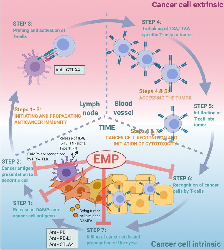
Page 8                     Lotsberg et al . Cancer Drug Resist 2020;3:[Online First] I http://dx.doi.org/10.20517/cdr.2020.41

recruit neutrophils through binding and activation of c-MET, wherein they gained immunosuppressive
properties[115]. Neutrophils are also frequently enriched at the invasive margin of gastric cancers, and in a
recent study, tumor-associated neutrophils were shown by in vitro co-culture experiments to promote EMT
in gastric cancer cells via IL-17a signaling[116]. Although the mechanisms are still unclear, NK cells may
increase the malignancy of melanoma cells by inducing an EMT-like switch[110].
The role of EMP in the cancer-immunity cycle
The finely tuned, step-wise process by which the immune system can efficiently recognize and eradicate
cancer cells was first conceptualized by Chen and Mellman and is now widely known as “the cancer-
immunity cycle”[117]. In the first step of the cancer-immunity cycle, cancer antigens are recognized and
captured by professional antigen-presenting cells (APCs), such as dendritic cells (DCs)[117]. Alarmins or
damage-associated molecular patterns (DAMPs) released by dying tumor cells may aid in the attraction
of APCs to the tumor bed[118,119]. This initiation of the cancer-immunity cycle is dependent on the APCs
capturing both cancer specific antigens expressed by the cancer cells (immunogenicity) as well as signals
that specifically evoke an immune response (adjuvanticity)[117,120]. Toll-like receptors (TLRs) are expressed
on DCs and are key mediators bridging innate and adaptive immunity once stimulated by foreign
pathogen associated molecular patterns (PAMPs) or DAMPs. TLR activation results in the secretion of
pro-inflammatory cytokines including IL-6, IL-12, TNF-α, and type I interferons, which in turn serve to
recruit immune effector cells. The APCs from step one of the cancer-immunity cycle travel from the tumor
microenvironment to the lymph node where they present captured cancer related antigens to T cells (step 2).
T cells can then be activated and programmed to specifically react to these antigens (step 3). Activated
cytotoxic T lymphocytes (CTLs) then travel back to the tumor site (step 4) where they are then able to
infiltrate the tumor (step 5), recognize (step 6) and kill (step 7) the antigen-presenting cancer cells[117]. All
these steps need to be intact for the cancer-immunity cycle to be successfully completed.

As discussed below, EMT may affect several steps of the cancer-immunity cycle and thus hinder
progression in this finely-tuned cycle in multiple ways [Figure 1]. One of the remaining challenges will be
to identify the “Achilles heel” of individual cancer cases and tailor the treatment to initiate a robust cancer-
immunity cycle. We suggest that targeting cancer EMP may represent a unique opportunity to bolster
several steps of the cancer-immunity cycle. Although the molecular mechanisms of EMP affecting the
various steps of the cancer-immunity cycle are still largely obscure, targeting EMP represents a promising
therapeutic addition within the overarching strategy of personalized cancer immunotherapy to re-activate a
halting cancer-immunity cycle and re-establish a robust host immune response against the cancer cells[53,121].

Using hypoxia-inducible factor 1-alpha (HIF1 alpha) and AXL inhibitors as examples, the rationale
behind clinical trials combining ICB and mediators of epithelial phenotypic plasticity are based on the
effects of these drugs on multiple steps of the cycle. First, cancer treatments that induce immunogenic
cell death (ICD) have been shown to contribute to anticancer immune attack, mediated by the exposure
of DAMPs. Hypoxia is a strong microenvironmental factor that supports coordinated induction of EMT
and also AXL expression and autophagy. The HIF1 alpha inhibitor PX-478 was also recently shown to
enhance gemcitabine-induced immune responses and eliminate pancreatic ductal adenocarcinoma (PDAC)
cells through induction of ICD[122]. EMT correlates with autophagy induction, and pre-mortem stress-
related autophagy is also linked to the release of DAMPs and induction of ICD[53,123]. Thus, the impact
of targeting autophagy to increase the efficacy of immune checkpoint inhibition is expected to be seen
particularly in cancers of an immune cold or immune-excluded immunophenotype[49]. We have recently
shown that the small molecule AXL inhibitor bemcentinib abrogates the autophagic flux and is a potent
inducer of ICD; this is of particular interest as it implicates a favorable adjuvant effect and alteration of
the TIME upon AXL inhibition in immunogenic but immunosuppressed tumors[53,124]. In this context,
it is important to note that the increased danger signaling per se is not sufficient to compensate for the
Lotsberg et al . Cancer Drug Resist 2020;3:[Online First] I http://dx.doi.org/10.20517/cdr.2020.41                              Page 9

Figure 1. EMP affects various steps of the cancer-immunity cycle. The therapeutic rationale for targeting EMP in combination with ICB
is based on the fact that EMP affects multiple steps of the cancer-immunity cycle described by Chen and Mellman[117]. Briefly, targeting
EMP can induce an increased release of DAMPs serving as an adjuvant during the release of cancer cell antigens (STEP 1). DAMPs
are further recognized by pattern recognition receptors (PRRs) including TLRs. EMP targeting can induce an M1 to M2 polarization of
macrophages and an activation of APCs, and thus aid in cancer antigen presentation (STEP 2). Targeting EMP and the EMP-associated
immunosuppressive tumor immune microenvironment (TIME) can enable the infiltration of educated T cells into the cancer (STEP 5).
EMP is associated with reduced recognition (STEP 6) and immune effector cell-mediated killing (STEP 7) of cancer cells, and targeting
EMP can therefore induce increased effector cell-mediated lysis of cancer cells and propagation of the cycle. Adapted courtesy of Chen
and Mellman[117]. EMP: epithelial-mesenchymal plasticity; ICB: immune checkpoint blockade; DAMPs: damage-associated molecular
patterns; APCs: antigen-presenting cells
Page 10                    Lotsberg et al . Cancer Drug Resist 2020;3:[Online First] I http://dx.doi.org/10.20517/cdr.2020.41

intrinsically low immunogenicity of some tumors, and that the effect of autophagy-dependent danger
signaling is solely expected to warm up immune cold tumors and elicit a strong immune response in
immunogenic tumors[125]. Increased immunoadjuvanticity, that is, the recruitment of professional APCs
to educate the naïve T lymphocyte population, is critical to maximize the potential therapeutic benefit
of ICB. Furthermore, AXL has been shown to suppress antigen presentation by downregulating major
histocompatibility complex I (MHC-I), and genetic ablation of AXL in the experimental PyMT-induced
tumor model was shown to induce MHC-I expression to even higher levels compared to IFN-γ-induced
parental cells [126]. AXL inhibition enhances cytokine release and increases CD8+ T cell response in
syngeneic models, resulting in an elevated cytotoxic T cell-dependent antitumor immune response after
radiation, which could be further enhanced by ICB[126].

Carcinoma subclones with pronounced mesenchymal phenotypes emerging from sustained hypoxic stress
of primary NSCLC cells in culture were shown to be less susceptible to NK and CTL cell-mediated lysis
compared to subclones with a more epithelial phenotype[96]. This difference was associated with reduced
expression of intercellular adhesion molecule 1 (ICAM1), UL16 binding protein 1 (ULBP1), and MHC-I
as well as increased TGF-β expression in mesenchymal subclones[96]. It was shown that expression of
AXL in the mesenchymal NSCLC clones was correlated with an increased cancer cell intrinsic resistance
to immune cell-mediated killing by NK cells as well as autologous CTLs while AXL targeting via small
molecule inhibition potently sensitized mesenchymal lung cancer cells to cytotoxic lymphocyte-mediated
killing[127]. These findings raise the intriguing possibility that phenotypic plasticity may enable tumor cells to
circumvent NK- and CTL-mediated cell killing. This has also been attributed to the associated cytoskeletal
remodeling in tumor cells, interfering with immune synapse formation[96,128,129]. This process may provide a
rationale to test EMP-targeting approaches to enhance antitumor immunotherapy[95,130,131].

The EMT-TF SNAI1 has been shown to have a strong impact on the cancer-immunity cycle. In human
mammary carcinoma MCF7 cells, Akalay et al.[132] observed that overexpression of exogenous SNAI1
correlated with increased mesenchymal and stemness traits in these cells while reducing their susceptibility
to CTL-mediated lysis. This was in part explained by reduced MHC expression in the SNAI1-transfected
MCF7 derivatives and concomitant reduced activity of TCR signaling at the site of cell-cell contact
(immunological synapse). Similar observations were reported using another MCF-7 derivative displaying
hyperactive TGF-β signaling. Likewise, SNAI1 overexpression in murine B16 melanoma cells resulted in
inhibition of CTL lysis activity, inhibition of DC maturation and expansion of suppressive Treg-like CD4+
Foxp3+ cells in a mechanism involving thrombospondin (TSP1) and TGF-β secretion[130]. Using PyMT
cells, a murine transplantable model of breast cancer carcinoma, Dongre and colleagues showed that
tumors arising with a more mesenchymal phenotype and high expression of SNAI1 expressed more PD-L1
and less MHC-I and had less cytotoxic T cells, but were enriched for immunosuppressive components
including M2 macrophages and Tregs[133]. The link between EMP and the presence of immunosuppressive
macrophages seems to be critical. Although the mechanisms are still unclear, it is tempting to speculate
the involvement of immunosuppressive chemoattractants (e.g., TGF-β) in the recruitment of macrophages
to the TIME. Another interesting observation made by Dongre and colleagues was that NK cells infiltrate
mesenchymal tumors more effectively compared to the epithelial tumors, which can be explained by
downregulation of MHC-I in the more mesenchymal tumors[133]. However, despite this NK enrichment,
the mesenchymal tumors rapidly grow in this model suggesting intrinsic or extrinsic resistance as well as
possible cooperative effects.

Converging preclinical evidence also suggests that tumors with a more EMP/mesenchymal phenotype
are less responsive to ICB. SNAI1+ melanoma tumors were unresponsive to immunotherapy, but
targeting SNAI1 with siRNA or anti-TSP1 monoclonal antibody could inhibit tumor progression and
induce systemic immune responses[130]. In ZEB1/miR-200-driven NSCLC models as well as in SNAI1-
Lotsberg et al . Cancer Drug Resist 2020;3:[Online First] I http://dx.doi.org/10.20517/cdr.2020.41      Page 11

driven breast cancer models, EMP was associated with PD-L1 expression[134]. Targeting PD-L1 in NSCLC
efficiently prevented tumor growth and metastasis. In PyMT breast cancer models, epithelial tumors were
susceptible to elimination by anti-CTLA-4 immunotherapy, whereas corresponding mesenchymal tumors
were refractory to such treatment[133]. By injecting E/M cells at various ratios, these investigators also
elegantly demonstrated that in the case of mixed epithelial-mesenchymal tumors, even the presence of
rare mesenchymal carcinoma cells could protect the epithelial counterpart from anti-CTLA-4 treatment by
promoting the recruitment of immunosuppressive cells including M2 macrophages and Tregs[133].

EMP as a mediator of primary and acquired ICB resistance
Sharma and colleagues have described three different categories of resistance against immunotherapy:
primary, adaptive and acquired resistance[37]. Primary, or innate, resistance to immunotherapy occurs when
the cancer does not have an initial response (i.e., it is refractory) to the therapy. Adaptive resistance occurs
when the tumor is initially recognized by the immune system, but the tumor adapts to escape immune
attack. Given the evolving nature of the interaction between immune cells and cancer cells in this process,
adaptive resistance can manifest clinically as either primary resistance, mixed responses or acquired
resistance. Thus, only clinical data describing the role of EMP in primary and acquired resistance will be
discussed here. Acquired resistance describes the clinical scenario in which the cancer initially responded
to immunotherapy but recurs and progresses after a period of time. However, as Sharma and colleagues
emphasize in their review, the immune response is dynamic and can evolve for each patient and tumor,
and could also be affected by environmental and genetic factors as well as by the cancer treatment[37].
Accordingly, it is important to note that the process of immunoediting mentioned above is anything but
a static process, as novel rounds of therapy and subsequent immunoediting affect the composition of the
TIME through clonal selection acting on pre-existing cancer cell populations or de novo acquisition of
resistant traits.

EMP in primary ICB resistance
The hypothesis that epithelial phenotypic plasticity could be a key determinant of the outcome of
immune checkpoint inhibition in cancer is fueled by pioneer transcriptional data[135]. Hugo and colleagues
performed exome and transcriptomic sequencing of melanoma pre-treatment samples, and a common
gene signature was shown to characterize tumors that were non-responsive to anti-PD-1 therapy[135].
This innate PD-1 resistance (IPRES) gene signature encapsulates the upregulation of EMT transcription
factors, immunosuppressive cytokines, and pro-angiogenic factors[135]. The IPRES signature, enriched in
non-responding patients, overlaps with signatures for wound-healing, angiogenesis, ECM remodeling,
cell adhesion, monocyte/macrophage chemotaxis and resistance to MAPK pathway inhibition[135]. Data
from The Cancer Genome Atlas (TCGA), PROSPECT, and BATTLE-1 showed that inflammatory changes
in the tumor microenvironment were strongly associated with induction of EMT signatures in lung
adenocarcinoma, which in turn correlated with upregulation of multiple suppressive immune checkpoint
receptors or their ligands, including B7-H3, CTLA-4, PD-1, PD-L1, PD-L2, B and T lymphocyte attenuator,
and T cell immunoglobulin and mucin-domain containing-3 (TIM-3)[45]. Taken together, these data suggest
that attenuating the mechanics underlying the IPRES signature may have the potential to enhance anti-
PD-1 responses in melanoma and other cancers. Intriguingly, the receptor tyrosine kinase AXL, whose
upregulation is associated with a reversible plasticity cell state[94] and is part of the “jackpot” signature
of pre-existing pre-resistant cells[91], was found to be a component of the IPRES signature among 532
upregulated genes including several EMT and EMP mediators such as EGFR, NGF, CDH11, EPHA3, HEY1,
HEY (NOTCH effectors), TWIST2, ID3, ID1, many interleukins including immunosuppressive IL10,
ITGA5/8; LOX, LOXL2, MMP1, MMP2, MMP3, MMP9, NRP1, THBS1 and THBS2, NUMBL (plasticity
factor), ROR2, RORB, RUNX2, SEMA3A SERPINE1 VEGFA, VEGFC and Wnt genes WNT11 WNT2
WNT5A WNT7B and DKK3. Among the 161 genes downregulated in the IPRES signature are HLA-A and
E-cadherin (CDH1)[135].
Page 12                   Lotsberg et al . Cancer Drug Resist 2020;3:[Online First] I http://dx.doi.org/10.20517/cdr.2020.41

In line with these findings from melanoma, Thompson and colleagues recently observed that NSCLC
tumors displaying a more mesenchymal phenotype showed reduced clinical responses to ICB[136]. In
this study, an EMT/inflammation-based signature had clinical utility in predicting clinical response[136],
supporting the view that the EMP signature is an adverse predictor of ICB response across tumor types.

Furthermore, in urothelial cancer patients with T cell-infiltrated tumors, a higher EMT/stroma-related
gene expression signature was associated with poor response and disease progression under anti-PD-1
therapy[137]. This study suggested that stromal elements are the major source of immune resistance in this
setting, thus providing a rationale for co-targeting PD-1 and the stromal elements mediating resistance[137].

EMP in acquired resistance to ICB
Potential mechanisms of acquired resistance to immunotherapy include loss of T-cell function,
downregulation of tumor-associated antigen presentation, and acquisition of mutations that enable immune
escape[37]. The IFN-γ pathway plays an important regulatory role at the center stage of primary, adaptive
and acquired resistance to ICB[37]. IFN-γ, which is secreted primarily by T cells and NK cells, is crucial to
initiate an effective antitumor immune response as it mediates increased MHC-I expression, recruitment of
immune cells and direct antiproliferative and pro-apoptotic effects in cancer cells[129]. However, constitutive
IFN-γ exposure might initiate immunoediting and subsequent immune escape by promoting alteration of
the molecules involved in IFN signaling pathways such as Janus kinase/signal transducers and activators
of transcription (JAK/STAT) and induction of EMT[37]. For example, an increased frequency of IFN-γ
pathway gene mutations in tumors of melanoma patients that did not respond to the anti-CTLA-4 antibody
ipilimumab has been shown[37,138]. Mutations in the IFN-γ pathway could also result in lack of IFN-γ-
induced PD-L1 expression causing primary resistance to PD-1 blockade[37,139].

Cancer cell activation of the Wnt/β-catenin pathway or activation of the phosphoinositide 3-kinase (PI3K)/
AKT signaling pathway, through, e.g., acquired loss of the tumor suppressor protein phosphatase PTEN,
have both been shown to regulate EMT, and both signaling pathways have been mechanistically linked
to immune resistance in preclinical studies[140-144]. Trujillo and colleagues performed a molecular analysis
of baseline and treatment-resistant tumor samples from two malignant melanoma patients who initially
showed a durable partial response to either a melanoma-peptide/interleukin-12 vaccine or combined
anti-CTLA-4 + anti-PD-1 therapy and subsequently developed new treatment-resistant metastases.
Transcriptional profiling and genomic sequencing for oncogenic alterations as well as histologic analysis for
T cell infiltration were performed to investigate mechanisms of acquired resistance to immunotherapy[144].
In the first case, the authors found increased tumor cell intrinsic activation of β-catenin in the patient’s
treatment-resistant metastasis, whereas in the second case genomic sequencing revealed acquired biallelic
PTEN loss, while both cases were associated with loss of T cell infiltration[144]. This study highlights that
continued analysis of acquired resistance samples is crucial to identify potentially targetable resistance
pathways amenable to therapeutic intervention.

Expression of IL8 by tumor cells has been shown to promote their EMP[108]. Thus, IL8 and its receptor
CXCR1/2 axis have been established as potential EMP regulating factors and subsequent targeting of
the IL8/CXCR1/2 axis has been explored as a promising EMP target. Preclinical studies have also been
performed combining CXCR1/2 blockade with bifunctional anti-TGF-β RII/PD-L1[145]. In this study, Horn
and colleagues were able to show that simultaneous inhibition of TGF-β, PD-L1 and CXCR1/2 in murine
breast and lung cancers synergized to reverse EMP and associated mesenchymal features, reduce infiltration
by immunosuppressive MDSCs, and enhance T cell infiltration and overall antitumor immune response[145].
Clinical trials: targeting EMP to improve immunotherapy of cancer
Combination therapies being tested that seek to enhance the immune response have been extensively
reviewed elsewhere[49,146]. From the discussion above, it is evident that it is not clear which combination of
Lotsberg et al . Cancer Drug Resist 2020;3:[Online First] I http://dx.doi.org/10.20517/cdr.2020.41                                  Page 13

Table 1. Clinical trials where EMP targets are being evaluated in combination with ICB targeting the PD-1/PD-L1 axis
                                                                                                                          Clinical trial
    Target                          Drug name                Type              Cancer type          Latest phase*
                                                                                                                           number**
    TGF-βRI                       Vactosertib +          Selective TKI       Urothelial                   II             NCT04064190
                                  durvalumab
    TGF-βRI                       LY3200882              Selective TKI       Advanced cancer              II             NCT04158700
    PD-L1/TGF-βRII                Bintrafusp-alfa        AntiPD-L1/          NSCLC                        III            NCT03631706
                                                         TGFbetaRII
                                                         fusion protein
                                                                             Biliary tract                III            NCT04066491
                                                                             Cervical                     II             NCT04246489
    (TYRO3, AXL, MER)/KIT/        Sitravatinib           Pan-TKI             NSCLC                        III            NCT03906071
    VEGFR2
                                                                             ccRCC                        II             NCT03680521
                                                                             Urothelial                   II             NCT03606174
    c-MET/VEGFR2/AXL/RET          Cabozantinib           Pan-TKI             RCC                          III            NCT03937219
    AXL                           Bemcentinib            Selective TKI       NSCLC                        II             NCT03184571
                                                                             Mesothelioma                 II             NCT03654833
                                                                             Breast cancer                II             NCT03184558
                                                                             Melanoma                     II             NCT02872259
*
 Only phase II or later trials are shown. Trials designated as phase I/II are listed as latest phase = II; **Trial identifiers and associated
information obtained from www.clinicaltrials.gov. TKIs: tyrosine kinase inhibitors; NSCLC: non-small cell lung cancer; RCC: renal cell
carcinoma; TGF-β: transforming growth factor-beta; PD-L1: programmed death-ligand 1

the multifaceted characteristics of EMP drives the prominent resistance to ICB, and thus it is also not clear
how EMP may be targeted most effectively in various cancer contexts. The multiple pathways involved
in the initiation and maintenance of epithelial plasticity and the redundancy between these overlapping
pathways makes EMP a challenging target. Attempts are being made to target EMP through the multitude
of soluble factors, receptors, and transcription factors involved. Several combinations with ICB have been
explored in preclinical models, and a few trials have been initiated based on the hypothesis that EMP
is one of the main mediators compromising the efficacy of ICB therapies and that targeting EMP could
lead to stronger and more durable ICB responses [Table 1]. Here, we will specifically highlight promising
therapeutic agents targeting EMP-mediated mechanisms of ICB resistance and discuss them in light of the
available mechanistic data to support their multifaceted mechanism of action within the TIME.

TGF-β is a major regulator of the tumor microenvironment, and also a potent inducer of cancer cell EMT
and immune suppression[61,86,97,98]. TGF-β is being explored as a target for combination with ICB. Recent
retrospective and preclinical reports highlight its role in ICB resistance, while mechanistic and clinical
studies continue to refine targeting strategies and synergy with checkpoint blockade. TGF-β is expressed
in cancer, stromal and immune cells in three isoforms, TGF-β1, β2, and β3. Several pan-cancer gene
expression analyses have shown that TGF-β1 is the primary isoform expressed in tumors, while TGF-β3
is less frequently observed and TGF-β2 is rarely detected and mainly serves to regulate normal cardiac
function and hematopoiesis, which helps explain the failure of earlier pan-TGF-β targeting therapies due
to toxicity-related adverse events[108,147,148]. The most common proposed mechanisms of TGF-β-driven ICB
resistance are CD8+ T cell exclusion via fibroblast differentiation and subsequent stromal remodeling
and inhibition of CD4+ T helper cell differentiation to the immune-activating T-helper (Th)1 effector
cell phenotype, as these conditions have been reproduced in various mouse tumor models and effectively
treated by combined targeting of TGF-β and the PD-1/PD-L1 axis[149-152]. Additionally, anti-TGF-β was
shown to act in synergy with ICB by suppressing immunosuppressive Tregs and EMT in cancer cells in a
murine squamous cell carcinoma (SCC) model[153]. However, not all tumor models respond equally well to
the combination since its efficacy seems to be highly dependent on CTL anti-tumor activity, suggesting that
the most immunogenic tumors are expected to benefit the most from this combination[150].
Page 14 Lotsberg et al . Cancer Drug Resist 2020;3:[Online First] I http://dx.doi.org/10.20517/cdr.2020.41 TGF-β-targeting therapies in combination clinical trials with ICB for advanced solid cancers include the small molecule tyrosine kinase inhibitors (TKIs) vactosertib and LY3200882, which specifically target TGF-βRI signaling via ALK5 inhibition (NCT04158700, NCT04064190, NCT02937272)[108]. The most advanced of these, galunisertib, has been shown to inhibit in vivo cancer cell invasion and metastasis in addition to the mechanisms mentioned above[154]; however, it also inhibits p38α due to its similarity to ALK5, and this may contribute to potentially serious side effects that have caused other TKIs in this class of drugs to fail[155]. It was recently announced that Eli Lilly discontinued the development of galusertinib. However, several alternative TGF-β-targeting strategies are also being tested in the clinic, including AVID200, a novel fusion protein that specifically traps free TGF-β1/3 and has recently shown promising safety in a phase I monotherapy trial[148], and bintrafusp-alfa (formerly M7824), an anti-PD-L1/TGF-βRII fusion protein that functions both as a checkpoint inhibitor and a TIME-localized TGF-β trap. Bintrafusp alfa has shown superior efficacy in several murine models compared to anti-PD-L1 and anti-TGF-β administered both alone and in combination while also reversing TGF-β1-induced tumor cell plasticity[108], prompting multiple clinical trials in various malignancies including lung (NSCLC and SCLC), breast, cervical, and colorectal (CRC) cancers as monotherapy or in combination with radio- or chemotherapy (NCT02517398, NCT03554473, NCT03620201, NCT03524170, NCT03579472, NCT04246489, NCT03436563). Notable among these is the trial in second-line advanced NSCLC (unselected for PD-L1 expression level), which has shown durable responses and encouraging long-term survival while maintaining a manageable safety profile over a two-year follow-up period, prompting a randomized phase III clinical trial vs. pembrolizumab in first-line PD-L1-high NSCLC (NCT03631706)[156]. AXL receptor tyrosine kinase (RTK) has been consistently linked to EMT-mediated drug resistance in a number of cancers[157], and thus represents a promising drug target in this context. AXL expression is prevalent in EMT- and stem cell-related gene expression profiles, highlighting its role in facilitating plasticity phenotypes[95,158-160]. AXL signaling has also been shown to contribute uniquely to both tumor intrinsic and microenvironmental immunosuppression and is initiated by its ligand Gas6 as well as heterodimerization with other RTKs. Antony and colleagues showed that GAS6-AXL signaling network is a mesenchymal molecular subtype-specific therapeutic target in ovarian cancer[161]. Of note, this paper also elegantly demonstrated the crucial role of AXL moving from cholesterol-rich lipid rafts in epithelial carcinoma cells to the normal plasma membrane domain where it multimerizes with other RTKs in mesenchymal carcinoma cells[161]. In a later study, Antony and co-authors were able to demonstrate that the tumor suppressor opioid-binding protein/cell adhesion molecule (OPCML), which is silenced in over 83% of ovarian carcinomas by loss of heterozygosity or by epigenetic mechanisms, is spatially restricted to the cholesterol-rich lipid rafts together with AXL, and when present, represses AXL-dependent oncogenic signaling through protein tyrosine phosphatase receptor type G (PTPRG) in a coordinated manner[162]. This study also serves to illustrate how two spatially restricted tumor suppressors, OPCML and PTPRG, coordinate to repress AXL-dependent oncogenic signaling in the Epi context, and where the loss of lipid raft organization upon EMT could lead to overactivation of AXL and more promiscuous heterodimerization with other RTKs in the M state. Thus, a therapeutic strategy to re-introduce OPCML represents an opportunity to target signaling networks, as opposed to more linear systems[158]. AXL signaling activates downstream MAPK/ERK and PI3K/Akt pathways, which are known to drive cell survival, proliferation, migration and invasion, both of which are crucial for cancer progression and associated with poor patient outcomes[157,163]. AXL inhibition in malignant tumors holds the potential to target AXL-mediated epithelial plasticity, stimulate antigen presentation, and block the recruitment of immunosuppressive macrophages to the TIME [164,165]. As such, several AXL-targeting therapeutics have emerged and are in various phases of clinical development in combination with ICB[108]. Although not initially developed as AXL- targeting agents, pan-TKIs that inhibit a spectrum of RTKs including AXL, most notably cabozantinib and sitravatinib, have shown promising activity in combination with ICB particularly in ICB-naïve renal cell carcinoma (NCT03937219, NCT03680521). The most advanced clinical stage selective small
You can also read