Spheres of Influence: Insights into Salmonella Pathogenesis from Intestinal Organoids - MDPI
←
→
Page content transcription
If your browser does not render page correctly, please read the page content below
microorganisms
Review
Spheres of Influence: Insights into Salmonella
Pathogenesis from Intestinal Organoids
Smriti Verma 1,2, * , Stefania Senger 1,2 , Bobby J. Cherayil 1,2     and Christina S. Faherty 1,2
 1   Mucosal Immunology and Biology Research Center, Division of Pediatric Gastroenterology and Nutrition,
     Massachusetts General Hospital, Charlestown Navy Yard, Boston, 02129 MA, USA;
     ssenger@mgh.harvard.edu (S.S.); cherayil@helix.mgh.harvard.edu (B.J.C.);
     csfaherty@mgh.harvard.edu (C.S.F.)
 2   Harvard Medical School, Boston, 02115 MA, USA
 *   Correspondence: sverma5@mgh.harvard.edu; Tel.: +1-617-726-7991

 Received: 28 February 2020; Accepted: 28 March 2020; Published: 1 April 2020

 Abstract: The molecular complexity of host-pathogen interactions remains poorly understood in
 many infectious diseases, particularly in humans due to the limited availability of reliable and specific
 experimental models. To bridge the gap between classical two-dimensional culture systems, which
 often involve transformed cell lines that may not have all the physiologic properties of primary cells,
 and in vivo animal studies, researchers have developed the organoid model system. Organoids
 are complex three-dimensional structures that are generated in vitro from primary cells and can
 recapitulate key in vivo properties of an organ such as structural organization, multicellularity, and
 function. In this review, we discuss how organoids have been deployed in exploring Salmonella
 infection in mice and humans. In addition, we summarize the recent advancements that hold promise
 to elevate our understanding of the interactions and crosstalk between multiple cell types and the
 microbiota with Salmonella. These models have the potential for improving clinical outcomes and
 future prophylactic and therapeutic intervention strategies.

 Keywords: organoids; enteroids; Salmonella; host-pathogen interactions; model systems; infectious
 diseases; organotypic culture system

1. Introduction
      Many basic life processes are conserved in all organisms, with similarities in molecular mechanisms
often increasing as the phylogenetic distances decrease. This conservation enables investigation into
various biological mechanisms and diseases, with the expectation that the knowledge gained from
one system will provide insight into the workings of related organisms. The similarity between
different organisms is particularly important for studying human biology and diseases, since human
experimentation is typically unfeasible or unethical. However, several factors must be considered before
employing an organism or derived cells as a model for humans, including ease of maintenance and
reproducibility, amenability to genetic and molecular manipulations, and perhaps most importantly,
similarity to the characteristics of the human biological process under investigation.
      The mainstays of research for many decades have been the cell culture and animal model systems.
Classically, the in vitro cell culture methodology involves either growing cell lines that are derived
from tumors or are genetically transformed to allow continuous propagation, or culturing primary
cells derived from the tissues of humans or experimental animals. Use of cell lines has a significant
limitation in that the cells do not recapitulate “normal” cellular physiology. Primary cells closely
represent the tissue of origin but are limited in availability, have a finite life-span in vitro without
being transformed, and display variability arising from differences in donors. Conventional cell

Microorganisms 2020, 8, 504; doi:10.3390/microorganisms8040504          www.mdpi.com/journal/microorganisms
Microorganisms 2020, 8, 504                                                                          2 of 23

culture comprises growing cells (cell lines/primary cells) either in liquid suspension or as adherent,
two dimensional (2D) monolayers on an impermeable solid surface. The cell culture model is also a
reductionist approach since the model cannot account for dynamic and complex interactions that occur
in vivo, particularly those that result from the interactions with multiple cell types. Although processes
such as immune responses, cell signaling, or crosstalk with microbiota have been studied using cell
culture models of various cell types either alone or in combination, the biggest disadvantage of this
system is that the cells may not fully recapitulate in vivo phenotypes and behaviors. More complex
model systems, such as Caenorhabditis elegans, Danio rerio, Drosophila melanogaster, mice, or primates
provide a whole organism perspective and allow compensation for some of the limitations of cultured
cells as the complexity of the models increase. However, these animal models resemble, but do not
completely represent, all the conditions encountered in humans, an issue that is especially important
in the understanding of disease and development [1]. Recently, many researchers have developed
sophisticated, yet tractable, non-cancerous tissue culture models that bridge the gap between simple
2D cell culture and complex in vivo experiments. Such models are broadly known as organotypic
culture systems, which can be composed of various types as detailed below. In general, these models
consist of a three-dimensional (3D) organ-like structure that is made up of organ-specific differentiated
cells of multiple lineages and that recapitulates the unique organizational and functional characteristics
of the corresponding organ in vivo.
      Historically, the term “organoid” referred to short-term in vitro cultures of tissues such as lung
and intestine [2], or 3D cell aggregates such as spheroids. More recently, it has been applied to
organized 3D structures generated in vitro from cells with a whole range of origins, such as tissue
segments [3,4] and their derived adult stem cells (ASCs) [5,6], transformed cell lines [7], and pluripotent
stem cells (PSCs) [8]. However, as research has progressed, the term “organoid” has taken on a more
restricted meaning. Organoids have the following characteristics: (1) self-organization: individual
cells arrange in vitro into a 3D structure that mimics the in vivo organ or tissue, (2) multicellularity:
organoids are composed of multiple cell types typically found in the organ or tissue in equivalent
proportions, 3) functionality: the organoid structure should be able to execute at least some of the
organ- or tissue-specific functions, and 4) sustainability: organoids can propagate indefinitely without
requiring transformation by maintaining a pool of progenitor cells. In 2012, the International Stem Cell
Consortium [9] set guidelines for the nomenclature to be used to define the 3D structures generated
in vitro depending on their origin and cellular composition. When referring to intestinal organotypic
models, 3D structures that are composed of just epithelial cell types are generally known as “enteroids”
if derived from the small intestinal epithelium, or “colonoids” if derived from colonic origin. The term
“organoid” is usually reserved for 3D structures containing more than one cell lineage. However, it
should be noted that these guidelines have not been uniformly adopted by the field. Many researchers
commonly use “organoids” as a blanket term for 3D structures derived from ASCs, PSCs, or comprising
transformed cell lines that resemble in vivo 3D architecture and physiology.
      Methodologies have been adapted to develop organoids from various normal and cancerous
mouse and human tissues including colon [6,10], stomach [11], liver [12–14], pancreas [15], and
kidney [16] to name a few. These techniques have been well reviewed elsewhere [11,17–21]. For the
purposes of this review, we will provide a historical context and examples of the different types of
intestinal organoid models (Figure 1), followed by a discussion of the application of the models to the
study of Salmonella pathogenesis.
Microorganisms 2020, 8, 504 3 of 23
Figure 1. Use of organotypic models in Salmonella biology. We highlight studies that have utilized
organotypic models to better understand Salmonella biology, as well as studies that do not directly
pertain to Salmonella research but have the potential to be deployed for better understanding of the
dynamics of host-Salmonella interactions.
2. Historical Background
The organoid model systems have been established on the basis of several studies that have
contributed to various aspects of the technology [22]. One key advance was the development of cell
culture methods that permitted cells to form 3D structures. Many of the non-physiological properties of
cells in the conventional 2D cell cultures are believed to be due to the loss of 3D geometry [23]. Several
techniques have been developed to deal with this problem, such as the hanging drop method [24],
3D micro-molds [25], and rotating wall vessel (RWV) bioreactors [26], a more advanced method
that permits larger-scale production of 3D assemblies. Nickerson, CA et al. (2001) [26] utilized a
RWV bioreactor developed by NASA that generates low shear and microgravity, allowing cells to
remain in suspension to aggregate and grow three dimensionally. The authors were able to establish a
3D culture of Int-407 cells (derived from a normal fetal intestine but later shown to have HeLa cell
contamination), thereby promoting cellular differentiation. The resulting 3D aggregates modeled
human in vivo differentiation with well-defined cell-to-cell borders, tight junctions, apical-to-basal
polarity, and microvilli development. These features were not present when the Int-407 cells were
grown as conventional monolayer cultures. Thus, the study helped to demonstrate that 3D organization
provides important physical and mechanical cues that facilitate organogenesis. A 3D platform is
generally achieved by using matrices such as Matrigel, which consist of proteins typically found in
the extracellular matrix (ECM). The ECM is the non-cellular component present within all tissues and
organs, and consists mostly of collagen, enzymes, and glycoproteins to provide both structural and
biochemical support to surrounding cells. ECMs have been shown to promote the maintenance of
structural and functional characteristics of the tissue of origin [27]. The other major events that propelled
the development of organoids were advancements in the understanding of stem cell properties andMicroorganisms 2020, 8, 504 4 of 23
developmental biology of the intestine. The defining characteristics of stem cells—i.e., the capacity for
clonal expansion and the ability to differentiate into daughter cells of multiple lineages—have been
vital for the development of organoid technology. In fact, with the right cues daughter cells differentiate
into multiple cell types and undergo two processes (1) cell sorting out—the formation of discrete
domains by different cell types, and (2) spatially restricted lineage commitment, the specialization of
function of a cell based on its position in the tissue architecture [28]. These processes result in complex
3D, self-propagating, multicellular, and functional structures [29]. The improved understanding of
intestinal development helped define some of these cues. Studies have shown that different molecular
gradients converge to create the micro-environment that shapes the intestinal epithelium. Epidermal
growth factor (EGF) and Wingless-related integration site protein (Wnt) are highly active in the crypt
and are necessary for proliferation, while bone morphogenetic protein (BMP) and Notch signaling
pathways active in the villus control cell fate programming [30–32].
Another milestone in the field came in 2007 when Barker and colleagues [33] demonstrated in
adult mice that intestinal crypt stem cells, which are responsible for proliferation and migration of
the epithelial layer towards the villus tip, express the marker leucine-rich repeat containing G-protein
coupled receptor 5 (Lgr5) [33]. Shortly thereafter, Ootani et al. [34] developed a long-term culture of
primary mouse intestines using air-liquid interface methodology [34]. Fragments of neonatal mouse
intestines containing epithelial and mesenchymal cells were cultured to generate cyst-like structures
containing all major cell types found in an adult mouse intestine, including Lgr5+ stem cells that the
authors demonstrated could be modulated by Wnt signaling [34]. The next critical step occurred
in 2009, when Sato and colleagues [5] realized the power of these organ-specific stem cells in 3D
cultures by isolating intestinal crypts from adult mice. The authors demonstrated that a single Lgr5+
cell could form 3D crypt-villus epithelial structures in the presence of appropriate cues, including an
environmental signal from a lamin-rich 3D matrix (Matrigel) and a physiological signal in the form
of growth factors (the Wnt agonist R-spondin1, EGF, and the BMP-inhibitor Noggin) [5]. The model
recapitulated the physiology and organization of the mouse intestine; and when transplanted into
mice, successfully reconstituted crypt-villus units [35,36]. Thus, the adult stem cells (ASCs) develop
self-organized structures that consist of various types of intestinal epithelial cells. The authors referred
to these structures as “organoids”; however, according to the guidelines set by the International Stem
Cell Consortium [9], the model should be called “enteroids” since it was derived from the small
intestinal epithelial cells.
Regardless of the definition, this work laid the groundwork for the utilization of pluripotent
stem cells (PSCs), either derived from embryonic tissue or generated by inducing stemness in mature
somatic cells (iPSCs) [37–40]. The latter offer the additional advantages that invasive procedures to
isolate intestinal or colonic biopsies are not necessary and also iPSCs bypass ethical concerns regarding
procurement of embryonic stem cells. The PSC-derived structures consist of epithelial cells surrounded
by mesenchymal stromal cells and are referred to as “organoids”. During embryonic development,
the mesenchyme induces and specifies epithelial identity and differentiation, and is critical for the
maintenance of tissue identity [41,42]. However, for interrogation of epithelial cell-specific functions,
the mesenchymal cells may serve as a hindrance. Mithal and colleagues [43] have recently identified a
directed differentiation protocol that generates mesenchyme-free human intestinal organoids (HIOs),
which were employed to measure cystic fibrosis transmembrane conductance regulator (CFTR) gene
function using cystic fibrosis patient-derived iPSC lines. The epithelial cells of organoids are not fully
mature and resemble fetal epithelial cells when transcriptionally profiled [44]. The maturation of
these organoids requires implantation in vivo, such as on the kidney capsule of immunocompromised
mice [8,39,44,45]. The fetal-like nature of PSC-derived organoids presents an advantage as it allows
for studies that would otherwise be difficult to conduct given the limited availability and ethical
concerns surrounding the use of human fetal tissues. PSC-derived organoids follow a differentiation
path observed during development [46] and take one to two months to develop, while tissue-specific
ASCs develop organoids by a process that recapitulates tissue repair and is complete within weeks [20].Microorganisms 2020, 8, 504 5 of 23
The epithelial cells derived from ASC (enteroids) represent adult-like mature epithelium. The cells of
the enteroids also preserve characteristics of the tissue of origin including a diseased phenotype if
derived from patients bearing an intestinal condition. These characteristics make enteroids from ASC
a faithful model for intestinal diseases [47–49].
3. Intestinal Organoids/Enteroids
Intestinal enteroids derived from the intestinal stem cells develop multi-lobed structures (Figure 2)
consisting of crypt-villus units, with a clearly defined proliferative, crypt-like zone and a large,
non-proliferative differentiated zone surrounding proliferative areas [50]. To generate organoids, the
pluripotent stem cells are first programmed towards endoderm development to form the epithelial
tube that eventually gives rise to the foregut, midgut, and hindgut in response to a combination
of anteriorizing and posteriorizing growth factors provided. Additional mesenchymal cells are
also present and consist of myofibroblasts and smooth muscle cells surrounding the epithelial cells,
presumably derived from remnant cells of mesodermal lineage after endoderm induction [20,51].
The epithelial cells reach homeostasis with a proliferative rate matching the rate of shedding of the
cells at the edge of the monolayer. All major epithelial cell types are generated, including goblet
cells, enteroendocrine cells, Paneth cells, antigen-sampling microfold cells (M cells), and columnar
enterocytes that have a brush border of apical microvilli. The cells of the intestinal enteoroids are
suitably polarized, and produce and secrete mucus from the apical surface. The cellular composition
of the organoids/enteroids can be varied depending on the factors that are supplemented during
the process of growth and differentiation [34]. Cell types not previously generated or maintained in
traditional cell culture have been produced and/or characterized in organoids/enteroids, including
the enteroendocrine cells and M cells [52,53]. In 2017, Haber and colleagues [54] profiled epithelial
cells from both the mouse small intestine and crypt-derived enteroids generated from the mouse small
intestine, with the aim of identifying novel subtypes and defining genetic signatures. As M cells
are a scarce cell type comprising only 10% of the rare follicle associated epithelium, the authors did
not detect M cells in the transcriptomes profiled from the mouse small intestines. However, small
intestine-derived enteroids treated with growth factor Receptor Activator of Nuclear factor Kappa-B
Ligand (RANKL) to generate M cells, displayed M cell specific signatures [54].
Figure 2. Bright field microphotograph depicting a representative field of human duodenum-derived
enteroids in Matrigel. Each of the structures in the figure represents an enteroid at 8 days of culture,
consisting of a 3D cellular aggregate organized into an epithelial monolayer resembling that of the
small intestine. The apical surface of the monolayer faces the center of the enteroid while the basolateral
surface faces the exterior. Bar scale indicates 1.0 mm; image from Stefania Senger, unpublished data.Microorganisms 2020, 8, 504 6 of 23
A unique property of the enteroids is that the 3D structure of the tissue is not required for
terminal cellular differentiation [55,56]. Thus, organoids/enteroids can be dissociated enzymatically
and reseeded onto Transwells to generate short-term 2D monolayer cultures consisting of various
differentiated epithelial cell types that can then be used for experimentation [55–58]. Moon and
colleagues [55] established epithelial cell monolayers from cultured intestinal spheroids derived from
the colon of mice. The differentiated monolayers displayed a robust transepithelial electrical resistance
(TEER, a measure of barrier integrity) and the ability to transcytose secretory immunoglobulin A
(sIgA) upon stimulation with microbial products [55]. The same group in another study (vanDussen
et al., 2015) [56] adapted the technique to generate polarized 2D monolayers from human enteroids
that were used to conduct adherence assays with enteropathogenic and diarrheagenic Escherichia
coli [56]. The monolayers generated from enteroids maintain apical-to-basal polarity and barrier
function while allowing easy access to the apical and basolateral compartments. The monolayers also
maintain the genetic and functional phenotypes of the organoid/enteroids of origin [56] and can be
manipulated to express a crypt-like or villus-like phenotype by changing media composition [49].
This system lends itself to many standard functional assays designed for 2D cells as well as to
high throughput assays that cannot be easily performed on spherical enteroids/organoids, including
but not limited to adhesion/invasion assays, transmigration assays, and inflammatory immune
responses [49,51,55,56,59]. These monolayers can be cultured for up to three weeks (Stefania Senger,
unpublished observations). Recent studies [60–62] have developed techniques to generate monolayers
that exhibited compartmentalization of proliferative crypt-like domains and differentiated villus-like
regions closely resembling in vivo distribution. Liu et al. [60] generated self-renewing 2D monolayers
by plating intestinal stem cells on a thin layer (10 µM) of Matrigel coated onto glass sheets, while
Wang et al. [63] relied on the presence of collagen hydrogels in tissue culture plates. Liu and
colleagues [60] also altered the combination of growth factors added to the medium, such as addition
of blebbistatin and removal of EGF to improve the survival and growth of the stem cell population.
These models do not provide access to the basolateral side of the monolayers. To overcome this
limitation, Altay and colleagues [62] cultured mouse-derived intestinal enteroids on Transwells coated
with Matrigel that provided proper mechanical stiffness. The authors also boosted the proliferation of
the stem cells by supplementing the culture medium for the basolateral compartment with conditioned
medium from intestinal sub-epithelial myofibroblasts. The resulting monolayers possessed all cell
types found in vivo as well as an effective barrier function. Thus, the 2D monolayers offer in vivo-like
structural and functional characteristics with the convenience of the 2D format and should prove quite
useful, particularly in studies interrogating effects on intestinal stem cells, including during Salmonella
infection [64,65].
Overall, organoids/enteroids have several advantages. The models maintain the in vivo tissue
architecture, cellular composition, and region-specific differentiation programming [28] while being
genetically stable [66]. Organoids/enteroids are amenable to many of the established techniques of
molecular analysis and manipulation, such as CRISPR/Cas9 technology [67], lentivirus transduction [42]
single-cell RNA sequencing [53], and mass spectrometry [68] to name a few. Organoids/enteroids
have been utilized for improving the basic understanding of tissue homeostasis, organogenesis, and
physiological functions [4,69]. The systems have also been used to model diseases [49,51], to study
host-pathogen interactions [59,65,70,71] and cancer [72], and to test potential vaccines and drugs [73–75].
The organoids/enteroids are also used as new tools for personalized medicine, where patient-derived
models can serve as platforms for testing treatment options. For example, Dekker and colleagues [76]
utilized intestinal enteroids derived from patients with cystic fibrosis to assess the responsiveness to
CFTR-modulating drugs. Several large repositories of organoids/enteroids from multiple patients have
been established and can be a source of both healthy and diseased cells globally, providing researchers
access to a varied genetic background to test their hypotheses [10]. The models even have the potential
to be a source for transplantation [35]. Below, we will highlight the use of organoids/enteroids for
studying Salmonella pathogenesis.Microorganisms 2020, 8, 504 7 of 23
4. Salmonella enterica
The genus Salmonella is a major foodborne pathogen [77]. It comprises two species: S. bongori
and S. enterica. Almost all Salmonella organisms that cause disease in humans and domestic animals
are serovars belonging to S. enterica subspecies enterica. Broadly, the diseases caused by Salmonella in
humans are of two kinds: 1) systemic febrile illness termed typhoid/enteric fever, and 2) an acute
self-limiting gastroenteritis. The serovars that cause typhoid are referred to as typhoidal Salmonella and
include S. enterica subsp. enterica serovar Typhi and S. enterica subsp. enterica serovar Paratyphi A, B,
and C. S. Typhi causes approximately 76.3% of global enteric fever cases [78]. S. Typhi and S. Paratyphi
are restricted to humans and higher primates, and clinical manifestations of the infection include
sustained high fever, abdominal pain, headache, weakness, malaise, and transient diarrhea/constipation.
Without appropriate and effective antibiotic therapy, the infection may lead to gastrointestinal bleeding,
intestinal perforation, septic shock, and death [79,80]. The Non-Typhoidal Salmonella (NTS) serovars
cause self-limiting gastroenteritis and include S. enterica subsp. enterica serovar Typhimurium and
S. enterica subsp. enterica serovar Enteridis, which are the most prevalent clinical isolates, according
to the World Health Organization (WHO). These pathogens are broader in host range and infect
humans and animals such as poultry, cattle, reptiles, and amphibians. Infections with NTS typically
involve self-limiting diarrhea, stomach cramps, headache, vomiting, and fever that resolve on their
own; however, the infection can be severe in children and the elderly and can sometimes be fatal [81].
NTS serovars have been reported to cause an invasive infection similar to typhoid, particularly in
sub-Saharan Africa, predominantly in children and HIV-positive adults, with several co-morbidities
such as ongoing or recent malaria infection and malnutrition contributing to higher mortality [82–84].
Salmonella infections represent a considerable economic burden and public health concern in both
developing and developed countries. The Center for Disease Control and Prevention (CDC), estimates
that 1.35 million infections by Salmonella occur in the United States. NTS causes more than 93 million
global infections per year, with 155,000 deaths [85]. A study examining the global burden of diseases,
injuries, and risk factors in 2017 reported 14.3 million cases of enteric fever globally, with a case fatality
of 0.95% resulting in approximately 135,000 deaths [78]. Children, elderly, and those residing in
lower-income countries account for the greatest incidences [86,87].
Salmonella is acquired via contaminated food and water. Luminal bacteria invade M cells and
absorptive enterocytes via a specialized apparatus called the type three secretion system (T3SS) [88]
encoded on the Salmonella Pathogenicity Islands (SPIs) [89]. The T3SS injects bacterial proteins
into host cells allowing the bacteria to essentially commandeer host cellular processes to induce
cytoskeletal rearrangements that engulf the bacteria into specialized vesicles called the Salmonella
containing vacuoles (SCVs) [90]. This invasion process, with subsequent translocation across the
epithelium, is followed by the uptake of the bacteria by macrophages and dendritic cells in the intestinal
sub-mucosa, where bacterial proteins interfere with phagolysosomal maturation and allow the bacteria
to survive inside the cells [91–93]. NTS serovars undergo prolific growth in the intestine, while T3SS
effectors induce fluid secretion and promote inflammation. Immune signaling via recognition of
pathogen-associated molecular patterns such as lipopolysaccharide (LPS) and flagella also induce
a robust inflammatory response, which actually provides Salmonella with a growth advantage over
resident microflora [81,94–96]. The immune response eventually limits Salmonella growth; nevertheless,
the short-term proliferation is sufficient to ensure propagation. Typhoidal Salmonella strains elicit
a more attenuated inflammatory response in the intestine, especially in terms of limited neutrophil
recruitment [79,97,98]. Bacteria migrate to the mesenteric lymph nodes (MLN) and systemically within
the reticuloendothelial cells, and as free bacteria in the blood or lymph, to establish new foci of infection
in the liver, spleen, bone marrow, and gallbladder [99]. At these new sites the bacteria replicate and
re-enter the intestinal lumen via secretion in bile, promoting shedding of the bacteria to continue
the cycle of new infections by contaminated food and water. In 3%–5% of cases, the bacteria can
persist for long durations in the gallbladder, which serves as a reservoir of chronic infection [100].Microorganisms 2020, 8, 504 8 of 23
Chronic infection with Salmonella has been found to be a risk factor for the development of malignant
neoplasms, including gallbladder cancer [101,102] and colorectal cancer [102,103].
The emergence of multi-drug resistance to conventional antibiotics complicates the treatment
of Salmonella infection [104–107]. Antibiotic treatment destroys the resident microflora, provides
a niche for Salmonella to proliferate, and may lead to increased levels of bacterial shedding [108].
Additionally, bacterial populations that express an antibiotic-tolerant phenotype can evade treatment
and persist, causing relapses of the infection as well as the evolution of bacterial virulence [109,110].
Asymptomatic carriers act as reservoirs, contribute to the continued propagation of the pathogen, and
are particularly important for food safety considerations. Currently, there are no effective vaccines
against gastrointestinal Salmonella infections. Several typhoid vaccines are available and licensed in
many countries; however, robust protection is limited and has been associated with injection site
reactions. Furthermore, the vaccines have not been widely adopted by public health programs [111].
With the significant aging populations in both developed and developing countries [112,113], more
people are at risk for severe consequences of Salmonella infections. Advances in the mechanistic
understanding of Salmonella infections will facilitate the development of improved control strategies,
particularly, safe and effective vaccines. The broad conservation of host responses as well as the
molecular machinery used by Salmonella strains during infection of various hosts, namely T3SSs
encoded by SPI1 and SPI2 that enable invasion of epithelial cells and subsequent intracellular survival,
allows several model systems to be applied for Salmonella research [114]. Each non-human model has
pros and cons, and varies in the ability to recapitulate natural infection. Given the host-specific aspects
of infection physiology, it is necessary to be cautious in applying the results from these model systems to
human patients. Additionally, with Salmonella being an important tool to understand host physiology,
metabolism, immune function, and interactions with microbes, human-specific investigations are
valuable to the research community.
5. Model Systems to Study Salmonella Biology
Transformed cell lines such as Cos-1, MDCK, HeLa, HepG2, CaCo2, and T-84 have been used
to carry out several fundamental studies on Salmonella pathogenicity, such as the identification of
the T3SS and the SPIs [88,115]. However, these cells have the drawback noted earlier inthat the
cells do not adequately represent the physiological characteristics of normal human tissue [116,117].
Explant tissue cultures have organotypic properties that can be vital for studies on development and
physiology, but are limited by culturing difficulties and short life span [118,119]. Classically, animal
models have offered solutions for several of these limitations, and have been used to corroborate
data obtained from other model systems as well as to investigate the deeper molecular mechanism of
infection. For example, S. Typhimurium is a natural pathogen of calves and causes a gastroenteritis with
clinical and pathological manifestations similar to humans, namely diarrhea, anorexia, fever, localized
infection, and neutrophil infiltration [120,121]. Meanwhile, the bovine ligated loop was instrumental
in characterizing fluid accumulation and host inflammatory responses following S. Typhimurium
infection. For example, mutants lacking the invasion proteins SipA, SopA, and SopD were shown to
have little to no effect on the ability of S. Typhimurium to invade epithelial cells, but were shown to
reduce the fluid accumulation and neutrophil immigration in bovine loops [122,123].
More importantly, the vast majority of studies on Salmonella pathogenesis have been conducted
in the murine model, including studies for the development of new vaccines. This model has
been useful in clarifying various aspects of in vivo Salmonella pathogenesis; however, it does have
limitations with respect to its ability to faithfully reproduce all aspects of Salmonella infection in
humans. S. Typhimurium causes two very different types of diseases in human and mice. In humans,
the infection is localized, dominated by the infiltration of neutrophils and self-limiting. In mice, S.
Typhimurium spreads systemically, with slow infiltration of mononuclear inflammatory cells and little
or no localized intestinal tissue injury. The host responses involved are also dramatically different.
Since the clinical manifestations and pathology of mice infected with S. Typhimurium resemble thoseMicroorganisms 2020, 8, 504 9 of 23
observed in humans with S. Typhi infection, the model has been used as a surrogate to study typhoid
pathogenesis [124]. A mouse model of S. Typhimurium-induced enterocolitis has been developed and
involves pre-treatment of mice with a single dose of streptomycin. This procedure diminishes the
colonization resistance by the commensal microbiota, allowing Salmonella to grow to high densities
in the cecum and large intestine and trigger acute gastroenteritis [125]. Mouse models have also
been developed to mimic chronic infections of Salmonella observed in certain carrier individuals,
which typically involve infecting susceptible mice with avirulent strains, or sub-lethal doses of S.
Typhimurium in resistant mice. Experimental interpretations from the mouse model may not translate
to human disease. S. Typhi and S. Typhimurium share about 89% of their genes, with approximately
500 genes unique to S. Typhimurium and 600 genes unique to S. Typhi, including the genes encoding
typhoid toxin and the immunoprotective capsule [126,127]. S. Typhi and S. Paratyphi also have
pseudogenes as well as small sequence differences in genes encoding the T3SS apparatuses and
related effectors that may have important implications in pathogenesis. It is possible that virulence
factors that may have no role in S. Typhi-mediated infection in humans may be important for mouse
infection by S. Typhimurium and vice versa. Finally, S. Typhi genes required for causing typhoid but
absent in S. Typhimurium cannot be studied easily using the latter. Typhoid fever can be induced
experimentally by oral infection in higher primates or human volunteers, but these studies come with
their own set of difficulties and ethical objections. Researchers have attempted to compensate for
the discrepancies by developing humanized mice, i.e., immunodeficient mice engrafted with human
hematopoietic cells [128,129], but these models are cumbersome, expensive, and still do not guarantee
that mouse-specific factors will not add complexities or variations to the data generated. In addition,
host genetic background has been found to play a role in susceptibility to invasive NTS infections, a
concept that borders on the realm of personalized medicine [130]. Thus, organoids offer a promising
model to mechanistically study host-specific aspects of infection.
6. Organoids/Enteroids in Salmonella Biology
One of the earliest studies that utilized organotypic 3D structures to investigate Salmonella
pathology was carried out by Nickerson, CA et al., in 2001 [26]. As described above, the authors
generated 3D organotypic cultures by growing human embryonic intestinal cell line Int-407 in RWV
bioreactors and subsequently infected the cells with S. Typhimurium. The resulting infection was
quite different from what had previously been observed in monolayer cultures. There was minimal
loss of structural integrity, lower ability of the bacteria to adhere to and invade epithelia, and lowered
expression of cytokine in 3D Int-407 aggregates as compared to infected Int-407 monolayers. Since
the authors observed that the 3D Int-407 aggregates more closely resembled in vivo characteristics
(tissue organization, tight junctions, apical-to-basal polarity, microvilli development, expression of
extracellular and basement membrane proteins, and greater M cell glycosylation pattern), the authors
concluded that the infection phenotypes observed in the 3D aggregates were likely representative of
an in vivo infection. This study laid the groundwork for the use of 3D organotypic cultures to study
Salmonella biology. The following section will highlight research performed in both mouse and human
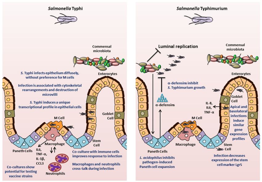
models that has improved our understanding of Salmonella pathogenesis (Figure 3).
6.1. Mouse-Derived Models
Following the establishment of protocols to generate crypt-derived mouse intestinal enteroids
(referred to as organoids by the authors) by Sato et al., Zhang and colleagues [65] in 2014 utilized
the system to analyze interaction of S. Typhimurium with epithelial cells. The authors visualized
bacterial infection, while also observing bacterial-induced disruption of tight junctions, activation of
the nuclear factor kappa-light-chain-enhancer of activated B cells (NF-kB)-mediated inflammatory
response, and a decrease in the stem cell marker Lgr5. The authors noted that these observations
were similar to findings in animal models. The caveat to this study is that the Salmonella were not
delivered into the lumen of the enteroids, the location of the initial contact of bacteria with epithelialMicroorganisms 2020, 8, 504 10 of 23
cells in vivo; instead, the bacteria were added to the medium and came in contact with the enteroids
basolaterally. Nevertheless, this study established mouse-derived enteroids as a model system for
studying Salmonella infection biology.
Since this initial study, enteroids have been used to interrogate various aspects of Salmonella
pathology, including investigating cell types that were previously not accessible to study in vitro.
Farin and colleagues [131], in a 2014 study, used mouse intestinal enteroids to study the control of
Paneth cell (PC) degranulation in response to bacteria or bacterial molecules such as LPS. The authors
found that PC degranulation did not occur upon stimulation with microbial molecules or Salmonella,
but was induced by a novel mechanism requiring only the presence of recombinant interferon gamma
(IFN-γ) [131]. In another study, Wilson and colleagues [132] interrogated the antimicrobial role of
Paneth cell α-defensin peptides. The authors developed small intestinal epithelial enteroids from
both wild-type mice or mice mutated for α-defensin production (Mmp7-/- mice, MMP7 is a matrix
metalloprotease that is required to generate bactericidally active α-defensins in mice [133]), and infected
the enteroids with S. Typhimurium by microinjecting the bacteria directly into the lumen. The absence
of mature α-defensins reduced the intra-luminal bacterial killing, which could be partially restored by
expression of the human defensin HD5 [132]. This study demonstrated the contribution of α-defensins
to the innate immune response to Salmonella, which previously had been a challenge to examine since
most of the earlier experimental systems inadequately recapitulated in vivo cellular processes [134].
Salmonella has been suggested to contribute to the development of cancer by epidemiological
studies [102]. Scanu and colleague [135] probed this phenomenon in a 2015 study using the case of
gallbladder cancer (GBC). The authors derived gallbladder enteroids from mice carrying mutations
that inactivate p53 and are known to be found in GBC patients in India, where the disease is
prevalent. When exposed to wild-type S. Typhimurium, single cells derived from the gallbladder
enteroids carrying these predisposing mutations generated new enteroids that exhibited growth factor
independence, which is one of the hallmarks of transformation, and had histopathological features
consistent with neoplastic transformation, thus establishing a direct association between Salmonella
and cancer. To delve into the mechanism of this transformation, the authors looked at the Salmonella
T3SS-mediated activation of AKT or mitogen-activated protein (MAP) kinase pathways, which have
been shown to be elevated in human cancers. The signals activated by AKT and MAPK were found
to be key in driving the cellular transformation and were sustained even after the eradication of the
Salmonella infection. Studies have shown that AKT and MAPK pathways are activated by other bacteria
and viruses that have been associated with various cancers [136–139]. Although the authors employed
a murine gallbladder enteroid model and S. Typhimurium to study GBC, the authors proposed that
the AKT and MAPK pathways are activated by both S. Typhimurium and S. Typhi serovars, and
contribute to development of cancer that is associated with chronic S. Typhi infection in humans.
Chronic infection by Salmonella has also been found to be a risk factor for developing cancers in the
ascending and transverse colon [103]. However, detailed mechanistic studies of Salmonella-associated
colon carcinogenesis need to be carried out and organoid model systems may prove to be extremely
useful for this purpose.
Finally, another important aspect of Salmonella is the interaction of the pathogen with the
host microbiome. Lu and colleagues [140] recently demonstrated that Lactobacillus acidophilus, a
well-established probiotic bacterium, can alleviate damage caused by S. Typhimurium. Earlier studies
had shown that L. acidophilus can inhibit adhesion of Salmonella to CaCo2 cells [141]. In this study, the
authors extended the mechanism of protection to include the effects on the host. L. acidophilus altered
the differentiation of epithelial cells in crypt-derived enteroids by impeding the Salmonella-mediated
expansion of Paneth cells [131], thus maintaining homeostasis and appropriate epithelial composition
during the infection. This study not only improved our understanding of the role of L. acidophilus
in protecting the epithelial lining, but also demonstrated the ability to include microbiota-specific
analyses to study Salmonella infection with enteroids/organoids.Microorganisms 2020, 8, 504 11 of 23
6.2. Human-Derived Models
The potential to gain significant insight into Salmonella pathogenesis is particularly relevant in
relation to the use of human-specific organoids, especially since these models possess human genetic
specificities absent in mice. Studies have interrogated the usefulness of intestinal enteroids/organoids
derived from human ASCs or PSCs to understand complex interactions between the epithelium and
Salmonella. In 2015 Forbester and colleagues [70] used RNA sequencing to examine the epithelial
transcriptional signature following injection of S. Typhimurium into the lumen of organoids derived
from human induced PSCs (hiPSCs). The analysis showed significant up-regulation of genes for
cytokine-mediated signaling, NF-κB activation, angiogenesis, and chemotaxis. Enhanced release of
pro-inflammatory cytokines IL-8, IL-6, and TNF-α was also confirmed. The findings were consistent
with prior studies in animal and mouse organoid models, thus establishing the human organoids
as a viable infection model for Salmonella. The study also demonstrated that a noninvasive mutant
strain (deficient in invA gene) could be used in the model to examine Salmonella pathogenicity and the
functionality of defined mutants [70]. Furthermore, the authors generated an RNA sequencing data set
following basolateral administration of Salmonella to the organoids. Interestingly, 49 of the 100 most
highly upregulated genes were also significantly induced in the data set obtained by microinjecting
the bacteria for apical infection. The data provide credence to the results of the Zhang and colleagues
study [65], which documented similar patterns of gene expression upon basolateral administration of
Salmonella to mouse organoids as had been observed earlier in literature with other model systems.
Thus, the hiPSC organoids maintain a conserved response to Salmonella infection and provide a
human-specific model for pathogenesis studies.
A subsequent study further demonstrated the validity of hiPSCs as a model to study human-specific
responses to Salmonella infection. Using the same model system, the role of the cytokine IL-22 in priming
intestinal epithelial cells towards a more effective response to S. Typhimurium was also explored [142].
The study showed that IL-22 pre-treated hiPSC-derived organoids increased phagolysosomal fusion
leading to enhanced antimicrobial activity. Thus, this study confirmed earlier observations made in
mouse organoids [143].
The fidelity of organoid-derived data in representing human disease was further demonstrated
in 2018 by Nickerson, KP and colleagues [71], who compared infection of human tissue biopsies
and human intestinal enteroid-derived monolayers seeded on a 2D Transwell system, and observed
that the enteroid-derived epithelial monolayers recapitulated S. Typhi infection observations made
in the tissue biopsy model. The authors also carried out transcriptional profiling of both the host
tissue and the bacteria in order to determine early critical interactions. Infection with S. Typhi
significantly down-regulated several host genes, including those involved in activation of the mucosal
immune response, bacterial clearance, and cytoskeletal rearrangement. Interestingly in this model,
a down regulation of SPI1 genes in S. Typhi was observed. This work demonstrated that S. Typhi
reduces intestinal inflammation by limiting the induction of pathogen-induced processes through the
regulation of virulence gene expression, which is a characteristic feature of human infection with S.
Typhi. Transmission electron microscopic comparisons of the tissues and human organoid-derived
epithelial monolayers showed that the monolayers reproduced the cytoskeletal arrangements, microvilli
destruction, and vesicle-bound bacteria observed in tissues. There were no changes observed in
paracellular permeability, increased death of host cells, or bacterial association with M cells, suggesting
divergence from S. Typhimurium infection in mice. This study highlights the ability of organoids
to compare human-specific responses to each Salmonella serovar, which is important in the context
of translational capacity for developing prophylactic or therapeutic intervention strategies against
S. Typhimurium versus S. Typhi infections.
Despite the multiple advantages of the organoids as an experimental system, the technology is
still in its infancy and has certain limitations. The complex structure of the organoids poses a practical
limitation in accessing the internal luminal compartment. Researchers have used microinjections
to access the apical epithelium. This approach may preserve the internal microenvironment, but isMicroorganisms 2020, 8, 504 12 of 23
resource-intensive, may not allow synchronous exposure and suffers from variability in volume that
can be injected due to heterogeneity of the organoid/enteroid sizes. In addition, the lumen of 3D
organoid/enteroid accumulates cellular debris, which may bind bacteria or hamper interactions with the
apical membranes. As noted above (in Section 3), researchers have turned to organoid/enteroid-derived
2D monolayers to better access the apical side of the model and enable more efficient, user-friendly
analyses in a multiple-well plate format. However, this modification can limit the number of processes
that can be interrogated, especially when considering the lack of 3D structure. Interestingly, Co and
colleagues [144] demonstrated in a recent study that the polarity of human enteroids could be reversed
such that the apical surface faced the medium and was readily accessible. The enteroids released mucus
and extruded cells outwards into the culture medium rather than having the cells embedded in the
basement membrane. Using enteroids with reversed polarity, the authors showed that S. Typhimurium
invades and induces actin ruffles more efficiently at the apical surface compared to the basolateral
surface. The authors observed a more diffuse process of epithelial invasion rather than invasion
only or predominantly at the M cells [144], which confirmed the S. Typhi observations by Nickerson,
KP et al. [71].
Current organoid/enteroid models are devoid of muscles, innervation, vascularization, and
immune cells. There are a couple of approaches being carried out to increase the complexity of organoid
models, including co-culturing techniques. In 2011, Salerno-Goncalves and colleagues [7] generated an
organotypic model using the human ileococal adenocarcinoma cell line HCT-8 and adding primary
endothelial cells, fibroblasts, and peripheral blood mononuclear cells (PBMCs), which they used in a
2019 study to probe the crosstalk between these cell types during infection with S. Typhi, S. Paratyphi A,
or S. Paratyphi B [145]. An ECM composed of collagen-I enriched with other gastrointestinal basement
proteins was embedded with the fibroblasts and epithelial cells, and transferred to a RWV bioreactor
containing epithelial cells. Under low microgravity and low shear conditions, the HCT-8 cells behaved
as multi-potent progenitor cells and gave rise to multiple cell types, including absorptive enterocytes,
goblet cells, and M cells. After one to two weeks, PBMCs were added to the system. The co-culture
model was then infected with the various Salmonella serovars to compare responses to the three strains.
The authors found that the presence of the immune cells in the model resulted in secretion of the
cytokines IL-1β and CCL3, while secretion of cytokines IL-6 and TNF-α was enhanced. Using depletion
experiments, the authors showed that macrophages were the PBMC cell type responsible for the
enhanced secretion of IL-6 and TNF-α. The authors further used the Transwell system to show that
supernatants from organotypic models built with whole or macrophage-depleted PBMCs infected
with the three Salmonella strains varied in their ability to elicit transmigration of macrophages and
neutrophils [145]. Interestingly, the two immune cells displayed crosstalk during infections with S.
Paratyphi A and S. Paratyphi B, such that the presence of macrophages in the co-culture reduced
neutrophil migration as compared to the system built without macrophages [145]. This study illustrates
that co-cultures can aid in probing the contribution of immune cells to Salmonella infection at the
mucosal surface. Finally, this model has also been used to assess the inflammatory response to several
candidate S. Typhi vaccine strains in comparison to the response elicited by the oral vaccine strain
Ty21a strain and its parent wild-type Ty2 stain [146]. Salerno-Goncalves and colleagues [146] found
that specific changes to the genetic makeup of the candidate vaccine strains (in the form of deletions
of specific metabolic genes) elicited host changes in intestinal permeability, inflammatory cytokine
secretion, as well as activation of innate immunity pathways. Higgins and colleagues [73] also used
the model to test the inflammatory response of an S. Typhimurium vaccine strain that they generated.
These studies highlight the usefulness of co-cultured organoid/enteroid models in assessing important
factors to be considered while designing vaccines.
Schulte and colleagues [147] generated a co-culture system of human intestinal epithelial cell line
(Caco-2), primary human microvascular endothelial cells, primary intestinal collagen scaffold, and
PBMCs in a Transwell set up. Using GFP-labeled S. Typhimurium, microscopy, and flow cytometry,
the authors demonstrated that the bacteria can be found in epithelial but not endothelial cells, thusMicroorganisms 2020, 8, 504 13 of 23
modeling the epithelium-restricted infection of humans with S. Typhimurium. These findings are in
contrast to those of Spadoni and colleagues [99] in the mouse model of S. Typhimurium infections
where a breach of the gut-vascular barrier by the bacteria was observed. The endothelial cells respond
to the infection process by bringing about changes in transcription of various genes and releasing the
phagocyte chemoattractant IL-8. Such models, ideally with enteroid/organoid-derived cells replacing
cell lines where used, should prove to be extremely useful and versatile in interrogating the role of
different immune cells, vasculature, and the related crosstalk with epithelial cells during infection with
Salmonella, especially for S. Typhi, where the bacteria spread systemically both as free bacteria and
within reticuloendothelial cells [148].
7. Future Directions
Currently, researchers have little or no control over how cells self-organize into organoids.
The physical environment of the enteroids/organoids in the form of the 3D scaffold provides cues such
as adhesive ligands and stiffness. The current ECMs are derived from animals, are poorly defined,
may show batch to batch variation that can lead to heterogeneous growth and differentiation between
the organoids generated at different times, and are not mechanically pliable after plating. Thus,
improvements to the 3D scaffolds are expected to yield better consistency, which would facilitate
mechanistic infection studies and even provide a better platform for clinical applications. Some recent
work [149] has focused on using chemically defined 3D scaffolds to improve the uniformity of the
environmental cues for growth and differentiation. Indeed, a better defined and mechanically dynamic
matrix would increase the potential of organoid technologies for therapeutic development (also
reviewed in [150] and [151]).
Another important requirement is the further characterization of organoids/enteroids to ensure
that the models faithfully represent the in vivo human physiology so that in vitro analyses with the
system will be relevant for subsequent clinical development. Recent work in brain organoids has shown
that important differences exist in the expression profiles between cells derived from organoids as
compared to human brain cells [130]. Similar studies must be carried out in intestinal organoid/enteroid
models, and indeed in all organoid models, to ensure fidelity of representation.
The enteric nervous system (ENS) carries out important functions in the gastrointestinal tract,
such as motility and contractility, regulation of blood flow, maintenance of epithelial barrier, and fluid
exchange [7]. Future investigations of the role of the ENS in Salmonella infection will be critical to
our mechanistic understanding of the pathogen. Recent studies have shown that the neurons in the
gut also protect against S. Typhimurium infections [152,153]. For example, Lai and colleagues [153]
demonstrated that the pain-sensing neurons that lie beneath Peyer’s patches in the gastrointestinal tracts
of mice, become activated and release the neuropeptide calcitonin gene-related peptide (CGRP) upon
detecting S. Typhimurium. This process results in a decreased number of M cells while increasing the
levels of segmented filamentous bacteria (SFB) that can protect against Salmonella infection [153]. In light
of studies like this, it becomes essential to probe and understand the interaction of Salmonella with the
ENS. To facilitate analyses with the ENS, researchers have developed an ENS-HIO model. Workman
and colleagues [154] generated vagal-like neural crest cells (NCCs, the precursors of ENS) from PSCs
and incorporated them into human intestinal organoids via direct co-culture. The NCCs subsequently
differentiated into multiple cell types including neurons and glia. The authors demonstrated that
the neurons were functional by observing a response to oscillations in calcium. The ENS-HIO was
then transplanted into mice and displayed contractile activity. Given the recent findings of the role of
neurons in potential protection against Salmonella infection, this model has tremendous promise to
further our understanding of the dynamics of the ENS-Salmonella interaction.
Methods to incorporate the microbiome into organoid-based models are essential to future
Salmonella research. It is well known that Salmonella must successfully compete with the resident
microflora in order to infect the intestinal epithelium, and several competitive mechanisms deployed by
Salmonella have been elucidated [155]. Most often, microbiome studies are carried out in germ-free orMicroorganisms 2020, 8, 504 14 of 23
humanized mice; however, there are significant differences between the composition of the microbiota
and host physiology when using these animals as a model of humans. The healthy gut contains several
anaerobic bacteria in the lumen given the decreasing oxygen gradient that extends from the anaerobic
lumen to the hypoxic epithelium. Despite the complexities of microbiota interactions and oxygen levels,
a few studies have made progress in incorporating the microbiota into organoid models or reproducing
anaerobic environments such as the 2020 study by Lu et al., described above [140]. Karve and
colleagues [156] in a 2017 study incubated induced human intestinal organoids (iHIOs) with human
neutrophils to model innate cellular responses to commensal and pathogenic Escherichia coli in which
neutrophil recruitment was monitored by microscopy. The authors were able to culture commensal
E. coli, despite the fact that the bacterial strain is a facultative anaerobe. Greater recruitment of the
neutrophils occurred when the organoids had been injected with pathogenic bacteria as compared to
saline or commensal bacteria. In another study by Leslie and colleagues [157], the authors demonstrated
that anaerobic Clostridium difficile could remain viable for at least 12h when injected into the lumen of
HIOs, indicating the reproducibility of the appropriate oxygen levels in the model. Finally, LeBlay and
colleagues [158] have devised a specialized bioreactor for cultivation of human intestinal microbiota
that will enable researchers to grow a wide range of commensal bacteria for analyses. The authors
immobilized fecal microbiota from a two year old child in gel beads and cultured under anaerobic
conditions with continuous flow of medium containing chyme. The bacteria grew as biofilms and
formed stable populations. S. Typhimurium also immobilized onto beads was added to the reactor
to simulate gut infection in children. This step was followed by addition of two concentrations of
amoxicillin, and the effects of the treatment on the microbial composition and the metabolites generated
were analyzed. The authors observed a strong disturbance in the microbial composition upon antibiotic
treatment, with Bifidobateria significantly decreasing in numbers while C. cocoides-E.rectales group
strongly increased. S. Typhimurium levels were also strongly decreased upon amoxicillin treatment,
but returned to previous levels upon interruption of the antibiotic treatment. Antibiotic treatment also
resulted in a decrease in the concentration of the metabolites acetate and butyrate, which remained
at lower levels even on the withdrawal of antibiotic treatment. Indeed, these studies show that the
incorporation of the microbiota into human-specific organoid systems will allow mechanistic studies
investigating the crosstalk between host physiology, microbiota, and Salmonella pathogenicity.
8. Conclusions
Infections caused by S. enterica remain a major health concern world-wide. Models used to
study the disease pathology so far have provided valuable advancements. However, there remains
a disconnect between what works at the bench versus at the bedside, particularly in the case of
vaccines. The development of organoids/enteroids offers a tremendous opportunity to bridge this gap
by bringing human-specific factors into the research models (Figure 3).
Gastrointestinal organoid and enteroid models have been shown to capture the cellularity,
organization, and complexity of the intestine in vivo along with providing the flexibility of an in vitro
system. These models have provided new and fundamental knowledge in human physiology,
pathology and the molecular basis of host-microbe interactions. In addition, recent studies have seen
efforts to build upon existing paradigms. Research is being conducted to improve the fidelity and
reproducibility of the organoid model systems. Co-culture systems have been developed allowing the
integration of various cell types, which allows us to better understand and interrogate the crosstalk
between a wide-range of potential combinations of lineages in vivo. Most importantly, the integration
of immune cell types is likely to help in understanding how Salmonella is viewed by and responds to
immune processes, particularly at the mucosal epithelium that is the first site of contact and thus an
important stage of the infection process for the development of vaccines. There have also been studies
that have integrated the microbiota into the organoid model and future work may delve into the role of
an individual’s microbiota in Salmonella pathogenesis. Despite its potential, it is important to keep in
mind certain limitations of organoid-based systems, the most significant being that the models have toYou can also read