Toward the Specificity of Bare Nanomaterial Surfaces for Protein Corona Formation - MDPI
←
→
Page content transcription
If your browser does not render page correctly, please read the page content below
International Journal of
               Molecular Sciences

Review
Toward the Specificity of Bare Nanomaterial Surfaces for
Protein Corona Formation
Fabio Vianello            , Alessandro Cecconello            and Massimiliano Magro *

                                          Department of Comparative Biomedicine and Food Science, University of Padua, Viale dell’Università 16,
                                          35020 Legnaro, Italy; fabio.vianello@unipd.it (F.V.); alessandro.cecconello@gmail.com (A.C.)
                                          * Correspondence: massimiliano.magro@unipd.it

                                          Abstract: Aiming at creating smart nanomaterials for biomedical applications, nanotechnology
                                          aspires to develop a new generation of nanomaterials with the ability to recognize different biological
                                          components in a complex environment. It is common opinion that nanomaterials must be coated
                                          with organic or inorganic layers as a mandatory prerequisite for applications in biological systems.
                                          Thus, it is the nanomaterial surface coating that predominantly controls the nanomaterial fate in the
                                          biological environment. In the last decades, interdisciplinary studies involving not only life sciences,
                                          but all branches of scientific research, provided hints for obtaining uncoated inorganic materials able
                                          to interact with biological systems with high complexity and selectivity. Herein, the fragmentary
                                          literature on the interactions between bare abiotic materials and biological components is reviewed.
                                          Moreover, the most relevant examples of selective binding and the conceptualization of the general
                                          principles behind recognition mechanisms were provided. Nanoparticle features, such as crystalline
                                          facets, density and distribution of surface chemical groups, and surface roughness and topography
                                          were encompassed for deepening the comprehension of the general concept of recognition patterns.


                                          Keywords: nanoparticles; biomolecules; protein corona; uncoated nanomaterials; surface recognition
Citation: Vianello, F.; Cecconello, A.;
Magro, M. Toward the Specificity of
Bare Nanomaterial Surfaces for
Protein Corona Formation. Int. J. Mol.    1. Introduction
Sci. 2021, 22, 7625. https://doi.org/
10.3390/ijms22147625
                                               Although the constant improvements in nanoscience and nanotechnology are rais-
                                          ing the bar of nanomaterial applications in biomedicine, the efforts of manufacturers are
Academic Editor: Mahmoud Ghomi
                                          very often frustrated by the adverse effects of biological environments on nanomaterials.
                                          The main reason is that, upon exposure to a biological environment, the nanomaterial
Received: 18 June 2021                    surface is subjected to competitive binding of different macromolecules, resulting in a
Accepted: 15 July 2021                    biological shell, called protein corona, characterized by a composition that can be extremely
Published: 16 July 2021                   variable depending on biological milieus and time. As a consequence, upon their intro-
                                          duction in vivo, engineered nanoparticles enter into an inevitable and hardly predictable
Publisher’s Note: MDPI stays neutral      progressive transformation that, in most cases, leads to their elimination.
with regard to jurisdictional claims in        The in-depth comprehension of the interactions and mutual influences between pro-
published maps and institutional affil-   teins and nanoparticles represents one of the main goals for the successful transfer of
iations.                                  nanotechnologies into medicine [1,2] and, more specifically, for the control over protein
                                          corona formation in vivo, which can be pivotal for determining opposite scenarios: the
                                          elimination or the effective internalization of nanocarriers [3].
                                               In recent years, attempts to modulate the nature of the protein corona were carried
Copyright: © 2021 by the authors.         out by designing tailored nanoparticle ligands and their distribution on the nanomaterial
Licensee MDPI, Basel, Switzerland.        surface [4–8]. Specific ligands can ideally control the nature of the protein shell by the incor-
This article is an open access article    poration of suitable functionalities for the selection and orientation of proteins during the
distributed under the terms and           formation of soft or hard corona shells [9]. As an example, hydrophobic coatings on gold
conditions of the Creative Commons        nanoparticles (AuNPs) induce protein denaturation and irreversible protein binding (hard
Attribution (CC BY) license (https://     corona), making proteins readily susceptible to proteolysis by endogenous enzymes [10].
creativecommons.org/licenses/by/          Conversely, AuNPs coated with tetra(ethylene glycol) (TEG), which prevents hydrophobic
4.0/).

Int. J. Mol. Sci. 2021, 22, 7625. https://doi.org/10.3390/ijms22147625                                         https://www.mdpi.com/journal/ijms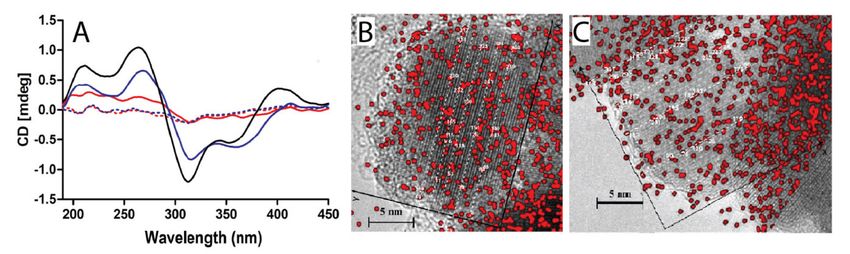
Int. J. Mol. Sci. 2021, 22, 7625                                                                                            2 of 20

                                   interactions, formed soft coronas with retained protein structure while slowing proteol-
                                   ysis [9,11,12]. Furthermore, the zwitterionic sulfobetaine surfactant has been shown to
                                   provide “stealth” properties to AuNPs, leading to corona-free nanoparticles in serum [5].
                                   In contrast, positively charged quaternary ammonium groups were shown to interact with
                                   proteins, readily forming protein coronas on NPs [6]. On this bases, surface modifications
                                   of nanomaterials would seem to be the only viable way for exerting a control over the
                                   protein corona formation. As this review will attempt to highlight, this is not entirely
                                   true, and a deeper comprehension should be dedicated to protein corona formation on
                                   bare nanomaterials.
                                         In our opinion, a novel set of opportunities could be at hand by filling the gap between
                                   nanomedicine, still suffering from a lack of comprehension regarding the interplay between
                                   proteins and nanoparticles, and the understanding of interactions between biotic-abiotic
                                   materials learned by other scientific areas. In fact, an impressive body of studies, spanning
                                   from bio-mineralization to prebiotic chemistry, suggests that the interaction of proteins
                                   with bare solid surfaces, such as inorganic nanomaterials, can be highly sophisticated and
                                   characterized by high selectivity. In this view, the idea of surface coating as an unavoidable
                                   prerequisite, mandatorily and indiscriminately applicable to any kind of nanomaterial,
                                   would preclude the actual application of bare nanomaterials in biomedicine, thus depriving
                                   this scientific branch of opportunities given by this class of nanomaterials.
                                         It should be considered that the surface of minerals played a crucial role in the chemical
                                   evolution of the prebiotic era and in the origin of life. Indeed, reactions at the mineral—
                                   water interface were proposed for the initial synthesis of biomolecules [13]. Thus, to living
                                   (biotic) systems inorganic (abiotic) materials are not alien.
                                         The present review, aimed at summarizing the principles governing the interactions
                                   between proteins and uncoated nanoparticles, pays particular attention to the abiotic–biotic
                                   interface and tries to integrate insights of scientific research involving prebiotic chemistry,
                                   biomineralization, chiral recognition, protein purification, and material science. Thus, hints
                                   from different contexts were reviewed and compared in order to provide the reader with a
                                   general view of the principles controlling protein-(bare) nanoparticle recognition. In this
                                   view, a variety of nanomaterial features were highlighted, such as crystalline structure and
                                   facets [14], density and distribution of surface chemical groups [15], and surface roughness
                                   and topography [16]. Furthermore, synthetic nanomaterials displaying selectivity toward
                                   biomolecules were reviewed.
                                         Beyond applications in nanomedicine, an overall vision on the interactions between
                                   proteins and bare nanomaterials is here presented, possibly offering a perspective for
                                   predicting the formation, the stability, and the biological properties of protein coronas.
                                   Considering the magnitude of the topic, the present review does not claim to be compre-
                                   hensive. On the contrary, it is aimed at stimulating the reader by summarizing selected
                                   points from apparently distant contexts.

                                   2. The Role of Protein Selectivity in Biomedical Applications of Nanomaterials
                                        Upon exposing a nanomaterial to a biological fluid, a protein corona shell is generated.
                                   Depending on the environment, an early-stage corona (soft corona) is gradually replaced
                                   by a shell comprising proteins that display a higher affinity for the nanoparticle surface
                                   (hard corona). This competitive displacement of earlier and less specific proteins is named
                                   “Vroman effect,” resulting in undesired layer instability which can be hardly controlled [17].
                                   Thus, the nanostructure surface is destined to evolve into a complex biological interface
                                   which is actually what the organism “sees” once the synthetic nano-object is introduced
                                   into the body [3]. It is well known that protein corona composition and structure are
                                   influenced by the size, shape, and chemical composition of the nanomaterial surface. In
                                   addition, the specific environment the nanoparticles are exposed to and the duration of
                                   the exposure play fundamental roles in the process [18]. Indeed, exposure time is crucial
                                   due to the aforementioned dynamic nature of the interaction [19], making the prediction
                                   of the corona composition and its biological properties even harder [4,20,21]. A further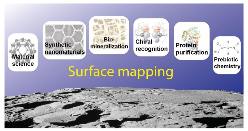
Int. J. Mol. Sci. 2021, 22, 7625                                                                                            3 of 20

                                   critical factor is protein modification induced by interactions with the nanomaterial surface.
                                   These structural alterations will eventually result in decreased protein flexibility and loss
                                   of biological activity. In the worst situation, the adsorption phenomenon leads to protein
                                   denaturation. Numerous studies that focused on the description of binding modes and
                                   the orientation of adsorbed proteins on nanomaterials, stressed the importance of the
                                   control of the biotic–abiotic interactions. As an example, it is crucial to ensure that the
                                   catalytic site of an enzyme remains accessible after adsorption on the nanomaterial, since
                                   steric hindrance and conformational changes are likely to affect biological activity [22–25].
                                   Again, proteins are flexible objects, and their internal dynamics play a central role in their
                                   biological function [26,27]. This last issue further exacerbates the complexity associated
                                   with designing nanomaterials, which ideally should be built for the binding of specific
                                   proteins. On this basis, for several decades protein corona was considered responsible for
                                   the triggering of the immune system and the final clearance of the nanomaterial. Actually,
                                   for most cases, protein corona formation is the very first step in the process of nanoparticle
                                   elimination [28]. Generally, coronas formed from the unspecific protein adsorption act as
                                   labels promoting nanoparticle recognition as “non-self” and, as a consequence, nanoparticle
                                   elimination by the immune system [29,30]. As a consequence, for a long time research has
                                   been focused on the development of antifouling coatings aimed at the complete suppression
                                   of biomolecule binding to nanomaterials. Noteworthy, antifouling coatings were believed
                                   to be the only coatings providing nanomaterials with the ability to avoid the immune
                                   system clearance. This phenomenon has been named the “stealth” effect and bio-inert
                                   coatings have been defined as “stealth materials.” Among them, the gold standard is
                                   represented by polyethylene glycol (PEG).
                                         Paradoxically, “stealth” materials present immunological drawbacks. For example,
                                   it is well-known that the prolonged use of PEGylated materials results in the production
                                   of anti-PEG IgM antibodies [31,32] in a phenomenon known as “accelerated blood clear-
                                   ance” (ABC) [33]. Thus, with time, PEGylated nanocarriers are recognized by IgMs and
                                   accumulated in the liver [34]. Moreover, these drug delivery stealth vectors, although
                                   providing advantageous features such as long-term circulation in the blood, are in turn
                                   detrimental for cell targeting, internalization, and accumulation [35]. Complete protein
                                   corona suppression, besides being an unreachable goal, was recently questioned by the
                                   demonstration that the “stealth effect” requires the binding of specific plasma proteins.
                                   Thus, a nanomaterial, in order to avoid the immune system reaction, does not need to
                                   be protein repellent but protein selective. Noteworthy, such a revolutionary principle
                                   was demonstrated by studying PEG-modified nanoparticles [36]. Indeed, the concept of
                                   controlling the phenomenon of protein corona formation by designing novel and specific
                                   nanoparticle surfaces is progressively replacing the illusion of zeroing macromolecular ad-
                                   sorption [37]. Thus, elucidating the molecular interactions at the nano-bio interface will be
                                   crucial for predicting protein corona formation and composition of designed nano-vectors
                                   characterized by high biocompatibility and specific targeting functions [38,39].
                                         An ideal protein corona should act as camouflage, promoting nanoparticle recognition
                                   as “self” by the organism and, at the same time, acting as a chaperone, promoting the
                                   crossing of biological barriers (Figure 1). In this view, the merging of research from different
                                   fields of science is becoming increasingly urgent in order to bridge in vitro technologies
                                   with in vivo biomedicine. Moreover, synthetic biology will benefit from the comprehen-
                                   sion of interactions occurring between biomolecules (biotic) and materials (abiotic) at the
                                   molecular level [40]. In this context, the concept of “epitope map,” proposed by Dawson
                                   and colleagues about twenty years ago [41], aimed at mapping the actual surface of the
                                   protein corona, ideally encompassing both protein composition and conformation. The
                                   epitope map is seen as the “biological fingerprint” of the particle–protein complex and
                                   defines its biological activity. Thus, the outer layer of the biomolecular envelop controls the
                                   interaction of the nanoparticles with cells and the nanoparticles trafficking to different cell
                                   compartments. Hence, this biological “mask” determines the fate of synthetic nanoparticles
                                   in biological systems.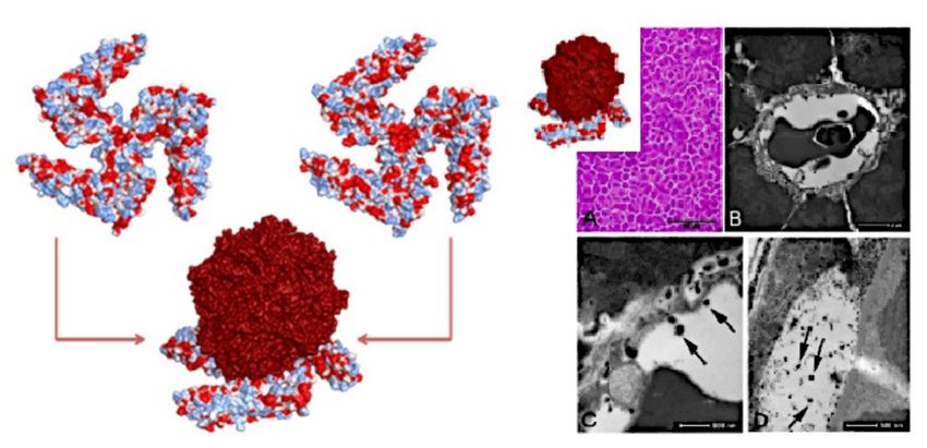
Int. J. Mol. Sci. 2021, 22, 7625                                                                                                 4 of 20

                                   Figure 1. In the schematic representation, upon exposure to a biological milieu, most of the nanopar-
                                   ticles reported in the literature (yellow) are subjected to the unspecific binding of proteins (red).
                                   On the contrary, nanoparticles displaying binding selectivity (grey) lead to the formation of a shell
                                   composed of specific proteins (blue) promoting cell internalization. This biological envelope makes
                                   the difference between nanoparticles being internalized or being cleared by the immune system.
                                   Arrows represent the Vroman effect.

                                        Among synthetic uncoated nanomaterials, carbon-based nanomaterials (CNMs) can
                                   count on a notable number of in vitro cell studies dealing with fundamental aspects, such
                                   as cell uptake, cytotoxicity, and drug delivery and, most importantly, providing valuable
                                   hints on the relationships linking nanomaterial surface, biomolecular interaction, and cell
                                   response. Thus, herein CNMs will be discussed first because, besides being at an advanced
                                   stage from the standpoint of biomedical applications, they represent an optimal example of
                                   how nanomaterial syntheses and post-synthetic modifications can modulate the selectivity
                                   of a bare surface toward proteins and stimulate, in this way, different cell responses.

                                   3. Carbon-Based Nanomaterials
                                         Surface features and defects are of great importance for the recognition of inorganic
                                   nanomaterials (vide infra) by peptides and proteins, and this paradigm was also confirmed
                                   for carbon-based nanomaterials. Sengupta and colleagues showed that in CNMs super-
                                   ficial defect-induced hydrophilicity strongly influenced the formation of protein coronas
                                   composed of albumin and fibrinogen [42].
                                         Due to their unique physicochemical properties, among CNMs, carbon nanotubes
                                   (CNTs) aroused great interest for their potential application in cancer treatments as nanocar-
                                   riers for drugs [43]. Besides their high surface-to-volume ratio, CNTs display delocalized
                                   π-electron clouds which are believed to promote protein corona formation. This peculiar
                                   feature has a potential application in antigen delivery and could be used for the develop-
                                   ment of strong and long-lasting antigen-specific immune responses. Bai and colleagues
                                   reported on carboxylated multiwalled CNTs (MWCNTs) as advanced carriers to deliver
                                   ovalbumin [44]. Combinatorial phage-display methods were successfully applied for se-
                                   lecting specific amino acid sequences interacting with a plethora of nanomaterials [45].
                                   For example, histidine- and tryptophan-rich protein motifs showing a selective affinity
                                   for carbon nanotubes were identified using phage-display technology. Intriguingly, these
                                   binding sequences were flexible and folded into a structure matching the geometry of
                                   CNTs [46]. Primary, secondary, and tertiary structures of peptides or proteins are believed
                                   to play a crucial role in their binding to CNTs [47], following a model based on shape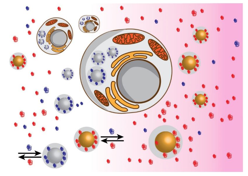
Int. J. Mol. Sci. 2021, 22, 7625                                                                                          5 of 20

                                   complementarity. For carboxylated MWCNTs, it was found that the higher the defect
                                   density on the nanomaterial surface, the greater the ovalbumin adsorption and consequent
                                   macrophage activation, indicating a new fascinating option for the development of next-
                                   generation vaccines. Again, it appears that the interaction of CNTs with biomolecules can
                                   be modulated by the introduction of structural defects. As reported in the ex vivo study
                                   by Raghavendra and colleagues, this can be obtained by mechanical stress, where protein
                                   corona composition on single-walled CNTs (SWCNTs) was evaluated as a function of the
                                   physicochemical alterations of the nanomaterial obtained by ball-milling. Furthermore, the
                                   phenomenon of protein corona formation was responsive to proteome variations and to
                                   the biological milieu [48].
                                         Machova and colleagues presented a systematic in vitro study on protein corona de-
                                   velopment using ultra-small (2 nm) hydrogenated or oxidized nano-diamonds, elucidating
                                   their interaction with human cells. The hydrogen- or oxygen-rich surfaces emerged as
                                   important discriminating factors orienting the surface chemistry toward protein selection.
                                   Different and specific protein coronas influenced the interactions of nano-diamonds with
                                   cells, resulting in different levels of cytotoxicity [49].
                                         Graphene-based materials (GBMs) represent other objects that are the focus of intense
                                   studies, due to their unique tunability, physico-chemical and surface properties, and great
                                   potential for biomedical applications. These are far from being fully exploited, and there
                                   is still much to be understood in the context of graphene/biomolecular interactions/cell
                                   responses. For example, graphene has been demonstrated to stimulate different cell
                                   responses and to influence the interactions with proteins in multiple ways [50].
                                         Several peptides have been identified as specific graphene binders by phage-display
                                   technology, opening a range of possibilities and challenges [51]. Indeed, the properties of
                                   graphene-based nanomaterials can be tuned by controlling structural and chemical param-
                                   eters, such as thickness and, most importantly, degree of oxidation. The latter emerged as
                                   the main factor affecting surface chemistry and material organization. Therefore, graphene
                                   surfaces can be produced with different wettability and roughness by controlling the de-
                                   gree of oxidation. Protein adsorption can be predicted and modulated by the intertwining
                                   of these factors [52]. As an example, single-layer graphene oxide (GO) and few-layer
                                   graphene oxide can form smooth hydrophilic and anti-fouling surfaces, which significantly
                                   resisted unspecific protein adsorption. Conversely, low or non-oxidized graphene, due to
                                   its degree of roughness and hydrophobicity, promotes macromolecule binding. Further-
                                   more, since peptides preferentially bind to the edge of planar graphene surfaces, the effects
                                   of graphene oxidation, number of layers, and underlying support were investigated using
                                   both experimental and computational methods [53].
                                         These authors believe that carbon nanomaterials could inspire researchers toward
                                   intensifying in vitro studies on bare metal/metal oxide nanoparticles.

                                   4. Molecular Biomimetics and Biomineralization
                                        Biomineralization, the natural process leading to the biosynthesis of bones, dental
                                   structures, and mollusk shells, is a worthful example of the level of complexity and
                                   specificity that can be reached by the interaction between proteins and inorganic surfaces.
                                   This process shows the fine biological control exerted by living organisms over biomineral
                                   formation [54].
                                        Thanks to their unique and specific interactions with small molecules, macromolecules
                                   and, even, inorganic materials, are smart building blocks controlling structures and func-
                                   tions of all biological hard and soft tissues in organisms. Regarding the interactions of
                                   biomolecules with abiotic materials, the recognition of surface features and defects is
                                   central to the absorption of peptides and proteins. On the surface of minerals such as
                                   calcite, the recognition and self-assembly of biomolecules on distinct surface kinks and
                                   steps can lead to changes in the overall shape and symmetry of the bulk crystal [55]. So
                                   and colleagues showed by in situ atomic force microscopy (AFM) that an acidic 20 kDa
                                   cement protein (MRCP20) from the barnacle Megabalanus rosa binds specifically to step edge
Int. J. Mol. Sci. 2021, 22, 7625 6 of 20
atoms on {1014} calcite surfaces, and, with time, it further assembles into one-dimensional
nanofibrils [55]. This selective surface interaction with step edge atoms directed a cooper-
ative calcite modification, where templated protein nanostructures re-shaped the calcite
crystal [55].
Combinatorial and directed evolution techniques led to the development of genetically
engineered proteins for inorganics (GEPIs), which can specifically bind to selected inor-
ganic materials. Combinatorial biological protocols, such as phage-display technologies,
demonstrated the possibility of selecting peptide sequences with the ability to specifi-
cally recognize solid surfaces, similarly to natural proteins, regulating crystal growth in
biomineralization [56]. As an example, Patwardhan and colleagues found sequence similar-
ities among peptides strongly interacting with amorphous silica nanoparticles of various
sizes (15–450 nm) [57]. Palafox and colleagues studied peptides selected as a function of
their affinity for Ag and Au surfaces [58]. The authors combined experimental binding
measurements, advanced molecular simulations, and selected nanomaterial synthesis,
to show that interaction selectivity and recognition of metallic surfaces by peptides oc-
curred by thermodynamically different binding modes [58]. In turn, the adsorption of
three homo-tripeptides (His-His-His, Tyr-Tyr-Tyr, and Ser-Ser-Ser) on Au surfaces was
investigated by Hughes and co-authors using molecular dynamic simulations [59]. The
authors’ findings suggested that Au facets affected the structure and energetics of the
adsorbed biomolecules, highlighting the possible impact in the field of the model used for
the interpretation of experimental binding data [59]. Among noble metal nanoparticles,
supposed to be characterized by similar surfaces, significant differences were reported in
the binding selectivity of silver and gold nanoparticles toward peptides, depending on
amino acid side chains. Tryptophan was singled out as the only aromatic amino acid with
the ability to produce a tight binding on gold surfaces [60]. Differently, using the same type
of peptides, AgNPs displayed no strong interaction with aromatic residues but showed
high affinity for lysine side chains [61]. Such a degree of specificity between peptides
and inorganic surfaces recalls the level of affinity existing among biological entities (i.e.,
antigen-antibody interactions).
Several human illnesses, such as Alzheimer’s disease, Creutzfeldt-Jacob disease,
and dialysis-related amyloidosis, involve the formation of amyloid fibrils due to peptide
aggregation. Fibril formation is a self-assembly process normally involving soluble pep-
tides and proteins as building blocks that develop macroscopic structures with important
connections with biological functions and human diseases. Fibril formation occurs by
nucleation-dependent phenomena characterized by a lag-phase, leading to a critical seed
produced from the free monomers in solution. This process represents the rate-determining
step of the phenomenon after which the formation of amyloid fibrils proceeds rapidly [62].
Moreover, amyloid fibrils act as nucleation surfaces for new peptide and protein aggre-
gation, leading to the autocatalytic growth of the fibril. These nucleation and elongation
processes occur at distinct “sites” on the fibril: while nucleation seems most frequent along
the sides of fibrils, elongation occurs at their ends. As nucleation leads to the formation
of toxic oligomers, nucleation inhibitors were proposed for therapeutic purposes [63,64].
Importantly, the fibril formation process of amyloid proteins can be affected by the pres-
ence of foreign surfaces and profound effects in the modulation of amyloid formation
by nanoparticles were reported [65–67]. In turn, the amino acid sequences have an effect
on both fibrillization patterns and nanoparticle modulation envisaging the existence of
specific interaction modes between particles and proteins [68]. Several types of nanopar-
ticles, such as co-polymer particles, cerium oxide particles, carbon quantum dots, and
carbon nanotubes, were found to enhance the rate of protein fibrillization. Linse and
co-authors [65] explained the reduction of the lag phase in the presence of nanomaterials
in terms of the increased probability of reaching a critical nucleation seed for protein fibrils.
Moreover, the observed shorter lag (nucleation) phase in the presence of nanomaterials
depends on the chemical nature of the particle surface. This nanoparticle-assisted nucle-
ation mechanism may increase the risk of toxic clusters and amyloid formation. On theInt. J. Mol. Sci. 2021, 22, 7625 7 of 20
other hand, it paves the way to new routes for the controlled self-assembly of proteins and
peptides and the production of novel nanomaterials [65]. A correlation between protein
stability and aggregation propensity has been documented by Szczepankiewicz et al. [69]
Moreover, Cabailero [68] and co-authors studied the development of amyloid fibrils by
comparing a series of five mutants of the single-chain sweet protein monellin, differing
for their intrinsic stabilities toward denaturation, and co-polymeric nanoparticles. A clear
correlation between intrinsic protein stability and nanoparticles on the aggregation rate
was reported and, in particular, mutants with a high intrinsic stability and low aggregation
propensity showed an accelerated rate of amyloid fibril formation induced by the presence
of nanoparticles. Conversely, nanoparticles led to a retardation of amyloid fibril formation
in mutants characterized by low intrinsic stability and high aggregation rates. Moreover,
both activating and inhibiting effects on amyloid fibril formation were particularly pro-
nounced in the presence of hydrophilic nanoparticles, which presented a large surface
accessibility for hydrogen bonding on the polymer backbone [68].
The selectivity of bare iron oxide nanoparticles for protein binding was exploited for
an application in biomedicine. Naked nanostructured iron oxide with selectivity toward
proteins was tested in vivo on zebrafish [70]. The nanomaterial was able to specifically
bind Apolipoprotein A1 (Figure 2, left) and to evade the clearance of the immune system
of zebrafish by mimicking, to some extent, high-density lipoproteins (HDL). The structural
analogy with HDL was also corroborated by the massive absorption of the nanomaterial
by the intestinal tract and, most importantly, by the presence of the nanomaterial in the
host ovaries (Figure 2, right). The tissue-specific delivery of an antibiotic and the absence
of an adverse outcome were demonstrated, substantiating the idea that the stealth effect is
a consequence of the formation of a shell of selectively bound functional proteins on the
nanomaterial, and opening a new avenue to the evaluation of metal oxide nanoparticles as
novel stealth nanomaterials for biomedicine.
Figure 2. Apolipoprotein A1 recognition of nanoparticle surface (left) and electron microscopy
images of biological samples showing nanoparticle uptake in Zebrafish (right): (A) photomicrograph
of a liver parenchyma cross-section showing the organization of hepatocyte cells (hematoxylin
and eosin staining); (B) transmission electron microscopy image of hepatocyte cells surrounding a
capillary; (C) high magnification of the region shown in (B) revealing nanoparticles (arrows) inside
the capillary; (D) transmission electron microscopy image of nanoparticles in the space between the
follicular epithelium and zona radiata of the zebrafish ovary (reproduced with permission from [70]).
5. Prebiotic Chemistry and Chirality Selection
Metal oxide surfaces are believed to have fulfilled a central role in the assembly
of early macromolecules and, therefore, of life. Nevertheless, these materials are not
sufficiently studied as models on the specificity of the interactions between proteins and
inorganic materials.
Metal oxide-based bioinorganic hybrids offer unique structures, physicochemical
features, as well as novel biochemical properties. In the last decade, extensive researchInt. J. Mol. Sci. 2021, 22, 7625 8 of 20
efforts were dedicated to TiO2 , ZnO, SiO2 , and GeO2 metal oxides, aimed at improving
methods of synthesis and surface functionalization of these nanomaterials, as well as at
their shaping and structural patterning, taking inspiration from natural processes. As an
example, the binding of the protein tyrosinase on birnessite, a manganese oxide mineral,
showed very high affinity. The enzyme molecule was not intercalated or adsorbed in the
mineral, but was immobilized on the external surface of the metal oxide [71,72].
Again, metal oxide surfaces are believed to have played an essential role in trigger-
ing the generation of biomolecules and, therefore, life, possibly by participating in the
concentration of biomolecules from dilute solutions, as well as at their organization into
structured biopolymers [73–79]. Cairns-Smith’s group proposed the involvement of defec-
tive clay crystals as the first “genetic” code (i.e., not based on nucleic acids) fulfilling an
essential function during the early stages of life evolution [80]. Moreover, it was shown that
metal complexes were sequestered from metal oxide surfaces by small organic molecules
(e.g., polypeptides) to form the precursors of active centers of enzymes. Russell’s group
proposed that iron oxides could bind proteins to form catalytic entities, possible precursors
of oxygenic photosynthesis [81]. On the other hand, it was proposed that mineral surfaces
can recruit and select peptides from their environment leading to novel macromolecules
with improved catalytic activities [82]. Thus, minerals had an important role in prebiotic
chemistry, and they can be thought of as templates, promoting structural organization in
disordered organic molecules. An ordered structure can be induced in a peptide contain-
ing a suitable amino acid sequence by a surface providing properly spaced reactive sites.
Lundqvist and colleagues demonstrated this hypothesis by showing that a disordered
peptide can be forced into a well-defined structure by silica nanoparticles [83]. In another
example, a peptide displaying a low helical content in solution was induced to adopt a
well-defined α-helix upon interaction with silica nanoparticles [84]. Another possibility
inspired by biological processes was the development of silica-based nanoparticles as smart
matrices for the auto-encapsulation and controlled release of functional molecules (e.g.,
proteins). This approach involved the use of silica-forming peptides to mediate the in vitro
generation of silica hybrids as potential therapeutic nanocarriers [85].
For many decades, the origin of homochirality in biological systems has aroused the
interest of the scientific community, and it has been in a core position in studies on the
origin of life [86]. The central question deals with how enantiomeric separation could
take place in the absence of a chiral symmetry operator. In this view, one fascinating
feature of metal oxide surfaces is that they can provide periodic environments prone to
select, concentrate, and possibly even organize chiral molecules into polymers and other
macromolecular structures [87]. In this field, several phenomena were identified, including
solid surface reconstruction, chiral imprinting upon adsorption of biomolecules, and the
enhancement or suppression of enantio-selectivity of surfaces in the presence of racemate
mixtures of chiral compounds [88]. Naturally occurring clay surfaces were described to
selectively adsorb amino acids and act as chiral amplifiers, as reported for vermiculite
clay, prompting the involvement of non-centrosymmetric chiral surfaces at the origin of
biological homochirality [89]. Additionally, the role of clays was already proposed for
RNA oligomerization [90] and in the organization of amphiphilic molecules and lipids on
mineral surfaces [91], further suggesting the importance of these minerals in the origin of
chirality in lagoonal environments. In this view, naturally occurring abiotic surfaces can
inspire the synthesis of novel nanostructures, which are usually highly symmetric. Not
surprisingly, in the last decades, a number of chiral nanostructures were reported in the
literature [92]. As an example, hints on the dichroic behavior of iron oxide nanoparticles
were provided [93]. Chiroptical activity, commonly considered a prerogative of tetrahedral
organic molecules, was an unprecedented feature for iron oxide nanoparticles. Crystalline
vacancies on the nanomaterial surface were identified as chiral centers and studied in
the presence of inorganic ligands. The dichroic signal of nanoparticles was differently
influenced by the coordination of different chelators (Figure 3, left) and this phenomenon
was attributed to the chelator ability to rescue defective crystalline sites on the nanomaterialInt. J. Mol. Sci. 2021, 22, 7625 9 of 20
surface. Furthermore, ligand distribution was strictly governed by the lattice of iron oxide
nanoparticles (Figure 3, right).
Figure 3. Circular dichroism (CD) and EDS chemical mapping on HR-TEM micrographs of arsenic (As(III) and As(V))
coated iron oxide nanoparticles. Panel (A): CD spectra of unmodified nanoparticles (black continuous line), As(III) or As(V)
modified nanoparticles (red and blue continuous lines, respectively), and As(III), As(V) as controls (red and blue dashed
lines, respectively). Panel (B) and (C): mapping of nanoparticle surface sites displaying chiroptical activity: the distribution
of As(III) (panel (A)) and As(V) (panel (C)) oxyacids from EDS chemical mapping on HR-TEM micrographs. Segmented
white lines represent the (220) planes of iron oxide crystalline lattice (reproduced with permission from [93]).
6. Protein and Peptide Purification by Bare Nanomaterials
As highlighted above, material selectivity toward protein binding represents an ad-
vantageous feature that can be exploited for the development of effective biomedical
applications of nanomaterials. Some nanoparticles came into the limelight for being ex-
tremely selective and were applied for the isolation and purification of proteins in complex
biological matrices.
Immobilized Metal Ion Affinity Chromatography (IMAC) uses metal ions such as Ti4+ ,
Fe , Ga3+ , Al3+ , and Co2+ immobilized by a linker to a solid matrix and it has become one
3+
of the most widely applied techniques for the selective enrichment of phospho-peptides
and phospho-proteins [94–97]. IMAC has been successfully applied to large-scale phospho-
proteome studies, but it still lacks satisfactory selectivity for separating mono-, di-, and
multi-phosphorylated peptides [98]. In this context, metal oxide materials, such as titanium
dioxide (TiO2 ), zirconium dioxide (ZrO2 ), iron oxides (hematite and maghemite, Fe2 O3 ,
or magnetite, Fe3 O4 ), and alumina (Al2 O3 ), have recently been proposed as alternatives
to IMAC to specifically separate phospho-peptides from complex samples [99]. This
application was defined as Metal Oxide Affinity Chromatography (MOAC) [100]. Among
these purification strategies, TiO2 is the most commonly used metal oxide for the selective
capture of target peptides because it is extremely tolerant toward most buffers and salts
used in biochemistry and cell biology [101–104]. TiO2 has been proven to exhibit high
affinity and good selectivity for binding phospho-peptides for mass spectrometry [104–106].
Although the exact physicochemical mechanism is largely unknown, several additives,
such as aromatic and aliphatic hydroxy-carboxylic compounds, have been reported to
reduce nonspecific binding on TiO2 leading to dramatic enrichment improvements [107].
Alternatively, tin dioxide (SnO2 ) nanoparticles were successfully synthesized and applied
to selectively purify phospho-peptides for mass spectrometry analysis [108]. Ma and
colleagues showed that octahedral SnO2 nanoparticles, characterized by abundant under-
coordinated Sn atoms, exhibited high affinity and selective binding ability for phospho-
peptides [108]. Thus, low-abundance phospho-peptides were selectively and efficiently
purified from complex biological samples. Most importantly, the authors tailored the
exposed facets of the nanomaterial for modifying its physical and chemical properties and
for improving its binding selectivity [108].Int. J. Mol. Sci. 2021, 22, 7625 10 of 20
Superparamagnetic Iron Oxide Nanoparticles (SPIONs) constituted of magnetite
(Fe3 O4 ) or maghemite (γ-Fe2 O3 ) have proved their usefulness in many biomedical appli-
cations, such as drug delivery, induced-hyperthermia, and as MRI (magnetic resonance
imaging) contrast agents [109]. Their wide use is mainly due to the favorable biocompatibil-
ity and their magnetic properties. Moreover, the use of bare iron oxides offers advantages
for industrial applications, mainly due to the low-cost and rapid synthesis. Generally, de-
pending on the specific application, several surface coatings have been used for stabilizing
colloidal suspensions and for tuning the properties of these nanoparticles [110]. Several al-
ternatives have been proposed for the surface modification of magnetic nanoparticles, such
as coating with metallic gold, organic polymers, and silica [110–112]. Nevertheless, the
introduction of a coating layer presents several drawbacks. Indeed, stabilizing coatings can
bind weakly to nanoparticle surfaces and eventually desorb or exchange with bulk solution,
affecting the stability of colloidal suspensions. In addition, coating processes are often
time-consuming, expensive, and characterized by low yield, limiting massive productions.
Furthermore, the coating reduces the magnetic moment of the nanomaterial by introducing
a non-magnetic component. At last, these coatings strongly modify the nanomaterial
surface and influence the interactions with proteins in the biological environment.
Blank-Shim and colleagues performed the first systematic study on the interactions
between peptides and bare iron oxide nanoparticles, aimed at developing peptide tags
to be genetically engineered into proteins for the recognition of nanomaterials [113]. The
strategy for designing high-affinity peptide tags required an in-depth understanding of the
surface-peptide recognition patterns [56]. A model considering a patchwork of electrostatic
sites on the iron oxide surface was used to describe the adsorption affinity of all 20 natural
amino acids [56]. Results indicated that the binding efficiency of peptides can be fully
explained by electrostatic interactions, providing the basis for the design of peptide tags
for biomolecule recognition of bare magnetic nanoparticles, which was experimentally
demonstrated with tagged Green Fluorescent Protein (GFP) variants [56]. The study paved
the way for a fast and simple protein immobilization procedure without the need for
unstable and expensive affinity ligands, currently in use for the isolation of recombinant
proteins [113].
A tool for the prediction of the propensity of peptides to interact with iron oxide
nanoparticles was presented by Schwaminger and colleagues [114]. The authors reported
on an effective implicit surface model (EISM) considering electrostatic interactions, van der
Waals interactions, and entropic effects for the theoretical calculations. However, the most
important parameter was a force field contribution term of the surface accessible area di-
rectly derived from experimental results on the interactions of nanomaterials and peptides.
EISM was verified by further peptide binding experiments in an iterative optimization
process where negatively charged peptides were identified as the best binders for iron
oxide nanoparticles. Besides electrostatics, bare iron oxide nanoparticles revealed the abil-
ity to effectively isolate His-tagged proteins from complex biological matrices [115]. This
property can be attributed to the availability of under-coordinated iron(III) sites on iron
oxide surfaces as a consequence of dangling bonds derived from crystal truncation [116].
In this view, the reactivity and binding selectivity of the uncoated iron oxide surface can
be explained by the behavior of iron(III) sites and their topographical distribution on the
nanoparticle surface. A comparison using different model proteins revealed the existence
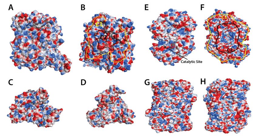
of patterned regions of carboxylic groups acting as recognition sites for naked iron oxide
nanoparticles. Readily interacting proteins display a distinctive surface distribution of
carboxylic groups, recalling the geometric shape of an ellipse (Figure 4B,F). This was inter-
preted as a morphological complementarity of nanoparticle curvature, compatible with the
topography of exposed iron(III) sites laying on the iron oxide nanomaterial surface [117].
The macromolecule recognition site, absent in non-interacting proteins, promoted the har-
boring of the protein on the nanomaterial surface and allowed the formation of functional
protein coronas.Int. J. Mol. Sci. 2021, 22, 7625 11 of 20
Figure 4. Three dimensional computational models of proteins displaying selectivity toward iron oxide nanoparticles
(A,B,E,F) and proteins with no affinity for iron oxide nanoparticles (C,D,G,H). Patterns of carboxylic groups can be
visualized on specific sides of the macromolecules. (A,B) = xanthine oxidase (XO); (C,D) = bovine serum albumin (BSA);
(E,F) = aminoaldehyde dehydrogenase 1 from tomato (SlAMADH1, from Solanum lycopersicum); (G,H) = bovine serum
amine oxidase (BSAO) (reproduced with permission from [117]).
Aiming at the diagnosis of threatening pathologies, such as cancer, the analysis of
protein recognized by nanomaterials represents an advantageous diagnostic strategy, as
it can simplify the analysis of the whole sample proteome [118,119]. Proteome variations
following the occurrence of a disease can be reflected by an alteration of the protein
corona composition in comparison with healthy controls. In this view, bare iron oxide
nanoparticles were successfully employed for the discrimination between milk coming
from healthy and mastitis-affected bovines [120].
7. Surface Energy and Protein Corona
Surface free energy is commonly considered the key factor for explaining the size-
dependent thermodynamic behavior of nanoparticles. Indeed, at the nanometer dimension,
nanomaterial structures and properties are governed by surface effects. This is due to the
nanomaterial’s high surface-to-volume ratio, resulting in the contribution of a significant
fraction of the nanostructure atoms to surface phenomena with respect to the bulk. As the
size of nanomaterials decreases, the ratio of surface atoms involved sharply increases.
The environment of surface atoms is different from the atoms in the bulk. In the
interior, each atom is surrounded in all directions by neighbor atoms, whereas surface
atoms present an anisotropic environment. Thus, no net force is exerted on the bulk
atoms, while surface atoms are subjected to an asymmetric force field, resulting in a
higher energy level associated with the surface. This excess energy at the surface is
named surface energy [121]. Surface energy is fundamental for understanding nanoparticle
thermodynamics, hence, different approaches were proposed that were aimed at the
prediction of this quantity, including classical thermodynamics calculations, molecular
dynamics simulations, and ab initio calculations [122]. Even if the concept of surface energy
is a hot topic in several applications, such as energy storage and catalysis, it can, as well, be
extremely useful for the comprehension of the interactions between, for instance, proteins
and bare inorganic nanoparticles. The lowering of surface energy is a natural process
and, from a thermodynamic standpoint, protein corona formation can be considered as aInt. J. Mol. Sci. 2021, 22, 7625 12 of 20
result of the tendency of nanoparticles to minimize their surface energy in the presence of
biological macromolecules [123,124]. Indeed, the net change of Gibbs free energy following
protein binding on the nanoparticle surface is negative, showing that the process occurs
spontaneously [125]. Protein corona formation competes with nanoparticle aggregation,
which lowers surface energy, as well, by reducing the surface-to-volume ratio [126].
Regarding bare crystalline nanomaterials, the reduction in surface free energy is
associated with surface reconstruction, namely to the re-organization of the lattice at the
boundary with the solvent, which, in turn, can induce conformational and functional
changes of bound proteins [28]. In this view, the surface energy of nanocrystals depends on
the high density of under-coordinated atoms at surface steps and kinks. It is well-known
that these features can determine exceptional catalytic properties [127]. In recent decades,
intensive efforts were dedicated to the synthesis of tailored crystalline materials with
different properties ascribed to differently exposed atoms at the material surfaces [128–130].
It should be considered that a plethora of structurally unprecedented motifs have been
recently developed, including prisms, polyhedrons, rods, plates, wires, and so on, and
the list of available shape-guiding synthetic processes is constantly increasing [131]. In
this context, surface energy excess and distribution are important topics to understand the
growth, reactivity, and stability of materials on the nanometric scale [127,132]. Chemical
species preferentially interact with specific crystal planes as a result of the different surface
energies [133].
As an example, a systematic investigation of the surface energy of cubic nanoparticles
was carried out using the modified embedded atom method (MEAM) [134]. Results showed
that both surface energy and dangling bond density increased with decreasing material
size [135]. Accordingly, Holec and colleagues observed that surface energy was correlated
to the number of dangling bonds (reduced coordination of the surface atoms) [136]. Ferrer
and colleagues proposed a morphology diagram reporting the quantitative energy distribu-
tion of nanomaterials, that can be used as a guide to understand the relationship between
crystal growth and surface interactions and to help experimental work [137]. Ma and Xu
(2007) proposed a quantitative calculation of the surface energy of nanomaterials [135].
Furthermore, a recent study by Visalakshan and colleagues stressed the importance of
taking nanoparticle shape into serious consideration as it can significantly influence protein
corona formation and the therapeutic potential of nanoparticles [138]. Indeed, authors
reported that different surface energies are correlated with different nanoparticle shapes, in
agreement with the previous literature [139,140]. A reachable goal should be the translation
of shape-guiding synthetic processes into the development of novel bare nanomaterials for
biomedical applications.
Energy distribution on a nanoparticle surface can be meant as a key aspect for the
interpretation and prediction of the selectivity toward protein binding. In the absence of an
optimal complementarity at the interface between the nanoparticle and the macromolecule,
the binding is thermodynamically unfavored. On the contrary, if the two surfaces suitably
match, the surface energy of the nanomaterial is strongly reduced, resulting in nanoparticle
stabilization, and the binding with the specific macromolecule is favored. Thus, ideally,
nanoparticles are designed for interacting with specific proteins. It is important to bear
in mind the strong link existing between shape-controlled synthesis, surface energy, and
surface coordination chemistry. Nevertheless, the bottleneck in the research on the sur-
face coordination chemistry of nanomaterials is mainly related to the lack of effective
tools to characterize coordination structures at surfaces [141]. With the comprehension
of surface coordination chemistry, the molecular mechanisms behind various important
effects of inorganic nanomaterials can be disclosed, including their actual potential for
biomedical applications.
8. Conclusions
The creation of protein-like nanoparticles represents the Holy Grail in nanoscience,
where the crucial challenge is to endow nanoparticles with protein-like specificity andInt. J. Mol. Sci. 2021, 22, 7625 13 of 20
enabling the abiotic material to interplay with biological systems [142,143]. In this view, as
nanoparticles and proteins are entities of the same size, protein–protein interactions could
inspire our comprehension of the selectivity between biological interfaces and synthetic,
abiotic objects. Although the recognition of peptides and proteins by bare inorganic
nanomaterials is at the core of a range of scientific branches, its potential in biomedicine
is far from being fully exploited. Possibly, this could be due to the limitations of many
reported synthetic inorganic nanomaterials, which do not present suitable characteristics
to be used in the absence of coating materials or surface stabilizers. On the contrary, novel
syntheses could bring to light unexpected and fascinating surface chemistries that would
merit the attention of the biomedical community.
The central question remains: how an abiotic surface of a generic inorganic nano-
material can be endowed with a protein-like specificity? We believe that the answer
could be hidden in the enormous body of studies related to, besides nanotechnology and
nanomedicine, prebiotic chemistry, biomineralization, and chiral nanomaterial selectivity
dealing with specific and sophisticated biotic-abiotic interactions (Figure 5). As an example,
it is important to mention that the success of biomedical implants depends on the interac-
tion between implant surfaces and the biological environment and, in this view, it is well
accepted that nanotopography is a key factor determining protein adsorption, as well as
cell growth onto a given biomaterial and, therefore, its biocompatibility [144].
Figure 5. Surface mapping emerges as the key to understanding the high level of complexity and specificity that can be
obtained in the interaction between proteins and bare inorganic surfaces.
Not surprisingly, phage-display technology, a technique largely employed for the
study of protein–protein, protein–peptide, and protein–DNA interactions, identified sev-
eral peptides as specific binders of naked abiotic material surfaces [45]. Furthermore,
computational methods have become increasingly reliable to understand the recogni-
tion mechanisms at inorganic–biological interfaces [145]. Based on rational design and
molecular dynamics simulations, a relatively simple computational method for screening
protein–nanoparticle interactions was provided by Penna and Yarovsky [146]. Noteworthy,
this method considered the entropic penalty due to protein binding as a loss of ligand flexi-
bility and interfacial water trapping, resulting in a restriction of macromolecule adsorption.
In this view, specific surface topographies and surface modifiers would determine protein
coronas with selected compositions [147]. Regarding protein orientation and structural evo-
lution upon corona formation, Bourassin and colleagues proposed a coarse-grained, elastic
network representation to implicitly model the impact of surface adsorption on proteinInt. J. Mol. Sci. 2021, 22, 7625 14 of 20
mechanics [148]. However, to obtain design rules for proteins and surfaces with deter-
mined binding characteristics, researchers often oversimplify the interaction mechanisms,
focusing on only one or a few of the properties influencing the affinity or the specificity of
the proteins for the surfaces. The transferability of simple design rules developed in one
study to other systems, under different conditions, has to be taken with caution [149].
Intriguingly different nanoparticles can display an affinity for specific amino acid side
chains, as in the case of CNTs and AuNPs, both displaying selectivity for peptides character-
ized by tryptophan-rich sequences [46,60]. However, the amino acid sequence of peptides
involving specific secondary structures and the surface distribution of functionalities of
proteins exert control over the interaction with nanoparticles.
As a general concept, nanoparticle–protein interactions would follow a scheme in-
volving a collection of different complementarities and affinities between the two surfaces,
each one displaying a specific topography of functional groups and binding moieties, as
well as shape complementarity [47]. One opportunity for improvement is to overcome the
difficulty to describe and represent the surface of naked inorganic nanomaterial with the
required details. The surface atomic layer of a solid is a discontinuity point, an interface
between the bulk structure (either crystalline or amorphous) and the surrounding medium
(air, water, or solvents). The surface of a solid is a complex and dynamic entity, that can ac-
tively interact with the surrounding medium, e.g., undergoing protonation/deprotonation,
adsorbing/desorbing solvent molecules, ions, and biomolecules, or assisting the precipita-
tion of compounds [150]. In this view, and taking inspiration from chemo-informatics, the
future possibility to encode multi-component structures of nanomaterials in a machine-
readable format was foreseen [151]. Information on size, shape, internal structure, and
surface characteristics, possibly including ligands and surface defects on uncoated nano-
materials, should be considered.
Finally, the present review suggests the presence of a common trait among different
bare surfaces displaying specificity for proteins. This is represented by specific patterns,
namely distributions of chemical groups, defects, and facets on nanomaterials. It is the
authors’ opinion that a crucial task for the successful application of these nanomateri-
als will be the development of approaches for mapping surfaces and, as a consequence,
identifying specific distributions of chemical functionalities on surfaces that are able to
mimic biomolecules (Figure 5). Furthermore, taking inspiration from studies on carbon
nanomaterials, bare inorganic nanomaterials displaying selectivity toward protein corona
formation should be an object of more intense in vitro evaluations. In contrast with the
body of literature highlighting the level of selectivity that can be reached by this class of
nanomaterials, their in vitro behavior lacks an adequate examination. This will be of crucial
importance for deepening the comprehension of the relationship between surface recog-
nition/protein envelope/cell responses, as well as for the enrichment of the biomedical
field with novel potential theranostic tools. Concluding, this approach would start a new
era in nanomedicine, bridging the gap between material science, nanomaterial synthesis,
structural biochemistry, biophysics, and biomedical applications in real-world scenarios.
Author Contributions: Conceptualization, Writing—original draft, visualization, supervision, F.V.
and M.M.; Writing—review& editing, A.C. All authors have read and agreed to the published version
of the manuscript.
Funding: The project was partially funded by the Veneto Region project “Safe, Smart, Sustainable
Food for Health (3S_4H)”—POR FESR 2014-2020, 1.1.4. DGR n. 1139.
Acknowledgments: Authors gratefully acknowledge the excellence department project of the Italian
Ministry of Education, University and Research (MIUR) “Centro di Eccellenza per la Salute degli
Animali Acquatici—ECCE AQUA” for the support.
Conflicts of Interest: The authors declare no conflict of interest.You can also read