Modeling and simulation of protein-surface interactions: achievements and challenges
←
→
Page content transcription
If your browser does not render page correctly, please read the page content below
REVIEW

                  Modeling and simulation of protein–
                  surface interactions: achievements
                  and challenges
                  Musa Ozboyaci1,2*, Daria B. Kokh1, Stefano Corni3 and Rebecca C. Wade1,4,5*

                  1
                    Heidelberg Institute for Theoretical Studies (HITS), Schloss-Wolfsbrunnenweg 35, 69118 Heidelberg, Germany
                  2
                    Heidelberg Graduate School of Mathematical and Computational Methods for the Sciences (HGS MathComp), Heidelberg University,
                  Im Neuenheimer Feld 368, 69120 Heidelberg, Germany
                  3
                    Centro S3, CNR Instituto Nanoscienze, via Campi 213/a, 41125 Modena, Italy
                  4
                    Zentrum für Molekulare Biologie der Universität Heidelberg, DKFZ-ZMBH Allianz, Im Neuenheimer Feld 282, 69120 Heidelberg, Germany
                  5
                    Interdisciplinary Center for Scientific Computing (IWR), Heidelberg University, 69120 Heidelberg, Germany

                  Quarterly Reviews of Biophysics (2016), 49, e4, page 1 of 45   doi:10.1017/S0033583515000256

                  Abstract. Understanding protein–inorganic surface interactions is central to the rational design of new tools in biomaterial sciences, nano-
                  biotechnology and nanomedicine. Although a significant amount of experimental research on protein adsorption onto solid substrates has
                  been reported, many aspects of the recognition and interaction mechanisms of biomolecules and inorganic surfaces are still unclear.
                  Theoretical modeling and simulations provide complementary approaches for experimental studies, and they have been applied for exploring
                  protein–surface binding mechanisms, the determinants of binding specificity towards different surfaces, as well as the thermodynamics and
                  kinetics of adsorption. Although the general computational approaches employed to study the dynamics of proteins and materials are similar,
                  the models and force-fields (FFs) used for describing the physical properties and interactions of material surfaces and biological molecules
                  differ. In particular, FF and water models designed for use in biomolecular simulations are often not directly transferable to surface simula-
                  tions and vice versa. The adsorption events span a wide range of time- and length-scales that vary from nanoseconds to days, and from nan-
                  ometers to micrometers, respectively, rendering the use of multi-scale approaches unavoidable. Further, changes in the atomic structure of
                  material surfaces that can lead to surface reconstruction, and in the structure of proteins that can result in complete denaturation of the
                  adsorbed molecules, can create many intermediate structural and energetic states that complicate sampling. In this review, we address the
                  challenges posed to theoretical and computational methods in achieving accurate descriptions of the physical, chemical and mechanical
                  properties of protein-surface systems. In this context, we discuss the applicability of different modeling and simulation techniques ranging
                  from quantum mechanics through all-atom molecular mechanics to coarse-grained approaches. We examine uses of different sampling meth-
                  ods, as well as free energy calculations. Furthermore, we review computational studies of protein–surface interactions and discuss the successes
                  and limitations of current approaches.

                  Key words: Biomolecular adsorption, protein-solid state interactions, bio–inorganic interface, molecular simulation, molecular modeling.

                      1. Introduction       2

                      2. Which types of surfaces can be modeled? 3
                         2.1. Elemental metals and alloys 3
                         2.2. Oxides and minerals 4

                    * Authors for correspondence: Musa Ozboyaci, Heidelberg Institute for Theoretical Studies (HITS), Schloss-Wolfsbrunnenweg 35, 69118 Heidelberg,
                  Germany; Heidelberg Graduate School of Mathematical and Computational Methods for the Sciences (HGS MathComp), Heidelberg University, Im
                  Neuenheimer Feld 368, 69120 Heidelberg, Germany & Rebecca C. Wade, Heidelberg Institute for Theoretical Studies (HITS), Schloss-Wolfsbrunnenweg 35,
                  69118 Heidelberg, Germany; Zentrum für Molekulare Biologie der Universität Heidelberg, DKFZ-ZMBH Allianz, Im Neuenheimer Feld 282, 69120 Heidelberg,
                  Germany; Interdisciplinary Center for Scientific Computing (IWR), Heidelberg University, 69120 Heidelberg, Germany. Tel.:+49-6221-533-247; Emails: musa.
                  oezboyaci@h-its.org, rebecca.wade@h-its.org

                  © Cambridge University Press 2016.
Downloaded from https://www.cambridge.org/core. IP address: 46.4.80.155, on 12 Feb 2021 at 13:21:10, subject to the Cambridge Core terms of use, available at https://www.cambridge.org/core/terms
. https://doi.org/10.1017/S0033583515000256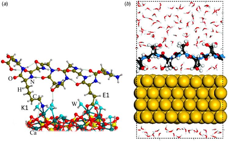
2.3. Self-assembled monolayers 4
                        2.4. Polymers 5
                        2.5. Carbon allotropes 5

                    3. Which surface properties need consideration in modeling?                        5
                       3.1. Ionization and hydration 6
                       3.2. Polarization 7
                       3.3. Reconstruction 7
                       3.4. Topography 7
                       3.5. Morphology 8

                    4. Which modeling and simulation techniques are applicable to protein–surface interactions?                                  9

                    5. Quantum mechanics studies of protein–surface interactions 9

                    6. Challenges in applying biomolecular molecular mechanics force fields to protein–surface interactions 12
                       6.1. Interaction potentials 12
                       6.2. Solvation models 14

                    7. All-atom molecular mechanics studies of protein–surface interactions 15
                       7.1. Metal surfaces 16
                       7.2. Titanium oxide surfaces 17
                       7.3. Silicon oxide surfaces 19
                       7.4. Mineral surfaces 20
                       7.5. Self-assembled monolayer surfaces 21
                       7.6. sp2-Carbon surfaces 21

                    8. Coarse-Grained molecular mechanics modeling of protein–surface interactions 23

                    9. Applications of sampling methods to protein–surface interactions 24
                       9.1. Molecular dynamics 24
                       9.2. Monte Carlo methods 26
                       9.3. Brownian dynamics 27

                  10. Applications of free energy calculation methods to protein–surface interactions 28
                      10.1. Equilibrium methods 28
                      10.2. Non-equilibrium methods 30

                  11. Outlook and future directions 30

                  Acknowledgements 31

                  References 31

                  1. Introduction
                  Protein–inorganic surface interactions have gained increasing attention owing to their widespread occurrence in nature, and
                  their broad range of applications in nanobiotechnology (Choi et al. 2009; Hill et al. 2007; Hu et al. 2005; Jackson et al. 2000;
                  Laera et al. 2011; Manecka et al. 2014; Millo et al. 2009; Park et al. 2008; Qin et al. 2007a, b; Slocik et al. 2011; Xu et al. 2010).
                  Adhesion of proteins on solid substrates is utilized by many organisms, e.g. sea urchins make use of the adsorption of matrix
                  proteins to specific crystal patches on endoskeletal calcite surfaces (Wilt, 1999), and has even been evolutionarily adapted to
                  enable some organisms to live in specific habitats, e.g. for the adhesion of mussels to mineral rocks (Yu et al. 2011). Humans
                  have long used inorganic materials that make direct interactions with proteins. For example, gold crowns as dental prosthetics
                  date back to the ancient Etruscan civilization (Demann et al. 2005), and man-made nanoparticles were used as pigments in
                  ointments by the ancient Romans (Casals et al. 2008). However, it is only quite recently that advances in science and tech-
                  nology have enabled the production of completely new or engineered surfaces and hence allowed new applications. For exam-
                  ple, the remarkable structural and mechanical properties of graphene, isolated in 2004 (Novoselov et al. 2004), have drawn
                  increasing attention to carbon allotropes and catalyzed further research into their interactions with proteins, motivated by
                  various biotechnological applications, including efficient biosensors (Alava et al. 2013).

                                                                                               2
Downloaded from https://www.cambridge.org/core. IP address: 46.4.80.155, on 12 Feb 2021 at 13:21:10, subject to the Cambridge Core terms of use, available at https://www.cambridge.org/core/terms
. https://doi.org/10.1017/S0033583515000256
Computational modeling and simulation of biomolecules can help scientists to unravel the mechanisms of molecular-level
events and predict the behavior of complex systems at a level of detail that cannot be directly measured in experiments.
Since the development of the first modeling and simulation methods for complex molecules, computational research in
the field has expanded enormously. Their importance is shown by the Nobel Prize in Chemistry being awarded in 2013
to Martin Karplus, Michael Levitt and Arieh Warshel “for the development of multiscale models for complex chemical systems”.
Proteins, nucleic acids, lipids and their interactions in aqueous environments have been widely studied computationally by
means of molecular mechanics (MM) force-fields (FFs) (Brooks et al. 1983; Cornell et al. 1995; Oostenbrink et al. 2004) de-
veloped and tailored specifically for these types of molecules. However, many of these FFs fall short in reproducing the
properties of protein-inorganic surface systems. To alleviate this problem, many useful models (Heinz et al. 2011; Iori &
Corni, 2008; Kokh et al. 2010) and FF parameters (De la Torre et al. 2009; Heinz et al. 2013; Iori et al. 2009; Schneider &
Colombi Ciacchi, 2010; Wright et al. 2013a) for material surfaces have been introduced that have been designed to be com-
patible with FFs for biomolecular systems. These FFs are still rather young and their improvement is an area of active research.
A number of reviews addressing different aspects of protein–surface interaction studies have been published previously.
Several of these provide a general overview of the adsorption of proteins at solid surfaces (Cohavi et al. 2010; Costa et al.
2013; Horbett & Brash, 1995; Rabe et al. 2011; Qu et al. 2013), whereas others focus on more specific aspects such as the
determination of the adsorption kinetics of protein–surface binding by weakly bound mobile precursor states (Garland
et al. 2012), and adsorption on various different surface types, such as metallic surfaces (Tomba et al. 2009; Vallee et al.
2010), polymer surfaces (Hahm, 2014; Wei et al. 2014) and protein repellent surfaces (Szott & Horbett, 2011). The physico-
chemical properties of nanomaterials, and their applications in medicine, biology and biotechnology, have also been reviewed
in several papers (see, e.g. Ansari & Husain, 2012; Dufort et al. 2012; Khlebtsov & Dykman, 2010; Mahmoudi et al. 2011;
Mahon et al. 2012; Mandal et al. 2014; Salata, 2004; Saptarshi et al. 2013; Shemetov et al. 2012). Various aspects of the com-
putational methods employed in modeling and simulation of protein–surface interactions are addressed in other reviews, in-
cluding issues in computational modeling of peptide–surface interactions (Di Felice & Corni, 2011), problems with these
simulation techniques (Latour, 2008) and approaches to multiscale modeling of soft matter that are transferable to protein-
surface systems (Praprotnik et al. 2008).
This review provides a discussion of the computational models and simulation techniques that have been used in studies of
protein–surface interactions. Due to the broad range of models used in these studies, only models of protein–surface inter-
actions based on chemical structures are discussed in this review and, therefore, more abstract models to describe these inter-
actions, such as that developed by Oberle et al. (2015) for the description of competitive adsorption of proteins to
nanoparticles, are not discussed further. A brief introduction to various types of material surfaces is provided and some of
the properties that need attention from a modeling point of view are discussed. We further give an overview of the different
modeling, sampling and free energy calculation techniques employed in recent studies. We discuss the properties that can be
computed by these methods and how they can assist and complement experiments. Some of the important findings from
applications of these methods are reviewed, and drawbacks and shortcomings of the available techniques are discussed.
The paper concludes with a discussion of the general limitations and future directions of the field.
2. Which types of surfaces can be modeled?
The interactions of proteins with inorganic materials are determined not only by the properties of the proteins, but also by the
chemical composition, molecular structure, size and shape of the material. Inorganic surfaces possess distinct physico-
chemical properties, such as reactivity towards different compounds, material stability and specific adsorption characteristics
for different adsorbents. These properties allow different types of surfaces to be employed for different applications, e.g. im-
plantation or chromatography. Understanding the basis of these physico-chemical properties usually requires atomic-level
investigation of the surfaces. The types of material surface modeled commonly in computational studies of protein–surface
interactions are elemental metals and alloys, metal oxides and minerals, self-assembled monolayers (SAMs), polymers and
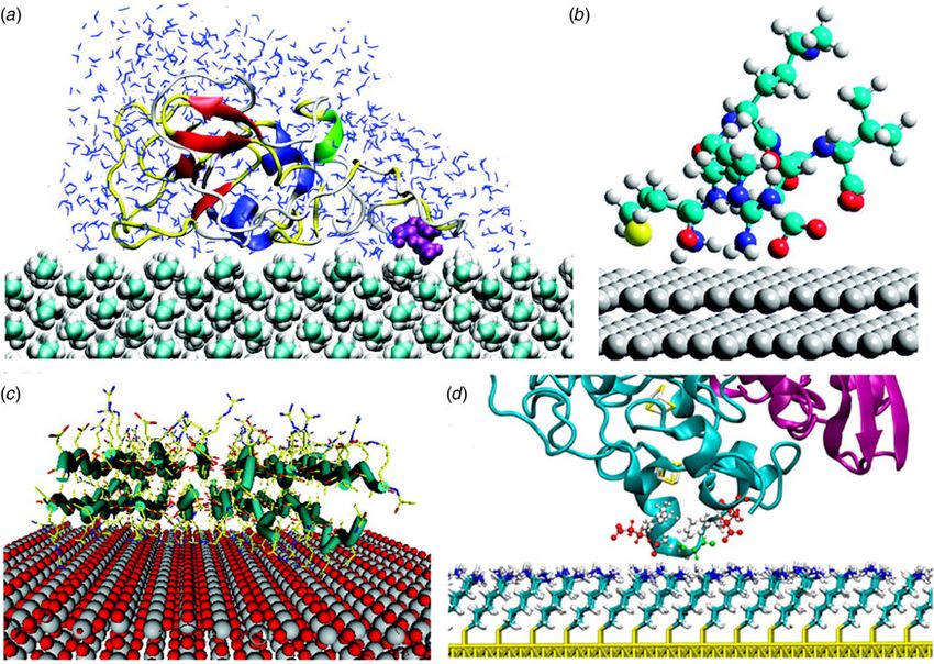
carbon allotrope surfaces, see Fig. 1.
2.1 Elemental metals and alloys
Protein–metal surface interactions can be studied with experimental techniques such as atomic force microscopy (AFM)
(Binnig et al., 1986), surface plasmon resonance (SPR) (Jönsson et al. 1991) and localized SPR (Stuart et al. 2005). Due to
their chemical inertness and unique optical properties (Jain et al. 2008), the noble metals, gold and silver, are the most com-
monly used metals employed as probes or sensors in these techniques. Along with well-known applications of metal surfaces,
such as biosensors and implants (Liu et al. 2004), metal surfaces are also used in bioelectronics as electrodes as they allow
3
Downloaded from https://www.cambridge.org/core. IP address: 46.4.80.155, on 12 Feb 2021 at 13:21:10, subject to the Cambridge Core terms of use, available at https://www.cambridge.org/core/terms
. https://doi.org/10.1017/S0033583515000256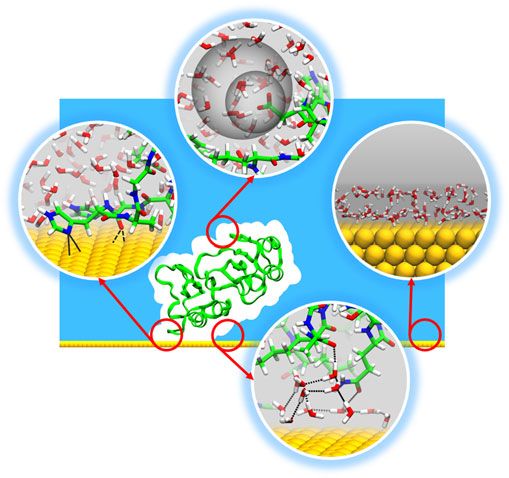
Fig. 1. Simulations of proteins with different types of surface: (a) lysozyme on a polyethylene surface (reprinted with permission from
                  (Wei et al. 2011). Copyright (2011) American Chemical Society), (b) the MRKDV peptide on a bare silver surface (adapted with per-
                  mission from (Aliaga et al. 2011). Copyright (2011) American Chemical Society), (c) RAD16II on a rutile surface (reprinted with per-
                  mission from (Monti, 2007). Copyright (2007) American Chemical Society), and (d) NiFe hydrogenase on a SAM surface (reprinted with
                  permission from (Utesch et al. 2013). Copyright (2013) American Chemical Society).

                  controlled exchange of electrons with metalloproteins immobilized on them (Alessandrini et al. 2005; Andolfi et al. 2004). The
                  interactions of proteins with bare metal surfaces have been the subject of many computational studies, which cover a wide
                  range of different elemental and alloy surfaces, such as Cu(100) (Chen & Wang, 2010), Au(111) (Bizzarri, 2006; Hoefling
                  et al. 2011; Siwko & Corni, 2013; Venkat et al. 2007; Zanetti-Polzi et al. 2014), Au(100) (Hagiwara et al. 2009),
                  Au nanoparticle (Todorova et al. 2014), Fe (Zhang et al. 2009b), Ni (Yang & Zhao, 2006), Pd (Coppage et al. 2011),
                  Pt (Kantarci et al. 2005), Ag (Aliaga et al. 2011; Ghosh et al. 2012) and steel (Imamura et al. 2003).

                  2.2 Oxides and minerals
                  Metal surfaces (excluding noble metals such as gold, platinum and palladium) are oxidized when exposed to water or air,
                  forming metal oxides that are very common in the Earth’s crust. Due to their good mechanical stability, catalytic properties
                  and biocompatibility (Andreescu et al. 2012), metal oxides and minerals are used in a wide range of applications that include
                  fabrication of biomaterials (Whaley et al. 2000), cellular delivery of drugs and biomolecules (Kievit & Zhang, 2011; Xu et al.
                  2006), tissue engineering (Shin et al. 2003) and proteomics (Sugiyama et al. 2007). Computational studies to investigate the
                  interactions of oxide and mineral surfaces with proteins or peptides have mostly been carried out for different forms of ti-
                  tanium dioxide, such as rutile and anatase (Carravetta et al. 2009; Kang et al. 2010; Köppen et al. 2008; Monti, 2007;
                  Monti et al. 2008; Sun et al. 2014a; Wu et al. 2013), silicon dioxides (Chen et al. 2009a; Nonella & Seeger, 2008;
                  Patwardhan et al. 2012; Rimola et al. 2009; Tosaka et al. 2010), calcite (Wierzbicki et al. 1994) and mica (Kang et al. 2013).

                  2.3 Self-assembled monolayers
                  SAMs are thin films that coat surfaces by spontaneous adsorption of organic molecules that form ordered molecular assem-
                  blies. Typically, in a SAM, molecules are chemisorbed onto a surface substrate through their reactive head groups and are,
                  therefore, very stable. SAMs can be categorized into two groups according to their head-group type: thiol-based and silate-
                  based (Schreiber, 2004). The head-group is attached to a tail group that, by following a fast adsorption phase (seconds),
                                                                                               4
Downloaded from https://www.cambridge.org/core. IP address: 46.4.80.155, on 12 Feb 2021 at 13:21:10, subject to the Cambridge Core terms of use, available at https://www.cambridge.org/core/terms
. https://doi.org/10.1017/S0033583515000256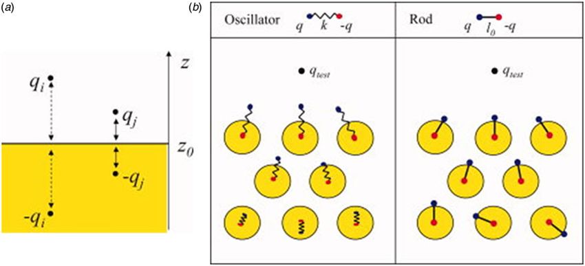
undergoes a slow reorganization (hours), in which interactions with other tail groups increase and the packing is improved
                  (Love et al. 2005). The tail group can be functionalized with small chemical groups or large molecules, such as peptides. These,
                  together with the length of the tail group, allow the physico-chemical interfacial properties, in particular, hydrophobicity and
                  ionization to be adjusted according to the desired application. Alkanethiols are the most common type of SAM. They have the
                  chemical formula S-(CH2)n-R, where R stands for a functional group, such as -CH3, -COOH, -NH2 or -OH.
                  Bare metals and oxide surfaces are prone to non-specific adsorption of proteins and other organic molecules. These adsorp-
                  tion processes may result in undesirable agglomeration of adsorbates on the surfaces. The self-assembly of organic molecules
                  on a metal surface in a SAM creates a physical barrier between the surface and the adsorbates, acting as an electrical insulator
                  and passivating the surface atoms (Love et al. 2005). The properties of SAMs have been reviewed in (Chaki & Vijayamohanan,
                  2002; Gooding et al. 2003; Love et al. 2005; Senaratne et al. 2005). Modeling of protein–SAM interactions has been reported,
                  mostly for alkanethiol SAMs, in (Alvarez-Paggi et al. 2010; Hsu et al. 2008; O’Mahony et al. 2013; Sun et al. 2005; Utesch et al.
                  2013; Wang et al. 2010a, b; Xie et al. 2012) and peptide–SAM interactions have been modeled by Nowinski et al. (2012).

                  2.4 Polymers
                  Polymer surfaces have attracted much attention in the nanotechnology field due to their mechanical stability, low cost and
                  their wide applicability (Nie & Kumacheva, 2008).
                  Particularly synthetic polymer-based biomaterials, due to their non-fouling properties, are currently being investigated inten-
                  sively for applications in controlled drug delivery (Hoffman, 2008), in highly sensitive biosensors (Anker et al. 2008), and in
                  bioelectronics (Senaratne et al. 2005). Nanostructured polymer materials used for bio-related and medicinal research include
                  electrostatic polymer brushes, micelles, layer-by-layer deposition and thin films (Stuart et al. 2010). Polymer brushes are sur-
                  face modifiers that share many properties in common with SAMs (Senaratne et al. 2005). They are prepared by grafting poly-
                  mers of the same or varying kinds on surfaces forming homopolymer and mixed brushes, respectively. Polymeric micelles are
                  formed through self-assembly of amphiphilic copolymers and typically have a diameter of size 30–50 nm (Otsuka et al. 2003;
                  Stuart et al. 2010).
                  These micelles are particularly important due to their lower critical micelle concentration (CMC), higher stability and slower
                  rate of dissociation than surfactant micelles. These properties have allowed polymeric micelles to act as effective cancer treat-
                  ment tools with high drug deposition at the target site (Otsuka et al. 2003). Polymer surfaces and their applications have been
                  reviewed by (Barbey et al. 2009; Kim et al. 2008; Nie et al. 2010; Otsuka et al. 2003; Senaratne et al. 2005; Stuart et al. 2010).
                  The limited number of computational studies of protein-polymer surfaces to date have been carried out for polymer types
                  including polystyrene, polyethylene and polydimethylsiloxane (Boughton et al. 2010; Jeyachandran et al. 2009; Liu et al.
                  2012; Lu et al. 1992; O’Brien et al. 2008; Raffaini & Ganazzoli, 2007; Zhang et al. 2009a; Wei et al. 2011).

                  2.5 Carbon allotropes
                  Pure carbon may exist in a number of different allotropes. Owing to their unique thermal, electrical, chemical and mechanical
                  properties, carbon-based nanomaterials have been the subject of numerous applications in analytical chemistry (Scida et al.
                  2011). These applications mostly focus on carbon nanomaterials with sp2-carbon bonding, such as fullerenes, carbon nano-
                  tubes (CNT), graphene and graphite. This is due to the extremely high surface areas of fullerenes and CNTs relative to their
                  size, which makes them suitable for design as highly efficient drug carriers. Furthermore, the excellent electrical properties of
                  graphene and CNTs make them suitable for biosensor applications (Liu & Liang, 2012). In all the four materials, the carbon
                  atoms make three chemical bonds with other carbons in the surface-plane with delocalized π electron clouds in the direction
                  perpendicular to the surface (Scida et al. 2011). This configuration makes the mutual van der Waals interactions between
                  CNTs very strong and hence leads them to be very hydrophobic (Guldi et al. 2006). To alter the hydrophobicity, surface mod-
                  ifications with surface defects and polar groups have been suggested, but these affect the stability of the materials as well as
                  their mechanical and electrical properties (Scida et al. 2011). Computational studies of protein–carbon surface interactions
                  have mostly focused on graphene/graphite (Mereghetti & Wade, 2011; Mücksch & Urbassek, 2011; Raffaini & Ganazzoli,
                  2010; Kang et al. 2013; Sun et al. 2014b; Yu et al. 2012b), CNT (Balamurugan et al. 2010; Chen et al. 2009b; Tallury &
                  Pasquinelli, 2010; Wang et al. 2003) and fullerenes (Durdagi et al. 2008; Kraszewski et al. 2010; Noon et al. 2002).

                  3 Which surface properties need consideration in modeling?
                  The modeling of protein-water-solid surface interfaces poses problems because a variety of distinct physical and chemical
                  properties may be associated with different surface types. Factors such as the size and shape of nanoparticles, the crystal
                                                                                               5
Downloaded from https://www.cambridge.org/core. IP address: 46.4.80.155, on 12 Feb 2021 at 13:21:10, subject to the Cambridge Core terms of use, available at https://www.cambridge.org/core/terms
. https://doi.org/10.1017/S0033583515000256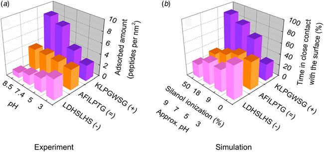
packing of a surface, the presence of surface defects, the density of SAM molecules, the change of the chemical state of the
                  surface, such as protonation, oxidation, or hydroxylation, due to the presence of water and the environmental conditions, such
                  as the presence of surfactants, all affect the interaction of biomolecules and the solid substrate. Therefore, it is very important
                  to choose the level of microscopic details to be included in the computational model carefully when modeling protein–surface
                  interactions, which must adequately describe the physical and chemical properties of the studied system under experimental
                  conditions.
                  Several important characteristics of the surfaces that should be considered in modeling protein adsorption are discussed in the
                  next sections: ionization and hydration, polarization, surface reconstruction upon binding, as well as the topography and mor-
                  phology of surfaces and nanoparticles.

                  3.1 Ionization and hydration
                  SAMs and metal oxide surfaces may be protonated and/or hydroxylated to varying degrees depending on the environmental
                  conditions and on the material itself, e.g. pH, material shape and size. Many theoretical studies on SAMs do not take into
                  account the ionization states of the functional groups of SAMs. However, a recent study (Utesch et al. 2013) reporting a ti-
                  tration curve of an amino-terminated alkanethiol SAM showed that the level of ionization was very sensitive at pH values
                  around 6 (with 16 ± 5% protonation at pH 7 and 52 ± 9% at pH 6). A positive correlation between the ionization level of
                  a SAM and the strength of adsorption of charged proteins was found (Zhou et al. 2004; Utesch et al. 2013). Therefore, it
                  is crucial to compute and model the surface ionization to obtain reliable results compatible with experiments. The protonation
                  states of SAMs in modeling studies are mostly represented by random assignment of protonated and deprotonated groups
                  (Alvarez-Paggi et al. 2013; Utesch et al. 2012; Zhou et al. 2004). In several studies, they are represented either by a uniform
                  distribution of small partial charges (Sun et al. 2005) or by large partial charges being assigned to functional groups in neutral
                  surfaces (Wang et al. 2010b).
                  As with SAMs, SiO2 and TiO2 surfaces have different levels of ionization of the surface groups depending on the pH of the
                  environment, and this has been shown to determine selectivity of the adsorption of proteins (Patwardhan et al. 2012). Studies
                  have shown that the concentration of silanol groups (Si-OH) and the degree of their ionization define the hydrophobicity of
                  silicon dioxide (silica) surfaces and govern their adsorption properties and thus also, the behavior of silica-based materials in
                  processes such as biomolecular adhesion and biomineralization (Sumper & Brunner, 2008; Voskerician et al. 2003). In com-
                  putational studies of protein–oxide surface interactions, ionization may be represented explicitly (Friedrichs et al. 2013;
                  Köppen & Langel, 2010; Patwardhan et al. 2012; Tosaka et al. 2010), or by assigning uniform partial charges to selected sur-
                  face atoms (Kubiak-Ossowska & Mulheran, 2012). Köppen & Langel (2010) showed that the adhesion energies of peptides on
                  titanium dioxide surfaces are sensitive to the values of the partial charges of the surface hydroxyl groups. Therefore, care must
                  be taken to ensure a reliable parameterization of the ionization charges.
                  Conventional simulations of proteins usually neglect changes in the protonation states of ionizable groups as a function of
                  time. However, an accurate simulation of a protein-surface system may require a more sophisticated approach, such as con-
                  stant pH simulation, to treat the variation of the protonation states of residues in the interfacial region, as well as of the surface
                  interaction sites. Although constant pH simulation techniques have been applied to various molecular systems, including
                  small chemotherapeutic drug molecules binding to nanodiamonds (Adnan et al. 2011), they have not, as far as we are
                  aware, been applied to simulations of peptide/protein–surface interactions.
                  The most important issue for modeling the adsorption of biomolecules on oxide surfaces is the treatment of the properties of the
                  surface hydration shell. Due to dissociation, the hydroxyl groups and hydrogen atoms form bonds with unsaturated surface
                  metal (e.g. Ti) and O atoms, respectively. The degree of water dissociation defines the physical properties of the surface and
                  strongly affects the binding properties of biological molecules. Kang et al. (2010) investigated the role of the water in the adsorp-
                  tion process on rutile surfaces and observed that human serum albumin (HSA) adsorbs on a rutile surface modified with -OH
                  groups more strongly than on an unmodified rutile surface. Hydrogen bond analysis in the same study showed that the bonds
                  formed between the structured hydroxyl groups on the modified surface reduced the possibility of hydrogen bond formation
                  between the surface and the water molecules, hence making it easier for the protein to adsorb onto the modified surface.
                  It should be noted that water dissociation is reversible on all oxides (Henderson, 2002). Therefore, the concentration and po-
                  sition of hydroxyl groups may change with time. Even though relying on the average distribution of hydroxyl groups on a
                  particular surface may be sufficient, it may not always give accurate results, especially if the protein adsorption kinetics
                  are determined by surface hydration kinetics. This poses a problem for conventional modeling techniques for biomolecules,
                  which ideally have to capture the dynamics of dissociative adsorption and associative desorption of water molecules on oxide
                  surfaces.
                                                                                               6
Downloaded from https://www.cambridge.org/core. IP address: 46.4.80.155, on 12 Feb 2021 at 13:21:10, subject to the Cambridge Core terms of use, available at https://www.cambridge.org/core/terms
. https://doi.org/10.1017/S0033583515000256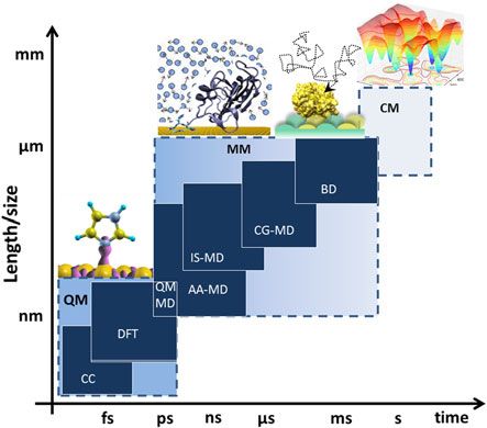
3.2 Polarization
                  The electrostatic potential of solutes and solvent molecules induces an attractive polarization of metal surfaces. Metal polar-
                  ization is negligible for neutral adsorbates that do not have large dipoles and, since proteins usually have a relatively small total
                  charge, it is often neglected in simulations (Braun et al. 2002). Early studies of metal surfaces also showed that the effect of
                  pure water on polarization of the metal is often negligible (Barabino & Marchesi, 1984; Shelley et al. 1997; Spohr, 1995) and
                  that charge-induced polarization does not cause any change in the structure of the water on the surface (Feng et al. 2011).
                  However, for the adsorption of biomolecules with considerable dipole moments, including some peptides and proteins,
                  the surface polarization contributes to the binding energy and influences the binding mode. Although studies have shown
                  that, for water-metal surface systems, the energy due to polarization is less than 10% of the total binding energy (Feng
                  et al. 2011; Neves et al. 2007; Siepmann & Sprik, 1995; Vila Verde et al. 2009, 2011), for proteins, in particular, on Au
                  (111) surfaces, the contribution from polarization has been estimated to be about 10–20% of the total binding energy
                  (Heinz et al. 2011). Further, on surfaces such as Au(100), where the van der Waals attraction is weaker, polarization was
                  found to tune the adsorption of proteins (Heinz et al. 2009) and act as a major contributor to the adsorption of highly charged
                  peptides (Heinz et al. 2011). For the simulation of amino acid adsorption on Au(111), polarization effects were also found to
                  be important for reproducing experimental binding propensities (Hoefling et al. 2011).
                  The polarization of surfaces of other types can also play an important role in the interactions of surfaces with their environ-
                  ments. Schyman & Jorgensen (2013) showed that while a non-polarizable FF is adequate for the description of interactions
                  between water and small hydrocarbons, such as benzene (C6H6) and coronene (C24H12), a polarizable FF is required for CNTs
                  and fullerenes in order to reproduce interaction energy values obtained from density functional theory (DFT) calculations.
                  Therefore, the effect of polarization should be carefully considered in computer simulations of biomolecules, in particular,
                  of charged proteins, with metal and carbon surfaces.

                  3.3 Reconstruction
                  The atomic structure of the surface of a material generally differs from that of its interior because of differences in the forces
                  acting on the atoms in the vicinity of the surface. The type and the degree of reconstruction of a surface is determined by
                  environmental conditions, such as temperature and pressure (Somorjai & Li, 2011), as well as the structure of the material
                  and may be affected by adsorbing molecules. Ideally, reconstruction of surfaces has to be taken into account in the simulation
                  of adsorption (Ghiringhelli et al. 2008): an enormous task in most cases. On the other hand, it has been reported in several
                  studies that the reconstruction of some surfaces in certain conditions is negligible (Feng et al. 2011; Iori et al. 2008; Raffaini &
                  Ganazzoli, 2012; Wright et al. 2013a). However, other experimental and computational studies show that large scale surface
                  reconstruction may take place after adsorption of small molecules (Eralp et al. 2011; Gibbs et al. 1990; Lal et al. 2004, 2006).
                  Therefore, care must be taken in modeling if a major reconstruction of the surface takes place upon adsorption.

                  3.4 Topography
                  The topographic characteristics of a surface or nanoparticle at the micro- to nanometer-scale are important determinants of
                  protein adsorption properties, such as binding affinities and surface saturation values (Fenoglio et al. 2011; Gagner et al. 2011,
                  2012; Roach et al. 2006). The topography of a surface can be characterized by its exposed crystal planes, its roughness and its
                  defects (due to locally varying chemical composition or the crystalline structure of the surface), as well as kinks, edges and
                  steps that occur during the growth of a crystal. Studies have shown that the same type of protein may have different adsorp-
                  tion energies, varying from highly favorable to highly unfavorable, for surfaces with different lattice structures but the same
                  chemical composition (Heinz et al. 2009; Oren et al. 2005). Although modeling of the intrinsic crystal structure is straight-
                  forward, it is important to consider that different crystal planes of a material (which can be kinetically or thermodynamically
                  favored) may be exposed during a surface adsorption process. For example, during the growth of a nanoparticle, different
                  surface planes of the same particle can display different structures at the same time, e.g. (100) and (111) (Korzeniewski
                  et al. 2011).
                  Similarly, protein binding properties depend on the material structure. Rechendorff et al. (2006) showed that adsorption of
                  fibrinogen on a tantalum surface can be induced by up to 70% by increasing the surface (root mean squared) roughness from
                  2·0 to 32·9 nm. The increase was much greater than the increase in the surface area due to the surface roughness of around
                  20%. On the other hand, Rechendorff et al. (2006) found that the adsorption of the more globular protein, bovine serum
                  albumin, to tantalum induced by the surface roughness was similar to the increase in the surface area, thus demonstrating
                  a selective effect of the material structure on the adsorption processes of different proteins. Finally, using molecular dynamics
                  (MD) simulations, Nada (2014) investigated the interactions of an aspartic acid with step edges and kinks, as well as flat
                  regions of a calcite crystal surface. They showed that aspartic acid binds preferentially to an acute step edge and not to an
                                                                                               7
Downloaded from https://www.cambridge.org/core. IP address: 46.4.80.155, on 12 Feb 2021 at 13:21:10, subject to the Cambridge Core terms of use, available at https://www.cambridge.org/core/terms
. https://doi.org/10.1017/S0033583515000256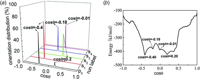
obtuse one, due to the ordered structure of water near the obtuse step edge that prevented the aspartic acid from
                  binding strongly (Nada, 2014). In summary, these studies demonstrate that the topography of a surface may determine its
                  binding characteristics to proteins and peptides. Although, many topographic features can be neglected in modeling studies
                  when comparing to experiments with well-characterized surfaces, the characteristics of the surface topography should be
                  taken into account to realistically model and simulate the interactions of proteins with the materials used in real-life
                  applications.

                  3.5 Morphology
                  The size of a nanoparticle, and therefore the curvature of its surface, has a strong impact on both the physico-chemical charac-
                  teristics of the nanoparticle itself, such as surface polarizability, surface charge (Chiu et al. 2009) and isoelectric point
                  (Suttiponparnit et al. 2010), and the properties of the layer of coating molecules which can determine its acidity (Wang
                  et al. 2011). The curvature of a nanoparticle may change the properties of its hydration shell as well. The solvation free energy
                  of a nanoparticle becomes more favorable as the surface curvature decreases (and the size of the particle increases) owing to
                  the increased water–particle interaction energy, which in turn determines its polarity (Chiu et al. 2009).
                  The effect of surface curvature on protein adsorption properties, i.e. the kinetics, thermodynamics and structural stability of
                  adsorbed proteins, increases with decreasing particle size (Lacerda et al. 2010), but also depends on the adsorbate. Moreover, a
                  strong difference in binding to spherical nanoparticles and nanorods is often observed (Gagner et al. 2011). A structural adap-
                  tion to the curvature of the nanoparticle surface upon adsorption may lead to a loss of enzymatic activity of some proteins
                  (Wu & Narsimhan, 2008), or it may lead to significant changes in secondary or tertiary structures of self-assembling peptides
                  (Shaw et al. 2012) or proteins (Tavanti et al. 2015; Yang et al. 2013) adsorbing onto the surfaces. It has been shown that while
                  surface curvature may help to retain the tertiary structure of some proteins with globular structures adsorbed on small nano-
                  particles (Vertegel et al. 2004; Lundqvist et al. 2004), it can also cause significant loss in the secondary structure of a protein
                  upon adsorption (Gagner et al. 2011).
                  The effects of the surface curvature of nanoparticles can be neglected in computational studies when the binding of relatively
                  small proteins to large nanoparticles is studied. However, the size and curvature of a nanoparticle may play a major role in
                  the adsorption patterns of proteins not only due to geometric adaptation of the protein to a nanoparticle of similar size, but
                  also due to changes in the physico-chemical properties of the nanoparticle itself. DFT calculations of amino acid adsorption
                  on a single-walled carbon nanotube (SWCNT) showed that glycine adsorbs more strongly on a nanotube (3,3) than on a flat
                  graphite surface, whereas phenylalanine adsorbs more strongly on a flat graphite surface, and the amino acids cysteine and his-
                  tidine, showed no significant change in their adsorption energies (Roman et al. 2006). Binding free energy calculations of amy-
                  loidogenic apoC-II(60–70) peptide on fullerene, CNT and graphene using MD simulations showed that the binding affinity was
                  weakest for the fullerene and strongest for the graphene due to reduced efficiency of π-stacking interactions between the aromatic
                  side chains of the peptide and the fullerene and CNT arising from the increased surface curvature (Todorova et al. 2013). Xie
                  et al. (2014) calculated the adsorption free energies of Alzheimer’s β-amyloid peptide fragments (Aβ) to two different fullerene
                  nanoparticle systems, C180 and three C60 (3C60). They found tighter binding of the peptides on the larger C180.
                  Raffaini & Ganazzoli (2013) showed that the binding strength of a protein to a carbon nanotube depends on the morphology
                  and that the interaction energy between the protein and the nanoparticle was larger for the concave interior surface of the
                  nanotube than for the convex outer surface. In a similar study, Chen et al. (2009b) showed that the length and the diameter
                  of CNTs affect the energetics of the interactions with a peptide drug. While longer CNTs provide more space to trap the pep-
                  tide inside the tube, a smaller diameter increases the interaction energy.
                  The surface morphology not only alters the binding characteristics of a peptide or protein to a surface but also the interactions
                  between biomolecules near a surface or a nanoparticle, thus leading to intermolecular structural changes. Li et al. (2011) per-
                  formed 3 separate sets of simulations of Aβ(16–22) peptides that abnormally self-assemble into β-rich aggregates: amorphous
                  peptide in solution, amorphous peptide with SWCNT, and prefibrillar peptide with SWCNT. They observed that without the
                  CNTs, the amorphous peptides form β-sheet structures. On the other hand, simulations with SWCNTs showed that the
                  amorphous peptides tend to form disordered coils, whereas the β-sheet structures formed by the prefibrillar peptides were
                  destabilized due to the interactions of the peptides with the CNTs.
                  Finally, noteworthy to mention is that a change in the morphology of a surface/particle will lead to changes in its physico-
                  chemical properties. In a study by Emami et al. (2014b), it was shown that the size of the silica nanoparticles determines their
                  surface ionization levels. The larger nanoparticles have higher ionization and therefore bind more strongly to peptides with
                  high net positive charge. Further, Baier et al. (2014) recently investigated the binding free energy profile of 12-mer peptides on
                  polar (001) and non-polar (100) ZnO surfaces. Employing an enhanced sampling approach (see Section 10.2), the authors
                                                                                               8
Downloaded from https://www.cambridge.org/core. IP address: 46.4.80.155, on 12 Feb 2021 at 13:21:10, subject to the Cambridge Core terms of use, available at https://www.cambridge.org/core/terms
. https://doi.org/10.1017/S0033583515000256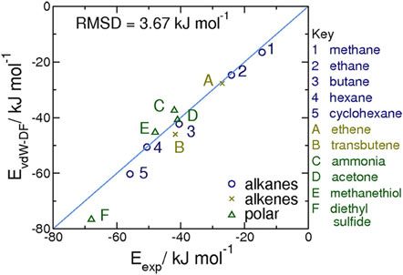
showed that there is a positive correlation between the ZnO particle sizes obtained in experiments in the presence of the pep-
                  tides and the calculated affinities of the peptides for the ZnO surface. Their result showed that the selective adsorption of a
                  peptide can impact the growth of certain nanocrystals. Therefore, one should consider modeling, not only the size and cur-
                  vature of the material, but also other properties that depend on the morphology, in particular for nanoparticles.

                  4. Which modeling and simulation techniques are applicable to protein–surface
                  interactions?
                  The approaches used for modeling protein-inorganic surface systems cover a broad range of scales from sub-atomic quantum
                  mechanical (QM) through classical atomic levels to mesoscopic descriptions and further to continuum descriptions on a
                  macroscopic scale. Figure 2 shows the most common techniques to model and to simulate molecular interactions.
                  Simulations at the QM level are applied to systems of a few hundreds of atoms at most and they do not reach the nanosecond
                  scale, hence they are directly applicable only to small nanoparticle-ligand systems (Mahmoudi et al. 2011).
                  Many physical processes at the protein-solid surface interface are driven by physisorption, i.e. proceed without formation of
                  chemical bonds between the adsorbate and the solid surface. In contrast to the relatively well-understood chemisorption, the
                  nature and behavior of non-covalent adsorption is often unclear since multiple factors, which depend strongly on the surface
                  type, influence the interactions that govern the adsorption process. Even if chemical binding takes place, physical adsorption
                  drives the first stages of molecular recognition and induces long-time scale structural adaptations of a protein to a solid sur-
                  face. Non-covalent binding processes can be described within a MM framework, which drastically reduces computational
                  costs compared with QM, and thus enables the simulation of the dynamics of systems that consist of millions of atoms
                  for up to microseconds with solvent molecules modeled explicitly. All-atom MM simulations are important, in particular,
                  for investigation of the dynamic and thermodynamic properties of protein adsorption (Mahmoudi et al. 2011; Utesch
                  et al. 2011). The nano length scale is usually appropriate for studying protein–surface interactions at the molecular level,
                  and hence, all-atom simulations are common methods of choice (Gagner et al. 2012).
                  In experiments, adsorption events typically take place over time periods from milliseconds to hours which are far from the
                  accessible time scales of all-atom simulations (Mücksch & Urbassek, 2011). The initial stages of protein adsorption to surfaces
                  occur on a sub-second time scale. These may be followed by a slow stage in which large secondary structural changes occur,
                  such as the transition from α-helix towards β-sheet of lysozyme on a SAM surface (Sethuraman & Belfort, 2005), and may
                  entail denaturation over periods lasting up to several hours, or even days (Gray, 2004; Pan et al. 2012). Capturing the time and
                  length scales of a complete adsorption process therefore necessitates employing mesoscopic, coarse-grained (CG) and multi-
                  scale approaches (Gray, 2004; Wei et al. 2011). Furthermore, hybrid approaches such as QM/MM to bridge typical time and
                  length scales of conventional approaches, and enhanced sampling simulation techniques to accelerate adsorption and desorp-
                  tion events have been proposed and successfully applied to study protein–inorganic surface interactions (Euston et al. 2008;
                  Utesch et al. 2011; Zhang & Sun, 2010).

                  5 Quantum mechanics studies of protein–surface interactions
                  QM-based methods are those where the quantum nature of electrons is explicitly taken into account while the much heavier
                  nuclei are usually considered as classical particles moving in the field generated by the electrons and the other nuclei. Several
                  approaches belong to this class, ranging from those that are relatively fast but rich in adjustable parameters, such as tightbind-
                  ing and semi-empirical methods, to very accurate but also computationally very expensive, parameter-free calculations, such
                  as coupled-cluster. The QM method that has been applied most extensively so far for studying biomolecules at surfaces is
                  DFT, as it represents the best compromise between accuracy and computational feasibility.
                  The fundamental idea behind DFT (Martin, 2004), based on the Hohenberg and Kohn theorems (Hohenberg, 1964), is that,
                  for non-degenerate systems, the ground state electron density, n(r), alone determines the entire behavior of the system, and
                  that such n(r) minimizes the energy of the system. The most useful approximation of such energy as a function of n(r) has
                  been proposed by Kohn & Sham (1965). It translates the minimization problem to a non-linear, independent particle ap-
                  proach akin to the Hartree–Fock one. A central quantity within this approach is the so-called exchange correlation functional
                  (fxc), i.e. the n(r)-dependent energy contribution due to quantum-mechanical and many-body deviations from a mean-field
                  description of the electrons. So far, there is neither an exact expression for fxc nor a one-fits-all approximation. An important
                  and often not-obvious choice to be made for any DFT calculation is therefore which fxc is the best for the system under study.
                                                                                               9
Downloaded from https://www.cambridge.org/core. IP address: 46.4.80.155, on 12 Feb 2021 at 13:21:10, subject to the Cambridge Core terms of use, available at https://www.cambridge.org/core/terms
. https://doi.org/10.1017/S0033583515000256
Fig. 2. Typical time and length scales of different simulation techniques: quantum mechanics (QM), including coupled cluster (CC) and
DFT methods (inset adapted with permission from (Iori et al. 2008). Copyright (2008) American Chemical Society); molecular mechanics
(MM) including all-atom molecular dynamics (AA-MD) simulations, implicit solvent and coarse grained MD (IS-MD and CG-MD), and
the Brownian dynamics (BD) technique; and continuum mechanics (CM). The ranges of time and length scales are approximate.
Given the geometry of the nuclei in space, DFT allows the forces acting on them to be calculated. This relation can be used to
find the nuclei geometries that provide local and global minima of the system energy (i.e. the optimal or relaxed geometry).
These are obviously the most important structures, since they are the structures in whose neighborhood the system fluctuates.
In the following, we shall refer to this kind of DFT calculation as static. In fact, forces can also be used to simulate the dy-
namics of the nuclei, and therefore, in principle, the thermodynamics as well, via different propagation algorithms, such as
Born-Oppenheimer and Car-Parrinello (Marx & Hutter, 2000). DFT ab initio molecular dynamics (AIMD) is often used to
collectively refer to this kind of simulations. Several software packages are available and maintained for performing static DFT
and/or AIMD calculations. They differ in the numerical approaches implemented to solve the Kohn–Sham equations (e.g.
using plane-waves or localized atom-centered functions), in the available fxc, in the level of parallelism (i.e. suitable for highly
parallel computers or for a few-core, high memory workstation), in the pre- and post-processing tools offered, and finally in
the distribution policy (e.g. open-source versus proprietary, free versus commercial).
Considering proteins on surfaces, static DFT calculations of single amino acids or even di- and tri-peptides, (Lee et al. 2014;
Muir et al. 2014) interacting with a surface, with no solvent present, are now affordable and widely used (Arrouvel et al. 2007;
Di Felice et al. 2003; Di Felice & Selloni, 2004; Ghiringhelli et al. 2006; Iori et al. 2008; Rimola et al. 2009). They may seem
rather distant from biophysically relevant systems, but they can actually provide important information by themselves, or be
preliminary to other approaches (e.g. provide the basis for a classical FF parameterization). Among the DFT calculations that
directly provide useful information, we mention those aimed at understanding whether a covalent bond is established between
an amino acid and an inorganic surface. In fact, when a covalent bond is present (chemisorption), it is unlikely that the pro-
tein and solvent environment missing in the calculations dramatically change its nature, although they do modify the details.
Notable examples of calculations of this kind are works on Cys–Au(111) interactions (Buimaga-Iarinca & Calborean, 2012; Di
Felice et al. 2003; Di Felice & Selloni, 2004; Fajín et al. 2013; Nazmutdinov et al. 2007), often used for protein immobilization
(Vigmond et al. 1994). Static DFT calculations on amino acids or simpler molecules representative of chemical groups in
natural amino acids on metals (Hong et al. 2009; Iori et al. 2008, 2009) have also highlighted the peculiar nature of the
amino acid–metal interaction. Depending on the partners, such an interaction ranges from clear non-bonding (e.g. alkyl
side chains on Au(111)) to clear chemisorption (Cys on Au(111)). It also encompasses border-line cases where the interaction
has some typical covalent bond characteristics, such as electron sharing and directionality, and yet it is only marginally stron-
ger than a non-bonded interaction (e.g. imidazole on Au(111), see Fig. 3).
DFT calculations have also been used to elucidate amino acid adsorption on silica (Rimola et al. 2013), hydroxyapatite
(Jimenez-Izal et al. 2012), alumina (Arrouvel et al. 2007) and titania (Carravetta et al. 2009; Koch et al. 2011) surfaces
where electrostatic interactions and hydrogen bonds are important, and where the formation of chemical bonds often implies
complex reaction mechanisms. The interaction of quartz and aluminosilicate structures with phospholipids was also studied
by DFT to understand why the enzyme phospholipase A2 digests phospholipids faster in the presence of quartz than
10
Downloaded from https://www.cambridge.org/core. IP address: 46.4.80.155, on 12 Feb 2021 at 13:21:10, subject to the Cambridge Core terms of use, available at https://www.cambridge.org/core/terms
. https://doi.org/10.1017/S0033583515000256aluminosilicates (Snyder & Madura, 2008). The interaction of amino-acids with graphene has also been the subject of recent
DFT studies (Akdim et al. 2013; Wang et al. 2014).
Static DFT simulations have been performed to parameterize classical FFs for protein–surface interactions in water
(Bellucci et al. 2012; Carravetta & Monti, 2006; Carravetta et al. 2009; Di Felice & Corni, 2011; Ghiringhelli et al.
2006; Iori et al. 2009; Schneider & Colombi Ciacchi, 2010; Wright et al. 2013a, 2013b). The general strategy here is to
perform energy calculations on the optimized geometries and/or along specific coordinates, for either entire amino
acids or simpler amino acid analogues, on the target surface, and to use the results as a training set. Classical parameters
are thus adjusted so as to reproduce as closely as possible the QM interaction energies and geometries in the classical cal-
culations. DFT calculations have also been used to generate partial atomic charges for the surface atoms to be used for
Coulombic interactions in the classical models.
AIMD is intrinsically much more expensive than static DFT simulations and, for this reason, its application in the protein-
surface field has been less popular. Recently, Motta et al. (2012) investigated the adsorption of glycine on a stepped boehmite
(AlO(OH)) surface in water, identifying inner-sphere adsorption, i.e. displacement of surface hydroxyl groups by glycine
molecules, as the most favorable. Wright & Walsh (2012) focused on ammonium and acetate ions, which are analogues of
common chemical groups in amino acids, at the water/quartz interface. Colombi Ciacchi and colleagues used AIMD to
build a model of a native silicon oxide surface (Colombi Ciacchi & Payne, 2005; Cole et al. 2007) that was afterwards
used for studying peptide–silica interactions (Schneider & Colombi Ciacchi, 2012).
The current limits of static and AIMD DFT calculations in the field of protein–surface interactions are well illustrated by some
recent examples shown in Fig. 4. (Rimola et al. 2012) exploited static DFT to study the adsorption of an entire dodecapeptide
on a hydroxyapatite surface, including some key water molecules (the system was composed of around 500 atoms). They dis-
cussed the driving force for the adsorption and the role of the surface in determining the peptide folding. A system of similar
size (a dodecapeptide on a graphene sheet) has also been investigated by Akdim et al. (2013) to confirm by DFT the adsorbed
peptide geometries obtained with a classical force-field.
Calzolari et al. (2010) considered a model polyserine β-sheet, periodically replicated, simulated by AIMD on Au(111) in liquid
water. This system was composed of approximately 500 atoms and its time evolution could be simulated for 20 ps. Such a
simulation time and the composition of the system allowed the investigation of some specific questions, such as the nature
of the local β-sheet/Au interactions, the competition between water and the serine side chain for gold, as well as the nature of
the β-sheet/water interface. However, it is apparent that several other important questions are inaccessible with this kind of
approach. Today, larger systems and longer AIMD simulations are affordable but the most expensive AIMD simulations are
still confined to a few thousand atoms and a few hundred ps.
Beside the current limitations related to the computational cost of DFT simulations, one notorious drawback of the fxc func-
tionals used so far is worth discussion here. It is the inability to account for long-range dispersion (London) interactions,
sometimes referred to as van der Waals’ interactions, that results in an underestimated interaction strength, and even no
solute-surface binding when such interactions are the only relevant ones (e.g. for inert metal surfaces and saturated, non-polar
molecules). Various corrections have been proposed to solve this problem (Tkatchenko et al. 2010), and some of them have
also been tested in the framework of protein–surface interactions with encouraging results. In particular, the DFT-Dn meth-
ods (Grimme, 2004; Grimme et al. 2010; Wu et al. 2001) (that add to the DFT energy, empirical atom-atom d−6 terms, where
d is the pairwise interatomic distance, suitably damped for small d) have been used for amino acid and peptide adsorption on
mineral surfaces (Folliet et al. 2013; Rimola et al. 2009, 2012) and on graphene (Akdim et al. 2013). The fxc functional,
vdW-DF, which does not contain empirical terms (Dion et al. 2004), has been tested against experimental desorption energy
data for some small molecules on Au(111) (Wright et al. 2013a). It was used to provide the main data (stable geometries and
the related energies for amino acid analogues on Au(111) and Au(100)) needed to parameterize the GolP-CHARMM FF
(Rosa et al. 2014b; Wright et al. 2013a). Tests confirmed the reliability of the vdW-DF adsorption energies, within a few
kJ/mol of the experimental values (Fig. 5), and pointed to an already documented tendency of vdW-DF to provide contact
distances and Au lattice parameters that are slightly too large by 0·1–0·2 Å (Lee et al. 2010). Other functionals akin to
vdW-DF have been proposed to correct this deficiency (Lee et al. 2010; Klimeš et al. 2010, 2011), and are awaiting testing
and validation in the field of molecular adsorption, and specifically for protein–surface interactions. The computational over-
head in using these functionals is modest, and they have also been applied to other biomolecules adsorbed on gold, such as
nucleic acids (Rosa et al. 2012, 2014a, b). AIMD is also possible with these functionals, as exemplified by a recent study of the
liquid water/gold interface (Nadler & Sanz, 2012). In this case, dispersion interactions do not change the picture provided by
conventional functionals (Cicero et al. 2011). In summary, the DFT limitations connected with the lack of dispersion inter-
actions are currently being overcome by recent methodological developments.
11
Downloaded from https://www.cambridge.org/core. IP address: 46.4.80.155, on 12 Feb 2021 at 13:21:10, subject to the Cambridge Core terms of use, available at https://www.cambridge.org/core/terms
. https://doi.org/10.1017/S0033583515000256Fig. 3. Isosurface plots for density functional theory (DFT) single electron states at the imidazole/Au(111) interface. (a) Bonding orbital
with σ-like shape; (b) antibonding orbital with σ-like shape. The atomic p-like character of the orbital on the imidazole N is visible in
both panels as density within the ring (red circle). Color scheme: the orbital density isosurface is represented in magenta; Au: orange, N:
grey; C: yellow; H: cyan. Adapted with permission from (Iori et al. 2008). Copyright (2008) American Chemical Society.
In the future, approaches other than DFT, such as Quantum Monte Carlo (QMC) (Austin et al. 2012), may also become
popular for investigating biomolecule–surface interactions. QMC is based on polyelectronic wavefunctions and naturally
accounts for long–range dispersion interactions. Such wavefunctions are determined by Monte Carlo (MC) algorithms,
which are highly parallelizable. While QMC calculations are currently too expensive to routinely study biomolecule–surface
interactions, its intrinsic high parallelism combined with the constantly growing size of modern supercomputers, make its
application to this field likely in the years to come.
To conclude, despite their size and time-scale limitations, DFT-based approaches are playing an important role in revealing
the physico-chemical basis of protein–surface interactions. They are used either to provide a detailed picture of the local
amino acid–surface interactions or as a basis for developing classical atomistic models, i.e. as a source of benchmark data
to train classical FFs or of model structures for complex materials such as amorphous silica.
6. Challenges in applying biomolecular molecular mechanics force fields to
protein–surface interactions
Modeling and simulation of protein–surface interactions brings along the challenges associated not only with modeling the
surfaces and proteins separately, but also with modeling the system as a whole (see Fig. 6 for a depiction of protein–surface
interactions in aqueous solvent). The FFs routinely used in modeling and simulation studies of proteins are parameterized for
interactions between biomolecular fragments or small chemical compounds in aqueous solution. Although the FFs developed
for protein simulations may provide a good approximation for modeling the interactions between a protein and a surface in
some cases, in general, the force field parameters to be used have to be derived and calibrated for the systems of interest to
obtain high quality results.
6.1 Interaction potentials
The classical potential energy functions employed in the all-atom molecular mechanics force fields (MM FFs) for bio-
molecules, such as AMBER (Cornell et al. 1995), CHARMM (Brooks et al. 1983), GROMOS (Oostenbrink et al. 2004),
and OPLS-AA (Jorgensen et al. 1996), are widely used and thoroughly evaluated for simulation of biomolecules in aqueous
solution. Most commonly used biomolecular FFs are expressed as a sum of pairwise interaction terms that represent changes
of the (i) chemical bond lengths and bond angles of a molecule as harmonic spring functions; and (ii) torsions as periodic
functions (dihedral angles, or torsional rotation of atoms around a central bond); and (iii) non-bonded electrostatic and van
der Waals intra- and inter-molecular interactions:
ETotal = Ebond + Ebond angle + Etorsion + Eelectrostatic + EvdW
Electrostatic interactions between the atoms in the system are approximated by Coulomb’s law with fixed point charges
assigned to each atom, whereas van der Waals interactions are typically described by the Lennard–Jones (12–6) potential:
σ ij 12 σ ij 6
ULJ = 4εij −
r r
where the two terms represent repulsive and attractive interactions, respectively, and the parameters, and σ are expressed as a
combination of parameters of atoms i and j. If this form of the FF is suitable for the solid surface to be studied, it can also be
applied for simulations of protein adsorption.
12
Downloaded from https://www.cambridge.org/core. IP address: 46.4.80.155, on 12 Feb 2021 at 13:21:10, subject to the Cambridge Core terms of use, available at https://www.cambridge.org/core/terms
. https://doi.org/10.1017/S0033583515000256You can also read