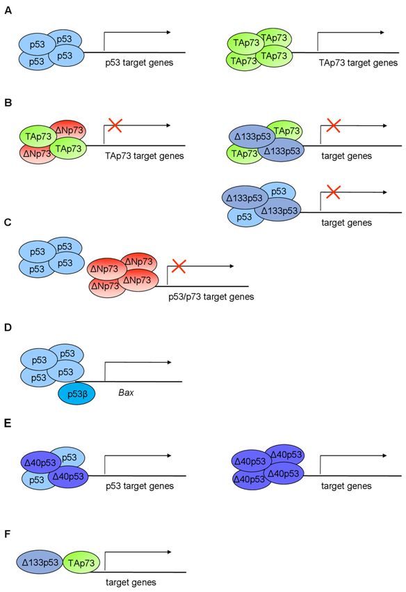
P53/p73 Protein Network in Colorectal Cancer and Other Human Malignancies
←
→
Page content transcription
If your browser does not render page correctly, please read the page content below
cancers
Review
p53/p73 Protein Network in Colorectal Cancer and Other
Human Malignancies
And̄ela Horvat † , Ana Tadijan † , Ignacija Vlašić † and Neda Slade *

                                          Laboratory for Protein Dynamics, Division of Molecular Medicine, Rud̄er Bošković Institute, Bijenička 54,
                                          10000 Zagreb, Croatia; Andjela.Horvat@irb.hr (A.H.); Ana.Tadijan@irb.hr (A.T.); Ignacija.Vlasic@irb.hr (I.V.)
                                          * Correspondence: slade@irb.hr
                                          † Contributed equally.

                                          Simple Summary: The p53 family of proteins comprises p53, p63, and p73, which share high
                                          structural and functional similarity. The two distinct promoters of each locus, the alternative splicing,
                                          and the alternative translation initiation sites enable the generation of numerous isoforms with
                                          different protein-interacting domains and distinct activities. The co-expressed p53/p73 isoforms
                                          have significant but distinct roles in carcinogenesis. Their activity is frequently impaired in human
                                          tumors including colorectal carcinoma due to dysregulated expression and a dominant-negative
                                          effect accomplished by some isoforms and p53 mutants. The interactions between isoforms are
                                          particularly important to understand the onset of tumor formation, progression, and therapeutic
                                          response. The understanding of the p53/p73 network can contribute to the development of new
                                          targeted therapies.

                                          Abstract: The p53 tumor suppressor protein is crucial for cell growth control and the maintenance

                                   of genomic stability. Later discovered, p63 and p73 share structural and functional similarity with
                                          p53. To understand the p53 pathways more profoundly, all family members should be considered.
Citation: Horvat, A.; Tadijan, A.;
                                          Each family member possesses two promoters and alternative translation initiation sites, and they
Vlašić, I.; Slade, N. p53/p73 Protein
                                          undergo alternative splicing, generating multiple isoforms. The resulting isoforms have important
Network in Colorectal Cancer and
Other Human Malignancies. Cancers
                                          roles in carcinogenesis, while their expression is dysregulated in several human tumors including
2021, 13, 2885. https://doi.org/          colorectal carcinoma, which makes them potential targets in cancer treatment. Their activities arise,
10.3390/cancers13122885                   at least in part, from the ability to form tetramers that bind to specific DNA sequences and activate
                                          the transcription of target genes. In this review, we summarize the current understanding of the
Academic Editors: Maja T. Tomicic         biological activities and regulation of the p53/p73 isoforms, highlighting their role in colorectal
and Thomas Efferth                        tumorigenesis. The analysis of the expression patterns of the p53/p73 isoforms in human cancers
                                          provides an important step in the improvement of cancer therapy. Furthermore, the interactions
Received: 13 May 2021                     among the p53 family members which could modulate normal functions of the canonical p53 in
Accepted: 3 June 2021
                                          tumor tissue are described. Lastly, we emphasize the importance of clinical studies to assess the
Published: 9 June 2021
                                          significance of combining the deregulation of different members of the p53 family to define the
                                          outcome of the disease.
Publisher’s Note: MDPI stays neutral
with regard to jurisdictional claims in
                                          Keywords: p53 isoforms; p73 isoforms; colorectal cancer; p53 family; isoform crosstalk
published maps and institutional affil-
iations.

                                          1. Introduction

Copyright: © 2021 by the authors.
                                                p53 has a central role in tumorigenesis; it regulates cellular response to stress signals or
Licensee MDPI, Basel, Switzerland.
                                          oncogenic cellular stimuli by inducing transient or permanent cell-cycle arrest, DNA repair,
This article is an open access article
                                          apoptosis, or senescence [1]. The inactivation of the p53 tumor suppressor is the single most
distributed under the terms and           common genetic defect in human cancer. The mutations of TP53 have been found in nearly
conditions of the Creative Commons        all tumor types and are estimated to contribute to more than 50% of all cancers.
Attribution (CC BY) license (https://           In the late 1990s, two novel family members TP73 [2] and TP63 [3] were described. Both
creativecommons.org/licenses/by/          encode several proteins whose structures and functions are similar to those of p53 but not
4.0/).                                    identical, each displaying peculiar functional features revealed by mouse model studies.

Cancers 2021, 13, 2885. https://doi.org/10.3390/cancers13122885                                                https://www.mdpi.com/journal/cancers
Cancers 2021, 13, x FOR PEER REVIEW 2 of 43
In the late 1990s, two novel family members TP73 [2] and TP63 [3] were described. Both
Cancers 2021, 13, 2885 encode several proteins whose structures and functions are similar to those of p53 but 2 ofnot
42
identical, each displaying peculiar functional features revealed by mouse model studies.
All members of the p53 family are evolutionarily conserved and have a very high
structural similarity within three main functional domains: transactivation domain
All members of the p53 family are evolutionarily conserved and have a very high
(TAD), DNA-binding domain (DBD), and oligomerization/tetramerization domain
structural similarity within three main functional domains: transactivation domain (TAD),
(OD/TD) (Figures 1 and 2). All family members share significant amino-acid homology in
DNA-binding domain (DBD), and oligomerization/tetramerization domain (OD/TD)
DBD (over 60%), which enables them to bind the p53-responsive elements (p53RE) and
(Figures 1 and 2). All family members share significant amino-acid homology in DBD (over
transactivate a large number of the same target genes. Accordingly, p63, p73, and p53
60%), which enables them to bind the p53-responsive elements (p53RE) and transactivate
proteins form a family of transcription factors. At the sequence level, p63 and p73 share
a large number of the same target genes. Accordingly, p63, p73, and p53 proteins form a
higher homology to each other than to p53 [4]. However, despite many overlapping roles,
family of transcription factors. At the sequence level, p63 and p73 share higher homology
each member has its unique identity and functions.
to each other than to p53 [4]. However, despite many overlapping roles, each member has
The family complexity has been enriched by the transcription from alternative pro-
its unique identity and functions.
moters,
Thealternative splicing, has
family complexity andbeen
diverse translation
enriched by theinitiation sitesfrom
transcription [4,5].alternative
Consequently,pro-
several
moters,protein isoforms
alternative withand
splicing, distinct N- and
diverse C- termini
translation are encoded.
initiation sites [4,5]. Consequently,
Theprotein
several roles isoforms
of each particular p53N-family
with distinct and C-member
termini are were determined by transgenic
encoded.
knockout mice. While the p53 knockout mice show high
The roles of each particular p53 family member were determined by susceptibility to transgenic
spontaneous and
knock-
induced carcinogenesis, thereby defining p53 as an important tumor
out mice. While the p53 knockout mice show high susceptibility to spontaneous and suppressor, total p63
and p73 knockout mouse models lack a cancer phenotype. Instead, p63-
induced carcinogenesis, thereby defining p53 as an important tumor suppressor, total p63 and p73-null mice
exhibit
and p73various
knockout developmental
mouse modelsdeficiencies
lack a cancer revealing
phenotype. theirInstead,
crucial p63-
functions in epithelial
and p73-null mice
and central nervous system formation, respectively [4]. Although p63
exhibit various developmental deficiencies revealing their crucial functions in epithelial and p73 are rarely
mutated
and centralin cancer,
nervous TP63 is situated
system within
formation, the locus frequently
respectively [4]. Althoughamplified
p63 and in squamous cell
p73 are rarely
carcinoma [6]. Their role in tumorigenesis is defined by several isoforms
mutated in cancer, TP63 is situated within the locus frequently amplified in squamous cell with opposed
functions,
carcinoma in [6].the same
Their cellular
role context [7]. is
in tumorigenesis However,
defined by their tumor
several suppressive
isoforms with potential
opposed
was not fully
functions, elucidated
in the until context
same cellular the establishment
[7]. However, of the
theirisoform-specific
tumor suppressiveknockout mice.
potential wasIn
this review,
not fully we summarize
elucidated until thethe structural and
establishment functional
of the properties
isoform-specific of different
knockout mice.p53
In and
this
p73 isoforms
review, and their the
we summarize rolesstructural
in tumorand formation withproperties
functional the emphasis on colorectal
of different cancer
p53 and p73
(CRC).
isoforms and their roles in tumor formation with the emphasis on colorectal cancer (CRC).
Figure 1. The TP53 gene
The TP53 gene architecture isoforms. (A) The scheme of the TP53
architecture and generation of the p53 isoforms.
gene structure. The human TP53 gene consists
consists of
of 11
11 exons
exons and
and two
two alternative
alternative exons
exons9β 9β and
and 9γ.
9γ. The
exons are shown as boxes of different colors and noncoding sequences in black. Introns 2 (i2) and 4
(i4)
(i4) are shown as
are shown as boxes
boxes with
with aa striped
striped pattern.
pattern. The
The TP53
TP53 gene
gene can
can be
be transcribed
transcribed from two different
from two different
promoters, the canonical promoter P1 upstream of exon 1 (giving rise to the
promoters, the canonical promoter P1 upstream of exon 1 (giving rise to the long p53/∆40p53 long p53/Δ40p53
isoforms) and the alternative P2 located in intron 4 (giving rise to the short Δ133/Δ160p53 isoforms).
isoforms) and the alternative P2 located in intron 4 (giving rise to the short ∆133/∆160p53 isoforms).
The TP53 mRNAs can be alternatively spliced at intron 2 or intron 9, producing isoforms with dif-
The TP53 mRNAs can be alternatively spliced at intron 2 or intron 9, producing isoforms with
ferent N- and/or C-termini. There are four possible different start codons for mRNA translation
differentATG40,
(ATG1, N- and/or C-termini.
ATG133, There are
and ATG160) four possible
resulting different
in protein startof
isoforms codons
varyingforlength.
mRNA(B) translation
Modular
(ATG1, ATG40, ATG133, and ATG160) resulting in protein isoforms of varying
structure of the p53 protein isoforms. Functional protein domains are shown in different length. (B) Modular
colors
structure of
matching the used
those p53 protein isoforms. exons
for the encoding Functional
of the protein
TP53 gene domains
in (A).are
Theshown in different
full-length colors
p53α protein
consists
matching ofthose
two transactivation domainsexons
used for the encoding (TAD1 andTP53
of the TAD2),
gene a proline-rich domain (PRD),
in (A). The full-length p53α aprotein
DNA-
consists of two transactivation domains (TAD1 and TAD2), a proline-rich domain (PRD), a DNA-
binding domain (DBD), a hinge domain (HD), an oligomerization domain (OD), and a C-terminal
domain (CTD). There are 12 distinct p53 protein isoforms differing in the composition of the structural
domains. Model adapted from [8–10].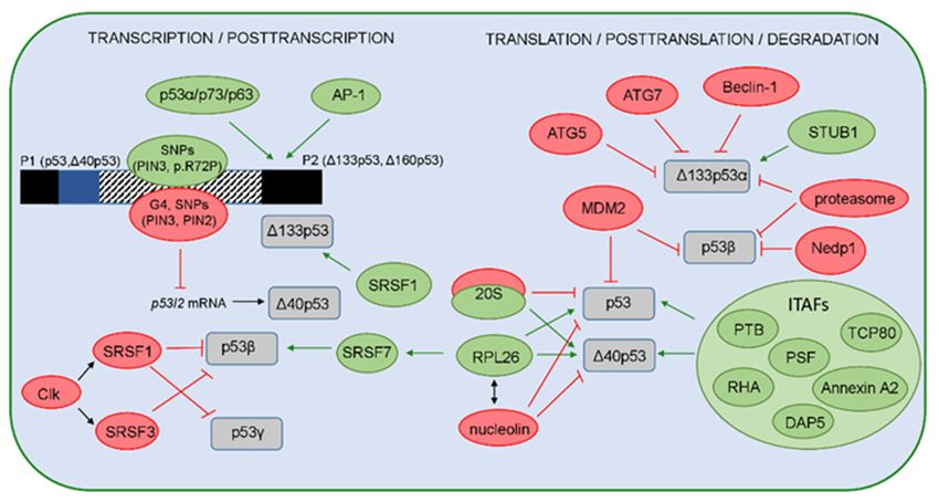
Cancers 2021, 13, x FOR PEER REVIEW 3 of 43
Cancers 2021, 13, 2885 binding domain (DBD), a hinge domain (HD), an oligomerization domain (OD), and a C-terminal 3 of 42
domain (CTD). There are 12 distinct p53 protein isoforms differing in the composition of the struc-
tural domains. Model adapted from [8–10]
Figure 2. TheTheTP73
TP73gene
genearchitecture
architectureandand
generation of the
generation of p73
the isoforms. (A) The
p73 isoforms. (A)scheme of the human
The scheme of the
human
TP73 TP73
gene gene structure.
structure. The human The human TP73
TP73 gene gene consists
consists of 14and
of 14 exons exons and additional
additional exon 30
alternative
alternative
exon
(shown 3′ as
(shown as gray
gray box). The box).
exonsThe exons as
are shown areboxes
shown as boxescolors
of different of different colors and
and noncoding noncoding
sequences se-
in black.
quences
There are two different promoters, the canonical promoter P1 upstream of exon 1 (giving rise to TAp731
in black. There are two different promoters, the canonical promoter P1 upstream of exon
(giving rise to TAp73 isoforms) and the alternative P2 located in intron 3 (giving rise to ΔNp73
isoforms) and the alternative P2 located in intron 3 (giving rise to ∆Np73 isoforms). The P1 transcript can
isoforms). The P1 transcript can be alternatively spliced leading to the expression of several N-ter-
be alternatively spliced leading to the expression of several N-terminally truncated isoforms (∆Ex2p73,
minally truncated isoforms (ΔEx2p73, ΔEx2/3p73, and ΔN′p73). Alternative splicing is also possible
0 p73). Alternative splicing is also possible at the C-terminus, giving rise to seven
∆Ex2/3p73,
at and ∆N
the C-terminus, giving rise to seven potential isoforms (α, β, γ, δ, ε, ζ, and η). (B) Modular struc-
potential
ture of the isoforms (α, β,isoforms.
p73 protein and η). (B) Modular
γ, δ, ε, ζ, Functional structureare
protein domains of the
shownp73 in
protein isoforms.
different colorsFunctional
matching
proteinused
those domains areencoding
for the shown inexons
different colors
of the TP73 matching
gene in those used
(A). The for the encoding
full-length TAp73αexons of the
protein TP73
consists
gene
of in (A). The full-length
a transactivation domainTAp73α(TAD), protein consistsdomain
a proline-rich of a transactivation domain (TAD),
(PRD), a DNA-binding a proline-rich
domain (DBD), a
nuclear-localization signal (NLS),
domain (PRD), a DNA-binding an oligomerization
domain domain (OD),signal
(DBD), a nuclear-localization a sterile α-motif
(NLS), (SAM), and an
an oligomerization
inhibitory
domain (OD), domain (ID).
a sterile There(SAM),
α-motif are 28 and
possible distinct p73
an inhibitory protein
domain (ID).isoforms
There arediffering
28 possiblein the compo-
distinct p73
sition of the structural domains (as Δ’Np73 and ΔNp73 mRNAs translate into
protein isoforms differing in the composition of the structural domains (as ∆’Np73 and ∆Np73 mRNAsidentical proteins).
translate into identical proteins).
2. Gene Architecture and Generation of the p53/p73 Isoforms
TheArchitecture
2. Gene human TP53 and geneGeneration
is located on ofthe
thechromosome
p53/p73 Isoforms17p13.1 and comprises 11 exons
and two alternative exons 9β and 9γ. It has a dual gene structure due to the presence of
The human TP53 gene is located on the chromosome 17p13.1 and comprises 11 exons
two functional,
and two distinct
alternative promoters,
exons 9β and 9γ. theItcanonical
has a dualP1genelocated upstream
structure of the
due to exon 1 and the
presence of
internal P2 that lies within intron 4, from which several TP53 mRNAs
two functional, distinct promoters, the canonical P1 located upstream of exon 1 and the can be transcribed
(Figure
internal 1A)
P2 that[11].lies
The TP53intron
within mRNAs can alternatively
4, from which severalbeTP53 spliced
mRNAsat intron
can be 2 or intron 9,
transcribed
resulting in variants with different N- or C-termini. In addition,
(Figure 1A) [11]. The TP53 mRNAs can alternatively be spliced at intron 2 or intron the TP53 mRNA can also9,
contain
resulting in variants with different N- or C-termini. In addition, the TP53 mRNA cana cell-
two internal ribosomal entry site (IRES) elements that mediate translation in also
cycle
containphase-dependent manner [12,13].
two internal ribosomal entry siteTranslation of the TP53
(IRES) elements thatmRNA
mediatecan start at differ-
translation in a
ent
cell-cycle phase-dependent manner [12,13]. Translation of the TP53 mRNAorcan
codons, e.g., codon 1 (ATG1), codon 40 (ATG40), codon 133 (ATG133), codon
start160
at
(ATG160) [8,14], resulting
different codons, e.g., codonin 1p53 isoforms
(ATG1), that40
codon differ in length
(ATG40), codon (Figure 1B).
133 (ATG133), or codon
Consequently,
160 (ATG160) [8,14],the p53 isoforms
resulting can be classified
in p53 isoforms that differasinlong or short
length depending
(Figure 1B). on the
initiation of transcription and translation. Transcribed from the
Consequently, the p53 isoforms can be classified as long or short depending on the canonical P1, the TP53
ini-
mRNA transcripts translate at codon 1 and/or codon 40 to encode
tiation of transcription and translation. Transcribed from the canonical P1, the TP53 mRNA the long isoforms
(p53/Δ40p53), whileattranscribed
transcripts translate codon 1 and/or from codon
the internal P2, translation
40 to encode can start (p53/∆40p53),
the long isoforms at codons 133
and/ortranscribed
while 160, giving from rise to
thetheinternal
short isoforms (Δ133p53/Δ160p53).
P2, translation can start at codonsThe transcription
133 and/orfrom 160,
the canonical
giving P1 generates
rise to the intron
short isoforms 2-spliced (also known
(∆133p53/∆160p53). Theas fully spliced,
transcription FSp53)
from or intron
the canonical
2-retained
P1 generates (also designated
intron 2-spliced as p53I2) TP53 mRNA
(also known as fullytranscripts.
spliced, FSp53)Translation at the
or intron codon 1
2-retained
(also designated as p53I2) TP53 mRNA transcripts. Translation at the codon 1 of intron
2-spliced TP53 mRNA transcript encodes the full-length p53 isoforms [8,14]. However, due
to the alternative translation initiation sites and the presence of IRES elements in the 50
untranslated regions (50 UTR) in the TP53 mRNA, IRES-mediated translation can produce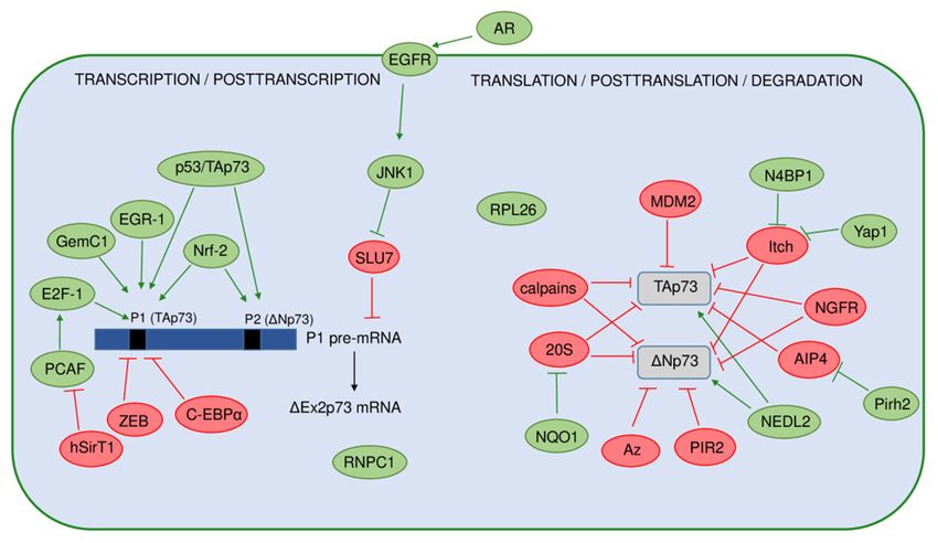
Cancers 2021, 13, 2885                                                                                             4 of 42

                         ∆40p53 isoforms from the codon 40 [12,13,15–17]. In addition, ∆40p53 isoforms can also
                         be synthesized from the TP53 mRNA transcripts with retained intron 2 that are translated
                         only at codon 40 [8,11,14]. Initiation of transcription from the internal P2 within intron
                         4 generates a TP53 mRNA transcript that can be translated either at codons 133 or 160,
                         encoding the ∆133p53 or ∆160p53 isoforms, respectively [8,14,18]. The alternative splicing
                         from the exon 9 to either exons 9β or 9γ generates the TP53 mRNA transcripts that encode
                         β or γ isoforms, respectively. Since both exons 9β and 9γ contain premature termination
                         codons (PTCs), exons 10 and 11 remain untranslated in β and γ isoforms [8,11,14]. To con-
                         clude, depending on alternative promoter usage (P1 or P2), alternative splicing of intron 2
                         or intron 9, and alternative initiation of translation, different p53 isoforms can be generated.
                         Consequently, nine different TP53 mRNA transcripts encoded by the TP53 gene can give
                         rise to 12 protein isoforms, known as p53α/β/γ, ∆40p53α/β/γ, ∆133p53α/β/γ, and
                         ∆160p53α/β/γ [8,14]. Essentially, there is an increased expression of the full-length p53 in
                         tumor tissue compared to corresponding normal tissue which is usually attributed to muta-
                         tions in p53. Depending on the p53 mutation status, the p53 isoforms can be differentially
                         expressed. The increased expression of different N- and/or C-terminal spliced variants has
                         been detected in various cancer entities, such as the ∆133p53α isoform in colon, lung, and
                         ovarian cancer, cholangiocarcinoma, and melanoma [9,14,19–22] or the ∆40p53α isoform in
                         both glioblastoma and breast cancer [14,23,24]. Furthermore, the p53β isoform is identified
                         and overexpressed in the head and neck squamous cell carcinoma and renal cell carcinoma,
                         respectively [25,26]. Interestingly, the expression profile of the N- and C-terminal spliced
                         variants can differ in premalignant lesion, tumor, and corresponding healthy tissue. For
                         example, premalignant lesions of colon adenomas are shown to express reduced levels of
                         N-terminal splice variant (e.g., ∆133p53α) and elevated levels of C-terminal splice variant
                         (e.g., p53β) compared with normal colon tissues. In contrast, the expression pattern of
                         these splice variants depends on the p53 status and is changed in colon carcinoma tissue
                         compared to colon adenoma, suggesting their role in cancer progression [19,27].
                               The p73 protein was discovered in 1997 as a product of the TP73 gene situated in the
                         region 1p36.33 that is often deleted in neuroblastoma and various other human tumors.
                         Frequent occurrence of loss of heterozygosity (LOH) and structural homology with the
                         canonical tumor suppressor p53 placed p73, together with almost concomitantly discovered
                         p63 protein, into the common p53 protein family [2,3]. The human TP73 gene consists of
                         15 exons (designated as exons 1–14, plus one alternative exon 30 ) and it can be transcribed
                         into different mRNAs, subsequently producing several different protein isoforms (Figure 2).
                         Multiple mRNAs are produced as a result of the existence of two promoters combined with
                         the alternative splicing at the 5’- and 30 -ends. The transcription from P1 located in the 50 UTR
                         upstream of exon 1 gives rise to a group of transcriptionally active TAp73 isoforms. Usage
                         of the alternative P2 situated in the intron 3 produces N-terminally truncated isoforms that
                         are, thus, referred to as ∆Np73 isoforms [28–30].
                               The transcript generated from P1 promoter can be alternatively spliced at the 5’-end,
                         producing more complexity among the N-terminally truncated isoforms. In this way, three
                         additional isoforms are produced, namely, ∆Ex2p73 (lacking exon 2), ∆Ex2/3p73 (lacking
                         exons 2 and 3), and ∆N0 p73 (containing alternative exon 30 ) [2,29–31]. Interestingly, the
                         ∆N0 p73 transcript and the ∆Np73 transcript generated from P2 promoter produce an
                         identical ∆Np73 protein isoform and can be distinguished exclusively on an mRNA level.
                         Specifically, the ∆N0 p73 transcript aberrantly contains 198 bp from the alternative exon 30
                         leading to a PTC in the regular reading frame, and, as a result, the translation starts at the
                         same codon in exon 30 used by the P2 transcript [30,32].
                               Similar to p53, an IRES element was discovered in exon 2 of the TP73 mRNA repre-
                         senting another possible mechanism of p73 expression regulation via CAP-independent
                         translation [33]. Alternative splicing is even more frequent at the 30 -end, producing seven
                         different transcripts named α, β, γ, δ, ε, ζ, and η [2,29,34,35]. Combining the usage of
                         two different promoters and splicing at 50 /30 -ends can theoretically result in 35 different
                         mRNAs, which can be translated into 28 different protein isoforms, but not all of them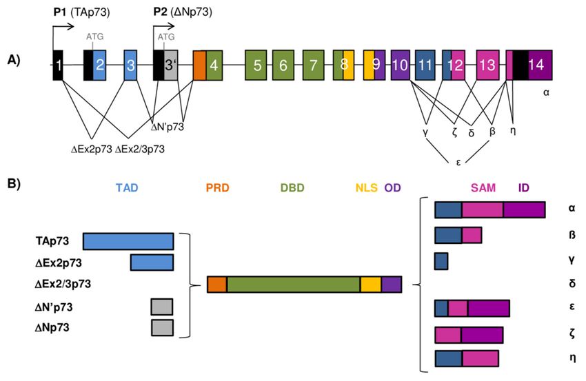
Cancers 2021, 13, 2885                                                                                           5 of 42

                         have been detected in cell lines or tissues so far. In general, p73 expression is often higher
                         in tumor tissues compared to the corresponding normal tissues. The increase in expression
                         is mostly attributed to the N-terminally truncated isoforms, but the TAp73 isoforms are
                         also found to be increased in several tumors. An increase in the variety of the C-terminally
                         spliced isoforms has been connected to certain tumor types. Thus, normal breast, colon,
                         and myeloid cells predominantly express the p73α and β isoforms, while, in breast and
                         colon cancer, as well as in acute myeloid leukemia, an increased level of the p73γ, δ, and ε
                         isoforms was observed [36–38].

                         3. Structure of the p53 Protein Isoforms
                               The canonical, full-length p53 (p53α) isoform displays a modular domain structure
                         and contains 393 residues that are organized in seven functional domains (Figure 1B). The
                         N-terminal region of the p53 protein contains two distinct acidic transactivation domains
                         (TAD1, residues 1–39 and TAD2, residues 40–61) that are intrinsically disordered, which
                         allows binding with high specificity to their interacting partners [39,40]. Each TAD can
                         transactivate genes independently and are required for the transactivation of different
                         target genes and effector pathways. Through its TAD, the p53 protein interacts with the
                         components of transcriptional machinery (e.g., TBP, TFIIH), proteins involved in DNA
                         metabolism (e.g., PCA, RPA), chromatin modifiers (e.g., p300/CBP, GCN5), and inhibitors
                         of p53 (e.g., MDM2, MDMX). Functional analysis based on in vivo mouse models showed
                         that TAD1 is important for the transactivation of the p53 target genes involved in cellular
                         responses to acute DNA damage, such as apoptosis and cell-cycle arrest, while both TAD1
                         and TAD2 can cooperate in the transactivation of genes associated with tumor suppres-
                         sion [41,42]. Furthermore, it has recently been shown that the induction of endoplasmic
                         reticulum (ER) stress-inducible genes (e.g., PTP4A1, PLK2) depends on the transactivation
                         activity of the TAD2 [43].
                               Following TA domains at the N-terminus lies a proline-rich domain (PRD, residues
                         62–93) that contains 15 proline residues, some as part of five PXXP motifs (where P rep-
                         resents proline and X any residue) important for growth suppression. These motifs have
                         the ability to bind SH3 (Src homology 3) domains of proteins such as c-Src and PI3K
                         (phosphatidylinositol 3-kinases) and mediate signal-transduction pathways [44,45]. The
                         PRD is shown to control modifications that influence p53 functions, e.g., activity and
                         stability. It contains conformationally flexible motifs that bind transcriptional coactivator
                         p300, involved in p53 activation through acetylation [46]. Furthermore, the PRD is impor-
                         tant for regulating p53-mediated apoptosis [47,48]; rs1042522 (p53 p.R72P) is a common
                         polymorphism in the PRD whose allele frequency differs in populations worldwide [49]
                         and was recently associated with the CRC risk among specific ethnic groups according to a
                         meta-analysis [50].
                               The p53 protein binds to DNA through its core domain, the conserved sequence-
                         specific DBD (residues 94–290). The DBD contains four conserved regions [51] and includes
                         the immunoglobulin-like β-sandwich that serves as a scaffold for the structural elements
                         involved in DNA-binding consisting of two large loops, which are held together by a zinc
                         atom through cysteine and histidine ligands, and a loop–sheet–helix motif [52]. It has been
                         recently shown that the DBD regulates conformation stability of the p53, and its absence, as
                         in the ∆133p53 and ∆160p53 isoforms, can destabilize the structure and trigger aggregation
                         propensity, ultimately causing abnormal protein function [53]. Most cancer-associated
                         mutations occur within the DBD, causing structural alterations (e.g., R175, Y220, G245)
                         or impacting DNA binding (e.g., R248 and R273) [54], which makes DBD crucial for the
                         tumor-suppressive functions of p53. Furthermore, p53-dependent tumor suppression and
                         apoptosis can be modulated by mutations in residues E180 and R181 that provide the
                         structural basis for cooperative binding of p53 to target promoters [55,56].
                               The p53 protein possesses a nuclear-localization signal (NLS, residues 305–322) that
                         facilitates the shuttling of p53 between the nucleus and cytoplasm. It has been shown
                         that the nuclear import of p53 is negatively regulated by ubiquitination of lysine residues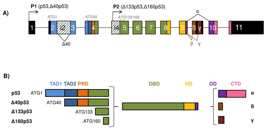
Cancers 2021, 13, 2885                                                                                               6 of 42

                         319–321 within the NLS, causing the retention of p53 in the cytoplasm. Upon stress, the level
                         of ubiquitination reduces, and the positive charges of NLS residues are rendered unmasked
                         and recognized by importin α3, an adaptor molecule that facilitates nuclear import of non-
                         ubiquitylated p53 [57,58]. The NLS is located in the hinge domain (HD, residues 291–324),
                         the short linker between DBD and OD (residues 325–356). Germline mutations in the HD
                         (e.g., p.K305M and p.G325V) have been shown to impair apoptotic functions of mutant (mt)
                         p53 protein that still retains the ability to induce cell-cycle arrest [59]. Additionally, specific
                         missense and deletion mutations in the HD (e.g., p.R306P, p.del300-308 and p.del300-327)
                         cause the loss of apoptotic function and reduced ability of p53 to transactivate the BAX
                         promoter, but do not affect the transactivation of p21 promoter [60], which would imply
                         that the HD is involved in allosteric regulation of DNA binding [61].
                               To fulfill its function as a tetrameric transcription factor, p53 needs to exhibit its capac-
                         ity to form tetramers, an active form needed for binding to RE and gene transactivation,
                         dependent on the OD. It has been revealed that the monomer, which consists of a β-strand
                         and an α-helix, connects with the second monomer to form a dimer in an antiparallel man-
                         ner by means of their β-strands and α-helices. Two dimers connect through their α-helices
                         to form a tetramer. On the other hand, β-strands are not included in the interaction between
                         the dimers since they are located outside of the tetramer [62]. In addition, the OD contains
                         a leucine-rich nuclear export signal (NES, residues 340–351) that regulates subcellular
                         localization of p53 and is masked in the tetramers, permitting their accumulation in the
                         nucleus. Consequently, there seems to be a coordinated regulation of p53 tetramerization
                         and its nuclear retention, which is dependent on the NES placement [63]. It has been
                         recently shown that the positively charged residues within the NES of OD are required for
                         proper regulation of the p53 target genes. For example, changing lysines 351 and 357 to
                         glutamines does not affect localization or tetramerization status of the protein. However,
                         p53 is impaired in the induction of cell-cycle arrest but retains the ability to induce cell
                         death [64]. Many other missense mutations in the OD have been reported [65], some of
                         which can impair the ability of p53 to form tetramers and to activate transcription of its
                         target genes. It has been shown that the p53 OD mutants with the alterations in L330 (i.e.,
                         p.L330R, p.L330E or p.L330P) can exhibit severe homotetramerization and ubiquitination
                         defects and the loss of transcriptional activity. However, when co-transfected with wt p53,
                         they could form heterotetramers and alter the expression of p53 target genes (BAX and
                         p21), thereby acting as dominant-negative mutants [66].
                               The C-terminal domain (CTD) (residues 357–393) is an arginine- and lysine-rich ba-
                         sic domain that recognizes and binds nonspecifically to DNA and RNA. The CTD is
                         intrinsically disordered and contains NLSs, as well as the sites with the most frequent
                         posttranslational modifications (PTM) that include phosphorylation, acetylation, ubiquiti-
                         nation, methylation, neddylation, and sumoylation [39,40,67,68]. Hence, the CTD regulates
                         the activity of the protein. Initially, the CTD was suggested to be a negative autoregulator of
                         sequence-specific DNA binding. However, later studies have described positive regulatory
                         features of the CTD that likely depend on low-affinity electrostatic interactions between
                         the DNA phosphate backbone and C-terminal lysine residues [40]. It has recently been
                         shown that the CTD is required for the DBD to recognize p53RE, and it is able to modulate
                         structural changes within the DBD, thus stabilizing the association of p53 to DNA-binding
                         sites [40,69].
                               To summarize, different p53 isoforms, except the canonical full-length p53, lack part of
                         the N- and/or C-termini and, consequently, are deficient in some of the functional domains
                         (Figure 1B). The isoforms with N-terminal deletions have a designation depending on the
                         length of the deletion; hence, considering the lack of the first 39, 132, or 159 residues, they
                         are called ∆40p53, ∆133p53, or ∆160p53 isoforms, respectively. Due to the lack of the first
                         39 residues, TAD1 is absent in the ∆40p53 isoforms that still retain TAD2. Both TAD and
                         PRD are absent in the ∆133p53 and ∆160p53 isoforms that, to some extent, retain the DBD.
                         The ∆133p53 isoforms lack a small part of the first conserved cysteine box of the DBD,
                         which is completely absent in the ∆160p53 isoforms. However, the ∆160p53 isoforms do
Cancers 2021, 13, 2885 7 of 42
retain the other three cysteine boxes of the DBD. The isoforms can exhibit differences in
the C-terminus due to alternative splicing of exon 9. The α isoforms contain exons 10 and
11 that encode the OD and CTD. Due to PTCs present in exons 9β and 9γ, both β and γ
isoforms lack part of the OD and complete CTD. The first seven residues of the OD are
present in all isoforms (α, β, γ); however, the β isoforms contain an additional 10 residues,
while γ isoforms contain an additional 15 residues that differ in sequence [8,14].
4. Structure of the p73 Protein Isoforms
The full-length p73 and p53 proteins show substantial degree of homology in the com-
position of the main functional protein domains (TAD, DBD, and OD) (Figures 1B and 2B).
The level of homology between p53 and p73 proteins is the highest in their DBDs (63%),
emphasizing their central role as transcription factors binding to the promoters of various,
many overlapping, target genes. A significant level of homology exists also between their
TADs (29%) and ODs (38%) [5]. However, at the C-terminus, there is more diversity between
the family members. While p53 contains the CTD at its C-terminus, the p73 protein possesses
a unique sterile α motif (SAM) domain, as well as an inhibitory domain (ID). Both p53 and
p73 can appear as numerous isoforms with different composition of protein domains, sus-
ceptible to different PTMs and regulatory protein interactions. This provides a broad area
for investigation in order to offer an explanation for their distinct transcriptional activity and
functions [61].
Only the TAp73 isoforms transcribed from P1 that are not subjected to additional
splicing at the 50 -end contain a complete TAD at the N-terminus, which makes them strong
transcriptional activators (Figure 2B). At first, it was considered that the p73 protein con-
tains only one unique TAD, in contrast to p53 which possesses two [70]. However, more
recent work on the interaction between the TAp73 N-terminus and different domains of
transcriptional activator p300 revealed the existence of two distinct p73 transactivation
subdomains, spanning residues 10–30 and 46–67 [71,72]. In contrast to TAp73, the ∆Np73
isoforms transcribed from P2 in intron 3 lack the first 62 residues and, consequently, do
not contain the TAD of the N-terminally intact isoforms. The presence or absence of the
TAD, defining these two groups of isoforms, is reflected in their ability to transactivate
different genes. While the TAp73 isoforms can activate the expression of various genes,
many of which are also induced by p53, the ∆Np73 isoforms are mostly transcriptionally
inactive. Moreover, the ∆Np73 isoforms act as dominant-negative inhibitors of p53 and
TAp73 [28,31]. However, inducible overexpression of the ∆Np73β isoform was found to
induce certain p53/p73 target genes such as p21, 14-3-3σ, and GADD45 causing cell-cycle
arrest, growth suppression, and apoptosis in different cell lines. This transactivation activ-
ity was dependent on the presence of 13 unique residues at the N-terminus of the ∆Np73
isoforms and the adjacent region with PXXP motifs (PRD), which together form a novel
transactivation domain specific for the ∆Np73 isoforms [73]. Similar activity was also
shown for ∆Np73γ, whereas ∆Np73α was inactive in the growth suppression and trans-
activation [73]. In another study, overexpression of both ∆Np73β and TAp73β induced
transcription of the antiapoptotic short caspase-2S isoform, possibly contributing to pro-
survival mechanisms in tumors. The induction of caspase-2S expression was dependent on
direct TAp73β/∆Np73β binding to a specific 18 bp site in the CASP-2S promoter region.
Neither ∆Np73α nor TAp73α was able to induce caspase-2S expression under the same
experimental conditions [74]. In contrast, ∆Np73α was able to induce several genes, in-
cluding EGR1 (early growth response 1) and CDC6 (cell division cycle 6), independently of
p53 in microarray analysis [75]. In addition, ∆Np73α enhanced the expression of the TGFβ
target genes PAI-1 and Col1a1, possibly through its interaction with Smad transcription
factors and subsequent binding to Smad-binding elements (SBEs) [76]. In more recent
research, ∆Np73β and, to a lesser extent, ∆Np73α were found to enhance the expression of
several keratinocyte genes in cooperation with ∆Np63α, having a role in skin development,
proliferation, and wound healing [77].Cancers 2021, 13, 2885 8 of 42
The PRD of the p73 protein comprises two PXXP motifs situated between residues
84–87 and 103–106 [78]. Between the two PXXP motifs, there is Y99, a residue that was
shown to be phosphorylated by c-Abl kinase after γ-irradiation, correlating with the
induction of p73 target genes and apoptosis [79,80]. In contrast to its importance for
∆Np73β transcriptional activity, neither Y99 nor the PXXP motifs were proven to be
indispensable for the activity of TAp73β isoform in the induction of p21 expression and
apoptosis [78].
The structure of the p73 DBD is highly similar to that of the p53 protein but differs in
the L2 loop important for protein–protein interactions [81]. The DBD of the p73 protein is
responsible for its binding to REs of more than 200 different genes involved in important
cellular processes such as apoptosis, DNA repair, and neuronal and epidermal differen-
tiation. It was recently shown using a biochemical approach that even small sequence
variations in the RE could affect binding affinity and velocity of p73 with its DBD [82].
The OD of p73 comprises residues 352 to 390 and enables tetramerization of p73 and
its activity as a transcription factor. In spite of the high degree of similarity in amino-acid
sequence with the OD of the p53 protein, the heterotetramers composed of the full-length
p73 and p53 have not been identified. This was explained by the discovery of an additional
α-helix located C-terminally of the core OD (homologous to p53) whose deletion causes
a conformational instability and dissociation of the p73 homotetramer into dimers. The
absence of the second α-helix is reflected in the reduced p73 transcriptional activity [83].
As intact OD is present in all identified p73 isoforms, including both N- and C-terminally
truncated, there is a complex network of possible protein interactions to be explored.
Next to the OD, there is a long stretch of 109 amino acids connecting it to an SAM
domain which is specific for the C-terminus of p73 [83]. The SAM domain constitutes
five helices (α1 to α5) forming a globular structure, and it is considered important for
modulation of the p73 transcriptional activity [84]. It is encoded by exons 11, 12, and 13,
a region that is subjected to intensive alternative splicing. There have so far been seven
different 30 -splice variants discovered (α, β, γ, δ, ε, ζ, and η), which makes p73 the most
complex among the p53 protein family members regarding alternative splicing at the
30 -terminus [85]. The C-terminally truncated p73 isoforms are produced as a consequence
of the PTCs created by alternative splicing. Only α isoforms of p73 contain a complete
SAM domain. As a consequence of alternative splicing, the p73β isoforms lack exon 13.
Although the p73γ isoforms contain all the exons coding for the SAM domain, the usage
of the long alternative frame due to splicing at exon 11 leads to a PTC. The p73δ isoforms
lack most of the p73-specific C-terminal region (exons 11–13), resembling p53 more than
other p73 isoforms [34]. The p73ε isoforms contain parts of the p73γ and p73α reading
frames lacking exons 11 and 13. The p73ζ isoforms are produced by internal deletion
of exons 11 and 12, lacking functionally important parts of the SAM domain [35]. The
p73η isoforms are closely related to p73α but differ at exon 14. Next to the SAM domain,
at the very C-terminus, the p73α, p73ε, and p73ζ isoforms have an additional ID. The
differences in the presence and functionality of the SAM and ID have been shown to
modulate the transcriptional activity of the C-terminally spliced p73 isoforms. In that
sense, the TAp73β isoform, which lacks both the SAM domain and the ID, is a stronger
transcriptional activator than TAp73α, which contains both. Although, in TAp73α, the ID
does not directly interact with the TAD, it prevents association with the transcriptional
coactivator p300/CBP, contributing to inhibition of its transcriptional activity [86].
The differential structure of multiple p53/p73 isoforms and their potential interactions
offer a broad area for investigation of the molecular basis for their diverse biological functions.
5. Regulation of the p53 Isoforms’ Expression and Activity
The expression and function of the p53 isoforms can be regulated on the transcriptional,
posttranscriptional, translational, and posttranslational levels (Figure 3).Cancers 2021, 13, x FOR PEER REVIEW 9 of 43
5. Regulation of the p53 Isoforms’ Expression and Activity
Cancers 2021, 13, 2885 9 of 42
The expression and function of the p53 isoforms can be regulated on the transcrip-
tional, posttranscriptional, translational, and posttranslational levels (Figure 3).
Figure 3. A A model
model representing
representing regulation of the p53 isoforms’ expression and stability. stability. The
The positive
positive
regulators are
regulators are shown
shown in green,
green, while
while the negative
negative regulators
regulators are
are shown
shown in in red.
red. On the transcriptional
transcriptional
level, expression
level, expression of of the
the p53 isoforms is
p53 isoforms is regulated
regulated by by usage
usage ofof two
two different promoters P1
different promoters P1 or P2,
or P2,
producing the long (p53, Δ40p53) or the short (Δ133p53, Δ160p53) isoforms,
producing the long (p53, ∆40p53) or the short (∆133p53, ∆160p53) isoforms, respectively. Several respectively. Several
regulators can influence the P2 activity. The canonical p53 and p53 family members (p63 and p73
regulators can influence the P2 activity. The canonical p53 and p53 family members (p63 and p73
and their isoforms) are shown to transactivate P2 with different efficiency. The transcription from
and their isoforms) are shown to transactivate P2 with different efficiency. The transcription from
P2 can be activated by the AP-1 transcription factor that mediates the expression of Δ133p53 in H.
P2 can be activated
pylori-infected by the
cells. The AP-1 transcription
single-nucleotide factor that mediates
polymorphisms (SNP) andthe expression
their haplotypes ∆133p53
of in in
the inter-
H.
nalpylori-infected
promoter region cells. The as
(shown single-nucleotide
box with a striped polymorphisms (SNP) and
pattern) that comprises their3,haplotypes
intron exon 4, and in the
intron
internal promoter
4 can affect the P2region (shown
activities. as box with
Furthermore, a striped
the pattern)PIN3
G4 structures, that comprises intron 3, exon 4, and
and PIN2 polymorphisms, can
decrease
intron theaffect
4 can level the
of theP2 p53I2 mRNA
activities. that encodes
Furthermore, thethe
G4 Δ40p53 isoforms.
structures, PIN3 andThePIN2
p53 isoforms are reg-
polymorphisms,
ulated
can on thethe
decrease posttranscriptional
level of the p53I2level mRNA by different
that encodes the ∆40p53
splicing factors. isoforms.
SRSF1 andThe SRSF3, activatedare
p53 isoforms by
Clk, promote
regulated on thecomplete exclusion oflevel
posttranscriptional intron 9 and, thus,
by different negatively
splicing factors. regulate
SRSF1 and theSRSF3,
level of p53β and
activated by
p53γpromote
Clk, isoforms. However,
complete SRSF1 upregulates
exclusion of intron 9 and,the thus,
Δ133p53α expression
negatively in human
regulate the levelaortic smooth
of p53β mus-
and p53γ
cle cells. In addition, the binding of RPL26 to the TP53 pre-mRNA allows the recruitment of SRSF7
isoforms. However, SRSF1 upregulates the ∆133p53α expression in human aortic smooth muscle
that prompts alternative splicing and, thus, generates p53β isoforms. Due to IRES, the level of p53
cells. In addition, the binding of RPL26 to the TP53 pre-mRNA allows the recruitment of SRSF7
and Δ40p53 is regulated by ITAFs (PTB, Annexin A2, PSF, DAP5, TCP80, RHA) or proteins such as
that
RPL26 prompts alternative
or nucleolin. splicing and,
Interestingly, Δ40p53 thus,
cangenerates p53βby
be generated isoforms. Due to IRES,that
the 20S proteasome thedegrades
level of p53
the
and ∆40p53 is regulated by ITAFs (PTB, Annexin A2, PSF, DAP5, TCP80,
full-length p53 protein. The level of the full-length p53 protein is regulated by MDM2 that RHA) or proteins suchwasas
RPL26
shown or to nucleolin.
promote the Interestingly,
degradation ∆40p53
of p53β.can In
be addition,
generatedtheby level
the 20Sof proteasome
p53β is alsothat degrades
regulated by the
the
MDM2-dependent
full-length p53 protein.neddylation,
The level ofproteasome, andp53
the full-length deneddylating enzymebyNedp1.
protein is regulated MDM2The that level of the
was shown
Δ133p53α
to promoteisoform is regulated
the degradation by the In
of p53β. proteasome,
addition, theas well
levelasofvia
p53βautophagic degradation,
is also regulated by theupon
MDM2-rep-
licative senescence,
dependent neddylation, where the proautophagic
proteasome, proteins (ATG5,
and deneddylating enzyme ATG7, Beclin-1)
Nedp1. act as
The level positive
of the regu-
∆133p53α
lators, while
isoform the STUB1/CHIP
is regulated acts as a negative
by the proteasome, as wellregulator of Δ133p53α
as via autophagic degradationupon
degradation, and senescence.
replicative
senescence, where the proautophagic proteins (ATG5, ATG7, Beclin-1) act as positive regulators,
As previously mentioned, the expression of p53 isoforms on the transcriptional and
while the STUB1/CHIP acts as a negative regulator of ∆133p53α degradation and senescence.
posttranscriptional levels is regulated by TP53 promoter usage (P1 or P2) and the alterna-
tive splicing of intron
As previously 2 or 9, which
mentioned, can be modulated
the expression by different
of p53 isoforms on thefactors. The expression
transcriptional and
of p53 isoforms is tissue-specific and can be precisely regulated. In addition
posttranscriptional levels is regulated by TP53 promoter usage (P1 or P2) and the alterna- to epigenetic
events
tive that influence
splicing of intron the
2 oractivity
9, whichofcanTP53 be promoters
modulated[11], several factors.
by different single-nucleotide poly-
The expression
morphisms
of p53 isoforms(SNP), more specifically,
is tissue-specific and cantheir
behaplotypes, can modify
precisely regulated. the activity
In addition of the in-
to epigenetic
ternal P2
events thatpromoter,
influencethereby affecting
the activity thepromoters
of TP53 expression[11],
of p53 isoforms.
several There are eight
single-nucleotide SNPs,
polymor-
including
phisms common
(SNP), morep53 p.R72P and
specifically, theirPIN3 Ins16bpcan
haplotypes, (16 modify
bp insertion in intron
the activity 3, rs17878362),
of the internal P2
in 11 different
promoter, haplotypes
thereby affectingidentified within
the expression ofthe
p53P2. Using specific
isoforms. There arereporter gene assay
eight SNPs, con-
including
structs, two
common p53ofp.R72P
the 11and haplotypes
PIN3 Ins16bpwere (16shown to increase
bp insertion the baseline
in intron expression
3, rs17878362), of
in 11
different
Δ133p53 haplotypes
isoform [87]. identified within
In addition, the P2. Usingcombination
a heterozygous specific reporter
of SNPsgene inassay constructs,
the P2 promo-
two of the (such
tor region 11 haplotypes were shown
as a combination of p53 top.R72P
increase theeither
with baseline
SNP expression ∆133p53
in intron 4,ofi.e., iso-
rs9895829
form [87]. In addition, a heterozygous combination of SNPs in the P2 promotor region (such
as a combination of p53 p.R72P with either SNP in intron 4, i.e., rs9895829 or rs2939430)
can affect the expression of ∆133p53, as well as the p53β isoform [88]. Furthermore, it has
been previously shown that different p53 family members and their isoforms can regulateCancers 2021, 13, 2885 10 of 42
the expression and function of ∆133p53 isoforms [89–92]. The transcription from P2 can
specifically be activated by the AP-1 (activator protein-1), a c-Jun/c-Fos transcription factor,
which upregulates the expression of the ∆133p53 isoform in Helicobacter pylori-infected
gastric epithelial cells [93].
On the posttranscriptional level, several factors have been shown to regulate the
alternative splicing of intron 2 or 9. The splicing factors, such as SRSF1 and SRSF3, which
are members of serine/arginine-rich (SR) proteins, have been shown to negatively regulate
the expression of p53β and p53γ isoforms [91,94]. SR proteins are essential for spliceosome
assembly and are activated by Clk (Cdc2-like kinases) [95,96]. In accordance, treatment
with Clk inhibitor TG003 and silencing of SRSF1 promote the generation of both TP53
mRNA β and γ variants and decrease the level of TP53 mRNA α variants, which would
suggest that SRSF1 regulates alternative splicing of TP53 intron 9 and favors its complete
exclusion (e.g., exons 9β and 9γ) [91]. Furthermore, downregulation of SRSF3 increased
the level of TP53 mRNA β variants and induced replicative senescence in early passages of
normal human fibroblasts, thus revealing that SRSF3 regulates alternative splicing of TP53
intron 9 and modulates the p53-mediated cellular senescence [94]. Alternative splicing and
the generation of p53β can be regulated by the DNA damage response (DDR) pathway
induced after external DNA damage (e.g., ionizing radiation (IR) and alkylating agent
MMS). IR was shown to suppress the kinase activity of the hSMG-1 protein, a member
of the PI3K family involved in the nonsense-mediated mRNA decay (NMD) pathway,
promoting the binding of RPL26 (ribosomal protein L26) to TP53 pre-mRNA. This allows
the recruitment of the splicing factor SRSF7 that prompts the alternative splicing of TP53
pre-mRNA to generate the p53β isoform, shown to be involved in the regulation of IR-
induced cellular senescence [97,98]. In addition to their role in regulating alternative
splicing, splicing factors can also regulate the transcription of p53 isoforms. Indeed, using
human aortic smooth muscle cells, it has been recently shown that SRSF1 upregulates
∆133p53α expression, but does not alter the expression of full-length p53 or the ∆40p53
isoform [99]. The ∆40p53 isoforms can be generated by alternative splicing of intron
2, which can be affected by the G-quadruplex (G4) structures located in the GC-rich
region of intron 3 in TP53 pre-mRNA [100]. The G4 structures are formed by stacking
G-quartets on top of each other, where each of G-quartet contains four guanine bases
linked via Hoogsteen hydrogen bonds stabilized by a specific cation, such as a potassium
K+ ion. The G4 structures can arise in both DNA and RNA sequences and, thus, can
affect gene transcription, mRNA splicing, and translation. Indeed, it has been shown
that G4 structures promote the splicing of intron 2, resulting in fully spliced TP53 mRNA.
In contrast, mutations that abolished G4 formation prefer retention of the intron 2 and,
consequently, increase the expression of intron 2-retained TP53 mRNA. Therefore, the
G4 structures influence the splicing of intron 2 and regulate the ratio between intron
2-spliced and intron 2-retained mRNAs [100]. In addition, the common polymorphism
PIN3 was shown to overlap with sequences included in G4 formation, and it can form a
quasi-identical G4 structure as the wt allele. PIN3 modulates the level of full-length p53
and ∆40p53 depending on the cell context and was shown to be associated with increased
cancer risk depending on the population and cancer entity [101–103]. Furthermore, the
presence of a polymorphism in intron 2 (rs1642785, PIN2) reduced the stability of p53I2
mRNA and TP53 pre-mRNA [101]. It has been recently shown that the presence of DNA
sequences prone to G4 structures adjacent to p53RE impact the transcriptional activity of
p53 family members and their α isoforms (p53/∆40p53/p73/∆Np73/p63/∆Np63); thus,
G4 could be an important transcriptional regulatory element [104,105].
On the translational level, p53 isoforms, such as full-length p53 and ∆40p53, can be reg-
ulated by IRES-mediated translation [12,17,106], which is increased under different stress
conditions that induce DNA damage (e.g., IR, etoposide, doxorubicin) [106–108], serum
starvation, ER stress [17], glucose deprivation [109], or oncogene-induced senescence [110].
IRES-mediated translation is regulated by ITAFs (IRES-interacting trans-acting factors) such
as PTB (polypyrimidine tract-binding protein), Annexin A2, PSF (PTB-associated splicingCancers 2021, 13, 2885 11 of 42
factor), DAP5 (death-associated protein 5), TCP80 (translational control protein 80), and
RHA (RNA helicase A) that drive translation of the full-length p53 and ∆40p53 [108,111–
114]. Interestingly, the SNPs that occur naturally in 50 UTR in TP53 can cause reduced
binding of PTB to the IRES element and weaker IRES activity [115]. Furthermore, TP53
translation is controlled by several other proteins such as RPL26 or nucleolin, which in-
teract with each other and utilize 50 or 30 UTRs of TP53 mRNA to enhance or suppress the
p53 translation after stress, respectively [107,116]. In addition to alternative translation,
∆40p53 generation has recently been shown to also be regulated on the posttranslational
level. A novel cellular mechanism has been described and includes the activity of the
20S proteasome, whose function is not restricted to complete degradation of proteins
but instead involves cleaving some proteins at specific sites, thereby forming functional
cleavage products. Indeed, the 20S proteasome can cleave the p53 protein precisely at
position 40, generating the ∆40p53 isoform [117,118] that is capable of forming functional
heterotetramers with p53 and TAp73, consequently modulating the transcriptional activity
of p53/p73 and attenuating the expression of the p53 target genes [117,119,120]. Further-
more, under oxidative stress conditions, enhanced p53 degradation by the 20S proteasome
results in the increased level of ∆40p53 isoform [117,118].
Many different PTMs, such as phosphorylation, acetylation, ubiquitination, methy-
lation, neddylation, and sumoylation, can occur at more than 50 sites located within the
TAD, DBD, OD, and CTD of the p53 protein where the TAD and CTD are known to be
the most affected [67,68]. The CTD contains carboxy-terminal lysines that are targets for
MDM2-mediated ubiquitination. MDM2 binds p53 at the N-terminus (i.e., residues 17–23
in the TAD1 [121], promotes ubiquitin-dependent proteasomal degradation, and is critical
for maintaining the p53 level, thereby regulating the stability and transcriptional activity of
the full-length p53 protein (p53α isoform) [67,122]. However, p53 isoforms are shown to be
differentially modified by MDM2 [123]. Although MDM2 can form a protein complex with
other p53 isoforms in addition to p53α, such as p53β and p53γ, it promotes ubiquitination
and degradation of p53β only. However, MDM2-promoted degradation of p53β seems
to be independent of ubiquitination. In addition, ubiquitination and degradation of the
p53 isoforms, such as p53β/γ and ∆133p53α/β/γ, can proceed in an MDM2-independent
manner, and these processes are in part regulated by the proteasome. Furthermore, MDM2
protects p53β from degradation by the proteasome through promoting neddylation, a
process that is negatively regulated by the deneddylating enzyme Nedp1 [123]. The level
of ∆133p53α isoform can be regulated through autophagic degradation upon replicative
senescence. Both pharmacological inhibition of autophagy by bafilomycin A1 and silencing
of proautophagic proteins, such as ATG5, ATG7 and Beclin-1, have been shown to restore
∆133p53α expression. Furthermore, the level of ∆133p53α expression is regulated by
the chaperone-associated E3 ubiquitin ligase STUB1/CHIP, which was shown to protect
the ∆133p53α from autophagic degradation and, thus, acts as a negative regulator of
autophagy and replicative senescence. Interestingly, STUB1 was reported to interact with
∆133p53α and take part in its ubiquitination [124]. Although both ∆133p53 and ∆160p53
isoforms are encoded by the ∆133p53 mRNA transcript [18], recent findings have shown
that the ∆160p53 isoform can also be translated from mutated full-length TP53 mRNA;
however, the underlying mechanism still needs to be elucidated [125].
To conclude, we present some regulators and mechanisms that, on different levels, can
modulate the expression and stability of p53 isoforms, thereby influencing their biological
activities and functions.
6. Regulation of the p73 Isoforms’ Expression and Activity
The p73 expression and activity are regulated at multiple levels, including the previ-
ously described usage of multiple promoters, alternative splicing, and translation initiation
sites, involving epigenetic and PTMs, as well as interactions with other proteins (Figure 4).You can also read