Non-Coding Variants in Cancer: Mechanistic Insights and Clinical Potential for Personalized Medicine - MDPI
←
→
Page content transcription
If your browser does not render page correctly, please read the page content below
non-coding
              RNA
Review
Non-Coding Variants in Cancer: Mechanistic Insights and
Clinical Potential for Personalized Medicine
Marios Lange 1,† , Rodiola Begolli 1,† and Antonis Giakountis 1,2, *

                                          1   Department of Biochemistry and Biotechnology, University of Thessaly, Biopolis, 41500 Larissa, Greece;
                                              mlangke@uth.gr (M.L.); rbegkolli@uth.gr (R.B.)
                                          2   Institute for Fundamental Biomedical Research, B.S.R.C “Alexander Fleming”, 34 Fleming Str.,
                                              16672 Vari, Greece
                                          *   Correspondence: agiakountis@uth.gr
                                          †   These authors contributed equally to this manuscript.

                                          Abstract: The cancer genome is characterized by extensive variability, in the form of Single Nu-
                                          cleotide Polymorphisms (SNPs) or structural variations such as Copy Number Alterations (CNAs)
                                          across wider genomic areas. At the molecular level, most SNPs and/or CNAs reside in non-coding
                                          sequences, ultimately affecting the regulation of oncogenes and/or tumor-suppressors in a cancer-
                                          specific manner. Notably, inherited non-coding variants can predispose for cancer decades prior
                                          to disease onset. Furthermore, accumulation of additional non-coding driver mutations during
                                          progression of the disease, gives rise to genomic instability, acting as the driving force of neoplastic
                                          development and malignant evolution. Therefore, detection and characterization of such mutations
                                          can improve risk assessment for healthy carriers and expand the diagnostic and therapeutic toolbox
                                          for the patient. This review focuses on functional variants that reside in transcribed or not transcribed

                                   non-coding regions of the cancer genome and presents a collection of appropriate state-of-the-art
                                          methodologies to study them.
Citation: Lange, M.; Begolli, R.;
Giakountis, A. Non-Coding Variants
in Cancer: Mechanistic Insights and
                                          Keywords: cancer; non-coding variability; SNPs; CNVs; lncRNAs; miRNAs
Clinical Potential for Personalized
Medicine. Non-coding RNA 2021, 7, 47.
https://doi.org/10.3390/
ncrna7030047                              1. Introduction
                                               Cancer specific regulation of transcription is manifested through ectopic activity of
Academic Editor: George A. Calin          proximal and/or distal Regulatory Elements (REs) [1–5]. REs are divided into proximal
                                          or cis-acting regulatory elements (CREs), such as promoters, and distal or trans-acting
Received: 28 June 2021
                                          regulatory elements (TREs) comprising of enhancers that establish physical contact with
Accepted: 1 August 2021
                                          the former via long range 3D chromatin loops [6,7]. Given their proximity to transcriptional
Published: 2 August 2021
                                          start sites, promoters predominantly function in a directional manner with regards to
                                          transcript orientation [8]. In contrast, enhancers can be located upstream or downstream of
Publisher’s Note: MDPI stays neutral
                                          the target gene as well as within intronic regions and they can operate from a distance and
with regard to jurisdictional claims in
                                          in a bidirectional fashion [9]. Promoter–enhancer communication is mainly established in
published maps and institutional affil-
                                          the form of intrachromosomal chromatin loops, while in some rare occasions enhancers may
iations.
                                          establish interchromosomal interactions with promoters [10,11]. Distinct epigenetic marks
                                          for each regulatory element facilitate dynamic chromatin accessibility and nucleosomal
                                          repositioning, which in turn dictate transcriptional status of the target locus.
                                               More specifically, nucleosomes in enhancer and promoter elements are decorated with
Copyright: © 2021 by the authors.
                                          histone acetylation H3 K27 Ac, which generally marks open chromatin, while histone methy-
Licensee MDPI, Basel, Switzerland.
                                          lation such as H3 K4 Me3 is indicative of active promoters [8]. Epigenetic modifications of
This article is an open access article
                                          enhancer loci can be subdivided in three main types according to their activity: nucleosomes
distributed under the terms and
                                          of neutral enhancers carry H3 K4 Me1 histone tag, poised/bivalent enhancers are decorated
conditions of the Creative Commons
Attribution (CC BY) license (https://
                                          with histone methylation active (H3 K4 Me1 ) and repressive (H3 K27 Me3 ) mark at the same
creativecommons.org/licenses/by/
                                          time, while active enhancers carry both H3 K4 Me1 and H3 K27 Ac histone marks [12–14].
4.0/).
                                          Moreover, promoters along with enhancers are the main binding sites of the Mediator

Non-coding RNA 2021, 7, 47. https://doi.org/10.3390/ncrna7030047                                                https://www.mdpi.com/journal/ncrna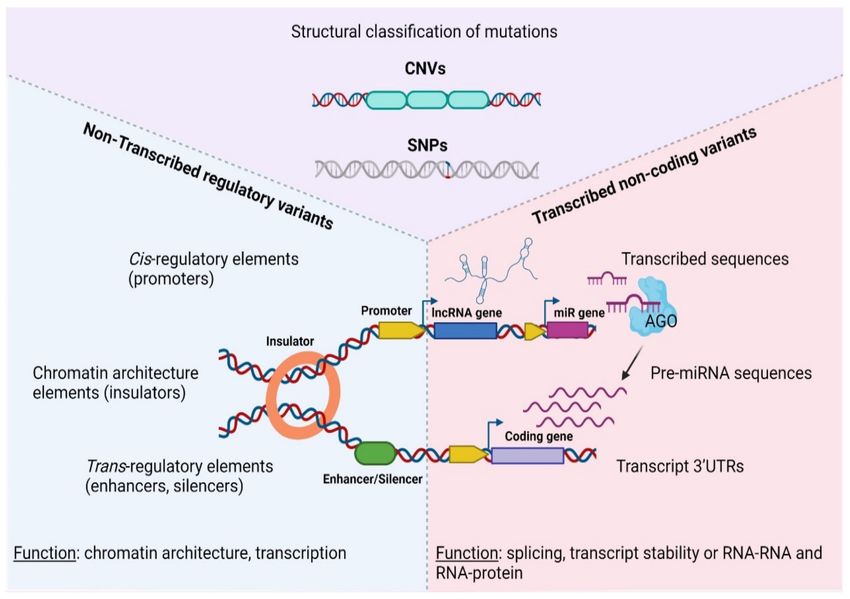
Non-coding RNA 2021, 7, 47                                                                                          2 of 38

                             Complex, which specializes gene expression patterns, recruits general transcription factors
                             and establishes transcriptional memory between tissues and across development [15]. At
                             a chromatin architecture level, promoter–enhancer communication is achieved through
                             the extrusion of chromatin loops leading to the stabilization of topologically associated
                             domains (TADs), which serve as functional genomic boundaries that restrict RE interactions
                             and specify gene expression in a spatiotemporal manner [16]. The adjacent genomic space
                             of TADs hosts specific motifs that facilitate binding of the CCCTC-binding factor (CTCF),
                             a key regulator of chromatin conformation as it interacts with the cohesin complex, which
                             acts as a chromatin loop stabilizer that dictates TAD formation [17–21]. Apart from chro-
                             matin architecture, enhancer transcription by itself may generate enhancer-RNAs (eRNAs-
                             referring to non-coding RNAs transcribed from enhancer loci) [22,23]. On many occasions,
                             eRNAs have a regulatory role in the establishment or maintenance of enhancer–promoter
                             loops. At the level of genomic organization, enhancers can exert their regulatory function
                             either individually or through the formation of clusters, known as super-enhancers, which
                             concentrate transcription factor binding, are characterized by extensive eRNA transcription
                             and serve as organizational centers for complex TAD formation [24–27].
                                  Distinctively, all this operational heterogeneity that underlies enhancer–promoter
                             communication ensures acute yet precise transcriptional responses at a spatiotemporal
                             level. Given the pivotal control of regulatory elements in diverse physiological processes,
                             such as cell-lineage specialization, differentiation, organogenesis and morphogenesis,
                             immune cell diversification and stroma cell interactions, deregulation of their physiological
                             function by mutations often serves as the molecular basis of pathological conditions like
                             cancer [26,28,29]. Identification of such variants in tumor progression can be used as
                             a diagnostic or prognostic tool to predict the clinical outcome for the patient and/or
                             tailor therapeutic strategy in a personalized manner [30,31]. It is therefore imperative
                             to identify and most importantly, functionally dissect genetic variability in the cancer
                             genome that is characterized by extensive variability, in the form of SNPs, or CNAs
                             across wider genomic areas [32,33]. International consortia document and metanalyze
                             functional genomic experiments, providing insights regarding the effect of tumor-specific
                             driver mutations on regulatory elements during neoplastic progression [34–37]. Such
                             consortia are the Encyclopedia of DNA Elements (ENCODE—a public research consortium
                             focused in identifying all functional DNA regulatory elements) [38], NONCODE which
                             is an integrated knowledge database dedicated to non-coding RNAs (excluding tRNAs
                             and rRNAs) [39], and The Cancer Genome Atlas (TCGA—a landmark program of cancer
                             genomics, containing genomic, epigenomic, transcriptomic, and proteomic data from
                             tumors, along with the clinical profile of the patients) [40].
                                  Although some genetic variants are stably inherited and occasionally predispose
                             for hereditary forms of cancer, carcinogenesis itself relies on inactivation of DNA repair
                             mechanisms, leading to genomic instability and extensive accumulation of a mutational
                             burden on a global scale [41]. Genomic variants can be further classified based on structural
                             (SNPs or CNAs), expression (transcribed or not transcribed sequences) or functional
                             (coding or non-coding regions) criteria, all of which ultimately reflect the mechanism
                             through which these genetic lesions are implicated in the development and progression
                             of human malignancies [42–45]. This review focuses on the mechanisms though which
                             non-coding regulatory variants in transcribed or non-transcribed parts of the genome
                             control carcinogenesis, together with the appropriate state-of-the-art methodologies to
                             identify and study them.

                             2. Genetic Variability in the Cancer Genome
                             2.1. Structural Classification of Mutations in Cancer
                                  At the molecular level, SNPs and/or CNAs can reside both in coding, as well as non-
                             coding sequences that are either transcribed or not inside tumors (Figure 1). Depending
                             on the nature of the mutation and the function of the underlining sequence, genetic
                             lesions fuel carcinogenesis through a diverse array of mechanisms, including but not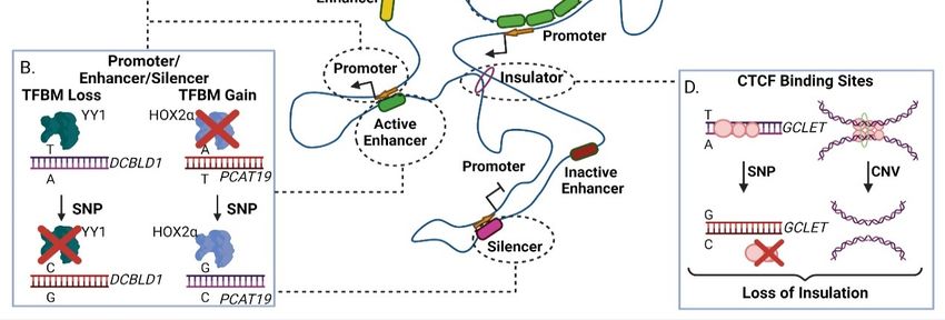
Non-coding RNA 2021, 7, 47                                                                                            3 of 38

                              limited to chromatin modification, transcriptional regulation and alternative splicing,
                              to altered transcript/protein structure or activity due to premature stop codons, non-
                              synonymous amino-acid changes and aberrant gene fusions [46–49]. Therefore, different
                              types of mutations or affected sequences predispose for cancer via an array of distinct
                              mechanisms that must be examined separately.

     Figure 1. Categorization of genetic variations in the non-coding coding cancer genome. AGO—Argonaute protein, CNV—
     Copy Number Variation, SNP—Single Nucleotide Polymorphism, UTR—Untranslated Region, lncRNA—long non—coding
     RNA, pre-miRNA, precursor microRNA. Created with BioRender.com, permission: 15 July 2021.

                                   Focusing on single nucleotide polymorphisms, genome-wide association studies
                              (GWAS) were successful in identifying their mechanistic interplay with normal develop-
                              ment or pathology [50]. Depending on the strength of their alleles, SNPs can be charac-
                              terized as high or low risk factors for complex traits, such as cancer [51]. Nevertheless,
                              association between SNP alleles and phenotypic impact can be confounding due to linkage
                              disequilibrium that segregates driver mutations (that directly control the trait of interest)
                              together with passenger mutations (that are passively inherited with the former but are
                              regulatory neutral) within populations. Therefore GWAS approaches should be accompa-
                              nied by extensive and careful mechanistic characterization with the aim of determining
                              which of the associated SNPs are the true causative factors of the disease [52].
                                   At the level of CNAs, two main categories can be identified: (i) germline CNAs that
                              include duplications or deletions and (ii) somatic copy number alterations of specific loci.
                              Germline CNAs range from 50 bp up to 1 Mb in length and predispose for hereditary
                              types of cancer, such as familiar breast cancer. Somatic CNAs are typically longer than
                              1 Kbp (100 Kbp on average [53,54]) and like germline CNAs, include duplications (known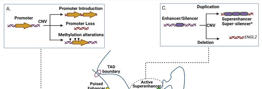
Non-coding RNA 2021, 7, 47                                                                                                4 of 38

                                  as Copy-Number Gains or CNGs), or deletions (representing Copy-Number Losses or
                                  CNLs) [55]. Both types of somatic CNVs (CNGs and CNLs) are prominent lesion types in
                                  tumors that are characterized by extreme Chromosomal INstability (CIN) [56].
                                       Presence of CNAs is heavily linked with malignant manifestation through three main
                                  mechanisms: (i) alterations in gene dosage, in which the copy number of at least one gene
                                  locus is affected [57,58], (ii) gene fusions (mainly due to genomic deletions) [59] and (iii)
                                  alterations of cis and/or trans regulatory elements [60–63]. In some cases, a correlation
                                  between copy number variations and DNA methylation status of the CpG islets for a given
                                  locus has been reported to negatively affect target gene expression [64]. Apart from cancer,
                                  germline CNAs also predispose for various developmental disorders and diseases, such as
                                  autism, schizophrenia with a parental-specific pattern of inheritance, for which they can be
                                  also potent biomarkers for prenatal diagnosis [65,66].

                                  2.2. Functional Classification of Mutations in Cancer
                                       Regardless of their structure, genomic variants (including both SNPs or CNAs) can be
                                  further classified according to their occurrence in transcribed or non-transcribed sequences
                                  (Table 1, [67]). This type of classification underpins the function through which different
                                  variants associate with disease etiology.

                                    Table 1. Structural and functional genetic diversity in cancer.

 Non-Coding Type             Variant ID            Target Locus                 Mechanism              Cancer Type    Citation
                                                Non-transcribed regulatory variants
                             rs11672691         PCAT19 promoter            NKX3.1, YY1 binding           Prostate
                                                                                                                        [68]
                              rs887391          PCAT19 promoter            NKX3.1, YY1 binding           Prostate
      Promoters              rs17079281         DCBLD1 promoter                YY1 binding                Lung          [69]
                              rs2267531        Glypican-3 promoter                    -                   HCC           [70]
                                                                             HSPH1 increased
                              rs2280059          HSPH1 promoter                                          NSCLC          [71]
                                                                               expression
                                                                          NKX3.1, YY1, HOXA2
                             rs11672691         PCAT19 Enhancer                                          Prostate       [68]
                                                                         interaction with PCAT19
                                                                         ONECUT, AR interaction
                              rs7463708          PCAT1 Enhancer                                          Prostate       [72]
                                                                             with PCAT1
                                               Enhancer between
                             rs35252396                                       Binding of HIFs             RCC           [73]
                                              MYC and PVT1 genes
      Enhancer
                                                Enhancer between                                         Prostate,
                              rs6983267                                       Binding of HIFs                           [74]
                                                 MYC and PVT1                                           Colorectal
                             EGLN2 CNV               Enhancer                Genomic deletion            Ovaries        [75]
                                                   Enhancer of
                             rs67311347                                      Binding of ZNF8              RCC           [76]
                                                  ENTPD3-AS1
                              rs4693608         Enhancer of HPSE            Regulation of HPSE            ALL           [77]
       Silencer               rs249473        Silencer in AKT locus           Binding of AKT           Endometrial      [78]
                                               Insulator at GCLET
                              rs3850997                                        CTCF binding              Gastric        [79]
                                                      intron
       Insulator
                                                                          Deletion, Loss of CTCF
                             MYCN CNV           Insulator of MYCN                                     Neuroblastoma     [80]
                                                                                 binding
                                                   Transcribed regulatory variants
Non-coding RNA 2021, 7, 47 5 of 38
Table 1. Cont.
Non-Coding Type Variant ID Target Locus Mechanism Cancer Type Citation
30 UTR region of
rs683/rs910 SNPs miRNA targeting Melanoma [81]
TYRP1
rs713065 miR-204 miRNA targeting of FZD4 NSCLC [82]
miR-96/miR-182 targeting
rs1071738 30 UTR of Palladin Breast [83]
of Palladin
miRNA rs1048638 30 UTR of CA9 miR-34a targeting of CA9 HCC [84]
pri-mir-30c-1 biogenesis
rs928508 miR-30c miR-30c interaction with Breast, Gastric [85,86]
SRSF3
rs6983267 Pre-miR-1307 pre-miR-1307 maturation Colorectal [87]
Maturation process of
rs11671784 miR-27a HOXA Gastric [88]
miR-27a
lncRNA interaction with
rs6983267 CCAT2 Colorectal [89]
CFIms25
LncRNA secondary
rs114020893 lncRNA NEXN-AS1 Lung [90]
structure
miR-194-5p interaction
rs664589 miR-194-5p Colorectal [91]
with MALAT1
miR-4658 interaction with
lncRNA rs1317082 CCSlnc362 Colorectal [92]
CCSlnc362
miRNA-149 interaction
rs11752942 LINC00951 ESCC [93]
with LINC00951
miR-1231 interaction with
rs11655237 LINC00673 PDCA [94]
LINC00673
Isoform selection via
rs10251977 EGFR-AS1 miR-891b and EGFR-AS Oral [95]
interaction
For example, functional variants that occur in transcribed portions of the genome
generally associate with altered transcript message or function (manifested as modified
exonic sequences, alternative splicing, modified UTRs, altered ncRNA folding and/or gene
fusions [96–98]). Interestingly, most GWAS/transcribed regulatory variants are not limited
to protein coding genes but primarily localize in transcribed non-coding sequences that
may generate regulatory transcripts with low or no protein coding potential [99–102]. Non-
coding RNAs (ncRNAs) are categorized based on their processed length, with transcripts
less than 200 nt referring to short non-coding RNAs (consisting mainly of microRNAs-
miRNAs [103], small nucleolar RNAs-snoRNAs [104] and piwi-interacting RNAs [105]),
in contrast to long non-coding RNAs (lncRNAs), which comprise transcripts with lengths
larger than 200 nt [106].
3. Non-Transcribed Regulatory Variants
Variability in non-transcribed regulatory sequences (e.g., promoters, enhancers, CTCF
sites) strongly associates with a mechanistic impact of non-coding variants during neoplas-
tic development [107]. Genome-wide studies revealed an extensive correlation of these
variants with conditionally deregulated spatiotemporal gene expression networks and
disrupted genomic organization in various tumor forms, thus highlighting the importance
of genetic non-coding variability in cancer onset and progression [67,108]. Rare SNP alleles,
associated with increased risk of carcinogenesis (and/or other diseases), are enriched
within expressed quantitative trait loci (eQTLs), with prominence in promoter regions of
oncogenes and tumor-suppressors [109–111].Non-coding RNA 2021, 7, 47 6 of 38
Non-Coding RNA 2021, 7, x FOR PEER REVIEW 6 of 40
Apart from SNPs, somatic CNAs act as the driving force of the CIN subtype that is typ-
ical for various neoplasms. For example, 65% of gastric adenocarcinomas are categorized
as CINtype
cancer andwith
sincehigh
CNAs are one of
percentage of CIN
the leading causes
is colorectal of extensive
cancer, in whichgenomic and tran-
CNVs contribute
to loss of heterozygosity in TP53 and APC, or amplification in KRAS and FGFR1,Another
scriptomic alterations, defining their functional role has a clinical interest [112]. leading
cancer type with high percentage of CIN is colorectal cancer, in which CNVs contribute
to poor prognosis due to drug resistance [113,114]. Despite of their discovery and statisti- to
loss of heterozygosity in TP53 and APC, or amplification in KRAS and FGFR1, leading to
cal association with diagnostic or prognostic markers, such variants often lack functional
poor prognosis due to drug resistance [113,114]. Despite of their discovery and statistical
characterization due to the small effect that a single SNP may have in gene expression,
association with diagnostic or prognostic markers, such variants often lack functional char-
together with tissue-specific restrictions in the expression of the target gene [115–117].
acterization due to the small effect that a single SNP may have in gene expression, together
Therefore, it is crucial to first stratify and subsequently present some of the elucidated
with tissue-specific restrictions in the expression of the target gene [115–117]. Therefore,
mechanisms through which non transcribed regulatory variants dictate neoplastic devel-
it is crucial to first stratify and subsequently present some of the elucidated mechanisms
opment.
through which non transcribed regulatory variants dictate neoplastic development.
3.1.
3.1. Genetic
Genetic Variability
Variability in
in Promoters
Promoters
There are numerous examplesofofgenomic
There are numerous examples genomicvariants
variantsinincis-regulatory
cis-regulatory regions
regions that
that af-
affect
fect transcription of coding or non-coding target genes [118–120]. Promoters
transcription of coding or non-coding target genes [118–120]. Promoters (especially the (especially
the
corecore promoter)
promoter) areprime
are the the prime regulatory
regulatory units
units of of transcription,
transcription, as theyas they transcription
embed embed tran-
scription factor
factor motifs motifs
that thatformation
enable enable formation of the Pre-Initiation
of the Pre-Initiation Complex Complex (PIC) adjacent
(PIC) adjacent to the
to the transcription
transcription start
start site sitegene
of the of the geneIn[121].
[121]. manyIncancer
manytypes
cancer types of promoter
of promoter activity isactivity
altered
is
byaltered by or
inherited inherited
somatic or somatic mutations,
mutations, leading
leading to the to the modulation
modulation of cryptic
of cryptic promoter pro-
activity,
moter activity, loss of promoter DNA methylation or alteration (including
loss of promoter DNA methylation or alteration (including loss or gain) of key regulatoryloss or gain) of
key regulatory motifs
motifs (Figure 2A, [122]).(Figure 2A, [122]).
2. The
Figure 2.
Figure The effect
effect of
of genomic
genomic variants
variants inin non-transcribed
non-transcribed gene
gene regulatory
regulatory elements within aa Topologically
elements within Topologically Associated
Associated
Domain (TAD). (A) Effects of Copy
Copy Number
Number Variations
Variations (CNVs)
(CNVs) in in promoters.
promoters. Duplication events may lead to ectopic
promoter introduction, while deletion event may result in loss of crucial promoter elements. Depending
Depending on on the
the occasion,
occasion,
duplication
duplication or
ordeletion
deletionmay
mayresult in in
result DNA
DNA methylation alterations,
methylation modulating
alterations, transcriptional
modulating activity.
transcriptional (B) Effect
activity. (B) of Single
Effect of
Nucleotide Polymorphisms (SNPs) in promoter, enhancer and silencer elements. Presence of SNPs may lead
Single Nucleotide Polymorphisms (SNPs) in promoter, enhancer and silencer elements. Presence of SNPs may lead to either to either
increased or decreased affinity in transcription factor binding motifs, thus altering the element’s function. (C) Effects of
CNVs at enhancer and silencer elements. Duplications may result in creation of super-enhancers or, hypothetically*, su-
per-silencers. Deletions may lead to loss of crucial transcription factor binding motifs, thus impairing regulatory element
function. (D) Presence of a risk SNP to a CTCF site may lead to loss of insulation due to CTCF site disruption. CNVNon-coding RNA 2021, 7, 47 7 of 38
increased or decreased affinity in transcription factor binding motifs, thus altering the element’s function. (C) Effects
of CNVs at enhancer and silencer elements. Duplications may result in creation of super-enhancers or, hypothetically*,
super-silencers. Deletions may lead to loss of crucial transcription factor binding motifs, thus impairing regulatory element
function. (D) Presence of a risk SNP to a CTCF site may lead to loss of insulation due to CTCF site disruption. CNV
occurrence may also lead to loss of insulation due to deletion of a CTCF site. TFBM-Transcription Factor Binding Motif.
CTCF-CCCTC-binding factor. Created with BioRender.com, permission: 15 July 2021.
For example, rs11672691 (G/A) and rs887391 (T/C/A), two risk-associated SNPs
for poor prognosis in prostate cancer, map to a genomic region with bifunctional role
acting either as promoter or enhancer. Presence of the cancer-predisposing alleles facilitates
promoter-to-enhancer switching, leading to reduced binding capacity of the transcription
factors NKX3.1 and YY1 to the promoter of the short isoform of the PCAT19 (Prostate
Cancer Associated Transcript 19) lncRNA transcript. This favors accumulation of the long
PCAT19 isoform that interacts with HNRNPAB and promotes the expression of cell cycle
genes that subsequently fuel tumor growth and metastasis [68]. Another example refers to
the high-risk SNP rs17079281 (C/T) that resides within the promoter of the DCBLD1 gene
and predisposes for lung cancer in Asian and European populations. The predisposing C
allele of this SNP reduces the binding affinity of the YY1 transcription factor that normally
represses transcription of the gene, ultimately leading to increased levels of the DCBLD1
oncogenic protein in the mutated tissues (Figure 2B) [69]. Another study in mice showed
a gene directly affected by SNPs and CNAs, Plekha5, which normally acts as a suppressor
of metastasis. In presence of SNPs or CNAs, Plekha5 is deregulated, leading to an increase
in the metastatic rate of the cells [123]. The rs2267531 SNP lies within the promoter of
Glypican-3 gene (in Xq26) and the CC/C genotype of which has been correlated with
susceptibility and reduced overall survival of patients with hepatocellular carcinoma
(HCC) [70]. In addition, the G allele of rs2280059 SNP, which lies within the promoter of
HSPH1, is able to increase its expression levels, leading to enhanced resistance of the cancer
cells to treatment in patients with advanced non-small-cell lung cancer [71]. Apart from
motif changes, CNVs may also alter the methylation status of oncogenic promoters (e.g.,
through the demise of methylation sites) leading to increased proliferation advantages for
the mutated cell subpopulations, evidence of which have been extensively found in lung
adenocarcinoma [124]. Collectively, genetic variability in promoter regions often associates
with altered gene expression that links to disease progression of various cancer types.
3.2. Genetic Variability in Enhancers
Most cancer enhancers show cell- and/or stage selectivity in their activation pat-
terns [125–127], therefore their associated genetic variability is ideal for assessing per-
sonalized predisposition or therapy. Since enhancers (and super-enhancers) function
through DNA binding motifs, their activity is vulnerable to variation that modulates the
binding capacity of transcription factor proteins, thus altering transcription of the target
gene [128]. Presence of CNA (and other architectural disarrangements) combined with
loss of insulation events can lead to ectopic enhancer creation or activity resulting into
metaplastic differentiation associated with malignancy (Figure 2C, [129–131]). For example,
genome-wide CNA studies have correlated a deletion in an ovarian-specific enhancer with
altered expression of EGLN2, an enzyme that mediates hydroxylation and subsequent
degradation of the HIF1A protein (a master regulator of oxygen homeostasis) in normoxia
(Figure 2C, [75]).
Parallel to CNAs, GWAS studies have also pinpointed the association between SNPs
and enhancer activity in cancer. For example, the variant rs11672691 (G/A), which resides
in an intronic enhancer at the lncRNA PCAT19 locus, correlates with prostate cancer pre-
disposition and aggressiveness [132–134]. More specifically, the risk allele rs11672691-G
enhances the binding activity of the novel transcription factor HOXA2, which in turn regu-
lates expression of PCAT19 in prostate cancer through enhancer–promoter loop formation
(Figure 2B) [135]. Single nucleotide editing in combination with ChIP-seq (Chromatin Im-Non-coding RNA 2021, 7, 47 8 of 38
munoprecipitation followed by Sequencing) experiments revealed that binding of HOXA2
positively regulates not only PCAT19 but also its neighboring locus CEACAM21. Thus, the
interplay of rs11672691 with the regulatory circuit of HOXA2, PCAT19 and CEACAM21 is
linked to advanced cell growth and invasion with a significant clinical impact on prostate
cancer disease aggressiveness and severity, highlight the role of enhancer mutations in the
regulation of neighboring coding and non-coding targets in cancer tissues [68,135].
With regards to SNPs, rs67311347 (G > A) shows a positive correlation with cancer
cell proliferation in patients with Renal Cell Carcinoma (RCC). The A allele creates a bind-
ing site for ZNF8 within an enhancer element regulating the tumor-suppressor lncRNA
ENTPD3-AS1, leading to its increased expression. ENTPD3-AS1 interacts with miR-155-5p
and activates the expression of HIF-1a in RCC [76]. The SNP rs4693608 lies within an
enhancer regulating the expression of HPSE, by affecting the self-regulation of the onco-
genic transcription factor in acute lymphoblastic leukemia (ALL), with the A allele carriers
escaping the methylation of the enhancer [77].
An independent study revealed another layer of complexity for this enhancer-like
regulatory region, which seems to have a bifunctional role. The presence of additional vari-
ants that also reside in the PCAT19 locus plays a crucial role in PCAT19 transcript isoform
generation (PCAT19-short and PCAT19-long isoforms respectively) with the PCAT19-long
elevated mRNA levels determining progression of prostate cancer. Specifically, the SNPs
variants rs11672691 and rs887391 that reside in the promoter region of the PCAT19-short
isoform can switch the regulatory identity of the element from promoter to enhancer.
Presence of these two risk alleles disturbs binding capacity of the transcription factors
NKX3.1 and YY1 to the promoter of the PCAT19-short isoform. At the same time the
same risk SNPs reinforce enhancer activity of the bifunctional regulatory element leading
to increased expression of PCAT19-long isoform through a promoter–enhancer interac-
tion. Subsequently PCAT19-long isoform interacts with HNRNPAB and thus influences
expression of cell cycle genes leading to acceleration of tumor growth and metastasis [68].
The expression of another prostate related lncRNA, PCAT1 (Prostate Cancer Associ-
ated Transcript 1), is also modulated at the transcriptional level by a cancer-associated SNP
with pivotal function in prostate cancer. Initially, PCAT1 was reported to be implicated
in early prostate cancer cell proliferation, yet recently it was shown to be involved also in
castration-resistant, advanced prostate tumors [84,136]. PCAT1 expression is modulated
by the risk SNP variant rs7463708 (T > G) located within an enhancer regulatory element
that lies 78 kb away from the PCAT1 Transcriptional Start Site (TSS). PCAT1 promoter
and its enhancer reside within a conserved TAD domain, which indicates the potential of
chromatin loop extrusion between them. The T allele intensifies the binding affinity of the
ONECUT and AR transcription factors, which in turn regulate PCAT1 transcription. Subse-
quently, the PCAT1 transcript interacts with the LSD1 and AR proteins facilitating their
recruitment to enhancer regulatory elements of GNMT and DHCR24 that are androgen-late
response genes that correlate with prostate cancer progression [72,137,138].
Apart from prostate, SNPs in enhancers also affect other forms of cancer. rs35252396
(AC > CG) refers to a two base pair substitution variant that is strongly associated with clear
cell renal cell carcinoma. This particular variant resides in an enhancer element at 8q24.21
between the genomic loci of MYC and PVT1 and along with the SNP rs6983267, whose
regulatory function is well characterized in colorectal and prostate carcinoma. rs35252396
affects chromatin accessibility in this area, increasing binding of hypoxia inducible factors
in this enhancer element [73,74,139,140]. rs6983267 together with rs35252396, highlight the
predisposing effect of neighboring yet separately segregating regulatory genetic lesions
in carcinogenesis.
3.3. Genetic Variability in Silencer Elements
Mutations in distal silencer elements are less understood due to the biased focus
on activating enhancers, even though the latter may also act as silencers and vice versa
in different tissues and cell types [141]. Silencer elements, just like enhancers, containNon-coding RNA 2021, 7, 47 9 of 38
transcription factor binding sites, that form chromatin loops with promoters (Figure 2B,C),
preferably those with high levels of trimethylation of Lysine 27 in Histone 3 (H3 K27 me3 )
epigenetic marker [142]. Supposedly, the formation of a super-silencer is possible, but so far
there are insufficient data that support their existence [143]. A putative silencer regulating
ESR1 and RMND1 expression can be found in 6q25.2, and the SNP rs910416 contained
within it shows allele specific binding of MYC. This disrupts the proper function of the
silencer, leading to breast cancer development [144]. Another example that highlights
the function of such repressive chromatin loops, refers to the regulation of Kit locus by
GATA1, which has a repressive role in hematopoietic differentiation [145]. Other silencers
are characterized by the presence of motifs of FRA1, USF1 and USF2, EBF1, BACH2, and
the RFX family among others, which display repressing activities [146–150]. In contrast
to the binding of activators in unmethylated or lowly methylated enhancer elements,
a proportion of these suppressors can bind to methylated sequences as well, indicating
that some silencers may show activity even in their DNA methylated form [151,152]. The
SNP rs249473 and especially the risk allele A, which lies within a silencer of the AKT1
locus (encoding for the AKT protein, part of the PI3K/AKT/mTOR signaling pathway),
abrogates its silencing activity by creating a binding site for YY1, which in turn activates
AKT1 transcription and elevates the risk of endometrial cancer [78].
3.4. Genetic Variability in Insulator Elements
Insulators are DNA elements which are recognized by CTCF and facilitate creation of
inter-domain boundaries, conferring separation of promoters and enhancers or insulation
against the spread of heterochromatin regions [153,154]. Loss of insulator elements may
occur due to the presence of SNPs that alter the CTCF binding site or the methylation status
of the region [155]. Moreover, CNAs that promote genomic rearrangements of CTCF sites
can lead to enhancer hijacking, that associates with increased levels of a putative oncogenes,
such as MYCN that is one of the main drivers for neuroblastoma (Figure 2D) [80,156]. The
SNP rs60507107 is correlated with increased risk of lung cancer, as the A allele reduces
the binding affinity of CTCF at a CTCF binding site in the first intron of DAGLA (in
11q12.2), leading to its altered expression in lung cancer [157]. Collectively, these examples
highlight the functional diversity through which genetic variability in regulatory elements
predisposes for neoplastic development and progression.
Apart from coding genes, genetic aberrations may also disturb transcriptional regula-
tion of lncRNAs at a chromatin architecture level. For instance, GCLET (Gastric Cancer
Low-Expressed Transcript) is a novel lncRNA with a gastric cancer related variant rs3850997
T > G at 16p13 in the third intron of the GCLET genomic locus. Expression analysis, includ-
ing eQTLs, revealed a strong association between high expression levels of GCLET and
improved patient survival. Moreover, in vitro experiments showed that the rs3850997-T
allele is bound by the CTCF transcription factor with higher affinity compared to G allele
(Figure 2D). CTCF exerts an inhibitory function, so when bound to the relevant intronic
region prevents chromatin loop formation between the intron/SNP variant and GCLET
promoter region, ultimately precluding lncRNA transcription [79]. Furthermore, GCLET
competes with miR-27a-3p to increase FOXP2 expression, therefore affecting lymph node
invasion and metastasis. Inferentially, the T allele of the rs3850997 variant represses tran-
scription of GCLET lncRNA and absence of the transcript contributes to gastric cancer
progression with a significant impact on patient clinical prognosis [79,158–160].
4. Transcribed Non-Coding Variants
Apart from regulatory elements, cancer-related genetic variability is also embedded
in transcribed, yet non-coding sequences. Transcribed non-coding Variants (referred to
as TncVs thereof) exist both in coding and non-coding transcriptional units and fuel car-
cinogenesis through a distinct set of mechanisms compared to their counterparts in coding
sequences. For example, TncVs can modulate the stability of the resulting transcript
through abnormal splicing patterns, UTR variations that create or disrupt miRNA bindingNon-coding RNA 2021, 7, 47 10 of 38
pockets, or through alterations in lncRNA secondary structure that influence interaction
with regulatory partners (both protein and RNA molecules) [107,161]. The latter can lead
to differential regulation of target gene expression, via loss of RNA-chromatin and/or
RNA–protein complex formation, concurrently with disruption of TAD architecture [161].
Such cancer-related transcribed variability is not restricted to the DNA level, but also arises
at the RNA level, giving rise to the very promising and largely unexplored field of epitran-
scriptomics which again may operate from within coding and non-coding transcripts in
a similar manner to inherited mutations [162].
4.1. Non-Coding Variants Affecting miRNA Targeting and Biogenesis
Small RNA sequencing efforts have identified hundreds of miRNAs involved in cancer
progression and tumorigenesis for a variety of cancer types and stages [163]. miRNA signa-
tures with significant prognostic and diagnostic properties often reflect the tissue- and/or
cancer-specific properties that characterize the expression of this class of non-coding tran-
scripts [164]. In terms of function, miRNAs act on the basis of sequence complementarity
with their cognate target-mRNA(s) [165–167]. Thus, any sequence variation, even in the
form of single nucleotide polymorphisms that occurs within the seed sequence of their
genomic loci, can alter targeting affinity [168]. Although GWAS approaches have revealed
the importance of SNPs in oncogenic or tumor-suppressing miRNAs, functional characteri-
zation for the majority of such alterations awaits experimental validation [168–170].
Apart from genetic lesions in miRNA transcripts, variability can also arise within
miRNA binding sites in 3’UTRs of their target genes [171–174]. Such variability may
ectopically create or disrupt a miRNA binding site in malignant or even pre-cancerous
tissues. The miR-155-5p is highly expressed in melanoma patients and targets the 30 UTR
region of TYRP1 (Tyrosinase Related Protein 1) mRNA in a SNP-dependent manner leading
to decreased TYRP1 transcript levels. It has been shown that different combinations of
AA/CC alleles of rs683/rs910 SNPs that lie in the 30 UTR region of TYRP1 mRNA affect
the expression of TYRP1 at a post-transcriptional level while there is also a correlation
with melanoma metastasis [81]. Another miR-SNP (rs713065, T to C change) in the 30 UTR
region of FZD4, which is a consequential epidemiological biomarker for non-small-cell
lung carcinoma (NSCLC), comprises a binding site for miR-204. The predisposing C
allele of this SNP enhances binding of miR-204 compared to the wild type allele (T),
leading to down-regulation of FZD4 through cleavage, uridylation and degradation of its
mRNA. Subsequently, the miR-204-SNP mediated loss of FZD4 induces deregulation of key
components of Wnt/Catenin signaling associated with impairment of colony formation
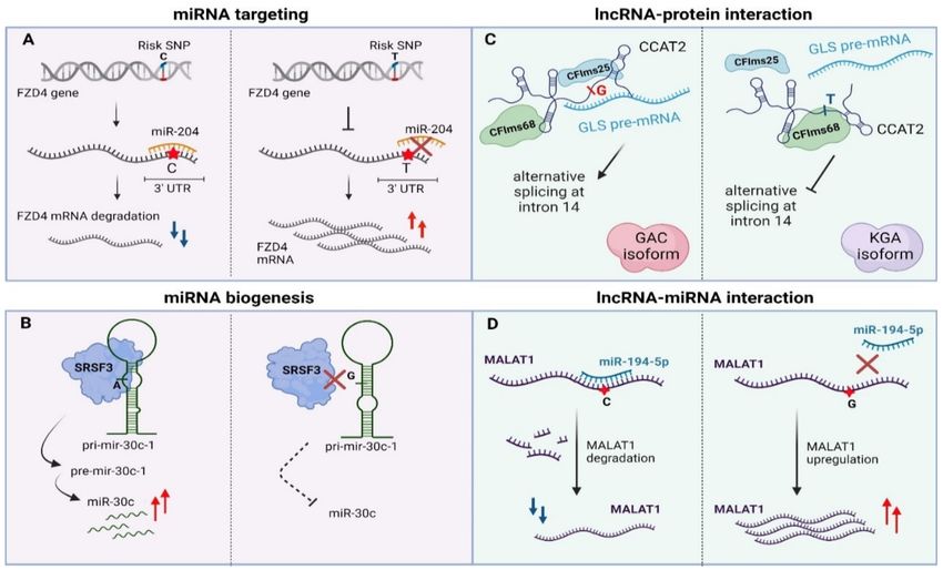
and cell migration of NSCLC cancer cells (Figure 3A) [82].
An analogous example of TncV refers to SNP rs1071738 (G common allele, C minor
allele in European individuals) at the miR-96/miR-182-binding site within the Palladin
30 -UTR with fundamental function in breast cancer metastasis. The ancestral C allele
allows miRNA:mRNA binding while the alternate G allele disrupts it. miR-96 and miR-182
have anti-migration and anti-invasion roles in breast cancer cells that is associated with
downregulation of Palladin, a phenotype which was confirmed by in vivo experiments.
At the therapeutic level, in vivo delivery of miR-96 or miR-182 (fully complimentary with
their binding site in Palladin-30 UTR) by using hydrogel-embedded gold nanoparticles
with efficient release of miRNAs, led to a remarkable decrease of cancer cells’ metastatic
capability [83]. Finally, the rs1048638 SNP that harbors within the 30 UTR of CA9 (Carbonic
anhydrase IX) mRNA is strongly correlated with clinical features (overall survival, poor
prognosis, recurrence) of HCC patients. The A allele of this SNP creates a binding site for
miR-34a targeting that declines CA9 mRNA levels and affects cell proliferation and metas-
tasis of HCC cells [175]. In conclusion, functional characterization of miRNA-associated
TncVs in cancer progression can offer novel therapeutic opportunities at the genetic basis
of cancer.Non-coding RNA 2021, 7, 47 11 of 38
Figure 3. Examples of genomic variants affecting non-coding RNAs function. (A) 30 UTR-occurring SNPs disturb miRNA
targeting via sequence-based mismatches. (B) SNPs in pri-miRNA transcript can alter miRNA biogenesis. (C) SNPs can
affect lncRNA–protein interactions via alterations in lncRNA secondary structure (D) SNPs can alter lncRNA stability via
modulation of miRNA binding sites. UTR—Untranslated region, pri-miRNA—primary miRNA, pre-miRNA—precursor-
miRNA, lncRNA—long non-coding RNA, miRNA—microRNA, GLS—glutaminase, GAC—glutaminase isoform C, KGA—
kidney glutaminase isoform. Created with BioRender.com, permission: 15 July 2021.
Parallel to miRNA binding sites, TncVs that reside in pri-miRNA sequences can affect
cancer progression through defects in miRNA biogenesis [88,176]. A thoroughly character-
ized example is rs928508, referring to a G to A substitution that is present in the terminal
loop of pri-mir-30c-1, perturbing its secondary RNA structure and subsequently leading
to increased levels of mature miR-30c in breast cancer. Particularly, the G/A substitu-
tion, which lies in a CNNC motif, facilitates interaction of the pri-miRNA with SRSF3,
a protein involved in alternative splicing and Drosha-mediated processing of pri-miRNA
maturation [85,177,178]. Experimental validation with SHAPE (Selective 2’-hydroxyl acy-
lation analyzed by primer extension, a technical approach for RNA structural analysis
at single-nucleotide resolution) and toeprint assays (an assay using a fluorescent-labeled
oligonucleotide to prime the reverse transcription step) proved that SRSF3 specifically
recognizes the CNNC motif in a dose-dependent manner in MCF7 cancer cells [179,180].
Importantly, G/A variation promotes formation of a particular tertiary RNA structure
of the pri-miRNA transcript, allowing stronger interaction with the SRSF3 protein and
ultimately proper biogenesis of the miRNA (Figure 3B) [85]. Of note, the same variant
has been previously linked to increased miR-30c expression in breast and gastric cancer
patients [86,181]. Finally, miR-30c has also been shown to be a tumor prognostic biomarker
for breast cancer, modulating chemoresistance through regulation of TWF1 and IL-11 [182].
In independent example of a mutation that affects miRNA biogenesis refers to
rs7911488 (T > C), located in pre-miR-1307 and ultimately interfering with progression
of colorectal cancer (CRC). Homozygous T alleles of this point mutation lead to elevated
expression of mature miR-1307, which in turn binds to 30 UTR of the PRRX1 mRNA and
diminishes its expression levels. Downregulation of PRRX1 enhanced proliferation andNon-coding RNA 2021, 7, 47 12 of 38
migration of CRC cells; however, the exact mechanism needs to be further clarified [87].
Finally, the A allele of rs11671784 SNP within the miR-27a is associated with high sus-
ceptibility to gastric cancer. Correlation studies indicated that this variant influence the
maturation process of miR-27a leading to decreased expression levels of miR-27a in gastric
cancer patients. The diminished levels of miR-27a activate the enhanced expression of
HOXA (miR-27a-target gene) affecting tumor growth of gastric cancer cells [88]. In conclu-
sion, genetic lesions that affect miRNA biogenesis/binding sites frequently associate with
site-specific cancer progression.
4.2. Non-Coding Variants Affecting lncRNA Function
The last decade, lncRNAs gained particular interest in cancer biology given their
cancer- and tissue-specific expression in various malignancies [161,183–185]. The intricate
non-coding nature of their function relies on interactions with i) protein complexes (tran-
scription factors, spliceosome, RNA binding proteins-RBPs, chromatin modifiers in the
nucleus and RBPs, ribosomes and other proteins in the cytoplasm), ii) other non-coding tran-
scripts (such as lncRNAs and miRNAs) or iii) DNA through triple helix formation [186,187].
As a result, genetic variability in lncRNA loci has also been associated with cancer predis-
position, yet few cases have been experimentally validated, given the increased difficulty of
functionally dissecting lncRNAs compared to miRNAs [188,189]. Among the most interest-
ing examples of variations that occur in lncRNAs are SNPs that disorder lncRNA transcript
functionality with a cancer-driving potential. Due to relaxed evolutionary constrains on
primary sequence [190], lncRNA loci can easily accumulate genetic variability in cancer
cells that can affect proper transcript folding, ultimately modulating lncRNA interactions
with their protein partners or other regulatory molecules [191].
More specifically, lncRNAs may contribute to post-transcriptional regulation by af-
fecting splicing [192], mRNA stability [193] or as precursors/regulators of miRNA bio-
genesis [194,195]. Presence of distinct sequence motifs in combination with a particular
secondary structure facilitates binding of splicing factors and other RBPs enabling the
lncRNA–protein functional interplay [161,196,197]. Cancer-risk variations that occur in
these motifs may disturb this interaction, leading to deviant molecular signaling pathways
and finally to malignant transformation [198]. An intriguing example is rs6983267 SNP
(G/T) that resides in the lncRNA locus CCAT2 (Colon Cancer Associated Transcript 2) and
is correlated with colon cancer metabolism (enhanced glutaminolysis) and cell proliferation.
This transcribed SNP can recruit two subunits of the cleavage factor Im complex (CFIm,
CFIms25 and CFIm68 subunits) in an allele-specific manner. CCAT2 transcripts containing
the G-allele, allow binding of CFIms25 with higher specificity compared to T-allele tran-
scripts, which in turn has stronger propinquity for the CFIm68 subunit. This dominant
effect regulates glutaminase (GLS) pre-mRNA alternative splicing. The interaction between
CCAT2 G-allele and CFIms25 directs binding of this RNA–protein complex to the poly(A)
site in intron 14 of GLS pre-mRNA, inducing in this way splicing of glutaminase isoform
C that associates with enhanced catalytic activity compared to the kidney glutaminase
isoform. Biotinylated RNA pull-down experiments revealed that CCAT2 directly binds to
GLS pre-mRNA, highlighting an example of RNA-RNA–protein complex which depends
on the secondary structure of a scaffold-lncRNA that is mainly affected by the rs6983267
SNP (Figure 3C, [89]).
Another example of lncRNA–protein interactions that are altered by the presence of
SNP variants is lncRNA NEXN-AS1 along with its associated SNP rs114020893 at 1p31.1.
This variant is correlated with increased lung cancer susceptibility and is predicted to
modulate the secondary structure of the NEXN-AS1 transcript [90,199]. These examples
highlight the potential of TncVs that alter lncRNA–protein interactions in disease progres-
sion, yet detailed functional insights are still required for the majority of association studies
that link SNP variation with lncRNA function in cancer [188].
Apart from affecting the interplay with protein partners, there are multiple levels of
lncRNA-miRNA interactions that rely on mutations with a role in cancer [200,201]. ThisNon-coding RNA 2021, 7, 47 13 of 38
type of mechanism is sequence-dependent, which means that any alteration in the base
sequence may influence base-to-base interplay. Similar to their role in mRNA:miRNA
interactions, some TncVs can affect lncRNA transcript levels through differential miRNA
binding. Such an example refers to MALAT, a thoroughly characterized lncRNA in many
cancer types (e.g., oral squamous cell carcinoma, melanoma) with a predominant onco-
genic activity, although a tumor-suppressive function has also been reported in breast
cancer [202–205]. The first functionally characterized SNP of MALAT1 was rs664589,
which is involved in colorectal cancer progression via its interaction with the miR-194-5p.
MiR-194-5p targets MALAT1 for degradation in a rs664589 allele-dependent manner. Bind-
ing of miR-194-5p to the MALAT1 transcript with the rs664589-C genotype targets it for
degradation in the nucleus, in contrast to the G allele that decreases overall binding affinity
of the miRNA, leading to accumulation of MALAT1 and ultimately poor patient survival,
increased distant metastasis and enhanced tumor growth (Figure 3D, [91]).
Another example of non-coding variant that affects miRNA-lncRNA interplay in
cancer is CCSlnc362. CCSlnc362 (RP11-362K14.5) is a recently identified tumor-promoting
lncRNA in colorectal cancer. Its expression correlates with the SNP variant rs1317082
(T > C), located at exon 1 of the CCSlnc362 locus. Functional experiments linked the
oncogenic role of the CCSlnc362 with acceleration of the cell cycle parallel to apoptotic
blockage. In vitro luciferase assays showed that miR-4658 binds to CCSlnc362 in an allele-
specific manner. Binding affinity of miR-4658 is increased in the presence of homozygous
C alleles in contrast to the T allele, highlighting an allele-dependent predilection of the
miR-4658 seed [92]. Finally, a correlation study of rs11752942 (A > G) SNP located in
LINC00951 (lincRNA-uc003opf.1) exon, conducted in 1493 Esophageal Squamous Cell
carcinoma (ESCC) patients, revealed a distinct association of the G risk-allele with the
reduced expression of LINC00951. The regulation of LINC00951 is miRNA-149 mediated
and is involved in ESCC cell proliferation and tumor growth [93].
An independent study focused on the long intergenic noncoding RNA (lincRNA)
LINC00673, which is correlated with an antitumor effect in pancreatic ductal adenocarci-
noma. Rescue experiments divulged the significant role of this transcript in cell prolifera-
tion mechanism of pancreatic cancer cells while in vivo xenografts experiments showed
its implication in pancreatic cancer tumor growth. LINC00673 promotes PTPN11 ubiq-
uitination and degradation via mediation of an PRPF19–PTPN11 interaction, resulting
into an elevated and STAT-dependent anti-tumor response. The function of LINC00673
was strongly linked to the germline variant rs11655237 (G > A transition), which creates
a binding site for the miR-1231 preventing LINC00673 from exerting its regulatory role.
Similar to the previous examples, miR-1231 acts with preference to the A allele of the
rs11655237 variant, serving as a decoy for LINC00673 function [94]. Another study of
lncRNA SNPs variants in a cohort of 505 nasopharyngeal carcinoma patients, uncovered
variants associated with chemoradiotherapy sensitivity of patients. Specifically, MEG3
rs10132552 CC genotype was linked to elevated toxicity, LINC-PINT rs1059698 CC had
a protective role against neutropenia and myelosuppression and pR-lncRNA-1 rs73594404
GA genotype patients had increased risk of toxic reactions. All the mentioned lncRNAs are
involved in p53 signaling network, a fact that highlights their SNP potential for reducing
treatment toxicity [206].
In the same context of ncRNA interactions, some polymorphisms indirectly modu-
late isoform selection [207]. An example of a miRNA:lncRNA interaction that relies on
transcribed SNPs and associates with isoform stabilization of a Receptor tyrosine kinase
(RTKs) target in cancer, is EGFR-AS1 (EGFR Antisense RNA 1). RTKs are of great impor-
tance in cancer progression with pivotal clinical and therapeutical applications [208–214].
rs10251977, which associates with the lncRNA EGFR-AS1, normally stabilizes isoform A of
its RTK target EGFR in oral cancer patients. EGFR-AS1 was suggested to act as a scaffold
for PTBP1 (member of the heterogeneous nuclear ribonucleoprotein family) to promote
EGFR-A stabilization. The minor allele A of rs10251977 creates a binding site for miR-891b
(which is downregulated in tumors), leading to degradation of EGFR-AS1 and thus isNon-coding RNA 2021, 7, 47 14 of 38
correlated with elevated levels of the alternative D isoform of the EGFR [95]. Although this
study needs further experimental validation, it represents a notable example of a natural
antisense transcript that regulates its mRNA target in cis through a genetic variant. This
type of genetic variation that alters isoform selection of well-defined oncogenic drivers like
EGFR may expand the prognostic toolbox of cancer or meliorate the personalized therapy
for the patient. Genetic variation could also affect ectopic biogenesis of miRNAs from
lncRNA loci cancer, however experimental validation of such cases in still pending.
5. Methodologies to Functionally Characterize Non-Coding Variants
There are many bioinformatic strategies and databases that take advantage of cancer
genomic data to conduct significant correlations of cancer risk and predisposition (Table 2).
Table 2. State-of-the art methodologies for functional genomic variant identification. Access: 15 July 2021.
Experimental
Advantages Disadvantages Publicly Available Databases/Software
Approach
Methodologies to study genomic areas in open-chromatin state
• HOMER (Hypergeometric
• Enrich in cis-acting Res
Optimization of Motif EnRichment)
DNase-seq • No need for specific TF targeting • Biased in favor of promoters
http://homer.ucsd.edu/homer/
• scDNase-seq improves sensitivity
download.html
• Simple application • ENCODE: Wiggler
• Low signal-to-noise ratio.
FAIRE-seq • Low bias https://sites.google.com/site/
• Requires high fixation efficiency.
• Sensitivity for intronic anshulkundaje/projects/wiggler
• Laborious protocol • http://compbio-zhanglab.org/
MNase-seq • Less noise from mtDNA
• Digestion-based NUCOME/
• Efficiency • ENCODE-DCC version 10
• Demands coupling with other
ATAC-seq • Simple, cost-efficient application https://github.com/ENCODE-DCC/
techniques
• Nucleosome and TF occupancy encoded/releases/tag/v101.0
Methodologies for non-transcribed functional variant identification
• High-throughput examination of • Episomal assay
• Shendurelab/MPRAflow
enhancer activity • Cell-type specific enhancer
MPRAs/CRE-seq https://github.com/shendurelab/
• Allows multiple independent activation profile
MPRAflow
examinations • False-positive ratio
• Gersteinlab/starrpeaker
• High-throughput examination of • Episomal assay
https://github.com/gersteinlab/
enhancer activity • Cell-type specific enhancer
STARR-seq starrpeaker
• Reduced false-positive ratio activation profile
• hyulab/eSTARR
• No barcoding • Reporter transcript stability
https://github.com/hyulab/eSTARR
• ChIA-PET Utilities-CPU
https://github.com/cheehongsg/CPU
• Complex data analysis
• Precise global interaction map • Mango
• Inefficient
ChIA-PET • Long-read ChIA-PETS has https://github.com/dphansti/mango
• Demands coupling with
improved mapping efficiency • TheJacksonLaboratory/ChIA-PIPE
RNA-targeted methodology
https://github.com/
TheJacksonLaboratory/ChIA-PIPE
• Efficiency
• FitHiChIP
HiChIP • Low false-positive ratio • Not available
https://github.com/ay-lab/FitHiChIP
• Simple workflow
• HPRep
• Efficiency
https://github.com/yunliUNC/HPRep
PLAC-seq • Specificity • Not available
• MAPS
• Simple workflow
https://github.com/HuMingLab/MAPS
• Increased false-positive ratio
ChIRP-seq • Commonly used • Not available
• Targets known RNA
However, validation and most importantly functional dissection of cancer-driver and
passenger mutation requires innovative experimental approaches [215–219]. Alongside
the advancement of genomic techniques and next generation sequencing technologies thatNon-coding RNA 2021, 7, 47 15 of 38
improved our understanding regarding the function of the genome, came pioneer research
strategies that identify, validate and finally characterize non-coding variability.
Discovery of functional variants in the non-transcribed portion of the genome is
inextricably bound to experimental approaches designed to unveil novel regulatory se-
quences. A hallmark of a regulatory sequence is chromatin accessibility that subsequently
allows functional activation of the region through binding of transcription factors [220].
Therefore, general approaches that scan the genome for open chromatin can serve as the
first step towards the identification of regulatory variants, especially in cis regulatory
elements [221–223]. When it comes to trans regulation (enhancers, silencers, insulators),
chromatin status needs to be complemented with experimental assessment of chromatin
architecture in the cancer genome in order to pinpoint the target(s) of the regulatory se-
quence [224]. Most importantly, the causative motif(s) within these cis or trans regulatory
sequences needs identification and experimental validation prior to any connection with
genetic lesions. Below some of these experimental approaches are presented based on
the function and position of the non-coding regulatory sequence that hosts the causative
variant with regards to it target(s).
5.1. Scanning for Regulatory Sequences Based on Open-Chromatin State
DNase was first used during the 1980s to map regulatory elements through the identi-
fication of global chromatin accessibility [225]. Following nuclei isolation, permeabilization
of the nuclear membrane and DNaseI incubation, DNA elements are enriched by size
selection either with gel extraction or by ultracentrifuge purification. The methodology can
then be coupled with next generation sequencing. The result of this process is the detection
of DNaseI Hypersensitivity Sites (DHS), which are sites located within open-chromatin
regions with median length of 300 bp that are protected from degradation by DNaseI due
to presence of transcription factors (TF) (Table 2) [226]. DNase-seq has the advantage of
detecting open-chromatin without requiring prior knowledge of the sequence of the TF
bound to the DHS. Additionally, it has higher sensitivity than other approaches (see below)
at promoters. Its drawbacks are linked to the sequence specific function of DNaseI, thus
DHS global identification is not bias-free. Moreover, the purification steps may lead to loss
of DNA sample, lowering overall sensitivity of detection [227–230]. Although DNase-seq
vastly enriches the sample in promoter regions, it demonstrates decreased representation
of regulatory elements in a condensed chromatin state. Such an example applies to cases of
SNPs within promoters of imprinted genes, where the methodology would show allelic
biases towards the imprinted allele [228].
Variants detected within DHS are strong candidates for having a key role in car-
cinogenesis. Such variants are the SNPs rs62331150 (within active promoter region) and
rs73838678 (within strong enhancer region), which are correlated with increased risk for
breast cancer [231]. Complementary to SNPs, presence of CNVs in DHS may result in large
scale chromatin accessibility changes. Such an example is deletion of the DHS chr8:579137-
581436, which leads to increased expression levels through enhanced promoter accessibility
of several tumorigenic protein-coding and non-coding genes [232].
An improvement of DNase-seq refers to single-cell DNase-seq (scDNase-seq). Ap-
plication of scDNase-seq enables study of gene promoters and enhancers at a single-cell
level with highly reproducible results. One SNP identified with this methodology is the
chr18:52417839 (G > C), the frequency of which increases in patients with thyroid carci-
noma, leading to decreased expression of TXNL1, due to disruption of a p53 binding motif
within its promoter [233].
Formaldehyde-Assisted Isolation of Regulatory Elements sequencing (FAIRE-seq) can
be used independently or in combination with other genomic approaches for the discovery
of accessible chromatin, as it enriches for nucleosome-depleted DNA [234]. Following
a cross-linking step, the non-crosslinked and therefore accessible DNA that may host
interactions with TFs is isolated. Coupled to next generation sequencing, this method
serves as a first, yet often crude, option for identifying accessible chromatin. The simplicityYou can also read


