Signaling Pathways in Cancer: Therapeutic Targets, Combinatorial Treatments, and New Developments - MDPI
←
→
Page content transcription
If your browser does not render page correctly, please read the page content below
cells
Review
Signaling Pathways in Cancer: Therapeutic Targets,
Combinatorial Treatments, and New Developments
Hon Yan Kelvin Yip                and Antonella Papa *

                                          Cancer Program, Monash Biomedicine Discovery Institute and Department of Biochemistry and Molecular
                                          Biology, Monash University, Melbourne, VIC 3800, Australia; kelvin.yip@monash.edu
                                          * Correspondence: antonella.papa@monash.edu; Tel.: +613-9902-9330

                                          Abstract: Molecular alterations in cancer genes and associated signaling pathways are used to inform
                                          new treatments for precision medicine in cancer. Small molecule inhibitors and monoclonal antibodies
                                          directed at relevant cancer-related proteins have been instrumental in delivering successful treatments
                                          of some blood malignancies (e.g., imatinib with chronic myelogenous leukemia (CML)) and solid
                                          tumors (e.g., tamoxifen with ER positive breast cancer and trastuzumab for HER2-positive breast
                                          cancer). However, inherent limitations such as drug toxicity, as well as acquisition of de novo or acquired
                                          mechanisms of resistance, still cause treatment failure. Here we provide an up-to-date review of the
                                          successes and limitations of current targeted therapies for cancer treatment and highlight how recent
                                          technological advances have provided a new level of understanding of the molecular complexity
                                          underpinning resistance to cancer therapies. We also raise three basic questions concerning cancer
                                          drug discovery based on molecular markers and alterations of selected signaling pathways, and further
                                          discuss how combination therapies may become the preferable approach over monotherapy for cancer
                                          treatments. Finally, we consider novel therapeutic developments that may complement drug delivery
                                and significantly improve clinical response and outcomes of cancer patients.


Citation: Yip, H.Y.K.; Papa, A.
                                          Keywords: signaling pathways; targeted therapies; combinatorial treatments; oncogenes and tumor
Signaling Pathways in Cancer:
                                          suppressors; cancer resistance; RTK; PROTACS
Therapeutic Targets, Combinatorial
Treatments, and New Developments.
Cells 2021, 10, 659. https://doi.org/
10.3390/cells10030659
                                          1. Introduction
Academic Editor: Frank Schnütgen               Cancer is a disease characterized by the presence of cells undergoing continuous
                                          cell divisions in a rapid and uncontrolled manner. While in the early days surgery and
Received: 24 February 2021                chemotherapies were the only means available to treat tumors, the discovery of oncogenes
Accepted: 13 March 2021                   and tumor suppressor genes has advanced the notion that single proteins can be targeted
Published: 16 March 2021                  pharmacologically for cancer therapy. Recent advances in next-generation sequencing
                                          and multi-omics analyses have further revealed how signaling pathways are intimately
Publisher’s Note: MDPI stays neutral      intertwined to form complex circuitries within cells that, if fully mapped, can be exploited
with regard to jurisdictional claims in   for more accurate targeted therapies.
published maps and institutional affil-        In this review, we discuss some of the recent advances in cancer drug discovery and
iations.                                  provide examples of signaling-based cancer therapies. We further discuss how synthetic
                                          lethality can be utilized to perturb “undruggable” targets, i.e., difficult to target proteins,
                                          and also to treat drug-resistant cancers. A large number of signaling nodes and molecular
                                          hubs have been implicated in the occurrence of cancer, and many of these, such as receptor
Copyright: © 2021 by the authors.         tyrosine kinases (RTKs) and downstream signaling pathways, are current targets of drugs
Licensee MDPI, Basel, Switzerland.        approved by multiple regulatory authorities (e.g., the U.S. Food and Drug Administration,
This article is an open access article    FDA; European Medicines Agency, EMA).
distributed under the terms and
conditions of the Creative Commons
Attribution (CC BY) license (https://
creativecommons.org/licenses/by/
4.0/).

Cells 2021, 10, 659. https://doi.org/10.3390/cells10030659                                                     https://www.mdpi.com/journal/cells
Cells 2021, 10, 659                                                                                           2 of 30

                      2. What Oncogenic Perturbations Are Currently Known and Available as Anti-Cancer
                      Targets?
                      2.1. Historical Perspective of Chemotherapy
                           Given the uncontrolled proliferative capacity distinctive of benign tumor and malig-
                      nant cancer cells, local surgical resection and radiotherapy appeared to be a logical and
                      effective way to treat tumors in the early days. However, tumors and cancers are more
                      than just a mass of rapidly proliferating cells; they are indeed a highly complex disease that
                      requires many elements to thrive, including a compromised microenvironment surround-
                      ing the primary tumor, as well as an adapted systemic response whereby a permissive
                      immune system [1,2] and microbiome [3] allow the tumor to evolve. Additional factors
                      such as intra-tumor heterogeneity and clonal evolution further challenge our appreciation
                      of the complexity of tumor biology and dangerously undermine our ability to choose the
                      most effective treatment at the point of care.
                           Modern chemotherapy began in the 1970s with the use of antimitotic drugs such as
                      cyclophosphamide and chlorambucil, which were prescribed for the treatment of blood
                      malignancies [4,5]. Methotrexate, an anti-folate that inhibits dihydrofolate reductase and
                      suppresses the synthesis of thymidine/purine, was prescribed to cancer patients in the
                      1980s [6,7]. However, despite the initial positive response, patients became resistant to
                      this antimitotic reagent and the disease inevitably relapsed. Since then, extensive drug-
                      screening programs have been performed with the goal to identify new candidate targets for
                      cancer treatment [8]. Furthermore, because of the toxicity associated with the administration
                      of poorly characterized drugs, it became clear that an understanding of the mechanisms
                      through which anti-cancer drugs function is a key element in the pursuit of safe and
                      effective cancer therapies.

                      2.2. Representative Signaling Pathways and Cancer
                           Since the early discovery of oncogenes (e.g., MYC, RAS, BRAF and KIT) and tumor sup-
                      pressor genes (e.g., TP53, BRCA1 and PTEN), cancer-associated genetic lesions have been
                      extensively documented [9,10]. Nowadays, signaling pathways and molecular networks
                      are recognized for their critical roles in executing and controlling important pro-survival
                      and pro-growth cellular processes and are therefore chiefly implicated in the onset of cancer,
                      and also in its potential treatment.
                           In this review, we will focus on a few signaling pathways and on selected types
                      of cancer for which targeted therapies have significantly contributed to improve clinical
                      outcomes. Two pathways in particular, the PI3K/AKT/mTOR signal transduction pathway
                      and the Ras/MAPK pathway, are frequently activated or mutated in cancer. These cascades
                      are highly interconnected in mediating upstream signals from receptor tyrosine kinases
                      (RTKs) to intracellular effector proteins and cell cycle regulators [11]. In addition to the
                      linear signals propagated from the extracellular space into the cytoplasmic and nuclear
                      compartments, the PI3K and MAPK pathways are strongly interconnected through a
                      number of positive and negative feedback loops. As an example, targeted inhibition of
                      mTORC1 through the administration of analogs of rapamycin, also known as rapalogs,
                      can cause MAPK reactivation and can lead to resistance to single mTORC1 inhibition.
                      Consequently, combinatorial targeting of mTOR plus MAPK has been shown to induce a
                      better response to therapies [12].
                           Signaling pathways that regulate cell cycle progression (e.g., RTK-CDK4/6) and
                      genomic stability (e.g., BRCA1, p53 and p21) are also frequently altered in cancer. Recent
                      pan-cancer analyses have shown that chromosomal rearrangements, occurring as a result
                      of genomic instability, are early events in various cancer types such as glioblastoma,
                      melanoma, and breast adenocarcinoma [10]. Consistently, CDK4/6 inhibitors and PARP
                      inhibitors have been tested in a number of clinical trials for the treatment of cancer cells
                      with compromised DNA repair, e.g., BRCA1/2 mutations. Furthermore, the growth of
                      cancers expressing hormonal receptors, including breast, ovarian, and prostate cancers,
                      has been shown to largely depend on the proliferative signal induced by their relative
Cells 2021, 10, 659 3 of 30
hormones, such as estrogen and androgen. The understanding of the mechanisms through
which these nuclear hormones and their receptors function has led to the development of
the diverse classes of inhibitors currently used as the standard of care for cancer treatment,
as we detail below [13–15].
Notably, these selected examples do not include successes achieved through therapies
directed at additional signaling pathways such as the apoptotic cascades [16]. Moreover,
emerging evidence suggests that the β-adrenergic receptor signaling, either at the level of
the tumor macro-environment (e.g., adrenal medulla) or within the micro-environment
(i.e., innervation by sympathetic nerve fibers within tumor tissues), can promote tumor
progression and can be targeted by β-adrenergic antagonists and beta-blockers [17,18].
Thus, these studies collectively show that after decades of intense genetic and biochemical
studies, we have been able to identify a number of critical oncogenic alterations and exploit
them for cancer treatment. This has tremendously improved our understanding of cancer
biology and has also contributed to improving the overall survival rates of a number of
cancer patients. Yet despite much progress, too often we find that even the most advanced
targeted therapies still fail to effectively put cancer in remission.
3. Why Do We Need to Identify Novel Molecular Perturbations?
3.1. Targeting Hormone Receptor Signaling through Endocrine Therapies
The first hormone therapy ever applied for cancer treatment was used in 1941 by
Dr Charles Huggins, who administered estrogen and removed the testicles to lower the
levels of testosterone in patients with prostate cancer [19]. Ever since, endocrine therapies
have been largely developed and used for the treatment and management of breast and
prostate cancer.
Breast cancer (BC) is the second most common type of cancer affecting women world-
wide. It is expected that almost 70% of newly diagnosed BC will depend on the pro-growth
effect driven by the hormone estrogen and its receptor. The estrogen receptor (ER), which
is a nuclear receptor able to transcribe pro-growth genes and to cross-talk with multiple
receptor tyrosine kinases (RTK) and signaling pathways to sustain tumor cell growth [20].
Given the high frequency of ER positive (ER+ ) BC [21], therapies suppressing the tumori-
genic activity of this hormone are in continuous evolution. Several treatment approaches
have been developed to inhibit estrogen action, and these include:
(1) aromatase inhibitors (e.g., letrozole, anastrozole, and exemestane), which inhibit
estrogen production and levels [22]
(2) selective estrogen receptor modulators (SERMs) such as tamoxifen and toremifene,
which bind to ER and form complexes with co-repressors instead of co-activators,
thus halting the ER transcriptional program [14,23]
(3) selective estrogen receptor degraders (SERDs) such as fulvestrant, which bind to ER, pre-
vent its nuclear translocation, and lead to its subsequent degradation (Figure 1) [24,25].
These therapies have dramatically improved outcomes and survival rates of breast
cancer patients, and the five-year survival rate for ER+ breast cancer is now higher than
90%. However, despite the prolonged positive response to treatments, ER+ BC patients
also develop resistance to hormone therapies over time, which can occur through multiple
mechanisms. Loss of ER expression occurs in 10% of breast cancers, leading to a loss of
treatment efficacy. Moreover, even when ER expression is still retained, molecular rewiring
of the signaling pathways through the engagement of alternative RTKs such as EGFR [26],
HER2 [27], and FGFR [28] can lead to the reactivation of ER-regulated transcription programs
and tumor cell growth.
Multiple clinical trials are currently underway to identify combination therapies able to
overcome resistance to ER targeted treatments. In an ongoing clinical trial (NCT01953926),
the HER2 inhibitor neratinib, administered together with fulvestrant, increased progression-
free survival compared to neratinib alone in ER+ /HER2-mutant metastatic BC patients
pre-treated with endocrine therapy [29]. The FGFR inhibitors erdafitinib (NCT03238196)
and TAS-120 (NCT04024436) have entered clinical trials in combination with fulvestrant toCells 2021, 10, 659 4 of 30
treat ER+ breast cancer with FGF amplification. The PI3K–AKT signaling pathway, which
acts downstream of many RTKs, is frequently found constitutively active in BC, owing to
mutations in PIK3CA and PTEN [30]. Consistently, combinatorial use of fulvestrant with
the PI3K p110α-specific inhibitor alpelisib has been approved for the treatment of ER+
metastatic breast cancer harboring PIK3CA mutations [31]. Finally, a recent clinical trial
testing the combinatorial effect of fulvestrant plus the AKT inhibitor capivasertib has also
started. [32].
Figure 1. Targeted therapies for cancer treatment. In physiologic conditions, ligands bind to receptor
tyrosine kinases (RTKs) at the cell membrane and induce the autophosphorylation of the RTKs’
catalytic domains and the activation of downstream effectors, e.g., p85 release and the activation
of the p110 catalytic subunit within PI3K; GDPase–GTPase conversion of Ras. Activation of the
PI3K and MAPK pathways initiates a series of phosphorylation events that promote cell growth
and proliferation and regulate cellular differentiation. In the presence of estrogen, the estrogen
receptor (ER) dimerizes and translocates into the nucleus where it activates pro-growth transcription
programs. Examples of FDA-approved monoclonal antibodies and small molecule inhibitors acting
on multiples molecular nodes are shown.
Similar to breast cancer, prostate cancer (PCa) can also be driven by high levels of
hormones such as androgens, whose synthesis is regulated by the hypothalamus–pituitary–
testicular axis [13]. PCa is one of the leading causes of death for men worldwide, and
various therapeutic approaches have been developed to monitor and treat this slow-
progressing tumor. After an initial active surveillance that can last several years, once the
disease progresses from a low-risk and slow-growing tumor to a high-risk aggressive dis-
ease, prostatectomy and radiotherapy are generally proposed as the first line treatment [33].
These initial treatments can be further followed up by androgen deprivation therapy (ADT)
plus chemotherapy. If prostate cancer progresses to metastatic castration-resistant prostate
cancer (mCRPC), this is then treated with antagonists of gonadotropin-releasing hormoneCells 2021, 10, 659 5 of 30
and androgen receptor (AR), which, altogether, lower testosterone activities; abiraterone
can be included in the treatment to further inhibit androgen synthesis [34–36].
Molecular profiling of PCa has identified three main mechanisms of resistance to ADT
in CRPC. Hotspot point mutations in the ligand-binding domain of AR, such as the L702H,
W742C, H875Y, and T878A mutations, are predominantly found in CRPC samples but
not in primary PCa samples. Together with AR amplification, these missense mutations
account for 60% of CRPC oncogenic mutations [37] and function by rendering prostate
cancer cells resistant to AR antagonists (e.g., hydroxyflutamide and enzalutamide) or by
imposing agonist-bound structural conformations, which lead to the reactivation of AR
signaling [38,39].
An additional mechanism of resistance to androgen deprivation is associated with
residual levels of androgens produced by the activation of the de novo steroidogenesis path-
way from cholesterol. Antiandrogen and steroidogenesis inhibitors such as enzalutamide
and abiraterone are currently approved agents for CRPC treatment [40,41].
Finally, reports have shown that the activation of other steroid receptors can also con-
tribute to treatment failure and PCa regrowth. The glucocorticoid receptor (GR)-regulated
transcriptome highly overlaps with AR gene signatures, and compensatory activation of the
GR signaling can lead to enzalutamide resistance in prostate cancer xenograft models [42].
Furthermore, PIK3CA mutations and/or loss of PTEN, as well as Raf activation and DNA
repair signaling pathways, have all been reported to contribute to the growth of metastatic
CRPC through AR-independent mechanisms. Because of these events, a number of combina-
torial treatments using ADT plus inhibitors directed at these signaling nodes are currently
being tested in several clinical trials [43].
3.2. Targeting Receptor Tyrosine Kinases
Under physiologic conditions, RTKs can transduce growth-promoting signals to the
cytoplasmic space. In cancer, RTKs can be found amplified, mutated, and constitutively
active, thus causing growth signals to be continuously transduced even in the absence of
upstream stimuli. To prevent this effect, monoclonal antibodies and targeted inhibitors
have been developed.
Monoclonal antibodies (mAb) directed at the ecto-domains of RTKs act by binding
and preventing RTKs interactions to their agonists. Cetuximab, a mAb binding EGFR, was
the first FDA-approved monoclonal antibody used for the treatment of metastatic colorectal
carcinoma [44]. It functions by inducing receptor dimerization and internalization, thus
reducing the overall EGFR protein levels on the plasma membrane. Given the frequency
of EGFR activation in cancer, additional tyrosine kinase inhibitors, TKIs, directed at the
cytoplasmic domain of EGFR have also been developed. To date, three generations of TKIs
have been approved for use in clinics, including:
(1) first generation TKIs, gefitinib and erlotinib, which compete with ATP for the kinase
domain of EGFR [45,46];
(2) second generation TKIs, e.g., afatinib and dacomitinib, with an improved affinity for
the EGFR kinase domain; and
(3) third generation TKIs, such as osimertinib, which covalently bind to cysteine residue
on EGFR [47].
The second generation TKIs were shown to prolong the overall survival of patients
with NSCLC treated with a first generation TKI, i.e., gefitinib plus chemotherapy [48].
Unfortunately, acquisition of the EGFR T790M missense mutation, occurring in 50% of
NSCLC, was found to cause structural changes in the EGFR binding pocket, thus rendering
tumor cells resistant to the second generation of TKIs and requiring new treatments [49].
The EGFR T790M-specific inhibitor osimertinib irreversibly binds to the C797 residue in
the ATP-binding pocket of EGFR and has been found to improve progression-free survival
(PFS) in patients with the acquired T790M mutation [50,51]. Resistance to EGFR-targeted
therapies, including osimertinib, can however still arise due to the activation of parallel
RTKs such as HER2 [52], MET, IGFR, FGFR, and PDGFR [53,54], or through the activationCells 2021, 10, 659 6 of 30
of downstream signaling pathways such as the PI3K–AKT and Ras–ERK pathways [55,56].
Combinatorial targeting of EGFR and components of these signaling pathways may be a
potential way of overcoming drug resistance to EGFR inhibition.
Human epidermal growth factor receptor 2 (HER2) is an EGFR family member fre-
quently overexpressed in breast and gastric cancer [57]. HER2 is an orphan receptor that
does not have a direct ligand. When overexpressed, HER2 can form homodimers or het-
erodimers with other members of the EGFR family, and transduces the oncogenic signal
to the PI3K–AKT and the Ras–ERK pathways. To target HER2 dimerization, and prevent
its activation, mAbs such as trastuzumab and pertuzumab have been developed [58,59].
HER2 can also be targeted with trastuzumab emtansine, which is a conjugate between
trastuzumab and the cytotoxic reagent emtansine used to treat HER2+ breast and lung
cancers [60,61]. Bispecific antibodies such as ZW25, which binds to two HER2 epitopes
(i.e., trastuzumab-binding domain and pertuzumab-binding domain) [62], and MCLA-128,
which targets HER2 and HER3 [63,64], are now in clinical trials. These bispecific antibodies
enhance HER2 internalization and better abrogate the HER2 oncogenic signal compared to
trastuzumab [65]. Finally, a more recent bispecific antibody, GBR1302, has been found to
bind HER2 and CD3, and to direct cytotoxic T cells to HER2+ breast cancer cells [66].
In addition to mAbs, reversible TKIs, such as lapatinib, and the irreversible neratinib
(pan-HER inhibitor) are also currently used in the clinic to halt signal transduction by
blocking the phosphorylation of the HER2 kinase domain [67,68]. However, due to the
redundancy in signal transduction mediated by the many members of the HER family,
incomplete inhibition of the signal can lead to resistance to targeted therapies. To over-
come this, a combination of dual TKI lapatinib, or pan-TKI neratinib, plus mAbs has been
shown to comprehensively block the known compensatory HER2 signaling network [69,70].
Recently, tucatinib, a highly selective HER2 TKI, has been shown to inhibit HER2/HER3-
mediated MAPK and PI3K/AKT signaling [71]. It suppresses tumor growth and leads to
tumor regression in HER2+ BT-474 breast xenograft models when used as monotherapy, or
together with trastuzumab. Tucatinib has been recently approved by the FDA in combina-
tion with trastuzumab and capecitabine, an anti-metabolite, for the treatment of metastatic
HER2+ breast cancer [72].
Activating HER2 point mutations (e.g., L755S, T862A, and T798M), and the truncated
p95HER2 mutation, has been associated with resistance to HER2 therapies in breast cancer
patients [73–75]. Trastuzumab deruxtecan, an antibody-topoisomerase I inhibitor conjugate,
effectively inhibits AKT phosphorylation, induces DNA damage, and suppresses the
growth of HER2+ patient-derived xenografts (PDX) resistant to trastuzumab emtansine.
Trastuzumab deruxtecan is now approved for the treatment of HER2+ breast cancer and
gastric cancer patients [76,77].
Finally, alterations in PI3K and MAPK pathways have been shown to cause resistance
to HER2-based therapies [78,79], and phase III clinical trials are now underway to treat
advanced HER2+ breast cancer harboring PIK3CA mutations using the PI3K p110α-specific
inhibitor alpelisib (NCT04208178).
3.3. Targeted Therapies Directed at the PI3K/AKT Signaling Pathway
The phosphoinositide 3-kinase (PI3K) signaling pathway plays an essential role in
the regulation of cell proliferation, cell growth, and survival downstream of many RTKs
(e.g., EGFR family; the insulin receptor, IR; and the insulin-like growth factor receptor,
IGFR). RTKs can bind directly to PI3K or can signal through adaptor proteins such as
the insulin receptor substrate 1 (IRS1). Multiple classes of PI3Ks exist but class IA PI3K
is the most frequently mutated in cancer [80]. Class IA PI3K (from now on just PI3K,
unless specified) is a heterodimer consisting of a catalytic subunit, p110; and a regu-
latory subunit, p85. Upon binding to phospho-tyrosine residues on RTKs or IRS, p85
releases its inhibitory effect and activates p110, which in turn binds and phosphorylates
the phosphatidylinositol-4,5-biphosphate (PtdIns-4,5-P2) to generate the second messenger
phosphatidylinositol-3,4,5-triphosphate (PtdIns-3,4,5-P3 or PIP3) [81,82]. At the membrane,Cells 2021, 10, 659 7 of 30
PIP3 recruits multiple downstream targets such as PDK1 and mTORC2, which catalyze
AKT phosphorylation on Thr308 and Ser473, respectively [83–86]. AKT is a central pro-
survival signaling hub that regulates a plethora of downstream effector proteins including
the mTORC1 complex, the FOXO family of transcription factors, and cyclin D1, and sup-
ports cell growth and proliferation. Multiple tumor suppressor proteins have been shown
to limit PI3K pathway activation, such as the lipid and protein phosphatase PTEN [80], the
promyelocytic leukemia protein PML [87], and the tuberous sclerosis complex TSC [88].
3.3.1. Targeting PI3K
Many members of the PI3K pathway are mutated in cancer, but PIK3CA and PTEN
are the two critical players with the highest frequencies of alterations. PIK3CA, the gene
encoding the p110α catalytic subunit of PI3K, is frequently mutated or amplified in en-
dometrial, breast, ovarian, and colorectal cancer [89]. Hotspot mutations in PIK3CA have
been mapped to the exon 20, encoding the kinase domain (e.g., PIK3CA H1047R mutation)
and the exon 9, encoding the helical domain (e.g., PIK3CA E542K, E545K mutations) [90].
These missense mutations render PI3K constitutively active and are frequently correlated
with AKT activation [91,92].
Alterations in the tumor suppressor PTEN [93] are found in somatic cancers such
as breast, endometrial, prostate, and brain cancer [57], but they can also occur in the
germline. where they cause various cancer-predisposition syndromes known as PTEN
hamartoma tumor syndromes (PHTS) [94]. In addition to genomic loss or silencing, loss
of PTEN function can frequently occur through acquisition of missense mutations [95].
Importantly, through the generation of PTEN knock-in (KI) mouse models, our group
and others have demonstrated that two frequent cancer-associated and loss-of-function
PTEN mutations (i.e., PTEN G129E and C124S) can dimerize with wild-type PTEN and
inhibit its function in a dominant negative manner [96–98]. In vivo, these PTEN mutations
cause more rapid tumor formation and progression than that observed in mice with PTEN
heterozygous condition (PTEN+/− mice) [96,99], suggesting that patients harboring these
PTEN alterations may display heightened sensitivity to therapies directed at PI3K and/or
AKT inhibition.
Early preclinical studies with the PI3K inhibitors Wortmannin [100,101] and LY294002 [102]
suggested that the complete inhibition of all PI3K isoforms could provide a positive
response to treatments in PIK3CA-mutant cancers. Over the years a number of pan-PI3K
inhibitors such as buparlisib [103], pictilisib [104], pilaralisib [105], and copanlisib [106]
have been generated and also entered clinical trials. However, due to off-target toxicities,
many of them have been discontinued, except for copanlisib, which has been approved
by the FDA for the treatment of B-cell lymphomas with an altered PI3K pathway [107].
Copanlisib is also in a phase II trial for the treatment of ER+ /HER2- metastatic breast cancer
patients (NCT03803761).
In order to reduce toxicity and increase treatment efficacy, isoform-selective PI3K in-
hibitors have been generated. The p110δ-specific inhibitor idelalisib, the first FDA-approved
PI3K-isoform specific inhibitor, is nowadays used to treat chronic lymphocytic leukemia [108].
More recently, the PI3Kα-specific inhibitor alpelisib has been approved for the treatment
of ER+ /HER2− PIK3CA-mutant and metastatic breast cancer in combination with fulves-
trant [31]. Taselisib is another PI3K inhibitor that equally inhibits p110α/γ/δ and has a 10-fold
lower activity towards p110β [109]. Preliminary results have suggested that taselisib sup-
presses the growth of PIK3CA-driven xenograft tumors and decreases both the activity and the
levels of mutant p110α in vitro [110]. Taselisib is now in a phase II clinical trial (NCT04439175)
for both lymphomas and solid cancers with PIK3CA mutations.
The PI3K signaling pathway is frequently activated in solid tumors that have become
insensitive to endocrine and RTK-targeted therapies. Because almost 40% of ER+ breast can-
cers harbor PIK3CA mutations [30,111], combination therapies using alpelisib or taselisib
with endocrine therapies have been extensively tested in clinical trials [112,113]. However,
targeted PI3K inhibition has been shown to induce systemic metabolic adaptation, whichCells 2021, 10, 659 8 of 30
impacts drug efficacy and limits tumor response to treatment [114]. Because of this, PI3K-
mutant specific inhibitors are now under development. GDC-0077, a PI3Kα inhibitor more
selective towards mutant PI3K than wild-type PI3K, was found to induce proteasomal
degradation of mutant PI3K, and caused tumor regression in PIK3CA-mutant breast cancer
xenografts [115,116]. This compound is now in clinical trial (NCT03006172) as a single
agent, as well as in combination with other TKIs and endocrine therapy for the treatment
of PIK3CA-mutant solid tumors [117].
Multiple mechanisms of resistance have been reported to cause treatment failure of
PI3K inhibitors. For instance, the rewiring of the downstream signaling cascade can involve
the reactivation of molecular targets such as mTOR [118], Pim1 [119], PDK1/SGK1 [120],
or SGK3 [121,122], all reported to support growth in an AKT-independent manner. Reac-
tivation of additional p110 isoforms such as p110β has also been shown to sustain PIP3
accumulation and attenuate the growth inhibitory effect induced by p110α inhibition in
PIK3CA-mutant and HER2-amplified breast cancer cells [123]. Consistently, pre-clinical
studies have shown that combined p110α and p110β inhibition can better suppress growth
and induce tumor regression in PTEN-deficient breast and prostate cancer models than
alpelisib alone [124,125].
PIK3CA mutations can co-occur with activating mutations in RTKs such as EGFR,
KRAS, and ALK in lung adenocarcinomas [126], as well as with the loss of PTEN in en-
dometrial and breast cancer [127–129]. Juric et al. reported that PIK3CA-mutant breast
cancer patients treated with alpelisib became refractory to treatment upon the acquired
loss of PTEN [130]. Treatments with the p110β inhibitor AZD6482 and alpelisib were
shown to resensitize tumor cells and suppress the growth of PTEN-mutant PDX xenograft
models [130]. However, a recent clinical trial testing the efficacy of alpelisib with aro-
matase inhibitors (letrozole or exemestane) for PI3K-mutant ER+ metastatic breast cancer
demonstrated that PTEN deletion, or loss-of-function PTEN mutations (e.g., PTEN A126S
and PTEN R130 *) together with ESR1 activating mutations can still cooperate to cause
treatment resistance [131].
Due to the high frequency of heterozygous deletion found in the PTEN genomic locus,
reactivation of the remaining PTEN wild-type proteins has been considered an attractive
approach for cancer therapy. The Pandolfi group has recently found that the ubiquitin E3
ligase 1 (WWP1) mediates PTEN poly-ubiquitination on lysine 27, which prevents PTEN
dimerization, plasma membrane translocation, and activity toward PIP3. In turn, this
abolishes PTEN tumor suppressive activity and favors cancer progression in a Myc-driven
prostate cancer model [132]. Authors further showed that indo-3-carbinol, a natural WWP1
inhibitor, can inhibit AKT activation and induces apoptosis in prostate organoids and
tissues derived from Hi-Myc mutant mice, suggesting that WWP1 inhibition through the
restoration of PTEN activity is synthetically lethal in Myc-driven tumors.
3.3.2. Targeting AKT
The proto-oncogene AKT, also known as protein kinase B, encompasses three isoforms
(i.e., AKT1, AKT2 and AKT3), and promotes mTOR activation while suppressing p21/p27-
mediated apoptosis [133]. Constitutive activation of AKT, due to activating mutations
in upstream RTKs, PI3K, and/or loss-of-function PTEN mutations, has been implicated
in multiple cancers [90,96,134,135]. Increased AKT activation has also been associated
with resistance to chemotherapy [136] and other TKIs [137], making AKT an attractive
target for combination treatments. Ipatasertib (GDC-0068), an ATP-competitive pan-AKT
inhibitor, synergizes with chemotherapeutic agents to inhibit growth and induce apoptosis
in xenograft models of a spectrum of cancer cell lines, including breast, prostate, ovarian,
and NSCLC cells [138]. Moreover, PTEN deletion has been shown to sensitize MCF-10A
breast cells and tumor xenografts to ipatasertib, suggesting that the loss of PTEN function
can be a biomarker predicting the response to AKT inhibitors [138]. Consistent with this,
we have recently demonstrated that loss-of-function PTEN mutations (PTEN C124S/+
and PTEN G129E/+) coupled with oncogenic PI3K (PIK3CA H1047R) lead to AKT hyper-Cells 2021, 10, 659 9 of 30
activation in a mouse model of breast cancer and that, although resistant to alpelisib, PTEN
and PI3K mammary organoids are sensitive to the AKT inhibitor MK-2206 [139].
Based on this large set of evidence, multiple clinical trials are now underway to evalu-
ate the efficacy of combination therapies using AKT inhibitors with additional targeted
therapies [140,141].
3.4. Targeted Therapies Directed at the Ras/MAPK Pathway
Activation of RTKs, such as the EGFR family, promotes activation of the guanosinet-
riphosphatase (GTPase) Ras proteins, which switch from their inactive Ras–GDP loaded
state to the active Ras–GTP state. The Ras guanine nucleotide exchange factors (GEFs)
catalyze the loading of GTP to RAS, whereas GTPase-activating proteins (GAPs) hydrolyze
GTP–Ras to the GDP-bound inactive state [142,143]. Ras activation recruits and activates
downstream targets such as Raf, including A-Raf, B-Raf, and Raf-1, which are recruited to
the plasma membrane through their Ras-binding domain (RBD) for dimerization and acti-
vation. Active Raf phosphorylates and activates MEK and ERK kinases [144–146], leading
to ERK1/2 nuclear translocation and the promotion of transcriptional programs supporting
cell growth, proliferation, or differentiation.
Multiple members of the MAPK pathway are mutated in cancer. KRAS is frequently
mutated in solid tumors such as pancreatic ductal adenocarcinomas (82%), colorectal carcino-
mas (41%), and lung adenocarcinomas (32%) [147]. BRAF is mutated in 8% of human cancers,
primarily melanoma, whereas MEK and ERK are rarely mutated. B-Raf/Raf-1 heterodimer
is the predominant form of heterodimers transducing the Ras signal in cancer [148,149].
The most prevalent activating mutation in BRAF occurs at the V600 position, which im-
poses an active conformation and can also transduce pro-growth signals as monomers in a
Ras-independent manner.
The GTPase K-Ras oncoproteins have long been considered undruggable targets; how-
ever, recent developments have shown that new selective compounds can form covalent
bonds with mutant KRASG12C , which accounts for 13% of all KRAS mutations, and inhibit
KRAS activity. AMG510 and MRTX849 are two KRASG12C inhibitors currently being tested
in clinical trials [150,151].
Three B-Raf inhibitors (i.e., dabrafenib, encorafenib, and vemurafenib) are available for
the treatment of melanoma and NSCLC harboring the BRAFV600E/K mutations [152]. B-Raf
inhibitors specifically inhibit monomeric BRAFV600 mutant while their inhibitory potential
towards dimeric Raf is reduced. Unexpectedly, B-Raf inhibitors have been reported to
activate, instead of inhibit, the MAPK signaling in non-BRAFV600E tumor cells. It was
found that by binding to B-Raf, these inhibitors increased the potential of wild-type
Raf-1 to heterodimerize with B-Raf [153], thus sustaining high levels of pERK1/2 in a
Ras-dependent manner [154,155].
In order to block the MAPK signaling in non-BRAFV600 tumor cells, type-II RAF
inhibitors have been developed. Type-II RAF inhibitors can bind to B-Raf dimeric kinases
and prevent Raf-1 transactivation, thus blocking downstream activation of MEK and
ERK [156–158]. Type-II Raf inhibitors such as PLX704 and PLX8394 are also called ‘paradox
breaker’. They disrupt the formation of B-Raf homodimers/heterodimers and block the
B-Raf-driven ERK signaling cascade in both BRAFV600 mutant and non-BRAFV600 mutant
tumor cells [159,160].
Type-II Raf inhibitors can also overcome the ERK reactivation caused by vemurafenib
treatments in vemurafenib-resistant melanoma cell lines [159]. A pre-clinical study has
shown that in combination with cetuximab, PLX8394 completely inhibited the growth
of colorectal cancer PDXs with BRAF K601E and G466V mutants and resistant to vemu-
rafenib [160].
Although the first generation of type-II RAF inhibitors (e.g., sorafenib and LY3009120)
did not show significant activities in clinical trials, a new generation of pan-RAF inhibitors,
e.g., AZ628, TAK632 and LXH254, have been developed and have been demonstrated to
reduce phospho-MEK and phospho-ERK levels in the non-BRAFV600 mutant lung cancerCells 2021, 10, 659 10 of 30
H1666 cell line [161], in BRAF mutant human melanoma xenograft models [162,163], and
human NSCLC xenograft models [164].
Resistance mechanisms to Raf inhibitors are usually due to genetic alterations within
the MAPK pathway, such as KRAS/NRAS mutations, BRAFV600E/K amplification, and
MEK1/2 amplification [165,166]. Since Ras–GTPase activates both the MAPK and PI3K
signaling pathways, activating mutations in the PI3K signaling pathways such as PTEN
deletion and the constitutive activation of PI3K and AKT, it has been associated with
resistance to Raf inhibition [167]. Because RAS mutations can co-exist with mutations in
members of the PI3K pathway [168], a simultaneous inhibition of both the MAPK and
PI3K signaling pathways may overcome resistance to Raf inhibitors. Pre-clinical studies
have shown that pan-PI3K inhibitors, AKT inhibitors, or mTOR inhibitors, in combination
with MEK inhibitors, may deliver promising tumor suppressive effects in KRAS or BRAF-
mutant tumors; however, the associated toxicity may still limit clinical benefits [169–172].
An alternative approach to target both the PI3K/AKT and MAPK pathways is to combine
the IGFR inhibitor, linsitinib, with the KRASG12C specific inhibitor, ARS-1620, and the
mTOR inhibitor, everolimus [173]. This three-drug combination was found to be better
tolerated in clinical settings when each drug was administered at low dose [173].
Currently, there are three MEK1/2 inhibitors (binimetinib, trametinib, and cobime-
tinib) available for the treatment of melanoma and NSCLC harboring the BRAFV600E/K
mutations [152]. Next-generation MEK inhibitors are also available, such as RO5126766,
which is defined as a dual Raf/MEK inhibitor that complexes with both proteins and
inhibits phosphorylation of ERK in NRAS melanoma cells and in KRAS mutant xenograft
models [174,175]. This compound is currently under clinical trials to treat RAS mutant
NSCLC (NCT03681483), and colorectal and ovarian cancer (NCT03875820). Since resis-
tance to MEK inhibitions is usually due to reactivation of downstream ERK1/2 [167,176],
multiple ERK inhibitors such as ulixertinib (NCT04566393) and LY3214996 (NCT04391595)
are also in clinical trials and may be considered as a valuable option to overcome resistance
to FDA-approved MEK inhibitors [177].
3.5. Targeted Therapies Directed at Cyclin-Dependent Kinases
Cyclin-dependent kinases (CDKs) are kinases that complex with cyclins to initiate and
regulate cell cycle progression. CDK activation is cell-cycle phase specific and is regulated
by a few inhibitory proteins such as p16INK4A (CDKN2A), p15INK4B (CDKN2B), p18INK4C
(CDKN2C), and p19INK4D (CDKN2D) [178]. Upon growth factor stimulation, the CDK4/6
complex binds to cyclin D1 and phosphorylates the retinoblastoma protein (RB), a master
regulator of cell cycle entry and critical tumor suppressor (Figure 2). RB phosphorylation
inhibits RB function and releases its inhibitory effect on the E2F transcription factor, thus
promoting G1-S cell cycle transition [179,180]. The CDK4/6 complex also activates FOXM1-
mediated transcription, which plays an important role in the regulation of cell cycle
progression and the suppression of cellular senescence [181].
Inhibitors targeting CDK4/6 activation (CDK4/6i) have made important progresses
in breast cancer treatment. To date, there are three FDA-approved CDK4/6i in clinics:
abemaciclib, ribociclib [182–184], and palbociclib [185]. Except for abemaciclib, which
can be used as monotherapy in refractory ER+ /Her2- breast cancers [186], ribociclib and
palbociclib are currently used in combination with aromatase inhibitors, or with fulvestrant
for ER+ luminal breast cancers [182,187]. Emerging evidence has shown that CDK4/6i can
also regulate the host immune response and could therefore be used in combination with
immune checkpoint inhibitors [188,189].Cells 2021, 10, 659 11 of 30
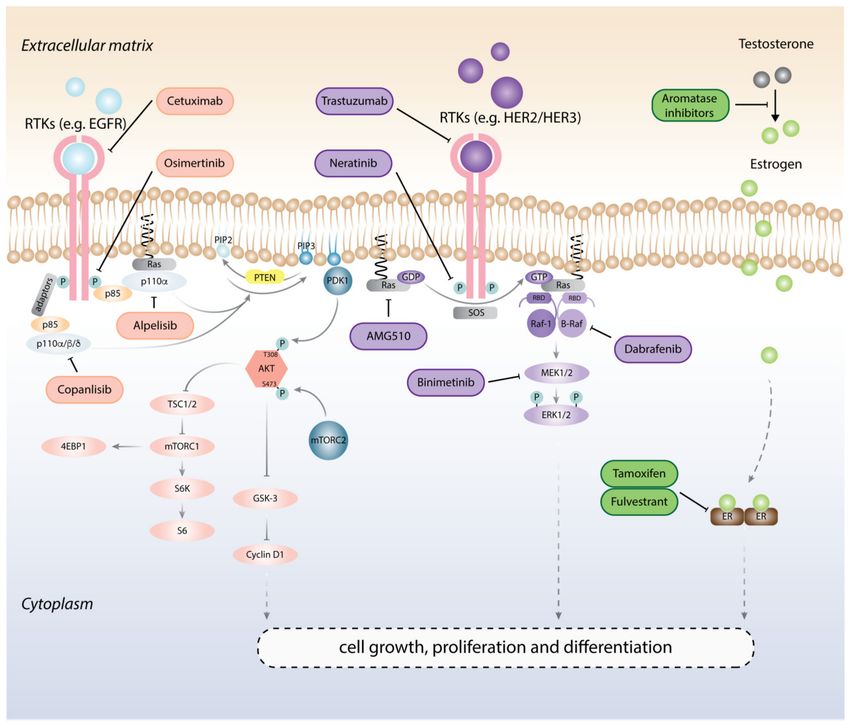
Figure 2. Targeted therapies directed at cell cycle and DNA damage repair pathways. Pro-growth
stimuli promote the formation of the CDK4/6-cyclin D1 complex, which phosphorylates and inac-
tivates the tumor suppressor Rb, and releases E2F. E2F then initiates a transcription program that
supports G1-S phase transition. Genomic stress activates DNA damage response (DDR) mechanisms,
e.g., ATM/ATR, and PARP signaling pathways, and triggers the Chk1/2-p53-p21 axis to induce
cell cycle arrest, senescence, or apoptosis as a means to halt the propagation of genetic lesions.
Accumulation of DNA double-stranded breaks (DSB) can be repaired by two mechanisms: (1) the
error-free homologous recombination, HR; or (2) the error-prone non-homologous end-joining, NHEJ.
FDA-approved PARP inhibitors, e.g., Olaparib, when used with standard treatments, can force HR
repair-defective cells, such as in BRCA1 mutant cells, to activate the error-prone NHEJ pathway, lead-
ing to genomic instability beyond repair and causing cell death. BARD1 (BRCA1-associated RING
domain protein 1); RIF1 (Rap1-interacting factor 1 homolog); and 53BP1 (TP53-binding protein 1).
CDK4/6 inhibition effectively reverses drug resistance to endocrine therapies and RTK-
based therapies. A screening of 47 breast cancer cell lines revealed that CDK4/6 inhibition
suppresses the growth of ER+ luminal cancer and HER2+ cancer cell lines by inhibiting
RB phosphorylation. More importantly, in combination with tamoxifen, palbociclib also
resensitizes resistant MCF7 cells to tamoxifen [190]. Miller and colleagues have shown
that chronic deprivation of estrogen can lead to the hyper-activation of the PI3K/AKT
signaling pathway and sustained CDK4-E2F activation, thus causing estrogen resistance
in breast cancer cells [191]. Consistently, a combinatorial drug screen of 42 compounds
found that CDK4/6 inhibition exhibits the most significant synergism with PI3K inhibitors
(BYL719 and GDC-0941) in a panel on PI3K-mutant breast cancer cell lines (T47D, MCF7,
and MDA-MB-453) resistant to PI3K inhibition [192]. Molecular studies found that high
levels of pRB S780 may contribute to the resistance to PI3K inhibition in resistant cells
in vitro, and in derived xenograft models. In agreement with this, combinatorial use of the
CDK4/6 inhibitor LEE011 plus BYL719 or GDC-0941 caused tumor regression in breast
cancer xenograft models [192].Cells 2021, 10, 659 12 of 30
Goel et al. have shown that CDK4/6 inhibition can also resensitize HER2-resistant
breast cancer cells to trastuzumab, and that a combination of CDK4/6i abemaciclib with
trastuzumab induces tumor regression in a HER2-driven breast cancer mouse model [193].
Similarly, CDK4/6i can resensitize therapy-resistant tumors to EGFR inhibitors, MET/TRK
inhibitors, and MEK inhibitors by suppressing the growth of tumor cells in multiple
pre-clinical models, in vitro and in vivo [194–197].
Rewiring of cell-cycle regulators or activation of alternative pathways can however
cause resistance to CDK4/6i [198]. Although the status of RB can be used to predict
sensitivity to CDK4/6i, genetic alterations in TP53 [199], AKT1 and RAS [200], as well as
overexpression of CDK4/6 [199,201], CNNE1/2 [200,202], and CDKN2A/2B [203], have been
associated with CDK4/6i resistance. Amplifying or activating mutations in FGFR1/2 and
ERBB2 [28,200] have also been found to cause resistance to CDK4/6i. Genetic alterations
in the Ras–ERK pathway and Hippo pathways may contribute to CDK4/6i resistance in
prostate [204] and breast cancer [199], respectively.
A recent study further revealed that loss of PTEN expression is a mechanism of
adaptive resistance to CDK4/6i in breast cancer patients [205]. Authors found that the
PTEN knock-out (KO) in T47D cells caused resistance to ribociclib and palbociclib, but
that administration of the AKT inhibitor MK-2206 resensitized PTEN-KO T47D cells to
CDK4/6i, suggesting that AKT activation mediates the resistance to CDK4/6i in PTEN-
deficient cells. Consistent with this, combinatorial treatment of ribociclib with MK-2206
induced tumor regression in PTEN-null T47D cells xenograft models [205].
3.6. Targeting Genomic Instability
DNA replication is a dynamic process under the active surveillance of cell cycle check-
point inhibitors, which maintain the integrity of the genetic information during cell division.
When DNA damage occurs in normal cells, multiple DNA damage response (DDR) path-
ways and mechanisms are induced. For instance, the base-excision repair (BER) and the
nucleotide-excision repair machineries mediate single base-pair repair [206]; homologous
recombination (HR) and non-homologous end joining (NHEJ) can mediate repair on DNA
double-strand breaks (DSB) [207–209]; and mitotic checkpoints ensure the correct centrosome
localization and chromosomal segregation during cell division [210]. Defects in these DNA
repair mechanisms can lead to the induction of failsafe mechanisms, which act as additional
checkpoints by inducing either a senescence response, or cell death [211].
However, upon the deletion of tumor suppressor genes or the activation of oncogenes,
genomic instability can accumulate within cells and malignant transformation occurs. Mu-
tations in DNA repair genes (e.g., BRCA1/2, PALB2, and RAD51C) and genome gatekeeper
genes (e.g., TP53, ATM and CHEK2) are frequently found in cancer [212,213], and cancer
cells with defective DDR or cell cycle checkpoints replicate damaged DNA to meet the
uncontrolled proliferative drive induced by oncogenic signals.
Nevertheless, we have learnt how to exploit DNA damage accumulation by targeting
DDR pathways and inducing mitotic catastrophe of cancer cells. Topoisomerase inhibitors
(e.g., topotecan and etoposide) and DNA alkylating agents (e.g., cyclophosphamide and
cisplatin) have long been used to induce cancer cell death [214], but targeted therapies
directed at DDR pathways are still limited in use.
3.6.1. Targeting PARP to Exploit Defective HR Repair in Cancer
DNA double-strand breaks (DSB) can be repaired through either the error-free HR [215,216],
or through the error-prone NHEJ repair mechanisms [217]. In BRCA-expressing cells, HR is
the predominant mode of DNA damage repair in the S/G2 phase. During DNA replication,
DSBs are repaired by RAD51 recombinase, which uses the homologous sequence of the
sister chromatid as template DNA. This process is facilitated by the BRCA1/BARD1
(BRCA1-associated RING domain protein 1) and BRCA2/PALB2 complexes [218]. Unlike
HR repair, the NHEJ machinery depends on the function of proteins such as Ku70/80 and
the DNA-dependent protein kinase catalytic subunit (DNA-PKc) complex, which recruitCells 2021, 10, 659 13 of 30
ligases to repair DNA breaks [217]. NHEJ is generally prone to introducing DNA deletions
or insertions and is not used as the preferred pathway when a wild-type BRCA gene is
expressed to ensure DNA integrity [219]. However, when HR repair is not functional,
such as upon BRCA loss, poly(adenosine diphosphate-ribose)ylation (PARylation) and
NHEJ repair become the main DNA repair mechanisms [220]. Defective HR repair is
associated with germline mutations in BRCA1 and BRCA2, which frequently occur in
hereditary breast and ovarian cancer [221]. These mutations increase the lifetime risk of
developing breast cancer from 12% to 46–87%, and ovarian cancer from 2% to 39–63%.
BRCA2 pathogenic mutations are also associated with a 3.7-fold increase in the risk of
developing prostate cancer.
PARylation is an early signal for sensing DNA damage. When cells undergo base-
excision repair, the PAR polymerases, PARP1 and PARP2, are mobilized to DNA single-
strand breaks where they synthesize branched PAR on acceptor proteins to stabilize the
replication fork and facilitate the recruitment of DNA repair factors [222,223]. PARP
inhibitors (PARPi), now in multiple clinical trials, act by blocking PARP1 polymerase
activity, which in turn traps and blocks the replisome from proceeding at the site of the
DNA damage [224]. This condition increases the load of the DNA damage in BRCA-
mutated HR-deficient tumor cells, and leads to chromosomal abnormalities and mitotic
catastrophe [220]. PARPi such as olaparib, niraparib, talazoparib, and rucaparib have been
approved for the treatment of HR-deficient breast and ovarian cancers [225], and since May
2020, they can also be used for the treatment of metastatic, castration-resistant prostate
cancer [226].
However, a large fraction of human cancers are HR competent and are therefore
unresponsive to PARP inhibition. To sensitize HR-proficient tumor cells to PARP inhibitors,
a new combination treatment with the PI3K inhibitor has been tested. The pan-PI3K in-
hibitor BKM120 has been shown to induce the ETS-mediated downregulation of BRCA1/2,
which depletes nucleotide triphosphate synthesis [227,228]. Reduced nucleotides synthesis
induces DNA damage and synergizes with PARP inhibitors in both HR-proficient and
-deficient breast cancer mouse models. Based on pre-clinical results, a recent phase 1b
trial has demonstrated synergism between olaparib and the PI3K p110α-specific inhibitor
alpelisib in treating HR-proficient epithelial ovarian cancers [229]. A recent study has
also shown that simultaneous inhibition on PARP and EZH2, which mediates epigenetic
silencing and suppresses NHEJ activity, can inhibit the growth of HR-proficient ovarian
PDX models by inducing the NHEJ pathway and chromosomal abnormalities beyond
repair, leading to apoptosis [230].
Resistance to PARPi is usually caused by the reactivation of the HR repair pathway.
For example, restoration of BRCA1/BRCA2 activity due to reversion mutations, promoter
demethylation [231,232], or the amplification of the mutant BRCA2 allele [233] have all been
associated with cancer resistance to PARPi. Additional proposed mechanisms of resistance
to PARP inhibition in HR-deficient tumor cells have been recently reviewed [234].
In light of the resistance to PARP inhibitors, researchers have tried to combine PARPi
with inhibitors that target the ATM (ataxia-telangiectasia mutated) and ATR (ATM- and
Rad3-related) pathways, in order to block DDR pathways on multiple levels [235,236].
Pre-clinical studies and clinical trials using replication/mitotic checkpoint inhibitors such
as CHK1 inhibitors (UCN-01, LY2606368) [237], ATR inhibitors (AZD6738), AURKA in-
hibitors (ENMD-2076), PLK inhibitors (Volasertib), and TTK inhibitors (BAY1161909) [238]
are currently being tested in combination with other inhibitors with the goal of causing
replication stress and cell death of cancer cells [239–241].
Finally, PARylation turnover is a process regulated by poly(ADP-ribose) glycohydro-
lase (PARG), which degrades PAR chains to promote the firing of replication fork and cell
cycle progression once DNA repair is complete [242,243]. Inhibiting PARG would lead to
the over-accumulation of PAR chains and eventually cause replication fork collapse and cell
death [244,245]. Pillay et al. [246] demonstrated that the PARG inhibitor PDD00017273 [247]
showed synthetic lethality with the inhibition of a number of G2/M checkpoint proteinsCells 2021, 10, 659 14 of 30
such as TIMELESS, HUS1 RFC2, ATR, WEE1, and CHK1. These combination treatments
can cause replication catastrophe independent of BRCA1/2 status and therefore could be
considered as an option to kill HR-proficient cells.
3.6.2. Targeting p53 and p21 to Induce Synthetic Lethality
TP53 is the most frequently mutated tumor suppressor gene in human malignancies,
and because of its essential role in regulating genome stability it is also known as the
guardian of the genome [57,248]. Under physiologic conditions, p53 protein levels are re-
stricted by the E3 ligase MDM2. However, upon cellular stress, the binding between MDM2
and p53 is disrupted, and p53 accumulation and activation occurs. The tumor suppressor
p53 is a transcription factor that regulates the expression of critical genes such as CDKN1A,
also known as p21, which induces cell cycle arrest and senescence, and BAX and PUMA,
which promote apoptosis. Because of its central role in regulating cell cycle progression,
forced p53 expression has long been considered a potential therapeutic approach for cancer
treatment. Small molecule inhibitors such as nutlins and di-hydroisoquinolinones can
disrupt the binding of MDM2 to p53, and their use has induced positive response in tumors
retaining wild-type p53. Some of these compounds have entered clinical trials [249].
An alternative approach to exploiting p53 expression levels for cancer treatment is to
identify contexts in which synthetic lethality can be achieved. For instance, ATR has been
shown to maintain genomic stability by delaying cell cycle progression and preventing
replication forks from collapsing [250]. However, when ATR is inhibited, its downstream
effector p53 becomes the predominant DDR mediator. Consistently, the ATR inhibitor
AZD6738 was found to cause DNA damage followed by mitotic catastrophe in a p53- or
ATM-defective context, as observed with chronic lymphocytic leukemia (CLL) cells [251].
Moreover, p53 can promote 53BP1 (TP53-binding protein 1) recruitment to sites of DNA
damage, thus excluding BRCA1 and inducing NHEJ [252]. In p53-defective cells, reduced
53BP1 recruitment is accompanied by an increase of BRCA1 recruitment to sites of DNA
damage, thus making HR the preferable mechanism of DNA repair. This suggests that
p53-mutant cells may be sensitive to HR inhibition [253].
The CDK inhibitor p21WAF1/CIP1 is one of the p53-regulated genes mediating cell cycle
arrest, apoptosis, and senescence in response to DNA damage [254]. Histone deacetylase
inhibitors can increase p21 expression and, when used in combination with cytotoxic
agents, can induce cell death or cell cycle arrest in cancer cells such as hepatocellular
carcinoma, ovarian cancer, and prostate cancer [255,256]. Synthetic lethality has also been
used to target p21. For example, p53-null colorectal cancer cells with low p21 expression
are more sensitive to CHK1 inhibition [257].
However, new and conflicting data have shown that high p21 levels may also occur
in cancer, which correlates with poor disease outcomes [258–260]. A recent study has
discovered a new population of Ki67+ dividing cells expressing high levels of p21 in head
and neck squamous cell carcinomas, lung squamous cell carcinoma, and urothelial carcino-
mas [261]. In addition, using the p53-null Saos-2 osteosarcoma cell line and p53-deficient
Li–Fraumeni cell lines, Galanos et al. showed that high p21 levels influence the function
of the CRL4–CDT2 ubiquitin ligase and increase levels of the licensing factors CDC6 and
CDT1, which lead to defective origin licensing and replication stress, fueling genomic
instability in cancer cells. It was found that cells with prolonged high p21 levels would
eventually escape senescence and display aggressive phenotypes such as increased in-
vasion and genotoxic drug tolerance. Mechanistically, high levels of p21 favored Rad52
recombinase-dependent break-induced replication (BIR) and single-strand annealing (SSA)
as the main DNA repair mechanism over error-free synthesis-dependent strand annealing
(SDSA) [262]. This suggests that targeting Rad52 may be synthetically lethal with high p21
levels in p53-null cancers. Additionally, clinically used treatments such as dexamethasone
have also been found to induce p21 expression independent of p53, suggesting a detrimen-
tal use of this compound in patients with p53 deficiency. Indeed, both p53 mutations andCells 2021, 10, 659 15 of 30
high p21 levels have been demonstrated to exert oncogenic activities in cancer in a context
dependent fashion [263,264].
3.7. Two Is Better Than One
Here we have summarized a number of known targeted therapies that affect the
function of essential molecular hubs and are used to deliver better anti-cancer treatments.
However, as we have detailed with various examples, signaling adaptation, acquired
resistance, and novel defense mechanisms still allow cancer cells to survive and relapse.
We propose that a better understanding of the “escape” mechanisms developing in cancer
cells under treatment can provide new insights that can inform more effective combination
therapies and will hopefully put cancer in a more stable remission.
4. How Can Technological Advances Assist with the Identification of New Cancer
Targets and the Delivery of Cancer Drugs?
4.1. Phosphoproteomics and Kinome Profiling
While genomic and transcriptomic sequencing projects have allowed the identification
of genetic mutations and gene expression profiles associated with tumor initiation and
progression, the molecular changes affecting the activation status of signaling pathways
during cancer evolution still remain to be characterized. To identify pathological alterations
of the signaling circuitries caused by reprogrammed signaling networks as a consequence
of targeted therapies, proteomics and kinome profiling can provide alternative, though
technically challenging, omics approaches.
Mass spectrometry-based phosphoproteomics is an unbiased approach to studying
the phosphorylation events of proteins, and can provide detailed views of the activation
status of various signaling nodes within cells. In contrast, kinome profiling uses multiple
kinase inhibitors that, arranged in a top-down fashion inside a column, present varying
specificities to protein substrates [265]. Endogenous protein kinases that bind to these
inhibitors are captured and analyzed by mass spectrometry to provide a global kinome
representation. Phosphoproteomics-based methods have also been used to determine
the activation states of kinases [266] and to establish new kinase–phosphosite relation-
ships [267]. Comprehensive use of proteomics approaches for cancer therapeutics can be
found in specialized reviews [268,269].
An understanding of how signaling networks adapt to perturbations induced by
drug inhibitors, and how kinase–phosphosite relationships contribute to generate adapted
signaling networks, can help design new combination therapies for cancer treatment.
Phosphoproteomics analyses have identified the CDK7/POLR2A axis as a new molec-
ular node in patient-derived epithelial ovarian cancer (EOC), and it was shown that
using the CDK7 inhibitor TZH1 suppressed the proliferation of EOC [270]. Furthermore,
Hijazi and colleagues used chemical phosphoproteomics to reveal that while PIK3CA
wild-type cells seem to depend on MAPK pathway activation, PIK3CA mutant cells more
predominantly rely on AKT and mTOR signaling outputs for survival [271]. Moreover,
phosphoproteomics analysis identified the protein kinase A (PKA)-regulated signaling
network as pro-tumorigenic circuitry in G-protein α subunit (GNAS)-mutated small cell
lung cancer cells [272]. Through phosphoproteomics, we recently found that the loss of
PTEN protein phosphatase activity leads to an enrichment of phospho-peptides regulated
by the glucocorticoid receptor, GR, and confirmed that GR activation sensitizes mutant
PTEN cells to death [139].
Kinome profiling can also be used to identify mechanisms associated with cancer
drug resistance. Lee et al. showed that the phosphorylation of Y397 and Y576 on the focal
adhesion kinase (FAK) is associated with resistance to docetaxel in a metastatic prostate
cancer cell line, and that a combination therapy with FAK tyrosine kinase inhibitor can
overcome the resistance [273]. Moreover, it has been demonstrated that AURORA kinase A,
AURKA, sustains mTOR phosphorylation levels in PIK3CA-mutated breast cancer cells
treated with a pan-PI3K inhibitor or the AKT inhibitor MK-2206 [274]. AURKA inhibitorsYou can also read