Conversion of Stem Cells to Cancer Stem Cells: Undercurrent of Cancer Initiation - MDPI
←
→
Page content transcription
If your browser does not render page correctly, please read the page content below
cancers
Review
Conversion of Stem Cells to Cancer Stem Cells:
Undercurrent of Cancer Initiation
Said M. Afify 1,2       and Masaharu Seno 1,3, *
 1   Department of Medical Bioengineering, Graduate School of Natural Science and Technology,
     Okayama University, Okayama 700-8530, Japan; saidafify@s.okayama-u.ac.jp
 2   Division of Biochemistry, Faculty of Science, Menoufia University, Shebin El Koum-Menoufia 32511, Egypt
 3   Laboratory of Nano-Biotechnology, Graduate School of Interdisciplinary Science and Engineering in Health
     Systems, Okayama University, Okayama 700-8530, Japan
 *   Correspondence: mseno@okayama-u.ac.jp; Tel.: +81-86-251-8216

 Received: 31 January 2019; Accepted: 6 March 2019; Published: 11 March 2019

 Abstract: Cancer stem cells (CSCs) also known as cancer-initiating cells (CIC), are responsible for the
 sustained and uncontrolled growth of malignant tumors and are proposed to play significant roles in
 metastasis and recurrence. Several hypotheses have proposed that the events in either stem and/or
 differentiated cells, such as genomic instability, inflammatory microenvironment, cell fusion, and
 lateral gene transfer, should be considered as the possible origin of CSCs. However, until now, the
 exact origin of CSC has been obscure. The development of induced pluripotent stem cells (iPSCs)
 in 2007, by Yamanaka’s group, has been met with much fervency and hailed as a breakthrough
 discovery by the scientific and research communities, especially in regeneration therapy. The studies
 on the development of CSC from iPSCs should also open a new page of cancer research, which will
 help in designing new therapies applicable to CSCs. Currently most reviews have focused on CSCs
 and CSC niches. However, the insight into the niche before the CSC niche should also be of keen
 interest. This review introduces the novel concept of cancer initiation introducing the conversion of
 iPSCs to CSCs and proposes a relationship between the inflammatory microenvironment and cancer
 initiation as the key concept of the cancer-inducing niche responsible for the development of CSC.

 Keywords: stem cell; cancer stem cells; induced pluripotent stem cells; cancer-inducing niche;
 chronic inflammation

1. Introduction
     Recent studies have revealed the heterogeneity of cell types that are present within malignant
tissues. As a result, tumors are comprised of a diverse collection of cells, with distinct molecular
signatures and different levels of sensitivity to treatment [1].
     Over the decades, there two major models that have been used to explain cancer heterogeneity:
the clonal evolution model (mutation hypothesis) and the cancer stem cell model (cellular hierarchy
organized in a tumor) [2–4].
     The first attempts to understand the basis for the mutation hypothesis originated in 1914, when
Boveri postulated that cancer could result from a combination of chromosomal defects [5]. After this
movement in research toward understanding the biology of cancer, the DNA double helix and genetic
information were discovered in the 1950s [6], which paved the way for the work of Carol O. Nordling,
who suggested that a number of mutated genes could cause cancerous cells to form a tumor [7].
After this hypothesis, the number of mutational changes required to cause cancer was extensively
been investigated. As a result, Ashley posited that approximately three to seven mutations might
be required for the development of cancer [8]. Furthermore, Weinberg confirmed that at least three

Cancers 2019, 11, 345; doi:10.3390/cancers11030345                             www.mdpi.com/journal/cancers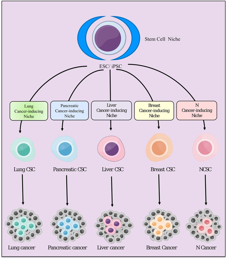
Cancers 2019, 11, x                                                                                        2 of 18
Cancers 2019, 11, 345                                                                                       2 of 19
required for the development of cancer [8]. Furthermore, Weinberg confirmed that at least three or
four mutation were required for the appearance of malignant phenotypes in vitro [9]. Stochastic
or four mutation were required for the appearance of malignant phenotypes in vitro [9]. Stochastic
models have suggested that serial mutation events generated tumor cell heterogeneity and
models have suggested that serial mutation events generated tumor cell heterogeneity and contributed
contributed to cancer progression [10]. In this model, most cancer cells should possess several
to cancer progression [10]. In this model, most cancer cells should possess several mutations that
mutations that give the cells malignant properties, and each mutation increases the probability of the
give the cells malignant properties, and each mutation increases the probability of the next (Figure 1).
next (Figure 1). The main concept of this theory was that cancer should result from time-dependent
The main concept of this theory was that cancer should result from time-dependent accumulation
accumulation of DNA mutations in a single cell. Accordingly, cancers were thought to be monoclonal,
of DNA mutations in a single cell. Accordingly, cancers were thought to be monoclonal, i.e., they
i.e., they were all considered to be derived from a single mutant cell, thereby generating a
were all considered to be derived from a single mutant cell, thereby generating a homogeneous tissue
homogeneous tissue composed of malignant cells [11]. Simultaneously, some scientists thought that
composed of malignant cells [11]. Simultaneously, some scientists thought that mutations occurred
mutations occurred in DNA, but without causing cancer. Mutational changes generally would be
in DNA, but without causing cancer. Mutational changes generally would be insufficient to cause
insufficient to cause cancer, because a minority of cancers were only triggered by about 5% mutations
cancer, because a minority of cancers were only triggered by about 5% mutations [12]. Others noted
[12]. Others noted that some cancers were not associated with any mutations whatsoever [13,14]. On
that some cancers were not associated with any mutations whatsoever [13,14]. On the other hand,
the other hand, many scientists demonstrated that carcinogenesis was a result of conversion of
many scientists demonstrated that carcinogenesis was a result of conversion of normal cells into CSCs.
normal cells into CSCs.

      Figure 1. Schematic
                Schematic illustration
                          illustration for
                                       for the
                                           the stochastic
                                               stochastic and
                                                          and cancer
                                                              cancer stem
                                                                     stem cell
                                                                          cell (CSC)
                                                                               (CSC) models
                                                                                     models of
                                                                                            of tumorigenesis.
                                                                                               tumorigenesis.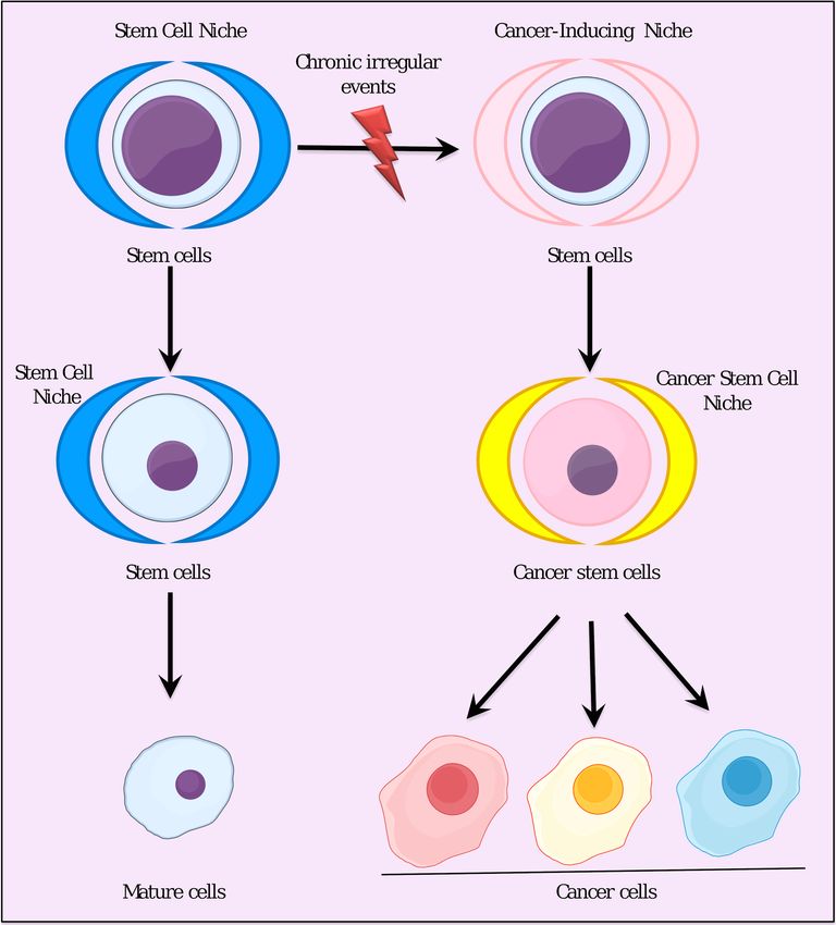
Cancers 2019, 11, 345                                                                                                     3 of 19

     Normal     stem
     Cancers 2019, 11, x cells are described as immature cells that have the double capability of self-renewal         3 of 18
and differentiation potential [15–17]. Stem cells were not discovered by a specific scientist or a
group, but Normal      stem cells
              the concept       was areestablished
                                        described asthrough
                                                      immaturethe cells that have the
                                                                      continuous       double
                                                                                     effort     capability
                                                                                             over   the past of self-renewal
                                                                                                                several decades
     and differentiation potential [15–17]. Stem cells were not discovered by a specific scientist or a group,
by many scientists. Alexander Maksimov, a Russian histologist, who developed and introduced a
     but the concept was established through the continuous effort over the past several decades by many
theory of hematopoiesis, was the first to propose the term “stem cell” in the early 20th century [18].
     scientists. Alexander Maksimov, a Russian histologist, who developed and introduced a theory of
Stem hematopoiesis,
      cells were at first      believed to be present only in certain tissues, such as blood, liver, and intestinal
                          was the first to propose the term “stem cell” in the early 20th century [18]. Stem cells
epithelia,
     were at first believed tothey
           but   nowadays                have been
                                     be present        recognized
                                                 only in              to be
                                                         certain tissues,    present
                                                                          such         in every
                                                                                 as blood,         tissue
                                                                                           liver, and       in theepithelia,
                                                                                                       intestinal    body [19,20].
Immature     cells were
     but nowadays        theyfirst
                               have isolated  from thetoinner
                                       been recognized            cell mass
                                                            be present         of the
                                                                         in every      mouse
                                                                                   tissue in theembryo       at blastocyst
                                                                                                  body [19,20].   Immaturestage
by Martin    Evans
     cells were    firstand   Matthew
                          isolated         Kaufman
                                     from the           [21]
                                               inner cell    andofGail
                                                          mass            R. Martin,
                                                                    the mouse     embryowho    named the
                                                                                           at blastocyst      cells
                                                                                                           stage  by“embryonic
                                                                                                                      Martin
stem Evans    and Matthew
     cell (ESC)”      [22] TheKaufman        [21] and
                                  first isolation       Gail R. ESCs
                                                    of human    Martin,   whofertilized
                                                                        from     named the    cells “embryonic
                                                                                          blastocysts     in vitrostem
                                                                                                                     was cell
                                                                                                                          done by
     (ESC)”    [22]   The   first  isolation  of  human    ESCs   from   fertilized  blastocysts
Thomson [23]. ESCs are defined by the capability to proliferate conservation of an undifferentiatedin  vitro  was   done   by
     Thomson     [23].   ESCs   are   defined  by  the capability  to  proliferate  conservation    of an
phenotype for prolonged periods [24,25], and by the pluripotency of differentiation into all lineages of   undifferentiated
     phenotype for prolonged periods [24,25], and by the pluripotency of differentiation into all lineages
the primary three germ layers, endoderm, ectoderm, and mesoderm (Figure 2) [26,27]
      of the primary three germ layers, endoderm, ectoderm, and mesoderm (Figure 2) [26,27]

            Figure 2. Representative scheme showing differentiation potential of pluripotent stem cells such as
      Figure 2. Representative scheme showing differentiation potential of pluripotent stem cells such as
          embryonic stem cells (ESCs) or induced pluripotent stem cells (iPSCs) into all cell types.
      embryonic stem cells (ESCs) or induced pluripotent stem cells (iPSCs) into all cell types.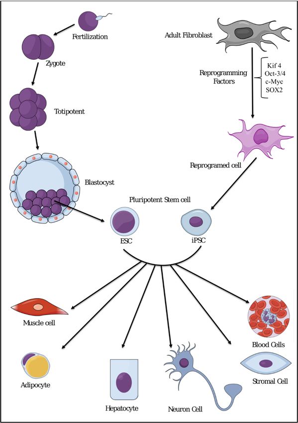
Cancers 2019, 11, 345                                                                                4 of 19

      During the conversion of the normal stem cells into CSCs, many changes can happen, like
abnormal cell division and epigenetic and genetic changes. This implies that the mutation hypothesis
is not always applicable in the formation of a cancer. The conversion is considered to be the causal
event in the origin of the vast majority of cancers. Most of the processes of carcinogenesis should
consist of sequences of steps that initiate cancer. Somatic mutations should follow these steps as the
later events occurring after carcinogenesis. Finally, somatic mutation theory seems to be inaccurate, as
mutations are not always the main causal event of cancer development [28]. Since somatic mutation
theory cannot explain the complexity/heterogeneity of cancer tissues, it should be dropped and
replaced with another theory/hypothesis [29]. The common properties of self-renewal potential and
pluripotency between CIC/CSCs and normal stem cells have led scientists to formulate a new concept
for abnormal stem cell disease, instead of the mutation theory for cancer [30]. The CIC/CSC model
suggests that only certain subpopulations of cancer cells have the ability to drive the progression of
cancer. These are specific and more aggressive subtypes of cells, which result in tumor progression
and recurrence [31]. This subset of tumor cells has the capacity to initiate and sustain tumors when
transplanted into immune-compromised animal hosts, due to its self-renewal and the generation of
differentiated progenies [32,33]. The differentiation of CSCs results in the cellular heterogeneity in
tumors, as well as exhibiting inherent drug resistance and enhancing invasive potential, which plays a
critical role in initial tumor formation and metastatic progression [34]. Therefore, CSCs should be a
pivotal target for the eradication of many cancers.
      According to the classical CSC hypothesis, there is a unidirectional differentiation hierarchy, where
non-CSCs cannot generate CSCs (Figure 1). However, it is increasingly apparent that differentiated
cells can be switched to CSCs [35]. This new hypothesis of heterogeneity opens the way for a CSC
plasticity model, which supports the idea that the differentiated stage of the cells can be converted
back to an undifferentiated stage or stem cell-like stage [36,37]. Collectively, CSCs could be considered
as a dynamic subpopulation of cancer cells.
      Enormous effort has been made to determine the origin of CSCs until now. However, little
is known about the origin of CSCs in tumor tissues. The field of stem cell research has attracted
more scientists than ever to study the stem cell biology and the possibility of the generation of CSCs.
However, the use of ESCs in research is still controversial from an ethical point of view. Therefore, the
discovery of induced pluripotent stem cells (iPSCs) opens a new page in stem cell research. iPSCs are
reprogrammed from completely differentiated cells to take on the characteristics of ESCs, including
the ability to give rise to all the cell types in the body (Figure 2) [38–40]. This reprogramming process
was first established by reprogramming differentiated fibroblasts with the four stemness genes, Oct4,
c-myc, Klf4, and Sox2 [41]. The scientific progress with iPSCs to date seems very promising. iPSCs
are considered to be an ideal replacement for ESCs, and many efforts have been made to understand
their nature. The most important contribution of iPSCs to medicine is the potential of generating
stem cells for clinical applications without sacrificing embryos [42]. In this review, we try to focus on
chronic inflammation, which can stimulate pluripotent stem cells or progenitor cells in each tissue to
generate CSCs.

2. Inflammatory Microenvironment Stimulation and Cancer
      The link between inflammation and cancer was first observed in the nineteenth century by
German physician Rudolf Virchow [43,44]. Virchow observed leukocyte infiltration inside tumors,
so he assumed that the cancer was originating at the sites of chronic inflammation. These leukocyte
infiltrates were initially assumed to be merely a sign of immune responses. In the last decades,
Virchow’s principles have been supported by considerable evidences that inflammation plays a critical
role in tumorigenesis [45,46]. The reason why leukocytes infiltrate tumors has not been well established.
What is the functional role of the leukocytes in the tumor observed by Virchow? Are they promoting
tumors in place of self-protecting or wound healing? Although the link between inflammation and
cancer is now largely accepted [47], the exact mechanism is not clear yet.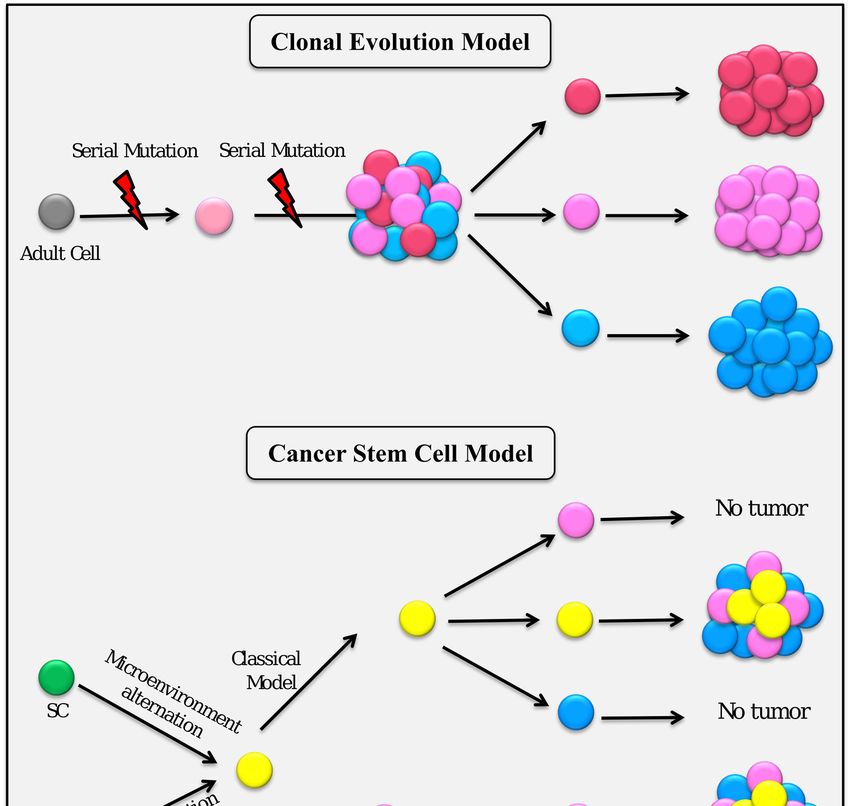
Cancers 2019, 11, 345                                                                                 5 of 19

      To understand this relationship between inflammation and cancer, we should understand first
the process of inflammation. Inflammation is generally considered to be an immune reaction against
a pathogen [48]. Sometimes a pathogen injures tissues, sometimes injured tissues are invaded by
pathogens, and, on other occasions, immune cells that respond to pathogens even without injuries,
e.g., activated immune cells producing reactive oxygen species (ROS), may injure the tissues.
      The first responses of the immune system against infection, which stimulate the release of
chemical factors by infected cells, establish a physical barrier against the spread of infection by clearing
pathogens. Resident immune cells present in all tissues, such as macrophages, histiocytes, Kupffer
cells, and mast cells, are primarily responsible for the initiation of this process. The chemical factors
such as histamine, serotonin, leukotrienes, and prostaglandins produced during the process cause local
vasodilation of the blood vessels and attract phagocytes, especially neutrophils [49,50]. Neutrophils
then trigger other parts of the immune system by releasing factors that attract additional leukocytes
and lymphocytes responsible for repairing.
      Acute inflammation is usually self-limiting; however, chronic inflammation develops, resulting
in organ dysfunction, when acute inflammation cannot be controlled or resolved. Chronic
inflammation results from continuous infection or delayed wound healing that keeps inflammatory
cells activated. These activated cells keep secreting pro-inflammatory mediators, such as cytokines
and chemokines [51], which in turn initiate cancer (Figure 3) [52,53].
      Chronic inflammation plays critical roles at different phases of tumor development. The initial
mechanism of tumor development could be triggered by the inflammation, which may enhance
the production of cytokines, chemokines, growth factors, proangiogenic factors, and extracellular
matrix-modifying enzymes, which should induce signal transductions essential for cell survival
and proliferation, facilitating chromosomal instability [54]. It is assumed that the production of
proinflammatory cytokines, which activate transcription factors, such as NF-κB and STAT3 in
pre-malignant cells, is the main tumor initiating mechanism [55,56]. Playing key roles in chronic
inflammation and tumor initiation, cytokines such as TNF, IL-1, and IL-6 and activated transcription
factors like NF-κB and STAT3 control the main pro-tumorigenic signaling [57,58], which induces
cellular transformation and malignancy [59].
      Tumor promotion is also considered to be the result of chronic inflammation through unusual
epigenetic modification. It is well known that epigenetic processes can control gene expression
without changes in DNA sequence [60]. These epigenetic changes occur in response to the
microenvironment [61], which may change in chronic inflammation and lead to unusual epigenetic
modification, causing irregular activation or silencing of genes. Collectively, the unusual epigenetic
modifications are considered to play critical roles in all types of cancers [62]. Taking these into
consideration, chronic inflammation should critically contribute to tumor initiation and promotion by
changing epigenetics, resulting in the alternative regulation of cancer associated gene expression.
      Inflammation leads to oxidative damage in the DNA of the infected cell, due to the response of
immune cells. NF-κB and STAT3, which are activated by proinflammatory cytokines and chemokines
during chronic inflammation, can cause the parenchymal cells to produce excess amount of reactive
oxygen species (ROS) and reactive nitrogen species (RNS), which induce genomic instability and
DNA mutations [63–65]. Mutations and chromosomal changes are thought to be involved in
tumor progression, which may be accelerated by a chronic inflammatory microenvironment, which
accumulates mutations as a result.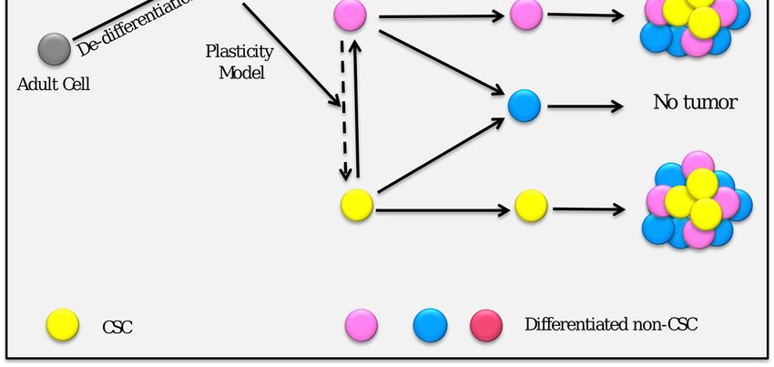
Cancers 2019, 11, 345                                                                                         6 of 19

 Cancers 2019, 11, x                                                                                           6 of 18

       Figure3. 3.Summary
      Figure       Summaryofofinflammation
                                inflammationinvolvement
                                              involvementinintumor
                                                                 tumorinitiation
                                                                        initiationand
                                                                                   andpromotion.
                                                                                        promotion.ROS:
                                                                                                   ROS:reactive
                                                                                                         reactive
       oxygen   species; DC: dendritic cell; NK: natural killer.
      oxygen species; DC: dendritic cell; NK: natural killer.
Cancers 2019, 11, 345 7 of 19
3. Stem Cell Niche and Cancer-Inducing Niche
Although the initial concept of stem cells is more than 100 years old, and its biology was largely
discovered during the previous semicentennial period, much about their nature remains unknown.
Stem cells are critical for tissue homeostasis; they are responsible for tissue regeneration, which replaces
dead cells with new cells according to cellular senescence. To maintain tissue homeostasis through
their lifetime, stem cells must keep an accurate balance between self-renewal and differentiation.
The essential mechanisms must be maintained in a delicate balance, together with the direction of
differentiation. These mechanisms must be understood in order to determine how stem cells are
regulated and contribute to human health and diseases [66,67]. Recent studies have revealed that the
microenvironment, or so called “niche,” is significant in various ways for stem cells.
The concept of niches specific to stem cells was first introduced almost five decades ago by
Schofield, who theorized that stem cell properties are dependent on their microenvironment [68].
Niche has usually been defined as the location surrounding the stem cells. However, this definition
of niche is being changed to involve the cellular components, such as fibroblasts, endothelial cells
and immune cells, which are rich in extracellular matrices, and secreting factors and receptors as well
as the signs of the microenvironment affected by the metabolism [69,70]. Secreted factors bound to
stem cell surface receptors stimulate the intracellular signaling cascade in order to direct stem cell
fates by controlling self-renewal and differentiation [71–73]. Therefore, the concept of seed and soil
is still feasible, with, the microenvironment significantly involved in the cancer development [74].
Chronic inflammatory conditions, as the surrounding niche, trigger stem cells to develop cancer [75,76].
In this process, the CSCs may appear from the normal stem cells affected by the cancer-inducing niche.
Characterization of the cancer-inducing niche will be the most important key to establishing the logic
of CSC development from normal stem cells.
The cancer stem cells concept is greatly changing the historic viewpoint on cancer, namely
mutation theory, which is now being challenged due to its failure to account for determinants
of cancer induction [77]. The cancer-inducing niche can be explained as the continuous irregular
environment that we typically recognize as chronic inflammation (Figure 4). This chronic inflammation
can be explained by the primary role of cytokines and other soluble mediators in the tumor
microenvironment. However, many questions remain to be answered. What are the exact components
of the cancer-inducing niche? Do they vary according to the type of cancer? What kinds of differences
lie between the cancer-inducing niche and the normal stem cell niche? In the next couple of years,
these critical questions will be answered by scientific researchers employing the new strategies that
will become available as stem cell technologies advance.
After CSC development, CSCs will establish their own niche as the CSC niche. Cells composing
the CSC niche are not only necessary for the maintenance of CSCs, but also for the generation of
factors and tumor associated cells that maintain the properties of CSCs, including invasion, metastasis,
and promotion of angiogenesis [78–80]. The CSC niche contains cellular components such as cancer
associated fibroblasts [81], tumor associated macrophages [82,83]. tumor associated neutrophils [84],
MSCs [85], and cell-mediated adhesion [86] and soluble-factors, which play critical roles in cell–cell
communication. The niche before the CSC niche, as a cancer-inducing niche, should be distinguished
from the CSC niche. The cancer-inducing niche can be studied experimentally only by the conversion
of ESCs or iPSCs to CSCs.Cancers 2019, 11, 345 8 of 19
Cancers 2019, 11, x 8 of 18
Figure 4. Stem cell differentiation in the stem cell niche and cancer-inducing niche.
Figure 4. Stem cell differentiation in the stem cell niche and cancer-inducing niche.
4. Inflammatory
4. Inflammatory MicroenvironmentStimulates
Microenvironment Stimulates the
the Generation
Generation of
of Cancer
CancerStem
StemCells
Cells
WeWe hypothesizethat
hypothesize thatchronic
chronic inflammation
inflammationstimulates thethe
stimulates generation of CSCs.
generation According
of CSCs. to our to
According
concept of the cancer-inducing niche, being developed from normal stem cells in chronic
our concept of the cancer-inducing niche, being developed from normal stem cells in chronic conditions,
CSCs are likely to be progenitor cells, which are destined to become cancer cells (Figure 5). Once
conditions, CSCs are likely to be progenitor cells, which are destined to become cancer cells (Figure 5).
CSCs have developed, the CSC niche, together with the cancer-inducing niche, provides a suitable
Once CSCs have developed, the CSC niche, together with the cancer-inducing niche, provides a suitable
microenvironment to maintain CSCs, which in turn develops malignant tumor. The phenotype of the
microenvironment to maintain CSCs, which in turn develops malignant tumor. The phenotype of the
malignant tumor appears to depend on both the tissue specific microenvironment and the CSC niche,
malignant tumor appears to depend on both the tissue specific microenvironment and the CSC niche,
as demonstrated by previous experiments [87,88]. We have demonstrated that iPSCs could acquire
as characters
demonstrated by previous experiments [87,88]. We have demonstrated that iPSCs could acquire
of CSCs when iPSCs were cultured in the presence of a conditioned medium prepared
characters of CSCs
from various when
cancer celliPSCs were cultured in the presence of a conditioned medium prepared from
lines [87,89].
various cancer cell lines [87,89].Cancers 2019, 11, 345 9 of 19
Cancers 2019, 11, x 9 of 18
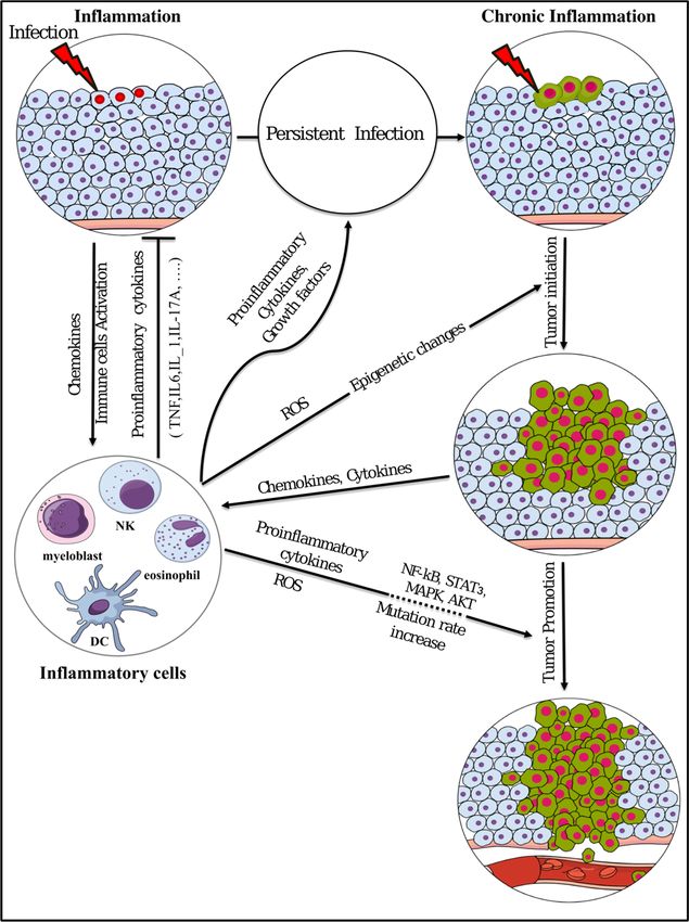
Figure5.5. TheThe
Figure hypothesis of stemofcellstem
hypothesis (ESCs/iPSCs) differentiation differentiation
cell (ESCs/iPSCs) under different cancer-inducing
under different
niches.
cancer-inducing niches.
4.1.4.1.
Lung Cancer
Lung Stem
Cancer StemCells
Cells
Lungcancer
Lung cancerisisthe themost
mostcommon
common causecause ofof cancer-related
cancer-related death
death worldwide
worldwide [90].[90].It It
is is
a a
heterogeneous disease with two different pathological classes: non-small cell lung
heterogeneous disease with two different pathological classes: non-small cell lung cancer (NSCLC), cancer (NSCLC),
which
which makes
makes upup80% 80%ofofcases,
cases,and
andsmall
small cell
cell lung
lung cancer
cancer (SCLC),
(SCLC),which
whichmakes
makesupup20% 20%ofof
allall
lung
lung
cancers
cancers [91].The
[91]. Thefirst
firstexperimental
experimental evidence
evidence forfor the
the presence
presenceofofaastem-like
stem-likesubpopulation
subpopulation in in
lung
lung
cancer was demonstrated by Carney [92,93]. Carney and colleagues described
cancer was demonstrated by Carney [92,93]. Carney and colleagues described a rare population a rare population
(Cancers 2019, 11, 345 10 of 19
H2170 cells, which have high tumorigenic potential in vivo [94]. Simultaneously, lung CSCs isolated
from patient primary tumors exhibited high resistance to standard anticancer agents in vitro [95].
The presence of lung CSCs helps in understanding the phenotype and characters of the sub-population,
but does not explain the origin. On the basis of our approach, iPSCs can be converted into lung CSCs,
which are very important to describe the origin of lung CSCs, as well as the process of the mechanism
of conversion.
Based on the cancer-inducing niche hypothesis, our group established a CSC model from mouse
iPSCs through epigenetic regulations affected by the conditioned medium (CM) from Lewis lung
carcinoma (LLC) cells for four weeks without any genetic manipulation [89,96,97]. These cells exhibited
self-renewal potential by their capacity of spheroid and differentiation potential of some specific
phenotypes. Furthermore, these cells sustained expression of genes associated with the stem cell
character and undifferentiated state, such as Nanog, Sox2, Oct4, and c-Myc. Simultaneously, the
tumorigenic potential was confirmed by the development of malignant tumors, which were recognized
by glandular epithelial hyperplasia, a high nuclear to cytoplasmic ratio, severe nuclear atypia, multiple
pathological mitotic figures, micro metastases, and hypervascularization, indicating angiogenesis [89].
4.2. Pancreatic Cancer Stem Cells
Pancreatic ductal adenocarcinoma (PDAC), the most common type of pancreatic cancer [98],
which constitutes 90% of pancreatic cancers, is one of the most aggressive solid malignancies [99].
PDAC solid tumors are comprised of a wide range of heterogeneous populations of cells, including
CSCs [100]. Rapid progress in identification and isolation of pancreatic CSCs has facilitated the
generation of new mouse models [101,102]. Unfortunately, most of these models are genetically
engineered, and may not be suitable to understand spontaneous tumor occurrence. In contrast, a
novel trial to produce pancreatic CSCs from iPSCs has been explored by the exposure to CM of two
different pancreatic carcinoma cell lines: PK8 and KLM1 cells [87]. The microenvironments provided
by the pancreatic cancer cell lines successfully converted iPSCs into CSCs. The two new pancreatic
CSCs were subcutaneously transplanted into immunodeficient Balb/c nude mice. Approximately
1 month later, miPS-PK8cm cells and miPS-KLM1cm cells generated tumors in 9 out of 9 mice for each
cell line, indicating tumorigenic potential. The histology of the tumors showed the characteristics
specific to the PDAC phenotype. Among the epithelial-like structures, pancreatic intraepithelial
neoplastic lesions (PanIN) were found, together with a moderate figure of poorly differentiated
ductal structures, in the tumors derived from both miPS-PK8cm and miPS-KLM1cm cells. These
cells were found to be metastatic to the liver, while the metastatic nodules exhibited the phenotype
of teratocarcinoma, containing very few structures corresponding to PDAC. Under treatment with
puromycin, the pancreatic CSCs in the primary cultures of the PDAC-like tumor were able to be
selected without the intrusion of cells from the host. The pancreatic CSCs, which were enriched from
the first primary culture, generated a specific cell population of well-defined colonies surrounded by
myofibroblast-like cells, most likely pancreatic stellate cells. The pancreatic CSCs from the primary
culture exhibited the specific feature of generating a spontaneous self-supporting system for integrity
and maintenance, implying their enhanced malignancy, which was not observed in iPSCs. This primary
culture showed significant elevation of CSC markers, such as CD24a, EPCAM, and CD133, compared
to iPSCs.
4.3. Liver Cancer Stem Cells
Liver cancer, also known as primary hepatic cancer, is a cancer that occurs in the liver. Liver cancer
is the second most frequent cause of cancer-related death [103]. In liver cancer, accumulating evidence
has confirmed the presence of a small subcategory of cancer cells with stem cell properties, which
are recognized as liver CSCs [104,105]. Liver cancer was formerly considered to be a disease derived
from dedifferentiation of hepatocytes. However, due to the enormous progress in stem cell research
during the last couple of years, liver cancer is currently believed to be derived from transformedCancers 2019, 11, 345 11 of 19
liver stem cells [106,107]. Therefore, any proliferative cells in the liver may be subjected to malignant
transformation, which is considered one possible origin of liver CSCs. However, the question is, “How
does this transformation happen?”
Many types of liver cancer will develop as a result of chronic hepatitis that is initiated by
viral infection, too much alcohol, or non-alcoholic fatty liver disease. During this process, the
microenvironment will initiate and promote liver cancer [108]. Furthermore, this process may be
explained by the transformation of liver stem cells into liver CSCs [109,110]. To explain this process of
liver CSC development more precisely, our group is currently working on the conversion of iPSCs into
liver CSCs with the CM from human hepatocellular carcinoma cell line Huh7 cells [88]. The malignant
tumor derived from the converted cells demonstrated high expression of glypican-3, alpha fetoprotein,
and cytokeratin19, which were considered liver cancer associated markers [111,112], as well as CD44,
CD133, and CD24, which were considered LCSC markers [113–115]. This model will be very important
and useful to identify the significant molecular mechanisms required for liver CSC development, as
well as to develop effective therapy for liver cancer.
4.4. Prostate Cancer Stem Cells
Similar to most solid tumors, prostate cancer is heterogeneous, containing many phenotypes of
carcinoma cells. The first experimental proof for the presence of prostate CSCs was reported by three
independent groups in 2005 [116–118], who manipulated different ways to isolate self-renewing and
tumorigenic cells from prostate tumors in patients and prostate cancer cell lines. Some scientists
hypothesized that prostate CSCs could be developed from cell fusion between stem cells and
surrounding cells, induced by irregular conditions [119]. This cell fusion may result in the combination
of abnormally differentiated cells with self-renewal properties of normal stem cells to accomplish
neoplastic transformation. Recently, prostate CSCs were reported to be derived from the malignant
transformation of normal stem cells [120]. Very recently, prostate cancer stem-like cells were developed
from mouse iPSCs, using CM derived from mouse prostate cancer cell line RM9 cells [121]. In this
study, miPSCs were induced to convert into prostate CSCs after treatment for 6 weeks. The converted
cells, named miPS-RM9cm cells, were subcutaneously injected into male C57BL/6 mice (6 to 8 weeks
old) and monitored for 2 weeks. As a result, malignant tumors were generated in 5 out of 5 mice after
injection of the cells. miPS-RM9cm cells expressed a high level of the CSC marker CD44, indicating
that miPSCs had obtained malignancy after the treatment with CM.
From human iPSCs, we have developed CSCs with the conditioned media of several cancer cell
lines [122]. These cells commonly expressed human embryonic stem cell (hESC)/hiPSC-specific genes
(POU5F1, SOX2, NANOG, LIN28, and SALL4) at a level equivalent to those of the control hiPSC 201B7.
The CSCs could be divided into three groups based on their culture conditions and original cancer
tissues from their gene expression profiles.
Collectively, it is conceivable to suppose the secretion of pro-inflammatory and/or inflammatory
cytokines, such as IL-6, IL-8, IL-17, TNF-alpha, IFNs, and so on, from the cancer cell lines and
conditioned medium, could be enriched with the cytokines, which could be used for the conversion of
iPSCs into CSCs. In this context, the cancer-inducing niche could experimentally be provided by the
conditioned medium from cancer derived cell lines.
5. Epigenesis or Mutagenesis?
5.1. Hyper- and Hypo- Methylation of DNA
Epigenetic events, such as DNA methylation, chromatin remodeling, noncoding RNAs, and
histone modifications, control gene expression [123], which determines the cell fate without changing
DNA sequences [124]. These events are pivotal during normal mammalian development [125].
Disruption of normal epigenetic processes leads to significant change of the cell fate from normal
to malignant. These epigenetic changes are believed to be responsible for the source of diversity inCancers 2019, 11, 345 12 of 19
cancer [126]. The chronic stimulation of the cancer-inducing niche affects the normal SC altering
the molecular signaling pathways; this abnormality could be explained by perturbed epigenetics.
The accumulation of such epigenetic abnormalities has been suggested to be an early event that prompts
and gives rise to CSCs [127]. Now, there is a strong argument about the source of CSCs, whether they
originate from adult SCs, from progenitors, or from differentiated cells that have dedifferentiated to
acquire self-renewal ability. Regardless of these different routes, the altered epigenetics represent the
first event of the generation of CSCs [128,129]. DNA methylation is a main epigenetic modification
that powerfully controls gene expression. The patterns of DNA methylation in cancer cells are well
known to be distinguished from normal cells [130]. Most tumors display a global hypomethylation
that causes chromosomal instability and re-expression of silenced genes that play critical roles in
tumorigenesis [131,132]. Furthermore, most tumors exhibit DNA hypermethylation that leads to
suppression of tumor suppressor genes [133]. The suppression of these tumor suppressor genes may
lead to the formation of CSCs [134,135].
Regarding the proposed model of CSCs generated from iPSCs with the CM of lung cancer
derived cells, regulation of gene expression by epigenesis rather than by mutagenesis may perform a
critical role in the CSCs’ initiation [89,97]. In this model, DNA hypomethylation was considered to
be the main cause in the conversion of iPSCs into CSCs. The differentially methylated regions were
identified, and hypomethylation was found to be greater than hypermethylation in the CSCs when
compared to miPSCs. Further bioinformatics analysis of KEGG pathways identified hypomethylated
genes in the category of the ‘PI3K-Akt pathway.’ The expression of the nominated genes in the
category was then assessed to find the responsible gene. Finally, PI3 kinase genes were found to be
overexpressed, resulting in the constitutive activation of the PI3K-Akt-mTOR signaling pathway, which
was considered to be a critical effector of carcinogenesis in several cancer types [136,137]. From these
results, we successfully demonstrated that the epigenetic changes were likely the first step in CSC
development from iPSCs, and that they have a critical role in CSCs as well as tumor initiation.
5.2. DNA Mutations
There is no doubt about the presence of mutations in cancer cells, and much evidence has been
reported that the mutation or gene introduction induces the transformation of the cells. However, no
evidence is available to demonstrate cancer initiation or CSC generation by mutation. Since cancer
initiation is more important than cancer progress, we should identify the critical events happening
in cancer initiation. To date, scientists have explained that mutations were mainly responsible for
cancer progression [138], while initiation could not be explained by mutation [139]. Furthermore, they
demonstrated that a mutation alone is unlikely to cause cancer, has a limited tendency to cause cancer,
and is unable, by itself, to transform a normal cell to a malignant cell. They hypothesized that signs
of transformation were perceived as DNA mutations after the cells changed from a normal state to
a cancerous state. They tried to explain that the phenotypes of cancer cells could be determined by
the order and points of mutations. The mutations, which have long been studied as the effectors and
the initiators of cancer, were considered to be the result of cellular transformation from normal to
malignant [140]. Consequently, the observed mutations in cancer cells were understood to produce
the malignancy.
Regarding cancer stem cell initiation from iPSCs, our group demonstrated that pancreatic cancer
stem-like cells, derived from iPSCs in the presence of CM of pancreatic cancer cell lines, represent
a realistic model for pancreatic CSCs, which provides a PDAC phenotype [87]. Furthermore, it was
mentioned that the conversion process of iPSCs has not been achieved under any genetic manipulation,
and, after analysis, the expected mutation in this model was not found. This result suggests that the
epigenetic events from chronic stimulation are likely to be the critical event in the conversion of iPSCs
to CSCs, and that mutation is not the cause of cancer initiation, but a result associated with the cancer
progress at the later stages of cancer development.Cancers 2019, 11, 345 13 of 19
6. Conclusions
Scientists in cancer research continuously provide new evidence of the critical events that initiate
cancer. Chronic inflammation has been considered to be the initial step of cancer stem cell generation,
as well as cancer initiation. However, the critical point of initiation has long been controversial.
Due to the recent availability of ESCs or iPSCs, the initiation of CSCs has made it possible to
demonstrate the presence of a cancer-inducing niche as a quintessential factor of cancer initiation
without mutations. Researchers will soon identify the inflammatory factors under chronic situations
and signal transduction paths closely related with the initiation of CSCs. This will help us study how
to defeat cancer in the future.
Funding: This research received no external funding.
Conflicts of Interest: The authors have no conflicts of interest to declare.
Abbreviations
CSCs Cancer stem cells
iPSCs Induced pluripotent stem cells
CICs Cancer-initiating cells
ROS Reactive oxygen species
DCs Dendritic cells
ESC Embryonic stem cell
MSCs Mesenchymal stem cells
CM Conditioned medium
PDAC Pancreatic duct adenocarcinoma
ROS Reactive oxygen species
SCLC Small cell lung cancer
References
1. Dagogo-Jack, I.; Shaw, A.T. Tumour heterogeneity and resistance to cancer therapies. Nat. Rev. Clin. Oncol.
2018, 15, 81–94. [CrossRef] [PubMed]
2. Ding, L.; Ellis, M.J.; Li, S.; Larson, D.E.; Chen, K.; Wallis, J.W.; Harris, C.C.; McLellan, M.D.; Fulton, R.S.;
Fulton, L.L. Genome Remodeling in a Basal-like Breast Cancer Metastasis and Xenograft. Nature 2010, 464,
999–1005. [CrossRef] [PubMed]
3. Greaves, M.; Maley, C.C. Clonal Evolution in Cancer. Nature 2012, 481, 306–313. [CrossRef] [PubMed]
4. Collisson, E.A.; Cho, R.J.; Gray, J.W. What are we learning from the cancer genome? Nat. Rev. Clin. Oncol.
2012, 9, 621–630. [CrossRef] [PubMed]
5. Boveri, T. Zur Frage der Entstehung maligner Tumoren; Verlag von Gustav Fischer: Jena, Germany, 1914;
pp. 29–32.
6. Cobb, M. When genes become “information”. Cell 2013, 153, 503–506. [CrossRef] [PubMed]
7. Nordling, C.O. A new theory on the cancer-inducing mechanism. Br. J. Cancer 1953, 7, 68–72. [CrossRef]
[PubMed]
8. Ashley, D.J.B. The two “hit” and multiple “hit” theories of carcinogenesis. Br. J. Cancer 1969, 23, 313–328.
[CrossRef] [PubMed]
9. Rangarajan, A.; Hong, S.J.; Gifford, A.; Weinberg, R.A. Species- and cell type-specific requirements for
cellular transformation. Cancer Cell. 2004, 6, 171–183. [CrossRef] [PubMed]
10. Nowell, P.C. The clonal evolution of tumor cell populations. Science 1976, 194, 23–28. [CrossRef] [PubMed]
11. Vaux, D.L. In defense of the somatic mutation theory of cancer. Bioessays 2011, 3, 341–343. [CrossRef]
[PubMed]
12. Tomlinson, I.P.; Novelli, M.R.; Bodmer, W.F. The mutation rate and cancer. Proc. Natl. Acad. Sci. USA 1996,
93, 14800–14803. [CrossRef]
13. Versteeg, R. Tumours outside the mutation box. Nature 2014, 506, 438–439. [CrossRef] [PubMed]Cancers 2019, 11, 345 14 of 19
14. Mack, S.C.; Witt, H.; Piro, R.M.; Gu, L.; Zuyderduyn, S.; Stütz, A.M.; Wang, X.; Gallo, M.; Garzia, L.;
Zayne, K.; et al. Epigenomic alterations define lethal CIMP-positive ependymomas of infancy. Nature 2014,
506, 445–450. [CrossRef] [PubMed]
15. Sherley, J.L. Asymmetric cell kinetics genes: The key to expansion of adult stem cells in culture. Stem Cells
2002, 20, 561–572. [CrossRef] [PubMed]
16. Inaba, M.; Yamashita, Y.M. Asymmetric stem cell division: Precision for robustness. Cell Stem Cell 2012, 11,
461–469. [CrossRef] [PubMed]
17. Monti, M.; Perotti, C.; Del Fante, C.; Cervio, M.; Redi, C.A. Fondazione IRCCS Policlinico San Matteo, Pavia
(Italia). Stem cells: Sources and therapies. Biol. Res. 2012, 45, 207–214. [CrossRef] [PubMed]
18. Maximow, A. Der Lymphozyt als gemeinsame Stammzelle der verschiedenen Blutelemente in der
embryonalen Entwicklung und im postfetalen Leben der Säugetiere (Demonstrationsvortrag, gehalten
in der außerordentlichen Sitzung der Berliner Hämatologischen Gesellschaft am 1. Juni 1909). Folia Haematol.
2009, 125–134.
19. Gronthos, S.; Mankani, M.; Brahim, J.; Robey, P.G.; Shi, S. Postnatal human dental pulp stem cells (DPSCs)
in vitro and in vivo. Proc. Natl. Acad. Sci. USA 2000, 97, 13625–13630. [CrossRef] [PubMed]
20. Cregan, M.D.; Fan, Y.; Appelbee, A.J.; Brown, M.L.; Klopcic, B.; Koppen, J.A.; Mitoulas, L.R.; Piper, K.M.E.;
Choolani, M.A.; Chong, Y.S.; et al. Identification of nestin-positive putative mammary stem cells in human
breast milk. Cell Tissue Res. 2007, 329, 129–136. [CrossRef] [PubMed]
21. Evans, M.; Kaufman, M. Establishment in culture of pluripotent cells from mouse embryos. Nature 1981, 292,
154–156. [CrossRef] [PubMed]
22. Martin, G.R. Isolation of a pluripotent cell line from early mouse embryos cultured in medium conditioned
by teratocarcinoma stem cells. Proc. Natl. Acad. Sci. USA 1981, 78, 7634–7638. [CrossRef] [PubMed]
23. Thomson, J.A.; Itskovitz-Eldor, J.; Shapiro, S.S.; Waknitz, M.A.; Swiergiel, J.J.; Marshall, V.S.; Jones, J.M.
Embryonic stem cell lines derived from human blastocysts. Science 1998, 282, 1145–1147. [CrossRef]
[PubMed]
24. Mountford, J. Human embryonic stem cells: Origins, characteristics and potential for regenerative therapy.
Transfus. Med. 2008, 18, 1–12. [CrossRef] [PubMed]
25. Smith, A.G.; Heath, J.K.; Donaldson, D.D.; Wong, G.G.; Moreau, J.; Stahl, M.; Rogers, D. Inhibition of
pluripotential embryonic stem cell differentiation by purified polypeptides. Nature 1988, 336, 688–690.
[CrossRef] [PubMed]
26. Cavaleri, F.; Scholer, H.R. Nanog: A new recruit to the embryonic stem cell orchestra. Cell 2003, 113, 551–552.
[CrossRef]
27. Patil, A.M. Embryonic Stem Cell Research Ethical and Legal Controversies. J. Indian Acad. Forensic Med. 2014,
36, 188–194.
28. Brücher, B.L.; Jamall, I.S. Somatic Mutation Theory—Why it’s Wrong for Most Cancers. Cell Physiol. Biochem.
2016, 38, 1663–1680. [CrossRef] [PubMed]
29. Sonnenschein, C.; Soto, A.M. Somatic mutation theory of carcinogenesis: Why it should be dropped and
replaced. Mol. Carcinogens. 2000, 29, 205–211. [CrossRef]
30. Wicha, M.S.; Liu, S.; Dontu, G. Cancer stem cells: An old idea—A paradigm shift. Cancer Res. 2006, 66,
1883–1890. [CrossRef] [PubMed]
31. Kreso, A.; Dick, J.E. Evolution of the cancer stem cell model. Cell Stem Cell 2014, 14, 275–291. [CrossRef]
[PubMed]
32. Reya, T.; Morrison, S.J.; Clarke, M.F.; Weissman, I.L. Stem cells, cancer, and cancer stem cells. Nature 2001,
414, 105–111. [CrossRef] [PubMed]
33. O’Brien, C.A.; Pollett, A.; Gallinger, S.; Dick, J.E. A human colon cancer cell capable of initiating tumour
growth in immunodeficient mice. Nature 2007, 445, 106–110. [CrossRef] [PubMed]
34. Gupta, P.B.; Chaffer, C.L.; Weinberg, R.A. Cancer stem cells: Mirage or reality? Nat. Med. 2009, 15, 1010–1012.
[CrossRef] [PubMed]
35. Chaffer, C.L.; Brueckmann, S.C.; Kaestli, A.J.; Wiggins, P.A.; Rodrigues, L.O.; Brooks, M.; Reinhardt, F.;
Su, Y.; Polyak, K.; et al. Normal and neoplastic nonstem cells can spontaneously convert to a stem-like state.
Proc. Natl. Acad. Sci. USA 2011, 108, 7950–7955. [CrossRef] [PubMed]
36. Marjanovic, N.D.; Weinberg, R.A.; Chaffer, C.L. Cell plasticity and heterogeneity in cancer. Clin. Chem. 2013,
59, 168–179. [CrossRef] [PubMed]Cancers 2019, 11, 345 15 of 19
37. Plaks, V.; Kong, N.; Werb, Z. The cancer stem cell niche: How essential is the niche in regulating stemness of
tumor cells? Cell Stem Cell 2015, 16, 225–238. [CrossRef] [PubMed]
38. Zhao, X.Y.; Li, W.; Lv, Z.; Liu, L.; Tong, M.; Hai, T.; Hao, J.; Guo, C.L.; Ma, Q.W.; Wang, L.; et al. iPS cells
produce viable mice through tetraploid complementation. Nature 2009, 461, 86–90. [CrossRef] [PubMed]
39. Kang, L.; Wang, J.; Zhang, Y.; Kou, Z.; Gao, S. iPS cells can support full-term development of tetraploid
blastocyst-complemented embryos. Cell Stem Cell 2009, 5, 135–138. [CrossRef] [PubMed]
40. Boland, M.J.; Hazen, J.L.; Nazor, K.L.; Rodriguez, A.R.; Gifford, W.; Martin, G.; Kupriyanov, S.; Baldwin, K.K.
Adult mice generated from induced pluripotent stem cells. Nature 2009, 461, 91–94. [CrossRef] [PubMed]
41. Takahashi, K.; Yamanaka, S. Induction of Pluripotent Stem Cells from Mouse Embryonic and Adult Fibroblast
Cultures by Defined Factors. Cell 2006, 126, 663–676. [CrossRef] [PubMed]
42. Wu, S.M.; Hochedlinger, K. Harnessing the potential of induced pluripotent stem cells for regenerative
medicine. Nat. Cell Biol. 2011, 13, 497–505. [CrossRef] [PubMed]
43. Virchow, R. Die krankhaften Geschwülste. Dreissig Vorlesungen, gehalten wahrend des Wintersemesters 1862–1863
an Der Universität Zu Berlin; Hirschwald: Berlin, Germany, 1863; p. 69.
44. Balkwill, F.; Mantovani, A. Inflammation and cancer: Back to Virchow? Lancet 2001, 357, 539–545. [CrossRef]
45. Karin, M. Nuclear factor-kappaB in cancer development and progression. Nature 2006, 441, 431–436.
[CrossRef] [PubMed]
46. Hussain, S.P.; Harris, C.C. Inflammation and cancer: An ancient link with novel potentials. Int. J. Cancer
2007, 121, 2373–2380. [CrossRef] [PubMed]
47. Mantovani, A.; Allavena, P.; Sica, A.; Balkwill, F. Cancer-related inflammation. Nature 2008, 454, 436–444.
[CrossRef] [PubMed]
48. Chen, L.; Deng, H.; Cui, H.; Fang, J.; Zuo, Z.; Li, J.D.Y.; Wang, X.; Zhao, L. Inflammatory responses and
inflammation-associated diseases in organs. Oncotarget 2018, 23, 7204–7218. [CrossRef] [PubMed]
49. Dassoler, M.; Schwanz, M.; Busseto, F.; Moreira, E.A.; Gutierrez, L. Perfil fitoquímico e ensaio farmacológico
de Averrhoa carambola L. (Oxalidaceae). J. Bras. Fitom. 2004, 2, 4–8.
50. Falcão, H.; Lima, I.O.; Santos, V.L.; Dantas, H.F.; Diniz, M.F.F.M.; Barbosa-Filho, J.M.; Batista, L.M. Review
of the plants with anti-inflammatory activity studied in Brazil. Braz. J. Pharmacogn. 2005, 15, 381–391.
[CrossRef]
51. Coussens, L.M.; Werb, Z. Inflammation and cancer. Nature 2002, 420, 860–867. [CrossRef] [PubMed]
52. Lewis, C.E.; Pollard, J.W. Distinct role of macrophages in different tumor microenvironments. Cancer Res.
2006, 66, 605–612. [CrossRef] [PubMed]
53. Grivennikov, S.I.; Greten, F.R.; Karin, M. Immunity, Inflammation, and Cancer. Cell 2010, 140, 883–899.
[CrossRef] [PubMed]
54. Hanahan, D.; Weinberg, R.A. Hallmarks of cancer: The next generation. Cell 2011, 144, 646–674. [CrossRef]
[PubMed]
55. Greten, FR.; Eckmann, L.; Greten, T.F.; Park, J.M.; Li, Z.W.; Egan, L.J.; Kagnoff, M.F.; Karin, M. IKKb links
inflammation and tumorigenesis in a mouse model of colitis-associated cancer. Cell 2004, 118, 285–296.
[CrossRef] [PubMed]
56. Liu, T.; Zhang, L.; Joo, D.; Sun, S.-C. NF-κB signaling in inflammation. Signal Transduct. Target Ther. 2017,
2, 17023. [CrossRef] [PubMed]
57. Kortylewski, M.; Xin, H.; Kujawski, M.; Lee, H.; Liu, Y.; Harris, T.; Drake, C.; Pardoll, D.; Yu, H. Regulation of
the IL-23 and IL-12 balance by Stat3 signaling in the tumor microenvironment. Cancer Cell 2009, 15, 114–123.
[CrossRef] [PubMed]
58. Shime, H.; Yabu, M.; Akazawa, T.; Kodama, K.; Matsumoto, M.; Seya, T.; Inoue, N. Tumor-secreted lactic acid
promotes IL-23/IL-17 proinflammatory pathway. J. Immunol. 2008, 180, 7175–7183. [CrossRef] [PubMed]
59. Zamarron, B.F.; Chen, W. Dual roles of immune cells and their factors in cancer development and progression.
Int. J. Biol. Sci. 2011, 7, 651–658. [CrossRef] [PubMed]
60. Holliday, R. Epigenetics: A historical overview. Epigenetics 2006, 1, 76–80. [CrossRef] [PubMed]
61. Bayarsaihan, D. Epigenetic Mechanisms in Inflammation. J. Dent. Res. 2011, 90, 9–17. [CrossRef] [PubMed]
62. You, J.S.; Jones, P.A. Cancer genetics and epigenetics: Two sides of the same coin? Cancer Cell 2012, 22, 9–20.
[CrossRef] [PubMed]
63. Wiseman, H.; Halliwell, B. Damage to DNA by reactive oxygen and nitrogen species: Role in inflammatory
disease and progression to cancer. Biochem. J. 1996, 313 (Pt 1), 17–29. [CrossRef]Cancers 2019, 11, 345 16 of 19
64. Ohnishi, S.; Ma, N.; Thanan, R.; Pinlaor, S.; Hammam, O.; Murata, M. DNA damage in inflammation-related
carcinogenesis and cancer stem cells. Oxid. Med. Cell. 2013, 387014. [CrossRef] [PubMed]
65. Yang, D.; Elner, S.G.; Bian, Z.M.; Till, G.O.; Petty, H.R.; Elner, V.M. Pro-inflammatory cytokines increase
reactive oxygen species through mitochondria and NADPH oxidase in cultured RPE cells. Exp. Eye Res.
2007, 85, 462–472. [CrossRef] [PubMed]
66. Li, L.; Xie, T. Stem Cell Niche: Structure and Function. Annu. Rev. Cell Dev. Biol. 2005, 21, 605–631. [CrossRef]
[PubMed]
67. Moore, K.A.; Lemischka, I.R. Stem Cells and Their Niches. Science 2006, 311, 1880–1885. [CrossRef] [PubMed]
68. Schofield, R. The relationship between the spleen colony-forming cell and the haemopoietic stem cell.
Blood Cells 1978, 4, 7–25. [PubMed]
69. Scadden, D.T. Nice neighborhood: Emerging concepts of the stem cell niche. Cell 2014, 157, 41–50. [CrossRef]
[PubMed]
70. Korkaya, H.; Liu, S.; Wicha, M.S. Breast cancer stem cells, cytokine networks, and the tumor
microenvironment. J. Clin. Invest. 2011, 121, 3804–3809. [CrossRef] [PubMed]
71. Van Es, J.H.; Sato, T.; van de Wetering, M.; Lyubimova, A.; Yee Nee, A.N.; Gregorieff, A.; Sasaki, N.;
Zeinstra, L.; van den Born, M.; Korving, J.; et al. Dll1+secretory progenitor cell srevertto stem cells upon
cryptdamage. Nat. Cell Biol. 2012, 14, 1099–1104. [CrossRef] [PubMed]
72. Greenbaum, A.; Hsu, Y.M.; Day, R.B.; Schuettpelz, L.G.; Christopher, M.J.; Borgerding, J.N.; Nagasawa, T.;
Link, D.C. CXCL12 in early mesenchymal progenitors is required for haematopoietic stem-cell maintenance.
Nature 2013, 495, 227–230. [CrossRef] [PubMed]
73. Ding, L.; Morrison, S.J. Haematopoietic stem cells and early lymphoid progenitors occupy distinct bone
marrow niches. Nature 2013, 495, 231–235. [CrossRef] [PubMed]
74. Paget, S. The distribution of secondary growths in cancer of the breast. Lancet 1889, 133, 571–573. [CrossRef]
75. Jones, D.L.; Wagers, A.J. No place like home: Anatomy and function of the stem cell niche. Nat. Rev. Mol.
Cell Biol. 2008, 9, 11–21. [CrossRef] [PubMed]
76. Sells, S. On the Stem Cell Origin of Cancer. Am. J. Pathol. 2010, 176, 2584–2594. [CrossRef] [PubMed]
77. Ciccarelli, F.D. Mutations differ in normal and cancer cells of the oesophagus. Nature 2019, 565, 301–303.
[CrossRef] [PubMed]
78. Oskarsson, T.; Batlle, E.; Massague, J. Metastatic Stem Cells: Sources, Niches, and Vital Pathways.
Cell Stem Cell 2014, 14, 306–321. [CrossRef] [PubMed]
79. Ye, J.; Wu, D.; Wu, P.; Chen, Z.; Huang, J. The cancer stem cell niche: Cross talk between cancer stem cells
and their microenvironment. Tumor Biol. 2014, 35, 3945–3951. [CrossRef] [PubMed]
80. Matsuda, S.; Yan, T.; Mizutani, A.; Sota, T.; Hiramoto, Y.; Prieto-Vila, M.; Chen, L.; Satoh, A.; Kudoh, T.;
Kasai, T.; et al. Cancer stem cells maintain a hierarchy of differentiation by creating their niche. Int. J. Cancer
2014, 135, 27–36. [CrossRef] [PubMed]
81. Nair, N.; Anna Sanchez Calle, A.S.; Zahra, M.H.; Prieto-Vila, M.; Oo, A.K.K.; Hurley, L.; Vaidyanath, A.;
Seno, A.; Masuda, J.; Iwasaki, Y.; et al. A cancer stem cell model as the point of origin of cancer-associated
fibroblasts in tumor microenvironment. Sci. Rep. 2017, 7, 6838. [CrossRef] [PubMed]
82. Wynn, T.A.; Chawla, A.; Pollard, J.W. Macrophage biology in development, homeostasis and disease. Nature
2013, 496, 445–455. [CrossRef] [PubMed]
83. Noy, R.; Pollard, J.W. Tumor-associated macrophages: From mechanisms to therapy. Immunity 2014, 41,
49–61. [CrossRef] [PubMed]
84. Broz, M.L.; Binnewies, M.; Boldajipour, B.; Nelson, A.E.; Pollack, J.L.; Erle, D.J.; Barczak, A.; Rosenblum, M.D.;
Daud, A.; Barber, D.L.; et al. Dissecting the tumor myeloid compartment reveals rare activating
antigen-presenting cells critical for T cell immunity. Cancer Cell 2014, 26, 638–652. [CrossRef] [PubMed]
85. Cabarcas, S.M.; Mathews, L.A.; Farrar, W.L. The cancer stem cell niche—There goes the neighborhood?
Int. J. Cancer 2011, 129, 2315–2327. [CrossRef] [PubMed]
86. Gilbertson, R.J.; Rich, J.N. Making a tumour’s bed: Glioblastoma stem cells and the vascular niche.
Nat. Rev. Cancer 2007, 7, 733–736. [CrossRef] [PubMed]
87. Calle, A.S.; Nair, N.; Oo, A.K.; Prieto-Vila, M.; Koga, M.; Khayrani, A.C.; Hussein, M.; Hurley, L.;
Vaidyanath, A.; Seno, A.; et al. A new PDAC mouse model originated from iPSCs-converted pancreatic
cancer stem cells (CSCcm). Am. J. Cancer Res. 2016, 6, 2799–2815. [PubMed]Cancers 2019, 11, 345 17 of 19
88. Afify, S.M.; Calle, A.S.; Kumon, K.; Nawara, H.M.; Khairani, A.C.; Mahmud, H.; Oo, A.K.K.; Juan, D.;
Zahara, M.H.; Seno, A.; et al. A model of CSC converted from iPSC in the conditioned medium of HCC
paving the way to establish HCC CSC [abstract]. In Proceedings of the American Association for Cancer
Research Annual Meeting 2018, Chicago, IL, USA, 14–18 April 2018.
89. Chen, L.; Kasai, T.; Li, Y.; Sugii, Y.; Jin, G.; Okada, M.; Vaidyanath, A.; Mizutani, A.; Satoh, A.; Kudoh, T.; et al.
A model of cancer stem cells derived from mouse induced pluripotent stem cells. PLoS ONE 2012, 7, e33544.
[CrossRef] [PubMed]
90. Knight, S.B.; Crosbie, P.A.; Balata, H.; Chudziak, J.; Hussell, T.; Dive, C. Progress and prospects of early
detection in lung cancer. Open Biol. 2017, 7, 170070. [CrossRef] [PubMed]
91. Jemal, A.; Bray, F.; Center, M.M.; Ferlay, J.; Ward, E.; Forman, D. Global cancer statistics. CA Cancer J. Clin.
2011, 61, 69–90. [CrossRef] [PubMed]
92. Carney, D.N.; Gazdar, A.F.; Minna, J.D. Positive correlation between histological tumor involvement and
generation of tumor cell colonies in agarose in specimens taken directly from patients with small cell
carcinoma of the lung. Cancer Res. 1980, 40, 1820–1823. [PubMed]
93. Carney, D.N.; Gazdar, A.F.; Bunn, P.A. Demonstration of the stem cell nature of clonogenic tumor cells from
lung cancer patients. Stem Cells 1982, 1, 149–164. [PubMed]
94. Ho, M.M.; Ng, A.V.; Lam, S.; Hung, J.Y. Side population in human lung cancer cell lines and tumors is
enriched with stem-like cancer cells. Cancer Res. 2007, 67, 4827–4833. [CrossRef] [PubMed]
95. Eramo, A.; Lotti, F.; Sette, G.; Pilozzi, E.; Biffoni, M.; Di Virgilio, A. Identification and expansion of the
tumorigenic lung cancer stem cell population. Cell Death Differ. 2008, 15, 504–514. [CrossRef] [PubMed]
96. Yan, T.; Mizutani, A.; Chen, L.; Takaki, M.; Hiramoto, Y.; Matsuda, S.; Shigehiro, T.; Kasai, T.; Kudoh, T.;
Murakami, H.; et al. Characterization of cancer stem-like cells derived from mouse induced pluripotent stem
cells transformed by tumor-derived extracellular vesicles. J. Cancer 2014, 5, 572–584. [CrossRef] [PubMed]
97. Oo, A.K.K.; Calle, A.S.; Nair, N.; Mahmud, H.; Vaidyanath, A.; Yamauchi, J.; Khayrani, A.C.; Du, J.; Alam, M.J.;
Seno, A.; et al. Up-Regulation of PI 3-Kinases and the Activation of PI3K-Akt Signaling Pathway in Cancer
Stem-Like Cells Through DNA Hypomethylation Mediated by the Cancer Microenvironment. Transl. Oncol.
2018, 11, 653–663. [CrossRef] [PubMed]
98. Valle, S.; Martin-Hijano, L.; Alcalá, S.; Alonso-Nocelo, M.; Sainz, J. The Ever-Evolving Concept of the Cancer
Stem Cell in Pancreatic Cancer. Cancers 2018, 10, 33. [CrossRef] [PubMed]
99. Adamska, A.; Domenichini, A.; Falasca, M. Pancreatic Ductal Adenocarcinoma: Current and Evolving
Therapies. Int. J. Mol. Sci. 2017, 18, 1338. [CrossRef] [PubMed]
100. Sainz, B., Jr.; Martín, B.; Tatari, M.; Heeschen, C.; Guerra, S. ISG15 is a critical microenvironmental factor for
pancreatic cancer stem cells. Cancer Res. 2014, 74, 7309–7320. [CrossRef] [PubMed]
101. Zagorac, S.; Alcala, S.; Fernandez, G.; Bou Kheir, T.; Schoenhals, M.; González-Neira, A.; Fernandez, F.M.;
Aicher, A.; Heeschen, C.; et al. DNMT1 Inhibition Reprograms Pancreatic Cancer Stem Cells via Upregulation
of the miR-17-92 Cluster. Cancer Res. 2011, 76, 4546–4558. [CrossRef] [PubMed]
102. Sancho, P.; Alcala, S.; Usachov, V.; Hermann, P.C.; Sainz, B., Jr. The ever-changing landscape of pancreatic
cancer stem cells. Pancreatology 2016, 16, 489–496. [CrossRef] [PubMed]
103. Kassebaum, N.J.; Bertozzi-Villa, A.; Coggeshall, M.S.; Shackelford, K.A.; Steiner, C.; Heuton, K.R. Global,
regional, and national levels and causes of maternal mortality during 1990–2013: A systematic analysis for
the Global Burden of Disease Study. Lancet 2014, 384, 980–1004. [CrossRef]
104. Chiba, T.; Zheng, Y.W.; Kita, K.; Yokosuka, O.; Saisho, H.; Onodera, M.; Miyoshi, H.; Nakano, M.; Zen, Y.;
Nakanuma, Y.; et al. Enhanced self-renewal capability in hepatic stem/progenitor cells drives cancer
initiation. Gastroenterology 2007, 133, 937–950. [CrossRef] [PubMed]
105. Wu, X.Z.; Yu, X.H. Bone marrow cells: The source of hepatocellular carcinoma? Med. Hypotheses. 2007, 69,
36–42. [CrossRef] [PubMed]
106. Kim, H.; Park, C.; Han, K.H.; Choi, J.; Kim, Y.B.; Kim, J.K.; Park, Y.N. Primary liver carcinoma of intermediate
(hepatocyte-cholangiocyte) phenotype. J. Hepatol. 2004, 40, 298–304. [CrossRef] [PubMed]
107. Zhang, F.; Chen, X.P.; Zhang, W.; Dong, H.H.; Xiang, S.; Zhang, W.G.; Zhang, B.X. Combined
hepatocellular cholangiocarcinoma originating from hepatic progenitor cells: Immunohistochemical and
double-fluorescence immunostaining evidence. Histopathology 2008, 52, 224–232. [CrossRef] [PubMed]
108. Yamashita, T.; Wang, X.W. Cancer stem cells in the development of liver cancer. J. Clin. Invest. 2013, 123,
1911–1918. [CrossRef] [PubMed]You can also read