Retinoblastoma from human stem cell-derived retinal organoids
←
→
Page content transcription
If your browser does not render page correctly, please read the page content below
ARTICLE
                  https://doi.org/10.1038/s41467-021-24781-7                 OPEN

                  Retinoblastoma from human stem cell-derived
                  retinal organoids
                  Jackie L. Norrie 1, Anjana Nityanandam1, Karen Lai1, Xiang Chen 2, Matthew Wilson3, Elizabeth Stewart 1,4,
                  Lyra Griffiths1, Hongjian Jin5, Gang Wu 5, Brent Orr 6,7, Quynh Tran 6,7, Sariah Allen6,7, Colleen Reilly2,
                  Xin Zhou2, Jiakun Zhang1, Kyle Newman1, Dianna Johnson3, Rachel Brennan 4 ✉ & Michael A. Dyer 1,3,6,7 ✉
1234567890():,;

                  Retinoblastoma is a childhood cancer of the developing retina that initiates with biallelic
                  inactivation of the RB1 gene. Children with germline mutations in RB1 have a high likelihood of
                  developing retinoblastoma and other malignancies later in life. Genetically engineered mouse
                  models of retinoblastoma share some similarities with human retinoblastoma but there are
                  differences in their cellular differentiation. To develop a laboratory model of human retino-
                  blastoma formation, we make induced pluripotent stem cells (iPSCs) from 15 participants
                  with germline RB1 mutations. Each of the stem cell lines is validated, characterized and then
                  differentiated into retina using a 3-dimensional organoid culture system. After 45 days in
                  culture, the retinal organoids are dissociated and injected into the vitreous of eyes of
                  immunocompromised mice to support retinoblastoma tumor growth. Retinoblastomas
                  formed from retinal organoids made from patient-derived iPSCs have molecular, cellular and
                  genomic features indistinguishable from human retinoblastomas. This model of human
                  cancer based on patient-derived iPSCs with germline cancer predisposing mutations provides
                  valuable insights into the cellular origins of this debilitating childhood disease as well as the
                  mechanism of tumorigenesis following RB1 gene inactivation.

                  1 Department of Developmental Neurobiology, St. Jude Children’s Research Hospital, Memphis, TN, USA. 2 Department of Computational Biology, St. Jude

                  Children’s Research Hospital, Memphis, TN, USA. 3 Department of Ophthalmology, University of Tennessee Health Science Center, Memphis, TN, USA.
                  4 Department of Oncology, St. Jude Children’s Research Hospital, Memphis, TN, USA. 5 Center for Applied Bioinformatics, St. Jude Children’s Research

                  Hospital, Memphis, TN, USA. 6 Department of Pathology, St. Jude Children’s Research Hospital, Memphis, TN, USA. 7 Howard Hughes Medical Institute,
                  Chevy Chase, MD, USA. ✉email: rachel.brennan@stjude.org; michael.dyer@stjude.org

                  NATURE COMMUNICATIONS | (2021)12:4535 | https://doi.org/10.1038/s41467-021-24781-7 | www.nature.com/naturecommunications                          1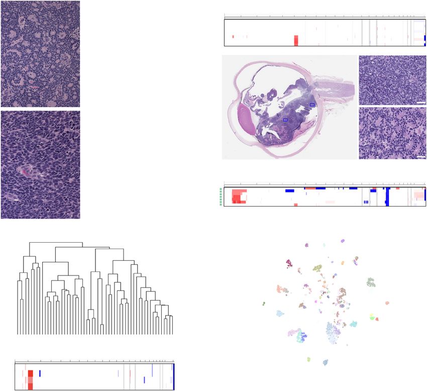
ARTICLE                                                                 NATURE COMMUNICATIONS | https://doi.org/10.1038/s41467-021-24781-7

R
       etinoblastoma is a rare pediatric cancer of the developing            Results
       retina that initiates in utero and is diagnosed within the            Isolation and characterization of patient-derived iPSC lines. In
       first few years of life1. The vast majority of retinoblastomas         total, 11 patients and 4 family members were enrolled on the
initiate with biallelic inactivation of RB1 and a small subset               RETCELL (NCT02193724) protocol at St. Jude Children’s
(1–2%) initiate with MYCN amplification in the absence of RB1                 Research Hospital (Supplementary Data 1 and Fig. 1A, B). Four
inactivation2,3. Approximately half of all retinoblastoma cases              samples for reprogramming were obtained by skin biopsy at the
involve a germline mutation in RB1 and 25% of germline                       time of anesthesia for examination of the eyes as part of routine
retinoblastoma patients inherited the mutant allele from a                   care, and eleven samples were obtained with the peripheral blood
parent4. Patients with germline RB1 mutations have an earlier                draw. Germline DNA samples were also obtained from 14 of the
age of onset because they only require inactivation of the                   15 participants. Participants were selected based on their clinical
remaining RB1 allele for tumor initiation1,5. Genomic studies                presentation to represent a broad spectrum of penetrance in
have indicated that RB1 inactivation is sufficient for                        heritable retinoblastoma disease. Four participants had no family
tumorigenesis6. Therefore, retinoblastoma is an important                    history and were diagnosed at less than 12 months of age
model of tumor initiation as a result of biallelic inactivation of a         (bilateral, n = 3; asynchronous bilateral, n = 1) and two partici-
single tumor suppressor gene.                                                pants had 13q deletions (manifested as a unilateral and bilateral
   Early attempts to model retinoblastoma in mice by                         disease). Four family cohorts (n = 9) consented to the protocol,
mutating the Rb1 gene failed to produce retinal tumors in Rb+/–              including two families where the parent was previously unknown
mice7–9. Subsequent conditional inactivation of both copies of               to carry the RB1 mutation until the children were diagnosed with
Rb1 in the developing murine retina also failed to produce                   bilateral or trilateral retinoblastoma (Supplementary Data 1 and
retinoblastoma10–13. Further molecular and genetic studies                   Fig. 1A, B). Offspring of the family cohorts presented with
demonstrated species-specific intrinsic genetic redundancies and              bilateral retinoblastoma (n = 4) and trilateral disease (n = 1) at
compensation among Rb family members prevent retinoblastoma                  diagnosis, and the parents had no evidence of tumors by oph-
in mice14. Conditional inactivation of Rb1 and p107 or Rb1 and               thalmologic screening.
p130 can lead to retinoblastoma10–14 in mice and more recently, a               Fibroblasts or blood were reprogrammed with the CytoTune
murine model of the MYCN amplified form of retinoblastoma                     Sendai reprogramming kit (Supplemental Information). In total,
was developed15.                                                             71 iPSC clones were isolated for 15 participants and the germline
   While these genetically engineered mouse models (GEMMs)                   DNA mutations in RB1 were verified by Sanger sequencing for
have provided important insights into retinoblastoma biology,                most lines, and fluorescent in situ hybridization for SJRB-iPSC-13
there are some differences in the molecular and cellular features            which had a RB1 locus deletion (Supplementary Data 2, Fig. 1A–E
of retinoblastoma across species11,14,16,17. For example, in a               and Supplemental Information). Only fully reprogrammed clones
recent epigenetic study of the developing retina and retino-                 with trilineage differentiation potential and a normal karyotype
blastoma from mice and humans, the human tumor epigenome                     were used for subsequent studies, hereafter referred to as SJRB-
mapped to a later developmental stage than that of the mouse                 iPSC-1-15 (Supplementary Fig. 1A–H and Supplementary Data 2).
tumors18. Those differences may be related to the initiation of              Whole-genome sequencing was performed on the iPSC clones,
tumorigenesis during retinal development, the cellular origin of             and the patient germline DNA to ensure additional deleterious
the tumors, or both19. Another important difference between                  gene mutations were not accumulated during the process of iPSC
human and murine retinoblastoma is their drug sensitivity. In                production (Supplementary Data 3).
side-by-side preclinical studies of murine retinoblastoma and                   To test the propensity of individual clones to form neurons, we
orthotopic patient-derived xenografts (O-PDXs) of retino-                    produced embryoid bodies (EBs) in neural induction medium for
blastoma, there were differences in the response to the conven-              2 days and then transferred them to Matrigel-coated plates to
tional systemic chemotherapy regimen currently used to treat                 form neural rosettes (Fig. 1F–H). We performed quantitative PCR
retinoblastoma patients20. Virtually, all of the GEMMs had a                 (qRT-PCR) with a panel of primers for early neurogenic genes
complete and durable response while few of the O-PDXs tumor-                 (Supplementary Data 2 and Fig. 1I). Using these morphologic
bearing mice had any response20. Therefore, a laboratory model               (rosette formation) and molecular (qRT-PCR) criteria, we
of human retinoblastoma could contribute to our understanding                identified multiple clones for each patient that had neurogenic
of the retinoblastoma cell of origin and provide patient-specific             potential (Supplementary Data 2). To determine if the iPSCs
tumors for preclinical testing.                                              could make retina in 3D organoid cultures, we used the protocol
   To produce a laboratory model of human retinoblastoma, we                 developed by Sasai22 and compared retinal formation for each
created iPSCs from 15 participants with germline RB1 muta-                   iPSC clone to that of the H9 ESC line using qRT-PCR and
tions or deletion. Multiple clones from each iPSC line were                  immunostaining (Fig. 1J–L and Supplementary Data 2). Overall,
validated for retention of the germline mutation, subjected to               we were able to isolate multipotent iPSC clones from each donor
molecular profiling, including whole-genome sequencing, and                   with normal karyotype and neurogenic/retinal differentiation
characterized for embryoid body formation, neural rosette                    competence that retained their germline RB1 alterations.
formation, and retinal differentiation using an improved
human retinal organoid procedure. Representative clones from
each participant were then differentiated into retinal organoids,            Optimization of retinal specification in 3D organoid cultures.
dissociated, and injected into the eyes of immunocompromised                 The original Sasai method for producing 3D retinal organoids
mice to monitor tumor formation. Parallel experiments were                   was developed and optimized using a highly neurogenic H9
performed with targeted RB1 gene inactivation using CRISPR-                  human ESC line23 and has not been systematically tested for
Cas9. Individual tumors derived from the organoids were                      patient-derived iPSCs. While each of our iPSC lines was able to
passaged by intraocular injection and cryopreserved, as done                 produce retinal organoids using the Sasai method, the efficiency
previously21. Complete molecular, cellular, and genetic profil-               varied from 4% (4/96 for SJRB-iPSC-12) to 27% (26/96 for SJRB-
ing were completed and the iPSC lines and tumors have been                   iPSC-7) across lines (Fig. 2A). In order to improve the retinal
made available to the biomedical research community free of                  specification, we carried out several rounds of optimization and
charge with no obligation to collaborate through the Childhood               made six changes to the procedure. In our modified 3D-RET
Solid Tumor Network21.                                                       protocol (Fig. 2A), we added the BMP signaling inhibitor

2                NATURE COMMUNICATIONS | (2021)12:4535 | https://doi.org/10.1038/s41467-021-24781-7 | www.nature.com/naturecommunications
NATURE COMMUNICATIONS | https://doi.org/10.1038/s41467-021-24781-7 ARTICLE
RB1 genomic locus
A 1 3 5 7 9 11 13 15 17 19 21 23 25 27 exons B
2 4 6 8 10 12 14 16 18 20 22 24 26
5’ UTR 3’ UTR
1 373 573 645 768 928
RB1 protein A B C
no ca no ca no ca no ca
SJRB-iPSC-1 Glu137
* A B C
SJRB-iPSC-2 158fs
SJRB-iPSC-3 Glu323
*
SJRB-iPSC-14,15 Tyr318
*
SJRB-iPSC-4 343fs SJRB-iPSC-10 RB1 wt
unaffected
SJRB-iPSC-11,12 381fs
A
SJRB-iPSC-8,9,10 AArg455 *B SJRB-iPSC-8 SJRB-iPSC-9
SJRB-iPSC-6,7 A B 726fs
trilateral RB bilateral RB
19 21 23 25 27 exon 1-17Δ (dx. 7 months) (dx. 1.5 months)
18 20 22 24 26
SJRB-iPSC-5
3’ UTR SJRB-iPSC-13
13q 14.11-14.3Δ
SJRB-iPSC-13 D SJRB-iPSC-3 E
C
phase DAPI POU5F1 SOX2
SJRB-iPSC-3
ATACGAAGAAATTTA
RB1
T chr 13
F qRT-PCR G DIC H DAPI I
H9 ESC
(normalized relative fold)
103 iPSCs
gene expression
7 days 102
ESCs
EBs 10
iPSCs 2 days
1.0
0.1
neural 400 μm
ZIC1
DMBX1
FOXG1
ASCL1
PAX6
EMX2
SOX17
induction
J K 3D retinal organoids L
H9
H9 ESC
100
normalized relative fold
SJRB-iPSC-1
SJRB-iPSC-7
gene expression
10 SJRB-iPSC-12
100 μm DAPI recoverin
1
SJRB-iPSC-15
0.1
0.01
100 μm
0.001 DAPI EdU
CRX RCVN PAX6 VSX2
gene
(dorsomorphin) and ROCK inhibitor to promote the neu- modified protocol called 3D-RET achieved 22–30% retinal orga-
roectodermal lineage24–26. We increased Matrigel to 2% to noid formation across all of our iPSC lines, and the individual
increase eye-field specification23. We change the medium every organoids from each line were more homogenous (Fig. 2B, C and
2 days rather than every 5 days during the early stages of the eye- Supplementary Fig. 2). Using the H9 ESC line and the iPSC lines
field specification to prevent depletion of growth factors. We produced from participants with germline RB1 alterations, we
moved the addition of smoothened agonist (SAG) earlier to day showed that molecular, cellular, functional, and anatomic features
12 based on a previous publication27. We replaced retinoic acid of retinal organoids produced by the Sasai method and the 3D-
with the more stable synthetic agonist EC23 (Stemgent Inc). Our RET method were indistinguishable in side-by-side comparisons
NATURE COMMUNICATIONS | (2021)12:4535 | https://doi.org/10.1038/s41467-021-24781-7 | www.nature.com/naturecommunications 3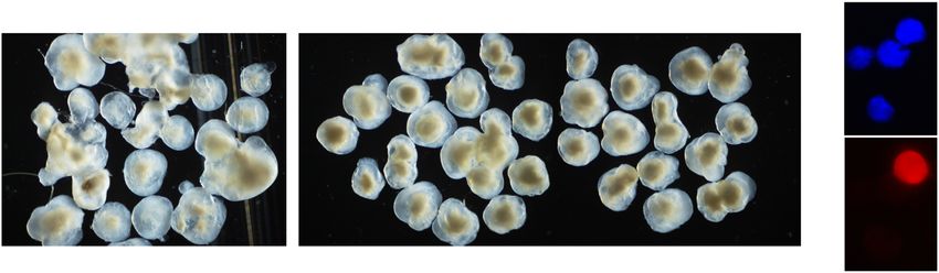
ARTICLE NATURE COMMUNICATIONS | https://doi.org/10.1038/s41467-021-24781-7
Fig. 1 Generation of iPSC lines from patients with germline RB1 mutations. A Map of the genomic locus showing each of the 27 exons of the RB1 gene and
the color-coded protein-coding domains. The germline mutations in each patient/family are shown below the full-length protein. B Representative pedigree
for an unaffected carrier (SJRB-iPSC-10) and two affected children (SJ-iPSC-8 and SJ-iPSC-9). C Representative micrographs of SJRB-iPSC-3 colony
immunostained for POU5F1 (green) and SOX2 (red) with DAPI (blue) nuclear stain. Staining was repeated on another iPSC line with the same results.
D Sanger sequencing chromatogram showing the heterozygous nonsense mutation in the RB1 gene (GAA→UAA). E Representative two-color fluorescence
in situ hybridization (FISH) of SJRB-iPSC-13 showing the 13q deletion (200 cells were analyzed). F Drawing of the neural rosette differentiation experiment.
G, H Differential interference contrast (DIC) and DAPI-stained colony from the neural rosette induction procedure. Arrows and the enlarged box indicate
neural rosettes. Three clones of each line were differentiated and each line was able to produce rosettes. I Boxplot of the normalized relative fold of
neurogenic genes for each iPSC line (n = 15) and H9 ESCs from qRT-PCR of the neural induction assay. J Micrograph of representative retinal organoid
from H9 ESCs and SJRB-iPSC-15 indicating retina (arrow). K GAPDH normalized relative fold gene expression for 3D retinal organoids from representative
iPSC lines using H9 ESCs as controls. Each dot represents the mean of two technical replicates for an individual organoid. L Micrograph of cryosection of
day 45 retinal organoid that was labeled with EdU for 1 h prior to harvest showing recoverin expressing photoreceptors and EdU+ retinal progenitor cells
(red) with DAPI counterstain. Each of the 15 lines was sectioned showing similar results in neural retina regions. Box and whisker plots include center line
as median, box as Q1 and Q3, and whiskers as 1.5× interquartile range. Scale bars: C, 50 μm; E, L, 10 μm.
0 10 20 30 40 50 200
A days
retinal specification retinal differentiation
SASAI
1% 1% SAG RMM RMM
MG MG CHIR SJRB-iPSC-7: 26 organoids SJRB-iPSC-4: 11 organoids
RA
FBS 40% O2 SJRB-iPSC-5: 22 organoids SJRB-iPSC-12: 4 organoids
3D-RET
DM
2% 1% RMM RMM
SJRB-iPSC-7: 27 organoids SJRB-iPSC-4: 29 organoids
MG MG CHIR
ec23 ec23 SJRB-iPSC-5: 24 organoids SJRB-iPSC-12: 22 organoids
FBS
ROCKi 40% O2 40% O2
SAG
SASAI 3D-RET Day 45
D 10
B C
recoverin+ (%)
* 8
* 6
* 4
2
3D AI n=8
ET n=8
SJRB-iPSC-4 SJRB-iPSC-4
0
DAPI
recoverin
S
-R
SA
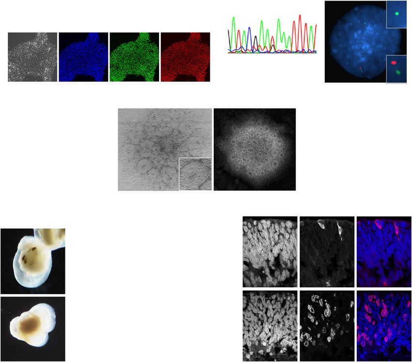
Fig. 2 The 3D-RET protocol for retinal organoid formation. A Drawing of the steps in the Sasai and 3D-RET retinal organoid protocol. The number of
retinal organoids produced from a 96-well dish is indicated for representative lines for each protocol. Red arrows indicate media changes. B, C Micrograph
of representative retinal organoids using the Sasai and 3D-RET protocols in a side-by-side comparison for SJRB-iPSC-4. All organoids from a 96-well dish
were analyzed. Arrows indicate retina organoids and (*) indicates cystic structures that are common in the Sasai protocol. D Micrographs of dissociated
cell immunofluorescence of retinal organoids and a dot plot showing the percentage of recoverin immunopositive cells from individual retinal organoids
(n = 8). MG Matrigel, SAG smoothened agonist, RMM retinal maturation medium, FBS fetal bovine serum, CHIR GSK3 inhibitor, ec23 retinoic acid analog.
Scale bars: B, C, 100 μm. D, 5 μm.
(Fig. 2D and Supplementary Fig. 2). While the quality of retinal retinal organoid cultures of the patient-derived stem cells, we grew
tissue was similar in each method, the increased efficiency of retinal organoids from iPSCs from each patient to day 45 when the
retinal organoid formation for iPSC lines and the more uniform tissue was dissociated and injected into the vitreous of immuno-
and consistent size and shape of the organoids, led us to use the compromised mice (Fig. 3A). As a negative control, we used RB1
3D-RET protocol for all subsequent experiments. wild-type stem cell (H9 ESCs and GM23710 iPSCs)-derived retinal
organoids and as a positive control, we induced RB1 mutations in
exon 4 with CRISPR-Cas9 in all 15 participant derived iPSC lines
Retinoblastoma from human retinal organoids. Four of the 15 and H9 cells, hereafter referred to as SJRB-iPSC-1CR - 15CR
participants in our cohort had surgical enucleation as part of their (Fig. 3B). The CRISPR-Cas9 RB1 inactivation was intentionally left
treatment. We were able to produce O-PDXs from two of those mosaic (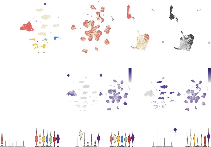
NATURE COMMUNICATIONS | https://doi.org/10.1038/s41467-021-24781-7                                                                      ARTICLE

Fig. 3 Retinoblastoma from 3D retinal organoids. A Schematic drawing of the retinoblastoma workflow. After 45 days of differentiation, retinal organoids
are dissociated and injected into the eyes of immunocompromised mice and they are held for 1 year to wait for tumor formation. B Drawing of the RB1
genomic locus with the location of the gRNA targeting exon 4 and the corresponding sequence. C Plot of mutation frequency in a representative iPSC line
for gRNA-3 with insertions (red) and deletion (blue) flanking the cut site (*). D Photograph of a mouse with retinoblastoma from a retinal organoid.
E Barplot of qRT-PCR for genes found in the retinoblastoma (SYK, SIX3, HMX1), human pluripotent stem cells (MYC, SOX2, FGFR2), and teratomas (WLS,
BMP2, COLA1) from one teratoma, one patient tumor (RB-169), two PDX tumors (RB116 and RB121), two spontaneous retinal organoids-derived tumors
(SJRB-iPSC-4 and SJRB-iPSC-8), and two CRISPR-modified retinal organoids (SJRB-iPSC-4CR, SJRB-iPSC-8CR). Each dot is the mean of technical
duplicates, the bar is the mean and standard deviation between replicates. All data are normalized to GAPDH and plotted relative to H9 ESCs (dashed line).
F, G Circos plot of representative organoid-derived retinoblastoma (SJRB-iPSC-4CR-T-E and SJRB-H9CR-T-C) showing somatic mutations acquired in the
tumor relative to the iPSC/ESC line. The copy number changes across the genome are shown below each tumor. H Principal component analysis (PCA) of
RNA-seq of organoid-derived retinoblastomas, O-PDXs, patient tumors, iPSC/ESCs, and retinal organoids. I, J Hematoxylin and eosin (H&E)-stained
organoid-derived tumor showing rosettes and IHC for SYK (brown) which is not present in the normal retina but is upregulated in retinoblastoma. Staining
was completed on three tumors with similar results. Scale bars: 25 μm.

iPSC-6-REV) using CRISPR-Cas9 and performed the same tumor                      injections of day 45 retinal organoids were teratomas based on
formation experiments in parallel with those lines (Supplementary               qRT-PCR (Supplementary Data 4). Two tumors (SJ-iPSC-15-T-A/
Data 4 and Supplemental Information). We also injected undif-                   B) that arose rapidly (37 and 100 days) had a pan-neuronal gene
ferentiated H9 ESCs to form teratomas in the eye as an additional               expression pattern but were not retinoblastoma based on gene
negative control. Each iPSC line and controls were tested with and              expression (Supplementary Data 4). All other tumors from the
without the RB1 CRISPR-Cas9 by two different technicians with                   patient-derived iPSC retinal organoids (iPSC-RBs) were retino-
three replicate injections per line for a total of more than 500                blastomas that were indistinguishable from patient retinoblastomas
individual eyes injected (Supplemental Information). After 1 year,              or orthotopic patient-derived xenografts (O-PDXs) (Fig. 3E and
we identified 13 independent tumors from iPSC lines SJRB-iPSC-                   Supplementary Data 4).
1,2,4,5,6,8 with CRISPR-Cas9 inactivation of RB1 and 7 indepen-                    Tumors were propagated and cryopreserved as done
dent tumors from SJRB-iPSC-3,8,15 lines without CRISPR-Cas9                     previously6,21,28. Next, we performed whole-genome sequencing
inactivation of RB1 (Fig. 3D and Supplementary Data 4). For H9                  and RNA-seq of each independent iPSC-RB when sufficient tissue
ESC retinal organoids, four independent tumors were formed with                 was available after initial propagation and screening. The patient-
CRISPR-Cas9 inactivation of RB1 and none was formed without                     derived iPSC-RBs showed inactivation of the 2nd allele of the RB1
RB1 inactivation (Supplementary Data 4). The two lines that had                 gene and no other mutations in known cancer genes (Fig. 3F, G,
the germline RB1 mutation reverted did not form tumors after                    Supplementary Data 4, and Supplementary Fig. 3). Importantly,
more than a year. We developed a custom Taqman qRT-PCR                          there were also copy number gains in MDM4 and MYCN which
microfluidic card to rapidly distinguish between teratomas, reti-                are common in retinoblastomas (Fig. 3F, G, Supplementary Data 4,
noblastoma, and other malignancies (Fig. 3E and Supplemental                    and Supplementary Fig. 3). In addition, the RNA-seq from iPSC-
Information). None of the tumors that formed from intravitreal                  RBs most closely matched retinoblastomas in principal component

NATURE COMMUNICATIONS | (2021)12:4535 | https://doi.org/10.1038/s41467-021-24781-7 | www.nature.com/naturecommunications                                5
ARTICLE                                                                  NATURE COMMUNICATIONS | https://doi.org/10.1038/s41467-021-24781-7

analysis (Fig. 3H and Supplementary Data 4). The difference in O-             from normal cells based on inferred copy number alterations
PDX and organoid-derived retinoblastomas are primarily due to                 (Supplementary Fig. 4A and Supplemental Information). For
differences in the tumor microenvironment. Vascular endothelial               each tumor sample, after pairs of cell correspondences between
cells, macrophages, and other normal human cells present in the               the reference and tumor dataset (anchors) were identified, the
patient tumors are murine in O-PDX and organoid tumors and                    cell-type classification was projected and transferred onto each
therefore filtered out in RNA-sequencing analysis. One of the                  tumor dataset to determine if there were cells with expression
hallmarks of retinoblastoma is the epigenetic deregulation of the             profiles similar to specific retinal cell types in the tumors
SYK oncogene which is required for tumorigenesis6. SYK RNA and                (Fig. 5B). Importantly, the cell cycle genes were removed before
protein were upregulated in iPSC-RBs as found in patient tumors               the label transfer to prevent bias toward proliferating retinal
(Fig. 3E, I, J and Supplementary Data 4). Taken together, these data          progenitor cells. Despite excluding the cell cycle genes, the most
suggest that spontaneous retinoblastoma can form from patient-                common cell identity from the label transfer was retinal
derived iPSCs differentiated into retinal organoids or be induced             progenitor cells at 52% (81,283/156,244) followed by rod
by CRISPR-Cas9 targeting of the RB1 locus in both hESCs and                   photoreceptors at 31% (48,591/156,244) (Supplementary Data 6).
patient-derived iPSCs.                                                        Indeed, for every patient tumor, O-PDX, and organoid-derived
                                                                              tumor, the most common cell identity was retinal progenitor cells
                                                                              (range 39–63%) and they were enriched in cell cycle genes even
iPSC-RBs recapitulate the epigenetic and clonal features of                   though the cell cycle genes themselves were not used for the label
retinoblastoma. High-density DNA methylation arrays (Illumina                 transfer (Fig. 5C, D and Supplementary Data 6). To determine if
Infinium 850 K) have been extensively used to classify tumors                  there was evidence of tumor cells with progenitor signatures
based on their genome-wide methylation signatures and copy                    giving rise to more differentiated tumor cells with photoreceptor
number variations29–31. The assay is robust even with small                   gene expression signatures, we performed RNA velocity analysis
amounts of formalin-fixed paraffin-embedded (FFPE) material.                    (Supplemental Information). While some tumors showed evi-
This has been particularly useful for pediatric tumors of the                 dence of the transition from progenitors to photoreceptors, others
central nervous system29. To establish a baseline for retino-                 showed the opposite pattern (Supplementary Fig. 4). A more
blastoma, we profiled 53 retinoblastoma patient tumors, includ-                definitive clonal analysis will be required to determine the
ing tumors with histopathological features of differentiation                 relationship between the cell populations in retinoblastoma.
(rosette formation) as well as less differentiated tumors (Fig. 4A,              Previous retinoblastoma single-cell gene expression array
B). Unsupervised hierarchical clustering of the patient tumors                analysis of a single O-PDX suggested that tumor cells may have
revealed that they separate based on differentiation evaluated                a hybrid gene expression signature of multiple cell types14.
from the histopathology and DNA copy number variation from                    Consistent with those data, we found that genes that are normally
the Infinium 850 K array (Fig. 4C). Indeed, the more differ-                   expressed in a mutually exclusive pattern in the normal retina
entiated tumors had fewer copy number alterations than those                  such as HES6 and AIPL1 are co-expressed in retinoblastoma
with more aggressive undifferentiated histopathologic features                tumor cells (Fig. 5E–H). This was also true for cone, rod, and
and some patient tumors were heterogeneous between regions of                 amacrine genes (Fig. 5I and Supplementary Fig. 4E, F). Therefore,
differentiated and undifferentiated tumor cells (Fig. 4D–F).                  individual retinoblastoma tumor cells express a hybrid gene
   Next, we performed the same DNA methylation array profiling                 expression signature that does not normally occur during retinal
on our collection of iPSC/ESC-derived retinal organoids,                      development. Taken together, our scRNA-seq analysis showed
retinoblastoma O-PDXs, and organoid-derived retinoblastomas                   that our organoid-derived tumors are indistinguishable from the
described above. Four of our organoid-derived tumors had                      O-PDXs and the patient tumors in terms of cell identity and
sufficient DNA for methylation array profiling and passed our                   proliferation (Fig. 5G and Supplementary Data 6) and all data are
quality control metrics (Supplemental Information). The                       available in a Cloud-based viewer (https://pecan.stjude.cloud/
organoid-derived retinoblastomas clustered more closely with                  static/rbsinglecell).
the undifferentiated patient tumors using the same unsupervised
methods (Fig. 4G and Supplemental Information). Based on copy                 Discussion
number variants, the organoid-derived tumors were intermediate                We have developed iPSCs from 15 participants with germline
between the differentiated and undifferentiated patient retino-               RB1 alterations and we have optimized a 3D retinal organoid
blastomas (Fig. 4G). In unsupervised methylation analysis, the O-             culture system for producing human retinoblastoma in the
PDXs and iPSC-RBs overlapped with retinoblastomas/pineal                      laboratory. We also developed the tools to induce RB1 mutations
tumors from a reference collection of 2901 brain tumor                        in a wild-type human stem cell and produce retinoblastomas
methylation profiles29 and they were clearly separated from the                indistinguishable from those of patient-derived iPSCs. The
normal retinal organoids and iPSCs in tSNE plots (Fig. 4H–K and               organoid-derived retinoblastomas have molecular, cellular, his-
Supplementary Data 5).                                                        topathologic, genetic, epigenetic, and clonal features that are
   To further validate the identity and heterogeneity of the                  indistinguishable from patient tumors and O-PDX models. In
retinoblastomas derived from iPSCs, we performed single-cell                  contrast with O-PDXs, this model is not reliant on patient tumor
RNA sequencing on 11 retinoblastoma O-PDXs in biological                      tissue, which in the case of retinoblastoma is usually only
duplicate for a total of (114,167 cells), 5 healthy adult human               available after enucleation. In addition, our model can be used to
retina (24,445 cells), 6 patient retinoblastomas (27,825 cells), and          derive multiple tumors from the same patient and can be used to
2 organoid-derived tumors (13,864 cells). We also included                    generate tumors from carriers who never developed retino-
human retinal progenitor cells from a publicly available scRNA-               blastoma. The process of producing retinoblastoma in our sys-
seq dataset on human fetal retina32. We combined the five                      tem was inefficient (
NATURE COMMUNICATIONS | https://doi.org/10.1038/s41467-021-24781-7 ARTICLE
1 3 5 7 9 11 13 15 17 19 21 X
A C differentiated/retinoma D 2 4 6 8 10 12 14 16 18 20 22 Y
E
region 1 region 1
B region 2
region 2
1 3 5 7 9 11 13 15 17 19 21 X
F 2 4 6 8 10 12 14 16 18 20 22 Y
undifferentiated
Molecularneuropathology.org
G H 200 n=4,046
differentiated retinoblastomas
pineal
undifferentiated tumors
100
organoid derived
tSNE-2
0
organoids
-100 iPSCs
1 3 5 7 9 11 13 15 17 19 21 X
2 4 6 8 10 12 14 16 18 20 22 Y
-200
-200 -100 0 100 200
tSNE-1
I J K
20 10
-115
0
tSNE-2
tSNE-2
tSNE-2
-120 0
-20
-125
-40 -10
-130
-60
-130 -120 -10 0 10 20 -20 -10 0 10 20
tSNE-1 tSNE-1 tSNE-1
retinoblastoma (patient-St. Jude) retinoblastoma (O-PDX) ESC/iPSCs
pineal tumor
retinoblastoma (patient-MNP) retinoblastoma (organoid) ESC/iPSC (organoid)
Fig. 4 DNA methylation profiling of retinoblastoma. A, B Hematoxylin and eosin (H&E) staining of differentiated and undifferentiated patient
retinoblastoma. C Unsupervised hierarchical clustering of retinoblastoma tumors showing separation of the differentiated and undifferentiated samples.
D–F Copy number variation in the differentiated retinoblastomas (D) and undifferentiated patient retinoblastomas (F) and H&E staining (E) of a patient eye
that has regions of both undifferentiated (region 1) and differentiated (region 2) tumor, suggesting intratumor cellular heterogeneity. G Copy number
variation and clustering of the retinal organoid-derived tumors. H t-distributed stochastic neighbor embedding (tSNE) plot of the St. Jude retinoblastomas,
retinal organoid-derived tumors, retinal organoids, and iPSC/ESCs integrated with the Molecular Neuropathology (MPN) database of brain regions and
pediatric brain tumors. I tSNE plot of the boxed region in (H) showing clustering of the organoid-derived tumors with patient retinoblastomas. J tSNE plot
of the patient, O-PDX, and organoid-derived retinoblastomas with iPSC/ESCs and normal retinal organoids. K tSNE plot of O-PDX and organoid-derived
retinoblastomas with iPSC/ESCs and normal retinal organoids. Scale bars: 25 μm.
selected for injection. The introduction of additional perturba- the same H9 ESC produced multiple independent tumors in
tions such as ectopic expression of MDM2/4, MYCN, or SYK 200–300 days. None of our tumors was teratomas, indicating that
may accelerate tumorigenesis. Previous attempts to generate retinal organoid differentiation prior to intravitreal injection was
retinoblastoma from genetically modified H9 ESCs may have sufficient to eliminate any residual stem cells. However, we did
failed because they did not allow enough time for tumors to grow identify two tumors using this method that had neuronal features
(60–90 days) or the lack of normal retinal development in the but were not retinoblastomas. Therefore, it is essential to
absence of RB134. Our approach using a mosaic approach with implement robust diagnostics for retinoblastoma from retinal
NATURE COMMUNICATIONS | (2021)12:4535 | https://doi.org/10.1038/s41467-021-24781-7 | www.nature.com/naturecommunications 7ARTICLE NATURE COMMUNICATIONS | https://doi.org/10.1038/s41467-021-24781-7
A normal human retina B human retinoblastoma C organoid tumor (SJRB-iPSC-1CR)
(n=35,487 cells) (156,244 cells) (6,595 cells)
20
progenitor (n=538)
progenitor S/G2/M
rod 71% S/G2/M G1/G0
bipolar (n=19,789) 10
10 10 10
amacrine
(n=7,727)
Müller
UMAP 2
5 rod 5
UMAP 2
cone
0 0 33% S/G2/M
horizontal
(n=1,134)
0 0
immune
-10 (n=1,173)
(n=179)
CD34
-10 -5 -5
VE (n=4,329)
RPE
-20 -5 0 5 10 -5 0 5 10
-10 0 10 -10 0 10 UMAP 1 UMAP 1
UMAP 1 UMAP 1
d
D E F
or oi
m n
tu rga
patient O-PDX
o
100
Retina Retinoblastoma 3 Retina Retinoblastoma 3
tumor cells (%)
2 2
HES6 HES6 AIPL1 AIPL1
1 1
50 0 0
10 10
UMAP 2
UMAP 2
0
0 0
100
progenitor signature rod signature
S/G2/M (%)
-10 -10
50
0
R 66
R 68
X 3
38
SC R
PD 121
R 69
R 4
R 5
PD 177
PD 116
X 2
PD 124
PD 128
PD 129
PD 130
PD 134
PD 135
0
R
-10 0 10 -10 10 0
PD 13
-10 10
7
7
PD 12
0
iP 9C
-10 10
1C
B1
B1
X1
B1
B1
B1
X
X
X
B
X
X
X
X
X
H
R
UMAP 1 UMAP 1 UMAP 1 UMAP 1
G H I
PDE6H expression
PDE6H expression
retinoblastoma retinoblastoma 6 retinoblastoma
AIPL1 expression
AIPL1 expression
HES6 expression
HES6 expression
retina retina retina
3 3 4
4 4 4
2 2
2
1 2 1 2 2
0 0 0 0 0 0
r
bi od
M ar
a er
riz ne
co l
ne
r
bi od
M r
a er
riz ne
co l
ne
r
bi od
M r
a er
riz ne
co l
ne
r
bi od
M ar
a er
riz ne
co l
ne
r
bi od
M ar
a er
riz ne
co l
ne
r
bi od
M r
a er
riz ne
co l
ne
ta
ta
ta
ito
ito
la
ito
la
ta
ta
ta
ito
ito
ito
la
l
am üll
am üll
am üll
l
am üll
l
am üll
am üll
on
on
on
r
po
ho cri
r
po
ho cri
r
po
ho cri
on
on
on
r
po
ho cri
r
po
ho cri
r
po
ho cri
en
en
en
en
en
en
og
og
og
og
og
og
pr
pr
pr
pr
pr
pr
Fig. 5 Cellular heterogeneity of retinoblastomas. A Uniform manifold approximation projection (UMAP) plot of scRNA-seq of normal human retinal cells
including retinal progenitor cells and all adult retinal cell types. B UMAP plot of scRNA-seq of human retinoblastoma from patient tumors, O-PDXs, and
organoid-derived tumors. The label transfer for cell identity is displayed, and the overall numbers are represented in the piechart in the upper left corner.
C Representative UMAP plot of one of the organoid-derived tumors showing cells with the progenitor cell signature and rod signature and the relative
distribution of gene expression profile for proliferation. D Barplot of the proportion of single cells with the retinal progenitor cell gene expression signature
and the rod signature for each tumor (upper panel) and the proportion of those cells that are proliferating (S/G2/M, lower panel) for each tumor analyzed
by single-cell RNA sequencing (n = 19). E, F UMAP plot of gene expression for normal retina and retinoblastoma for a representative retinal progenitor cell
gene (HES6) and a photoreceptor gene (AIPL1). G, H, I Violin plot of the distribution of expression of a progenitor gene (HES6), a photoreceptor gene
(AIPL1) and a cone gene (PDE6H) across retinal cell types and retinoblastomas. The colors match the colors in (A, B).
organoids that include molecular, cellular, genetic, and epige- Methods
netic features. This study also provided new insights into the Patients. RETCELL (NCT02193724), a protocol to establish the feasibility, vali-
dation, and differentiation of induced pluripotent stem cells produced from
cellular identity within retinoblastoma demonstrating a bias
patients with heritable retinoblastoma, was approved by the St. Jude Children’s
toward retinal progenitor cells and rods. It is possible that the Research Hospital Institutional Review Board and open to accrual in July 2014.
highly proliferative tumor cells with the retinal progenitor cell Written informed consent was obtained from each participant or participant’s
identity give rise to more differentiated tumor cells that have parent/guardian. Eligibility criteria included a family history of retinoblastoma with
features rods and other neurons but lineage tracing will be an identified germline RB1 mutation, diagnosis of bilateral retinoblastoma, or
diagnosis of unilateral retinoblastoma with germline RB1 mutation, MYCN
required to test that hypothesis. This complex process of the amplification, or 13q deletion identified. Samples obtained from participants
tumor cells progressing through some aspects of retinogenesis included skin biopsy or collection of peripheral blood mononuclear cells. Skin
may have confounded previous attempts to identify the retino- biopsies were performed while the patient was under sedation for clinical purposes
blastoma cell of origin from gene expression analysis of bulk (e.g., exam under anesthesia). Peripheral blood samples were obtained at any time
the participant was scheduled for routine labs; if the participant was not an active
tumors. Indeed, scRNA-seq shows a hybrid cellular phenotype, patient, the blood draw occurred as a separate, research-only blood draw.
which may simply reflect the multipotency of retinal progenitor Specimens were de-identified after collection in the St. Jude Biorepository and
cells. Our retinal organoid system will be an important tool for shipped overnight to the Waisman Center for further processing. One sample required
determining if those tumor cell populations are distinct clones or a repeat blood draw due to difficulties encountered with the shipping process.
A total of 11 patients were enrolled (4 female), with 4 parental samples obtained
if they represent the dynamic changes in gene expression of simultaneously due to positive family history. Four samples were obtained by skin
individual clones over time. The tumor modeling described here biopsy at the time of anesthesia for examination of the eyes as part of routine care,
may also be useful for testing novel therapeutic combinations for and 11 samples were obtained with the peripheral blood draw. Germline blood
individual patients. samples were obtained from 14 of 15 participants; one participant is not available
8 NATURE COMMUNICATIONS | (2021)12:4535 | https://doi.org/10.1038/s41467-021-24781-7 | www.nature.com/naturecommunicationsNATURE COMMUNICATIONS | https://doi.org/10.1038/s41467-021-24781-7 ARTICLE for germline testing at this time. Participants were selected and approached for Fluorescence in situ hybridization. Two fosmid clones (WI2-1468C23 and WI2- inclusion in this study based on their clinical presentation to represent a broad 0655F03) that localize to the region that is expected to be deleted were combined spectrum of penetrance in heritable retinoblastoma disease: four with no family with an internal control probe (Pan3) located 20 megabases centromeric to the RB1 history diagnosed at
ARTICLE                                                                           NATURE COMMUNICATIONS | https://doi.org/10.1038/s41467-021-24781-7

incubated on ice for 30 min in a darkened hood then washed by adding 5 mL of the       additional 0.5 mL of NIM was added, and the plates were incubated at 37 °C, 20%
chilled blocking solution to each tube, centrifuging at 300 rcf, 4 min, at 4 °C. The   O2, 5% CO2 O/N. Each day, 1 mL of NIM removed and 1 mL fresh NIM
wash was repeated once. iPSCs were resuspended in iPSC dilution media and left         was added.
on ice. Cells were analyzed by flow cytometry (FACSDiva 8.0.1), at least 50,000
events were collected.                                                                 Neuronal gene expression assay. On day 7, a wide bore p1000 tip was used to collect
                                                                                       the EBs. The EBs were pipetted onto a 40-µm filter and washed with 1 mL of the
Immunocompromised mice. All animal procedures and protocols were approved              NIM. The filter was then flipped, and the EB’s were washed off the filter with 3 mL
by the St. Jude Laboratory Animal Care and Use Committee. All studies conform to       of the NIM. The EBs were allowed to sink to the bottom of the tube, and the media
federal and local regulatory standards. Female C57BL/6 SCID mice were purchased        was removed. for preparation for the Taqman Array (see below).
from Jackson Laboratories (strain code 001913). Mice were housed on ventilated
racks on a standard 12-h light–dark cycle.                                             Rosette-formation assay. On day 5, a wide bore p1000 tip was used to collect the
                                                                                       EBs. The EBs were pipetted onto a 40-µm filter and washed with 1 mL of the NIM.
                                                                                       The filter was then flipped, and the EB’s were washed off the filter with 3 mL of the
Trilineage assays. iPSC lines were cultured to 40–50% confluency in mTESR in a          NIM. The EB’s were plated 3 mL per well onto one well of a matrigel-coated six-
six-well dish then media was changed to Nutristem media (Biological Industries         well dish. The media was changed on days 7–11, and rosette formation was
#05-100-1A). Cells were incubated at 37 °C, 5% O2, 5% CO2, O/N. In total, 1 mL of      observed.
Accutase was added to the well-containing iPSCs at 60–80% confluency. Plates
were incubated at 37 °C for 3 min. In all, 2 mL of plating medium (Nutristem
medium + 2 µM Thiazovivin) was added to the well and pipetted 8–10 times to            Retinal differentiation. In all, 1 mL of Accutase was added to one well of a six-well
obtain a single-cell suspension. Cells were centrifuged at 300 rcf for 4 min and       plate containing iPSCs at 60–80% confluency. Plates were incubated at 37 °C for 3
resuspend in 2 mL plating medium and counted. In total, 1 × 10⁶ cells were ali-        min. In total, 2 mL iPSC dilution medium (mTESR + 2 µM ROCKi Stem Cell Tech
quoted, and the volume was brought to 10 mL with a plating medium and pipetted         #72304) was added to each well and pipetted 8–10 times to resuspend cells and
with a p1000 tip to create a homogenous cell suspension. In total, 100 µl of cell      centrifuged at 300 rcf for 4 min. iPSCs were resuspend in 2 mL GMEM Retinal
suspension was plated per well into one Nunclon Sphera 96 U plate. The plate was       media (GMEM, 20% KSR, 1× NEAA; Gibco #11140-050, 1× sodium pyruvate;
spun at 100 rcf for 2 min, the bucket orientation was switched, and the plates were    Gibco #11360070, 1× ß-mercaptoethanol; Gibco #21985-023, 1× anti anti; Gibco
spun again at 100 rcf at 2 min. Plates were incubated at 37 °C, 20% O2, 5% CO2 O/      #15240-062, 2 µM ROCKi) supplemented with 3 µM IWRe (Selleck Chem #S7086)
N. Four columns of embryoid bodies (EBs) (32 EBs each) were used for each of the       and counted. In total, 0.9 × 10⁶ cells were aliquoted, and the volume was brought to
three lineage differentiation protocols outlined below.                                10 mL with GMEM Retinal media supplemented with 3 µM IWRe and pipetted
                                                                                       with a p1000 tip to create a homogenous cell suspension. The cell suspension was
Ectoderm differentiation. On days 1–4, 50 µL of media was removed and replaced         plated at 100 µL per well into one 96 U bottom plate. The plate was spun at 100 rcf
with 50 µL of Neural Induction Media (NIM, StemCell Tech #5835). On day 5, a           for 2 min, the bucket orientation was switched, and the plates were spun again at
wide bore p1000 tip was used to collect EBs from all 32 wells. EBs were pipetted       100 rcf at 2 min. The plates were incubated at 37 °C, 20% O2, 5% CO2 O/N.
onto a 40-µm filter and washed with 1 mL NIM. The filter was then flipped and the             On day 2, 30 µL of GMEM Retina supplemented with 3 µM IWRe, 2 µM
EB’s were washed off the filter with 3 mL NIM. The EB’s were plated 1.5 mL per          Dorsomorphin (Tocris #3093), and 2% v/v GFR-Matrigel (Corning #354230) was
well onto two Matrigel-coated wells of a 12-well plate. On days 7 and 9, the media     added to each well. On days 4, 6, 8, and 10, 50 µL per well of media was changed
was changed with 1.5 mL NIM. On day 11, the cells were harvested by adding 1 mL        with GMEM Retina media + 2% v/v GFR-Matrigel. On day 12, a wide bore p1000
of Accutase to the well and incubating at 37 °C for 3 min. In all, 2 mL NIM was        tip was used to collect the EBs and transfer them to two wells of a six-well low-
added, and the cells were centrifuged at 300 rcf for 4 min. The media was removed,     attachment plate. In all, 2 mL of GMEM Retina media supplemented with 10% v/v
and the cells were frozen for preparation for the Taqman Array (see below).            ES-FBS, 100 nM SAG, 1% v/v GFR-Matrigel was added. On day 15, the media was
                                                                                       replaced with GMEM Retina media supplemented with 10% v/v ES-FBS (Gibco
                                                                                       #15240-062), 100 nM SAG (StemCell Tech #73414), 3 µM CHIR (StemCell Tech
Mesoderm differentiation. On days 1 and 2, 50 µL of media was removed and              #72054), 1% v/v GFR-Matrigel. On day 18, the media was replaced with Retinal
replaced with 50 µL of Mesoderm Day 1 media (GMEM; Sigma #G5154, 20% KSR;              Maturation media (DMEM:F12 Gibco #11330-032, 1× N2 Gibco #17502-048, 10%
ThermoFisher #10828028, 6 µM CHIR; StemCell Tech #72254). On day 3, 50 µL of           ES-FBS, 1× anti anti, 0.5 µM EC23 ReproCell #SRP002) and incubated at 40% O2
media was removed and replaced with 50 µL of Mesoderm Day 3 media (GMEM,               (37 °C, 5% CO2). The media was replaced every 3 days until the retina was
20% KSR, 50 ng/µL BMP4; ThermoFisher #PHC9534). On day 5, a wide bore                  harvested.
p1000 tip was used to collect EBs from all 32 wells. EBs were pipetted onto a 40-µm
filter and washed with 1 mL of the Mesoderm Day 3 media. The filter was then
flipped, and the EB’s were washed off the filter with 3 mL of the Mesoderm Day 3         Retinal differentiation scoring. The hESC H9 and a selection of iPSC were dif-
media. The EB’s were plated 1.5 mL per well onto two Matrigel-coated wells of a        ferentiated using the method above and the original method from Nakano et al.23
12-well plate. On days 7 and 9, the media was changed with 1.5 mL of Mesoderm          On day 45 of culture, organoids were dissociated individually. Each organoid was
Day 3 media. On day 11, the cells were harvested by adding 1 mL of Accutase to the     placed in an Eppendorf tube with 200 µL of PBS−/− and 20 µL of trypsin (10 mg/
well and incubating at 37 °C for 3 min. In total, 2 mL Mesoderm Day 3 media was        mL Sigma T9935) for 10–15 min at 37 °C then 20 µL of soybean trypsin inhibitor
added, and the cells were centrifuged at 300 rcf for 4 min. The media was removed,     (STI 10 mg/mL, Sigma T6522) and 20 µL DNase (Sigma D4513) was added, and
and the cells were frozen for preparation for the Taqman Array (see below).            the cells were incubated for 5 min at 37 °C. For all cells, a portion of the cells was
                                                                                       added to a coated slide and allowed to sit for 1 h before immunostaining. The
Endoderm differentiation. On days 1 and 2, 50 µL of media was removed and              remaining cells were spun down, and RNA was extracted using Trizol (LifeTech
replaced with 50 µL of Endoderm Day 1 media (GMEM, 20% KSR, 6 µM CHIR,                 #15596018), cDNA was made using Superscript III (Invitrogen 18080-051), and
100 ng/mL Activin A; PerproTech #120-14 P, 0.5% FBS). On day 3, 50 µL of media         qPCR (Table S3) was run using SYBR Select Master Mix for CFX (ThermoFisher
was removed and replaced with 50 µL of Endoderm Day 3 media (GMEM, 20%                 #4472942) with standard cycling conditions (95 °C for 2 min and 40 cycles of 95 °C
KSR, 6 µM CHIR, 100 ng/mL Activin A, 1% FBS). On day 5, a wide bore p1000 tip          for 30 s, 60 °C for 1 min).
was used to collect EBs from all 32 wells. EBs were pipetted onto a 40-µm filter and
washed with 1 mL of the Endoderm Day 5 media (GMEM, 20% KSR, 6 µM CHIR,                Taqman qRT-PCR. RNA was extracted from samples using Trizol. cDNA was
100 ng/mL Activin A, 2% FBS). The filter was then flipped, and the EB’s were             prepared using a High-Capacity RNA-to-cDNA Kit (ThermoFisher #4387406).
washed off the filter with 3 mL of the Endoderm Day 5 media. The EB’s were plated       Custom TaqMan Arrays were created for neural gene expression assay, the trili-
1.5 mL per well onto two Matrigel-coated wells of a 12-well plate. On days 7 and 9,    neage assay, and to identify retinoblastoma (Table S4).
the media was changed with 1.5 mL of Endoderm Day 5 media. On Day 11, the
cells were harvested by adding 1 mL of Accutase to the well and incubating at 37 °C
                                                                                       Multielectrode array. For recording evoked response to optical stimulation in
for 3 min. In total, 2 mL Day 3 media was added, and the cells were centrifuged at
                                                                                       retinal organoids, the retinal ganglion cells inside the organoids need to be in
300 rcf for 4 min. The media was removed, and the cells were frozen for pre-
                                                                                       contact with the electrodes on the MEA plate. Therefore, retinal organoids, no
paration for the Taqman Array (see below).
                                                                                       matter the size, should first be sliced before plating. Slice the retinal organoids
                                                                                       using a pair of fine forceps by pinching the organoid below a smooth, translucent,
Neural/Rosette assay. In all, 1 mL of Accutase was added to 2–3 wells of iPSCs at      and laminated-looking surface along an axis parallel to the surface of the organoid.
60–80% confluency. Plates were incubated at 37 °C for 3 min. In total, 2 mL NIM         DO NOT pinch perpendicular to the surface of the organoid, and DO NOT pinch
was added to the well and pipetted 8–10 times to obtain a single-cell suspension.      too far from the laminated surface of an organoid such as through the center of the
Cells were centrifuged at 300 rcf for 4 min and resuspend in 1 mL NIM and              organoid.
counted. In total, 2.7 × 10⁶ cells were aliquoted and the volume was brought to 1          Growth factor reduced Matrigel (Corning) was diluted 1:3 in chilled media
mL with NIM and pipetted with a p1000 tip to create a homogenous cell sus-             containing retinal maturation media and 10% ES-FBS and kept on ice. Each slice
pension. The cell suspension was plated per well into one well of a pre-prepared 24-   was then plated onto one well of a 24-well Cytoview plate (Axion Biosystems),
well Aggrewell 800 plate. The plate was spun at 100 rcf for 2 min, the bucket          where each well contained a grid of 16 electrodes. After placing the organoid slice
orientation was switched, and the plates were spun again at 100 rcf at 2 min. An       onto a dry and empty well using a sterile flat spatula,15 µL of the chilled and

10                    NATURE COMMUNICATIONS | (2021)12:4535 | https://doi.org/10.1038/s41467-021-24781-7 | www.nature.com/naturecommunications
NATURE COMMUNICATIONS | https://doi.org/10.1038/s41467-021-24781-7 ARTICLE
diluted Matrigel was added onto the slice. A P10 tip was used to position the slice “minfi” Bioconductor package. Unsupervised hierarchical clustering (Euclidean
to the center of the well over the grid of electrodes. Three such plates were Distance and “Ward.D2” linkage) of DNA methylation profiling was performed
prepared. The plates were left at 37 °C for 3 h for the Matrigel to solidify over the based on the 5000 most variable methylation probes across all samples (or a subset
organoid slices. After 3 h, 500 µL of retinal maturation media freshly supplemented of samples), which were selected by the variance of the beta values. For dimen-
with 0.5 µM EC23 was added slowly to each well. Leave at 37 °C and 40% O2 sionality reduction and visualization, PCA was performed in the initial steps using
undisturbed for 3 days. On day 3, replace 300 µL media in each well with 1:1 the top 5000 most variable probes and the first 50 dimensions were retained to run
Retinal maturation media: complete BrPhys (BrPhys basal media + 10% ES-FBS + tSNE with perplexity values in the range (5–20) and 5000 iterations (“Rtsne”
100× N2 + 50× B27 w/o Vit.A + 10 ng/mL BDNF + 10 ng/mL GDNF), freshly package, version 0.15, https://github.com/jkrijthe/Rtsne). Copy number variation
supplemented with 0.5 µM EC23. On day 6, replace 300 µL media in each well with (CNV) analysis from methylation array data was performed using the “Conumee”
complete BrPhys, freshly supplemented with 0.5 µM EC23. Henceforth, change package (version 1.16.0). Most differentially methylated regions were detected with
media every 3 days. DMRcate (version 1.18.0)43.
We used the MaestroEdge (Axion Biosystems) to record evoked activity to optic
stimulation. Organoids were illuminated using Lumos that allow stimulation with
the following wavelengths: 475 nm (BLUE LED), 612 nm (ORANGE LED), 530 nm Whole-genome sequence analysis. The paired-end sequencing reads were
(GREEN LED), and 655 nm (RED LED). Light pulses at different wavelengths were mapped with bwa44. The in-house somatic mutation detection procedure was
provided for 1 s duration every 5 s. Data were plotted using the Neural Metric tool. described previously6. In addition, we also used an ensemble approach to call
somatic mutations (SNV/indels) with multiple published tools, including Mutect2
(v4.1.2.0)45, SomaticSniper (v1.0.5.0)46, VarScan2 (v2.4.3)47, MuSE (v1.0rc)48, and
RNA-seq. RNA was extracted using Trizol and sequenced on an Illumina HiSeq Strelka2 (v2.9.10)49. The consensus calls by at least two callers were considered as
2500 or 4000. RNA sequencing was aligned using STAR aligner35 and quantified confident mutations. The consensus call sets were further manually reviewed for
using Cufflinks36. the read depth, mapping quality, and strand bias to remove additional artifacts.
In terms of somatic copy number alternations (SCNA), in addition to
DNA extraction. DNA was extracted from iPSC lines using the DNeasy blood and CONSERTING, which is described previously50, they were also determined by
tissue kit (Qiagen #65904). DNA from small samples was extracted using CNVkit51 and cn.Mops52.
phenol–chloroform extraction (Invitrogen #15593-031). Cells were lysed in 480 µL For somatic structural variants, five SV callers were implemented in the
Lysis Buffer (10 mM Tris pH 8.0, 10 mM NaCl, and 10 mM EDTA), with 25 µL workflow for SV calling, including Delly (v0.8.2)53, Lumpy (v0.2.13)54, Manta
10% SDS, and 10 µL Proteinase K for 2 h at 55 °C. In all, 5 µL RNAse was added (v1.5.0)55, Gridss (2.5.0)56, and novoBreak (v1.1)57. The SV calls passing the
and incubated at 37 °C for 10 min. In total, 10 µL 5 M NaCl, 1 µL glycogen, and default quality filters of each caller were merged using SURVIVOR58 and
500 µL phenol:chloroform:isoamyl alcohol was added, and the solution was mixed genotyped by SVtyper59. The intersected call sets were manually reviewed for the
by shaking. The solution was centrifuged at 16,000 rcf for 5 min. The top aqueous supporting soft-clipped and discordant read counts at both ends of a putative SV
phase was kept and another 500 µL phenol:chloroform:isoamyl alcohol was added site using IGV.
and mixed repeat phenol extraction. In all, 0.9 mL cold absolute ethanol was added
to the aqueous phase and mixed by inverting 50 times. The solution was incubated Single-cell RNA-sequencing analysis. Tumor and xenograft samples were dis-
at −20 °C for at least an hour. The solution was centrifuged 20,000 rcf for 20 min at sociated using an enzymatic tumor dissociation. The tumor was added to 10 mL of
4 °C. The ethanol was removed, and the pellet was washed with 1 mL 70% EtOH RPMI with 600 μL of trypsin (10 mg/mL, Sigma Cat#T9935) and incubated at 37 °C
and centrifuged for 5 min at 20,000 rcf. The wash was repeated and centrifuged at for 10 min. In total, 600 μL of each Soybean Trypsin Inhibitor (10 mg/mL) DNase I
16,000 rcf for 5 min. The pellet was air-dried and resuspended in water. (2 mg/mL) and magnesium chloride (1 M) were added and the tumor suspension
was filtered through a 40-µm cell strainer and centrifuged at 500 rcf. The pellet was
CRISPR/Cas9 targeting of RB1. gRNAs were targeted to the 4th Exon of RB1 resuspended in 5 mL of red blood cell lysis solution (5 Prime Cat#2301310) and
(Table S5). incubated at RT for 10 min. 5 mL PBS −/− with 10% FBS was added and the cell
iPSC lines were transfected in two 20–30% confluent six-well wells using the suspension centrifuged at 500 rcf for 5 min. The supernatant was discarded, and
Lipofectamine 3000 Reagent kit (ThermoFisher L3000001). Cells were transfected the cell pellet was resuspended in RPMI. The cell suspension was then layered on
with both gRNAs, Cas9, and a GFP reporter. The cells were incubated for 48–72 h top of a BSA cushion (4% BSA in REM) and centrifuged at 500×g for 10 min. The
until 80–90% confluent. Cells were dissociated at 80% confluency by adding 1 mL supernatant was removed and the cells were resuspended at ~1000 cells/µL and
of Accutase to 1 6 ww of culture and incubating at 37 °C for 3′. In total, 2 mL of counted.
mTesr was added, and the cells were combined and spun down at 300 rcf, 4′. Cells The human retina was obtained from the MidSouth EyeBank. The tissue was
were resuspended in mTesr and sorted by flow cytometry for GFP-positive cells. dissected and added to 400 µL papain buffer (1 mM L-cysteine with 0.5 mM EDTA
The lines were purposefully left the lines mosaic (~10% insertions/deletions) so in PBS−/−) with 40 U of papain. The tissue was incubated for 10–20 min at 37 °C
that the line would not be a complete knockout of RB1 for retinal differentiation. with agitation every 5 min until dissociated. In total, 40 µL of DNase was added to
To quantify the indels created by the CRISPR system, DNA was extracted from the the sample and it was incubated at 37 °C for 5 additional minutes. The dissociated
targeted iPSC lines and PCR was run using tagged primers for exon 4. Indels were cells from both methods were then passed through a 40 µM filter and washed with
quantified using CRISPResso37. 3 mL REM (DMEM:F12, 10% FBS, 1% HEPES; ThermoFisher #35050-061, 1×
penicillin–streptomycin; ThermoFisher #15140-122, 1% GlutaMAX, 0.05% insulin;
Sigma G4386). The cell suspension was then layered on top of a BSA cushion (4%
RNA-seq analysis. The paired-end sequencing reads were subjected to mouse read
BSA in REM) and centrifuged at 500×g for 10 min. The supernatant was removed,
cleansing with “bbsplit” (https://sourceforge.net/projects/bbmap/) if the sample
and the cells were resuspended at ~1000 cells/µL and counted.
was derived from xenografts. The adapters in sequencing reads were trimmed with
Approximately 10,000 cells from each sample were taken and loaded onto the
“trim_galore” (v0.4.4, https://www.bioinformatics.babraham.ac.uk/projects/
10× chromium controller for single-cell RNA-sequencing analysis which was
trim_galore/, -q 20 –phred 33 -paired). The trimmed sequencing reads were
completed according to the 10× genomics protocol. Barcoded RNA was sequenced
mapped with STAR35 to the human genome GRCh38.
according to 10× Genomics protocol on an Illumina HiSeq 2500 or 4000. Cell-type
The expected gene counts calculated using RSEM38 for each sample were
recognition was determined using SingleR (v1.0.1)60, and copy number variation
compiled to one gene count matrix. Only genes annotated as level 1 or 2 by
(CNVs) were identified using inferCNV (v1.2.1, inferCNV of the Trinity CTAT
GENCODE (v31) were kept in the downstream analysis. In addition, only genes
Project. https://github.com/broadinstitute/inferCNV).
with count per million (CPM) more than 0.5 in at least one sample were kept. The
In human fetal retina datasets, only cells with more than 500 genes and less
normalization factor for each sample was calculated using “calcNormFactors” in
than 3000 genes expressed and withYou can also read