SAMHD1 restrains aberrant nucleotide insertions at repair junctions generated by DNA end joining
←
→
Page content transcription
If your browser does not render page correctly, please read the page content below
Nucleic Acids Research, 2021 1
                                                                                                                        doi: 10.1093/nar/gkab051

SAMHD1 restrains aberrant nucleotide insertions at
repair junctions generated by DNA end joining
Ekaterina Akimova1,2,3 , Franz Josef Gassner1,2 , Maria Schubert1,2 , Stefan Rebhandl1,2 ,
Claudia Arzt1,2 , Stefanie Rauscher1,2,3 , Vanessa Tober1,2,3 , Nadja Zaborsky1,2 ,
Richard Greil1,2 and Roland Geisberger 1,2,*

                                                                                                                                                              Downloaded from https://academic.oup.com/nar/advance-article/doi/10.1093/nar/gkab051/6138595 by guest on 23 February 2021
1
 Department of Internal Medicine III with Haematology, Medical Oncology, Haemostaseology, Infectiology and
Rheumatology, Oncologic Center, Paracelsus Medical University, Salzburg, Austria, 2 Salzburg Cancer Research
Institute - Laboratory for Immunological and Molecular Cancer Research (SCRI-LIMCR); Cancer Cluster Salzburg,
5020 Salzburg, Austria and 3 Department of Biosciences, Paris Lodron University of Salzburg, 5020 Salzburg, Austria

Received August 13, 2020; Revised January 15, 2021; Editorial Decision January 18, 2021; Accepted January 19, 2021

ABSTRACT                                                                        GRAPHICAL ABSTRACT
Aberrant end joining of DNA double strand breaks
leads to chromosomal rearrangements and to in-
sertion of nuclear or mitochondrial DNA into break-
points, which is commonly observed in cancer cells
and constitutes a major threat to genome integrity.
However, the mechanisms that are causative for
these insertions are largely unknown. By monitoring
end joining of different linear DNA substrates intro-
duced into HEK293 cells, as well as by examining
end joining of CRISPR/Cas9 induced DNA breaks in
HEK293 and HeLa cells, we provide evidence that
the dNTPase activity of SAMHD1 impedes aberrant
DNA resynthesis at DNA breaks during DNA end join-
ing. Hence, SAMHD1 expression or low intracellular                              INTRODUCTION
dNTP levels lead to shorter repair joints and impede                            DNA double strand breaks (DSBs) are the most severe
insertion of distant DNA regions prior end repair. Our                          form of DNA damage and require a quick and proper re-
results reveal a novel role for SAMHD1 in DNA end                               sponse by the DNA repair machinery to avoid the occur-
joining and provide new insights into how loss of                               rence of structural chromosomal rearrangements by illegit-
SAMHD1 may contribute to genome instability and                                 imate end joining of distant DNA ends. Any perturbations
cancer development.                                                             of the DNA DSB repair pathway are contributing to ge-
                                                                                netic instability, the acquisition of mutations and chromo-
                                                                                somal aberrations, which in turn facilitate the development
                                                                                of cancer (1). Generally, DNA DSBs are fixed by diverse re-
                                                                                pair pathways, depending on the availability of homologous
                                                                                DNA regions for template-guided repair. Presence of ho-
                                                                                mologous DNA regions stimulates DNA repair by homol-
                                                                                ogous recombination, whereas in G1 phase of the cell cycle,
                                                                                where no homologous sister chromatids are present, DNA
                                                                                DSBs can only be repaired by non-homologous end joining
                                                                                (NHEJ) (2–4). However, while homologous repair is thus
                                                                                largely restricted to S/G2 phase, NHEJ is still the predom-
                                                                                inant repair pathway throughout the cell cycle (5,6). While
                                                                                DNA ends with perfectly matching cohesive overhangs can
                                                                                simply be re-ligated by the ligase IV complex (7), repair of

* To   whom correspondence should be addressed. Tel: +43 57255 25847; Fax: +43 57255 25998; Email: r.geisberger@salk.at


C The Author(s) 2021. Published by Oxford University Press on behalf of Nucleic Acids Research.
This is an Open Access article distributed under the terms of the Creative Commons Attribution License (http://creativecommons.org/licenses/by/4.0/), which
permits unrestricted reuse, distribution, and reproduction in any medium, provided the original work is properly cited.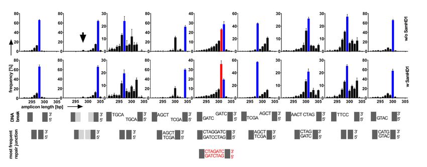
2 Nucleic Acids Research, 2021
non-cohesive ends relies on a set of specialized enzymes me- individual plasmid substrates PCR-amplified from the plas-
diating joining with or without prior resection, blunting or mid pool. Analyses of read length, frequency, and sequence
extension of DNA ends, which can be assigned to classical were performed using custom BASH and PERL scripts as
or alternative NHEJ (c-NHEJ or a-NHEJ) (2). Both NHEJ previously described (16).
pathways prefer the usage of short patches of microhomol-
ogy (≥1 nt) within DNA ends for end joining, with a-NHEJ
RT-PCR, western blotting and microscopy
typically depending on longer regions of homology between
the two DNA ends (>4 nt). Thus, a-NHEJ is also termed Total RNA was extracted from HEK293 cells (RNeasy
microhomology-mediated end joining (MMEJ) (4). Mini Kit, Qiagen) and subjected to first strand cDNA syn-
Downloaded from https://academic.oup.com/nar/advance-article/doi/10.1093/nar/gkab051/6138595 by guest on 23 February 2021
SAMHD1 (SAM domain and HD domain-containing thesis (iScript). RT-PCR was performed on first strand
protein 1) was initially discovered as protein with deoxynu- cDNA using specific primers for endogenous SAMHD1
cleoside triphosphate triphosphohydrolase (dNTPase) ac- and transgenic SAMHD1. Western blots were performed
tivity and putative nuclease activity. It was reported that on cell lysates using antibodies specific for SAMHD1
SAMHD1 is mainly a restriction factor for retroviruses and (ab67820, Abcam) and Flag-tag (clone M2, Sigma).
retroviral elements by reducing the pool of available dNTPs For the subcellular localization analysis,
to a level incompatible with reverse transcription of the HEK293SAMHD1-KO cells were transferred onto poly-L-
virus genome (8–12). In addition, SAMHD1 protein was lysine coated microscopy slides 4 hours after transfection
found to be associated with foci of DNA double strand with mCherry-SAMHD1 fusion constructs and nCas9
breaks (DSBs), proposing a possible role for SAMHD1 in constructs expressing zsGreen1 from a separate promoter.
DNA DSB repair (13). Indeed, it was shown that SAMHD1 Twenty four hours later, the cells were fixed with 4% PFA
promotes DSB repair by homologous recombination inde- and stained with anti-fade reagent with DAPI solution
pendent from its dNTPase activity but dependent on its in- (DAKO). Microscopy was performed on an Olympus IX81
teraction with the endonuclease CtIP. In this model, CtIP is using 60× magnification, DAPI, FITC and Cy3 channels
recruited to DSBs via interaction with SAMHD1, thus re- for DAPI, ZsGreen1 and mCherry detection.
secting DNA ends to generate 3 overhangs, which facilitate
strand invasion for homologous recombination (14).
Measurement of cellular dNTP levels
In this study, we asked whether aside of involvement in
homologous DNA repair, SAMHD1 may have an addi- Cellular dNTPs were quantified as recently described else-
tional effect on NHEJ. Our own results reveal a novel role of where (17). In brief, HEK293 SAMHD1-KO cells were
SAMHD1 for DNA DSB repair by NHEJ and give new in- transiently transfected with 1 g of SAMHD1 variant (wild
sights into its implication in genome stability and cancer de- type, K11A, K312A or K484T) using GeneJuice Trans-
velopment, and corroborate a very recent report on a novel fection Reagent (Novagen) or cells (HEK293, HEK293
role of SAMHD1 in regulating class switch recombination SAMHD1-KO and HeLa) were treated with DMSO or
in B lymphocytes (15). 2.5 mM dNs (Sigma Aldrich). Forty-eight hours post
transfection/treatment the cells were harvested in ice-cold
MATERIALS AND METHODS PBS, counted and the same cell number was aliquoted for
all samples. The harvested cells were resuspended in 60%
Cell lines and transfection ice-cold methanol, denatured at 95◦ C for 3 min and cen-
HEK293 and HeLa cell lines were cultured in RPMI trifuged at 18 500 ×g for 6 min at 4◦ C. Subsequently, the su-
medium supplemented with 10% FBS, 1% L-glutamine pernatants were transferred into new tubes and evaporated
and 1% Pen/Strep. For transient transfections, GeneJuice in Eppendorf Concentrator 5301 for 1 h at 60◦ C. dNTP
Transfection Reagent (Novagen) was used. extracts were resuspended in 100 l ice-cold nuclease-free
water. The extracts were stored at –80◦ C up to 1 week.
The extracts were subjected to a Q5 DNA polymerase and
Plasmid based DNA repair assay
EvaGreen-based assay for dNTPs using dNTP-specific 50-
Repair assays on linearized plasmids were performed ac- nt templates with conserved primer binding sites as de-
cording to our recent publication (16). In brief, HEK293 scribed in (17). The baseline and the end-point fluorescence
cells were either non-transfected or transfected with an ex- were read above the melting temperature of RNAse HII-
pression construct encoding human SAMHD1-Flag (Sino- nicked DNA as determined according to melt curve analy-
biological) or mutants using GeneJuice (Novagen). One day ses: 75◦ C for dATP and dCTP, 78◦ C for dTTP and 73.5◦ C
post transfection, a respective pool of linear plasmids (pool for dGTP. The higher the concentration of available dNTPs,
1: substate #1–#9; pool 2: substrate #11–#18; sequences the higher is the amount of dsDNA synthesized from the 50
provided in Supporting Table S1) was transfected and to- nt template and the higher is the fluorescence, measured on
tal DNA was extracted from cells 72 h post transfection of a ViiA7 Real-Time PCR system (ThermoFisher).
the plasmid pool (DNeasy Blood & Tissue Kit, Qiagen). Re-
paired plasmid-junctions were PCR-amplified using specific
CRISPR/Cas9 constructs and cloning
primers with different 5 tags specific for each sample. Am-
plicons were pooled and sequenced on the Illumina MiSeq SAMHD1 mutants were cloned using site directed mutage-
platform (MWG eurofins).The unique 6-bp region down- nesis with standard protocols using In-Fusion HD cloning
stream of the conserved primer-binding sites from each (Takarabio). mCherry fusion constructs were generated
plasmid was used for assigning relative frequencies of the by cloning RG1275 (5 - CCGCCACCAAGCTTG gccacc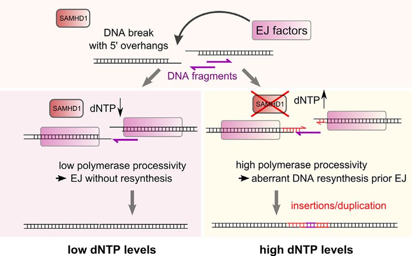
Nucleic Acids Research, 2021 3
ATGGTGAGCAAGGG-3 ) and RG1276 (5 - tcggctcgctgc amplified breakpoint junctions from independent experi-
atG TACTTGTACAGCTCGTCCATGCCG-3 ) amplified ments (as indicated by n-values) were purified from agarose
mCherry into KpnI linearized SAMHD1 constructs using gels, pooled and re-amplified using primers RG1309 (5 -
In-Fusion HD cloning (Takarabio). TCGTCGGCAGCGTCAGATGTGTATAAGAGACA
HEK293SAMHD1-KO cells (knockout of SAMHD1 in GTTCAGCCATGGTAGAATACAGCACTAC-3 ) and
HEK293 cells) were generated by cloning annealed oligos RG1310 (5 -GTCTCGTGGGCTCGGAGATGTGTATA
RG1075 5 -CCGGGTCATCGCAACGGGGACGCT-3 AGAGACAGCTGGAGTGCAGTAAACCTAGGAAC-
and RG1076 5 -AAACAGCGTCCCCGTTGCGATGAC- 3 ) followed by indexing and AMPure XP bead purification
3 (guide RNA for exon 1 of SAMHD1) into pGuide-it- (Beckman Coulter). For dN supplementation, cells were
Downloaded from https://academic.oup.com/nar/advance-article/doi/10.1093/nar/gkab051/6138595 by guest on 23 February 2021
ZsGreen1 Vector (Clontech) and transient transfection of transfected with del11q inducing nCas9 constructs. Four
HEK293 cells using GeneJuice (Novagen). Three days post hours post transfection, cells were mock-treated with
transfection, Zsgreen1 positive single cells were sorted into DMSO or treated with 2.5 mM dNs (dA, dT, dC, dG;
96-well plates (FACS ARIAIII, Beckton Dickinson). Two Sigma-Aldrich) and incubated for further 48 h. To de-
weeks post sort, colonies were expanded, genomic DNA termine contribution of DNA-PKc to del11q, cells were
was isolated (Qiagen), Cas9 target region was PCR ampli- mock-treated (DMSO) or treated with 10M DNA-PKc
fied using Phusion polymerase (ThermoFisher) and primers inhibitor NU7441 (Selleckchem) at time of transfecting
RG1105 (5 -CTACCTCGGATGTTCTTCAGCAG-3 ) nCas9 constructs. DNA was isolated 24 h post transfection
and RG1106 (5 -AATAGGCTGCCAATACTCCTTGG- and presence of del11q was determined by PCR using
3 ) and sequenced. Knockout clones were expanded and primers RG1209 and RG1210 using 60ng template DNA.
several stocks frozen for further experiments. For analysis of PCR-amplified junctions sequenced
For induction of chromosomal deletion on chr11q, two on the Illumina platform (MiSeq), demultiplexed paired
pairs of nCas9 constructs were generated using cloning end fastq files were adapter- and quality-trimmed using
of the following annealed oligos into ZsGreen1 Vector Trimmomatic (19), v0.33, default settings) and stitched
(Clontech): RG1195 5 -CCGGGGCTTGTGCCCTTCCC using FLASh (20), v1.2.11, -r 300, -f 350, -s 35). Sequences
TTCA-3 and RG1196 5 -AAACTGAAGGGAAGGGCA and length of reads containing exact matches of forward
CAAGCC-3 ; RG1197 5 -CCGGCTGCTGACTGAAGA and reverse primer plus five 3 bases of the chr11 target
GCCTTC-3 and RG1198 5 -AAACGAAGGCTCTTCA site were extracted (grep TTCAGCCATGGTAGAATA
GTCAGCAG-3 ; RG1199 5 -CCGGTTTCACCATGTT CAGCACTACTTAGAT.*GTGGGAAGGTTCCTAG
GCCCAGGC-3 and RG1200 5 -AAACGCCTGGGCAA GTTTACTGCACTCCAG) and used for further anal-
CATGGTGAAA-3 ; RG1201 5 -CCGGACAAAACTTA ysis. For both, Sanger and amplicon-sequencing, only
GCTGGGCGTG-3 and RG1202 5 -AAACCACGCCCA unique sequences were considered for analysis in order
GCTAAGTTTTGT-3 . Cas9 was mutated to nCas9 by gen- to examine individual del11q junctions. For microhomol-
erating D10A variants (18) of these vectors by PCR ampli- ogy analysis, junctions that contain insertions were not
fying a 786 bp fragment using Phusion polymerase (Ther- included.
moFisher) and primers RG1191 (5 -CCATGGTGGCGA To evaluate insertions at the nickase site, the extracted
ATTCTCCAGGCGATCTGACGG-3 ) and RG1192 (5 - sequences were filtered for unique sequences longer than
CAGCCCACGCTGTTGGTACCGATGGCCAG-3 ) and the expected wild type sequence (≥376nt). These sequences
cloning this fragment into KpnI/EcoRI (ThermoFisher) di- were transformed to fasta format and a search for se-
gested vectors using HD-in fusion cloning (Clontech). quence homologies was performed using NCBI blast+
(v2.10.0) (21), the hg19 human genomic database was
completed by adding sequences of used plasmids and the
nCas9-based DNA repair assay
del11q junction sequence. Sequence homologies to the nick-
For nCas9-based DNA repair assay HEK293, ase site at chr11 (chr11:106892584-107762754) were ex-
HEK293SAMHD1-KO and HeLa cells were transiently cluded. Furthermore, homologies that map to nickase site
transfected with four del11q inducing nCas9 constructs or del11q junction were selected to define intervals of ho-
using GeneJuice Transfection Reagent (Novagen), 250 mologies (alignment ≥87%). All other homologies (align-
ng each, according to manufacturer’s protocol. Simul- ment ≥ 99%) were checked for overlaps with these inter-
taneously, the cells were co-transfected with 1 g of vals. The overlapping parts were then trimmed, whereas the
SAMHD1 variant (wild type, K11A, K312A or K484T). remaining homology sequences (parts that do not overlap
Forty-eight hours post transfection the cells were har- with del11q junction) were used for further analysis if their
vested and DNA was isolated using DNeasy Blood & length was ≥15 bp. In the next step, we checked if single
Tissue Kit (Qiagen). The del11q junctions were PCR reads comprise several homologies that do not overlap with
amplified using 100ng input DNA and primers RG1209 each other. If homologies within the same read did not over-
(5 -TTCAGCCATGGTAGAATACAGCACTAC-3 ) and lap, both or all of them were further analysed. If homolo-
RG1210 (5 -CTGGAGTGCAGTAAACCTAGGAAC- gies overlapped, the one with the highest bit score was se-
‘3). PCR products were gel excised (Qiagen) and lected (i.e. the longest one). All homologies passing this fil-
TOPO cloned (Invitrogen) followed by Sanger se- ter were manually re-blasted for alignment to the del11q
quencing of individual clones (eurofins genomics). junction using NCBI BLAST tool (https://blast.ncbi.nlm.
Inserted DNA was mapped using BLAT search nih.gov/Blast.cgi), and del11q junction homologies were ex-
(https://genome.ucsc.edu/cgi-bin/hgBlat). For amplicon se- cluded. Since some of the sequences produced homologies
quencing on the in-house MiSeq platform (Illumina), PCR on multiple loci with equal bit score, one hit was selected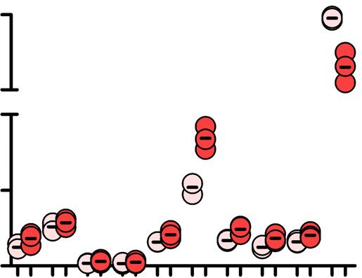
4 Nucleic Acids Research, 2021

randomly, and ambiguous blast-hits of reads were indicated        scribed (16). Thereby, we found that SAMHD1 overexpres-
in red within the circus plots.                                   sion severely affected the pattern of inter- and intramolecu-
   Moreover, genomic positions of detected homologies             lar repair frequencies (Figure 1C–F). Particularly, we found
were compared to the list of aphidicolin-sensitive genes gen-     that cohesive rejoining of the compatible ends on substrate
erated by Crosetto et al. (22), which was downloaded from         #6 occurred more frequently in presence of Flag-SAMHD1
the BLESS supporting website http://breakome.utmb.edu/            overexpression (Figure 1E). Concurrently, intermolecular
Home.html.                                                        repair events between different substrates occurred less fre-
   For statistical analysis of insertions, the numbers of reads   quently in presence of Flag-SAMHD1 overexpression, in-
containing homologies versus reads without homologies             dependent of the end structure (Figure 1E and F).

                                                                                                                                   Downloaded from https://academic.oup.com/nar/advance-article/doi/10.1093/nar/gkab051/6138595 by guest on 23 February 2021
were evaluated by Fisher’s exact test.
   All analyses were performed on a Linux Redhat system
                                                                  SAMHD1 facilitates joining of protruding 5 DNA ends car-
using custom bash or R (v3.6.1) scripting.
                                                                  rying short regions of complementarity
   The following R-packages were used: BiocMan-
ager,     BSgenome,         BSgenome.Hsapiens.UCSC.hg19,          To further corroborate this finding, we analyzed the se-
dplyr, IRanges, GenomeRanges, stringr, openxlsx, spgs,            quences of the individual intramolecular repair junctions
Biostrings, ggplot2, EasyGgplot2, devtools, circlize (23).        of substrates #1 to #9 (Figure 2). By ranking the sequences
                                                                  according to abundance, we found that all repair junctions
Statistics and data availability                                  except substrate #5 (non-cohesive 5 overhangs) shared the
                                                                  same predominant sequences, independent from presence
No explicit power analysis was used for sample size esti-         or absence of SAMHD1-Flag (Figure 2A–D). However, in
mation. N-values indicate biological replicates, which were       case of SAMHD1-Flag overexpression, we observed skew-
independent experiments (cells handled separately) but se-        ing towards a 1bp G:C microhomology within the repair
quenced on the Illumina platform as pooled (indexed) sam-         junction of non-cohesive 5 overhangs (substrate #5, Fig-
ples. All indices are provided together with sequencing data,     ure 2A–D). While in wt cells non-cohesive 5 overhangs
which have been deposited in the ArrayExpress database at         were preferentially blunted before joining, presence of over-
EMBL-EBI (www.ebi.ac.uk/arrayexpress) under accession             expressed SAMHD1-Flag led to incomplete filling of the
number E-MTAB-8382.                                               ssDNA overhangs prior joining, resulting in a 1bp shorter
                                                                  predominant repair junction, based on usage of a 1 nt mi-
RESULTS                                                           crohomology.
                                                                     For cohesive 5 overhangs, we detected a preference for
SAMHD1 affects DNA DSB repair by NHEJ
                                                                  simple rejoining of the sticky ends. The second most fre-
As previous work suggested involvement of SAMHD1 in               quent junction was generated by filling up the staggered
response to DNA damage, we aimed to investigate the con-          ends prior joining of blunted DNA ends, resulting in a 4 nt
tribution of SAMHD1 to accurate and efficient DNA DSB             longer repair junction. Nevertheless, we found that simple
repair by NHEJ. To this end, we first wanted to test whether      rejoining of cohesive 5 overhangs was also more effective
overexpression of SAMHD1 affects in vivo joining of arti-         in SAMHD1 transfected cells (substrate #6, Figure 2A–D).
ficial DNA substrates. Therefore, we used HEK293 cells,           This became apparent after analyzing the ratio of direct co-
which express low levels of SAMHD1 protein, and trans-            hesive joining to sequences derived from joining of blunted
fected them with Flag-tagged SAMHD1 to induce over-               ends (by filling up overhangs) prior joining (Figure 2E, F).
expression (Supplementary Figure S1). Twenty-four hours           However, we could not detect a general increase in MMEJ,
post transfection, mock transfected cells or Flag-SAMHD1          as end structures carrying a 6 bp microhomology were not
transfected cells were subjected to a previously reported         repaired at higher frequency in SAMHD1 transfected cells
plasmid based DNA DSB repair assay to analyze in vivo             (substrate #2 in Figure 2G, H).
joining of a pool of different DNA end structures in three           To more thoroughly investigate the effect of SAMHD1
independent experiments (16). In this assay, a pool of nine       on repair of 3 and 5 overhangs, we constructed a second
plasmids, which harbor individual 350-bp inserts flanked by       pool of linear plasmid substrates, carrying 1–3 complemen-
conserved primer-binding sites, is digested in vitro with dif-    tary C:G nts at the end of non-cohesive 3 and 5 4 nt over-
ferent restriction enzymes. This yields nine linearized DNA       hangs (substrates #11–#13, #15–#17, Figure 3 and Supple-
plasmids with defined DNA break structures ranging from           mentary Table S1). Here, we measured intramolecular re-
blunt ends with and without a 6-bp microhomology to co-           pair in mock versus SAMHD1-Flag transfected HEK293
hesive and noncohesive 3 and 5 protruding ends with 4           cells from two independent experiments as described above.
nt overhangs (substrate #1 to #9; Figure 1A, Supplemen-           By analyzing the sequences of the respective repair junc-
tary Table S1). Upon transfection into cells, the linear DNA      tions, we again could not detect an impact of SAMHD1
substrates are recognized by the cellular DNA DSB repair          overexpression on repair of 3 overhangs (substrate #11–
machinery and rejoined. Seventy-two hours post transfec-          #13, Figure 3). In contrast, for repair of 5 protruding ends,
tion, we isolated DNA from cells and PCR-amplified repair         we confirmed a clear bias towards usage of microhomolo-
junctions of pooled linear plasmid substrates using the con-      gies within the ssDNA overhangs, which was most pro-
served primer binding sites and subjected them to ampli-          nounced for 3 nt microhomologies with the most frequent
con sequencing (Figure 1B). We then calculated relative re-       repair junction being 3 nt shorter in presence of SAMHD1-
pair frequencies of the respective DNA ends by mapping se-        Flag (substrate #17, Figure 3). Corroboratively, repair junc-
quence reads to the nine repair substrates as previously de-      tions corresponding to partial or complete re-synthesis of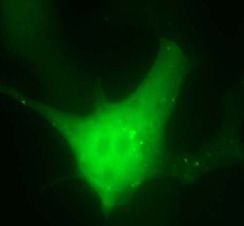
Nucleic Acids Research, 2021 5

                                                                                                                                                               Downloaded from https://academic.oup.com/nar/advance-article/doi/10.1093/nar/gkab051/6138595 by guest on 23 February 2021
Figure 1. DNA Repair of plasmid substrates is skewed by SAMHD1. (A) A pool of linear DNA plasmids (substrate #1-#9, with the respective DNA end
structures indicated) was transfected into SAMHD1-Flag expressing or non-expressing HEK293 cells. (B) Repaired plasmid junctions were PCR-amplified
from extracted DNA 0 and 72 h post transfection from 3 independent experiments. A control PCR on TP53 is shown on the bottom. Bands corresponding
in size to repair junctions are indicated with an asterisk. (C, D) Repair frequencies of inter- and intramolecular repair events (substrate #1–#9) are shown
as circos plots for SAMHD1-Flag non-expressing and expressing HEK293 cells. The size of the segments reflects the occurrence of the nine different DNA
plasmids serving as substrate for repair. Repair frequencies are indicated by size of ribbons (mean values from three independent experiments). Start of
ribbon denotes forward primed arms; arrow of ribbon denotes reverse primed arm of substrates. Repair junctions significantly overrepresented (C vs D
and vice versa) are colored red (P < 0.05) and light red (0.05 < P < 0.1), and the correspondingly underrepresented repair junctions are colored blue and
light blue, respectively. Significances were calculated by two-tailed t-tests with unequal variances, n = 3. (E) Repair frequencies from C and D are shown
separately for intramolecular (#1–#9) and intermolecular (IM) repair in HEK293 cells either expressing or not-expressing SAMHD1-Flag. (F) Repair
frequencies from C and D are shown separately for intermolecular repair in HEK293 cells either expressing or not-expressing Flag-SAMHD1. Means are
indicated; significances were calculated by two-tailed t-tests with unequal variances, n = 3.

cohesive overhangs from substrate #17 were markedly re-                          SAMHD1-Flag was present, the internal 2 nt microhomol-
duced in presence of Flag-SAMHD1 (Figure 3).                                     ogy was preferentially used for repair (substrate #18, Figure
    We next analyzed the impact of SAMHD1 on the re-                             3).
pair of 4 nt 3 and 5 overhangs harboring an internal 2
nt CC:GG microhomology (substrate #14 and #18). Inde-
                                                                                 End joining of Cas9 nickase induced chromosomal breaks is
pendent from SAMHD1 expression, microhomology was
                                                                                 affected by physiologic SAMHD1 levels and requires cat-
preferentially used for repair of 3 overhangs with the 3 -
                                                                                 alytic activity
terminal non-matching nt being resected and the gap re-
filled by the complementary nt (substrate #14, Figure 3).                        Our previous experiments revealed that superphysiologi-
For 5 overhangs, in absence of SAMHD1-Flag, the ss-                             cal levels of SAMHD1 affect EJ of artificial DNA sub-
DNA region was preferentially refilled and converted to ds-                      strates with 5 overhangs. Next, we wanted to test whether
DNA followed by joining of blunted ends. However, when                           physiological levels of SAMHD1 affect EJ of endogenous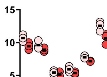
6 Nucleic Acids Research, 2021

                                                                                                                                                             Downloaded from https://academic.oup.com/nar/advance-article/doi/10.1093/nar/gkab051/6138595 by guest on 23 February 2021
Figure 2. Repair junctions are modulated by SAMHD1. (A, B) PCR-amplified repair junctions from SAMHD1-Flag non-expressing (A) or expressing (B)
HEK293 cells were sequenced and the frequencies of the lengths of the respective sequences are shown for each DNA plasmid substrate (#1-#9). The arrow
within the graph for substrate #2 denotes the size of the repair junction corresponding to junctions repaired by MMEJ using a 6bp microhomology. (C) The
respective DNA break structures for DNA plasmid substrates #1- #9 is shown. (D) The sequences of the most frequent junctions for cells not expressing
Flag-SAMHD1 are shown and framed in blue. The most frequent repair junctions for Flag-SAMHD1 expressing cells (if different to non-expressing cells),
are shown below and framed in red. The color of the frame allocates the sequences to the respective bars shown in (A, B). Bars are showing mean + SD
from three independent experiments. (E) DNA break structure of DNA plasmid substrate #6 carrying cohesive 4 nt 5 overhangs (top) and DNA sequences
of two potential joining events (bottom) are shown. (F) The ratio of repair junctions deriving from direct cohesive joining (dcj) of DNA ends to junctions
deriving from blunting prior joining by filling up ssDNA overhangs (bpj) for HEK293 cells either expressing or non-expressing SAMHD1-Flag is shown.
(G) DNA break structure of DNA plasmid substrate #2 carrying a 6 bp microhomology (top) and DNA sequences of two potential joining events (bottom)
are shown. (H) The ratio of repair junctions deriving from direct joining (dj) of DNA ends to junctions deriving from MMEJ for HEK293 cells either
expressing or non-expressing SAMHD1-Flag is shown. (mean values are indicated; significances were calculated by unpaired two-tailed t-tests from three
independent experiments).

DNA breaks. Therefore, we generated a SAMHD1 knock-                            mentary Figure S2C, treatment of cells with NU7441 prior
out cell line in HEK293 cells (HEK293SAMHD1-KO ). To mon-                      transfection with paired nCas9 constructs impeded induc-
itor an effect on EJ of endogenous 5 overhangs, we gen-                       tion of del11q, although nCas9 expression levels were not
erated pairs of Cas9 nickases, which induce two 5 stag-                       affected (Supplementary Figure S2C). In line with previ-
gered ends on chromosome 11q, which span a distance of                         ous reports (24), these experiments pointed to a NHEJ-
0.67 Mbp (Figure 4A). To analyze repair of these breaks                        dependent mechanism as the major pathway contribut-
by EJ, we characterized repair junctions descending from                       ing to nCas9 induced del11q. To examine the impact of
joining of the two distant DNA breaks, resulting in a chro-                    SAMHD1 on del11q, we performed amplicon-sequencing
mosomal deletion (del11q). Therefore, we PCR-amplified                         of PCR-amplified del11q junctions from HEK293 ver-
breakpoint junctions from nCas9 induced del11q and an-                         sus HEK293SAMHD1-KO cells and HEK293SAMHD1-KO cells
alyzed individual junctions from amplicon sequencing ap-                       transfected with wt SAMHD1 or catalytically dead K312A
proaches and from cloned PCR fragments by Sanger se-                           mutant, lacking dNTPase and nuclease activity (8). We
quencing. First, we determined the mechanism by which                          found that in presence of endogenous SAMHD1, junctions
nCas9 induced DNA breaks lead to del11q. We found that                         were significantly shorter than in the knockout cells or in
del11q was detected only when paired nicks on both sites                       cells expressing the catalytically dead K312A variant (Fig-
(and thus two DSBs) were present, whereas a single nick                        ure 4B). In line with our plasmid based repair assays, we
combined with a paired nick was not sufficient for generat-                    observed that overexpression of SAMHD1 induced an even
ing a chromosomal deletion (Supplementary Figure S2A,                          more pronounced skewing towards shorter repair junctions
B). In addition, we tested the sensitivity of the genera-                      compared to physiological levels, showing that SAMHD1
tion of del11q towards NU7441, an inhibitor of DNA-                            expression levels predict its effect on EJ (Figure 4B). To
PKcs, which is required for NHEJ. As shown in Supple-                          validate results from amplicon-sequencing, we cloned indi-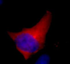
Nucleic Acids Research, 2021 7

                                                                                                                                                              Downloaded from https://academic.oup.com/nar/advance-article/doi/10.1093/nar/gkab051/6138595 by guest on 23 February 2021
Figure 3. Repair of staggered ends with microhomologies by SAMHD1. (A, B) PCR-amplified repair junctions from SAMHD1-Flag non-expressing
(A) or expressing (B) HEK293 cells were sequenced and the frequencies of the lengths of the respective sequences are shown for each DNA plasmid
substrate (#11–#18). (C) The respective DNA break structures for DNA plasmid substrates are shown. (D) The sequences of the most frequent junctions
for cells not expressing Flag-SAMHD1 are shown and framed in blue. The most frequent repair junctions for Flag-SAMHD1 expressing cells (if different
to non-expressing cells), are shown below and framed in red (substrate #17 and #18). The color of the frame allocates the sequences to the respective bars
shown in (A, B). (Bars are showing mean + SD from two independent experiments). (E) DNA break structure of DNA plasmid substrate #17 carrying
cohesive 4 nt 5 overhangs with 3bp microhomologies (top) and DNA sequences of two potential joining events (bottom) are shown. (F) The ratio of repair
junctions deriving from direct cohesive joining (dcj) of DNA ends to junctions deriving from blunting prior joining (bpj) by filling up ssDNA overhangs
(bpj) for HEK293 cells either expressing or non-expressing SAMHD1-Flag is shown. (G) DNA break structure of DNA plasmid substrate #18 carrying
an internal 2bp microhomology (top) and DNA sequences of two potential joining events (bottom) are shown. (H) The ratio of repair junctions deriving
from direct cohesive joining (dcj) of DNA ends to junctions deriving from blunting prior joining (bpj) for HEK293 cells either expressing or non-expressing
SAMHD1-Flag is shown. (mean values are indicated; significances were calculated by unpaired two-tailed t-tests from two independent experiments).

vidual PCR-amplified del11q junctions and analyzed them                         plementary Figure S3C; Supplementary Table S2). Some
by Sanger sequencing. Thereby, we could confirm the re-                         junctions featured complex arrangements of repetitive du-
sults from amplicon-sequencing and found that junctions                         plications and insertions, which were not observed in junc-
from knockout cells or K312A expressing cells were sig-                         tions from HEK293 cells or SAMHD1 deficient cells com-
nificantly longer than wt-expressing or wt-overexpressing                       plemented with wt SAMHD1 expressing plasmids (Supple-
samples (Supplementary Figure S3, Supplementary table                           mentary Figure S4; Supplementary Table S2).
S2). Furthermore, we analyzed microhomologies at cloned
unique del11q junctions, which revealed a typical NHEJ-
dependent pattern with predominant 0–1 bp microhomolo-                          Increase of the cellular dNTP pool enhances insertions at
gies independent of SAMHD1 expression (Supplementary                            nickase induced chromosomal junctions
Figure S3B and Supplementary Table S2). Western blotting                        To further investigate whether the effect of SAMHD1 on
confirmed similar expression levels between wt and K312A                        EJ is solely depending on its catalytic dNTPase activity and
mutant of SAMHD1 from transfected samples (Figure 4C).                          independent from its scaffold function at DNA ends, we re-
This shows that catalytic activity is important for the ob-                     peated our nCas9 experiment including two catalytically ac-
served effect on EJ. Strikingly, by analyzing cloned, Sanger-                   tive SAMHD1 mutants: one that fails to recruit the CtIP
sequenced del11q junctions we found many duplications                           endonuclease to DNA breaks due to a K484T mutation
and insertions at breakpoint junctions from SAMHD1 de-                          (14) and another that is excluded from the nucleus due to
ficient or K312A expressing cells. The duplications derived                     a K11A mutation within the nuclear localization sequence
from resynthesized DNA overhangs at nCas9 target sites,                         (NLS) (25,26). In addition, we performed this nCas9 as-
whereas the insertions mapped either to distant genomic                         say also in wildtype HEK293 cells and a second cell line
regions from other chromosomes or to transfected plas-                          (HeLa cells) in presence of high amounts of deoxynucleo-
mid DNA (SAMHD1 or nCas9 expressing plasmids; Sup-                              sides (dNs) as metabolic dNTP precursors to artificially in-
8 Nucleic Acids Research, 2021
Downloaded from https://academic.oup.com/nar/advance-article/doi/10.1093/nar/gkab051/6138595 by guest on 23 February 2021
Figure 4. Induction of a chromosomal deletion at chr11q using nCas9. (A) Schematic representation of chr11 indicating nCas9 sites and primers for PCR
amplification of breakpoint junctions. (B) Length of PCR-amplified amplicon-sequenced breakpoint junctions from HEK293 and HEK293SAMHD1-KO
cells transfected with the indicated SAMHD1 variants (K312A or wt; n = 4 for all samples). A length corresponding to joining of blunted (filled up) 5
overhangs is set to 0 bp. (C) Western blots of HEK293 and HEK293SAMHD1-KO cells (left), transiently transfected with the indicated constructs (right). For
(B), median with interquartile range is shown in boxes, with whiskers extending the boxes with the largest/smallest value no further than 1.5 times of the
interquartile range and other points plotted individually; significances were calculated by Mann-Whitney test and are indicated above the graph, medians
are given above the x-axis.
crease the intracellular dNTP pool, mimicking high dNTP breakpoint junctions, showing that the observed effect on
levels upon SAMHD1 deficiency (27). In line with previ- insertions had reached a plateau (Supplementary Figure
ous reports, a SAMHD1 K11A mutant fused to mCherry S7). Finally, we wanted to examine all sequences for pres-
showed primarily cytoplasmic localization, also in presence ence of insertions that mapped to distant genomic regions
of nCas9 endocucleases (Figure 5A). Next, we PCR am- or to transfected plasmids. To systematically detect inser-
plified breakpoint junctions from nCas9 induced del11q tions mapping to distant DNA sites at del11q junctions
(schematically depicted in Figure 5B) in HEK293SAMHD1-KO from our amplicon-sequencing dataset, we developed au-
cells either expressing wt SAMHD1 or mutants K11A, tomated blast searches on all unique sequences longer than
K484T or K312A and in HEK293 cells as well as HeLa cells 376nt, corresponding to repair joints descending from re-
supplemented with dNs to increase dNTP levels. PCR am- filled overhangs. Thereby, we found a significantly higher
plified junctions were amplicon-sequenced on a MiSeq next percentage of junctions with insertions mapping to distant
generation sequencing platform and amplicon lengths were sites in samples lacking SAMHD1 or expressing K312A
calculated. Again, absence of SAMHD1 as well as presence variant compared to SAMHD1 proficient cells or cells ex-
of a catalytically dead K312A variant resulted in signifi- pressing the catalytically active SAMHD1 K11A or K484T
cantly longer repair junctions, whereas expression of the cy- mutant (Supplementary Figure S8; Supplementary table
tosolic K11A mutant and mutant K484T induced shorter S4). Corroboratively, increased dNTP levels upon dN ad-
junctions similar to what we observed in wt SAMHD1 ex- dition in HEK293 and HeLa cells also yielded a higher
pressing cells (Figure 5C). Again, expression of low lev- percentage of junctions with insertions mapping to distant
els of endogenous SAMHD1 in wt HEK293 cells resulted genomic regions or to transfected plasmids (Supplemen-
in intermediate junction lengths (Figure 5C). These results tary Figure S5E and F; Supplementary table S4). Strik-
were confirmed by analyzing Sanger-sequences from cloned ingly, many of these genomic regions were previously de-
PCR-amplified del11q junctions (Supplementary Figure fined as common fragile sites, which are sensitive to replica-
S5; Supplementary table S3). Corroboratively, increasing tion stress and vulnerable for DNA breakage, with some of
dNTP levels by dN addition in two cell lines (HEK293 these sites mapping to cancer related genes (Supplementary
and HeLa) also yielded significantly longer junctions, sim- Table S5) (22).
ilar to loss of SAMHD1 (Figure 5D). Increased intra-
cellular dNTPs in SAMHD1 knockout HEK293 cells as
DISCUSSION
well as upon dN supplementation in HEK293 and HeLa
cells were confirmed by an EvaGreen-based detection as- SAMHD1 is a remarkable enzyme with an increasing num-
say (Supplementary Figure S6) (17). Notably, dN addi- ber of attributed biologically important tasks. Originally
tion in SAMHD1 knockout HEK293 cells did further in- detected as dNTPase, SAMHD1 was implicated in HIV-
crease dNTP levels (Supplementary Figure S6) but did restriction by minimizing the dNTP pool to a level incom-
not lead to a further increase in the length of amplified patible with reverse transcription of the viral genome. AsNucleic Acids Research, 2021 9
A C D
Downloaded from https://academic.oup.com/nar/advance-article/doi/10.1093/nar/gkab051/6138595 by guest on 23 February 2021
B
E F
Figure 5. Elevated dNTP pool increases insertions at repair junctions. (A) HEK293SAMHD1-KO cells were transiently transfected with constructs encoding
SAMHD1 and SAMHD1 K11A mutants N-terminally fused to mCherry together with del11q inducing nCas9 constructs encoding zsGreen1 as separately
transcribed gene. (B) Schematic representation of nCas9 induced del11q and primers for PCR amplification of breakpoint junctions. (C) Length of PCR-
amplified amplicon-sequenced breakpoint junctions from HEK293 and HEK293SAMHD1-KO cells transfected with the indicated SAMHD1 variants (K11A,
K484T, K312A or wt) (n = 4 for all samples, except K484T n = 3) and (D) from HEK293 and HeLa cells grown in excessive dN to increase intracellular
dNTP pools (n = 2 for HEK293 cells and n = 3 for HeLa cells). Median with interquartile range is shown in boxes, with whiskers extending the boxes
with the largest/smallest value no further than 1.5 times of the interquartile range and other points plotted individually; significances were calculated by
Mann–Whitney test and are indicated above the graph, medians are given above the x-axis. (E) Circos plots were generated from data in Figure 5D, which
show the genome as circle with ribbons indicating the homologies of inserted DNA at the del11q repair junction to the respective distant genomic regions.
Ribbons in red indicate that the insertions map to the genome ambiguously, with only one randomly chosen homology shown (detailed mapping results
are summarized in Supplementary table S4; n = number of junctions with distant homologies/total number of unique reads analyzed). (F) Bars show
the percentage of all amplicon-sequencing reads with insertions mapping to distant genomic sites or to transfected plasmids. Significances in (E, F) were
calculated by Fisher’s exact test.
even small variations in the level of dNTP concentrations DNA to the cytosol, which otherwise would induce a ro-
can affect precision and accuracy of DNA polymerase ac- bust interferon (IFN)-response (33). Germline mutations in
tivity, a further role for SAMHD1 in replication fidelity and the SAMHD1 gene have been associated with the Aicardi-
cell cycle regulation was suggested (28–31). Based on its Goutières syndrome (AGS), a hereditary autoimmune en-
dNTPase activity, SAMHD1 was also reported to detox- cephalopathy, suggesting that constant immune-stimulating
ify DNA base analogs, currently used in cancer treatment, IFN signaling in absence of SAMHD1 is the underlying ba-
thus, serving as biomarker predicting low response to these sis for aberrant inflammation in AGS (33,34). Furthermore,
drugs (32). SAMHD1 is an important factor for homologous DNA re-
In addition to this function, SAMHD1 was recently re- pair, as it binds to DSBs and recruits the endonuclease CtIP,
ported to be recruited to stalled replication forks, degrad- which generates 3 ssDNA overhangs for strand invasion
ing nascent DNA by attracting Mre11 exonuclease, which during homologous recombination. Notably, SAMHD1 ex-
enables efficient replication restart and avoids release of ss- ecutes its role in homologous DNA repair independent10 Nucleic Acids Research, 2021
from its dNTPase activity (14). Many of these functions, sis in human cells (44). As our study reveals that insertions
particularly its role in homologous repair and in regulating were increased in absence of catalytically active SAMHD1
the dNTP pool are important for genome integrity, suggest- or in presence of elevated dNTP pools, we propose that
ing an important role in cancer development. Indeed, down- super-physiological dNTP levels may increase DNA poly-
regulation or recurrent SAMHD1 mutations were found merase resynthesis rates leading to increased refilling of
in some cancer entities like chronic lymphocytic leukemia, staggered DNA ends and synthesis over inserted DNA tem-
colon cancer and T-cell prolymphocytic leukemia and were plates prior end ligation. Strikingly, our data are in line with
particular increased in chemorefractory cases, which clas- a parallel report, addressing SAMHD1 in class switch re-
sifies SAMHD1 as cancer gene (13,30,35,36). Our own combination of antibody constant regions in B cells, which
Downloaded from https://academic.oup.com/nar/advance-article/doi/10.1093/nar/gkab051/6138595 by guest on 23 February 2021
data show for the first time a previously undefined role for was published during finalization of our manuscript (15).
SAMHD1 in DNA end joining of DSBs. We provide ev- In this parallel work, it was concomitantly shown that ab-
idence that in presence of catalytically active SAMHD1, sence of SAMHD1 and high intracellular dNTP levels lead
breakpoint junctions are shorter, featuring less DNA resyn- to aberrant insertions at EJ dependent switch junctions.
thesis. This effect was restricted to DNA ends with 5 over- Summarizing, our data further support a previously un-
hangs, as only these structures provide 3 hydroxyl-ends defined role for SAMHD1 in preserving genome integrity
for polymerase-dependent gap filling. This effect was more and draw light on how loss of SAMHD1 and aberrant
pronounced in presence of overexpressed SAMHD1 and dNTP levels could contribute to cancer formation, clonal
depended on its catalytic activity, suggesting that the lev- cancer evolution and treatment resistance promoted by
els of dNTPs regulated by SAMHD1 dictate the veloc- aberrant insertions at repair joints.
ity of DNA end resynthesis by DNA-polymerases. This
assumption is corroborated by our finding that increas- DATA AVAILABILITY
ing the pool of dNTPs by dN supplementation enhances
the extent of nucleotide insertions at breakpoint junctions All sequencing data, which have been deposited in the
similar to SAMHD1 deficiency. Furthermore, a SAMHD1 ArrayExpress database at EMBL-EBI (www.ebi.ac.uk/
K11A mutant, that is excluded from the nucleus while re- arrayexpress) under accession number E-MTAB-8382.
taining catalytic functions (25,26), as well as a K484T mu-
tant that fails to interact with CtIP (14) are also leading SUPPLEMENTARY DATA
to short repair junctions, similar to those observed in wt
SAMHD1 expressing cells, which suggests that solely intra- Supplementary Data are available at NAR Online.
cellular dNTP levels regulate the extent of insertions at EJ
dependent repair junctions. In line with this, elevated dNTP FUNDING
levels were reported to increase DNA replication rates and
polymerase activities, particularly in presence of DNA le- SCRI-LIMCR, the City of Salzburg, the Province of
sions (28,37,38). Salzburg [20102-P1509466-FPR01-2015, 20102-P1601064-
Most strikingly, many of the insertions observed upon FPR01-2017 to R.Gr.]; Austrian Science Fund FWF
SAMHD1 loss were homologous to distant genomic sites or (P28201 to R.Ge.]. Funding for open access charge: Aus-
to transfected plasmids. Insertion of nuclear or mitochon- trian Science Fund FWF.
drial DNA into breakpoints from chromosomal rearrange- Conflict of interest statement. None declared.
ments is commonly observed in cancer cells and consti-
tutes a threat to genome integrity (39–41). Even in normal REFERENCES
cells, insertions of mitochondrial DNA into genomic re-
1. Khanna,K.K. and Jackson,S.P. (2001) DNA double-strand breaks:
gions were discerned, pointing to an important mechanism signaling, repair and the cancer connection. Nat.Genet., 27, 247–254.
that shapes the evolution of genomes (42). Insertions can 2. Chiruvella,K.K., Liang,Z. and Wilson,T.E. (2013) Repair of
either emerge from direct incorporation of DNA fragments double-strand breaks by end joining. Cold Spring Harb. Perspect.
from distant sites, or by copying distant DNA (fragments) Biol., 5, a012757.
without causing a deletion at the originating position, which 3. Lieber,M.R. (2010) The mechanism of double-strand DNA break
repair by the nonhomologous DNA end-joining pathway.
is therefore called templated insertion. According to the Annu.Rev.Biochem., 79, 181–211.
current view, templated insertions originate from DNA re- 4. Pannunzio,N.R., Li,S., Watanabe,G. and Lieber,M.R. (2014)
leased from damaged genomic or episomal/mitochondrial Non-homologous end joining often uses microhomology:
sites or otherwise, it may derive from RNA transcripts, implications for alternative end joining. DNA Repair (Amst.), 17,
74–80.
which were reversely transcribed by LINE-1 (long inter- 5. Karanam,K., Kafri,R., Loewer,A. and Lahav,G. (2012) Quantitative
spersed element-1) endonuclease and reverse transcriptase live cell imaging reveals a gradual shift between DNA repair
(ORF2) (41,43). In either case, the DNA fragments can mechanisms and a maximal use of HR in mid S phase. Mol. Cell, 47,
be directly inserted at the repair joint or can serve as tem- 320–329.
plate for DNA polymerases at DSB sites. Corroboratively, 6. Rothkamm,K., Kruger,I., Thompson,L.H. and Lobrich,M. (2003)
Pathways of DNA double-strand break repair during the mammalian
many insertions in our analysis mapped to fragile sites, cell cycle. Mol. Cell. Biol., 23, 5706–5715.
which are chromosomal regions susceptible to DNA break- 7. Gu,J., Lu,H., Tippin,B., Shimazaki,N., Goodman,M.F. and
age (22). As yeast experiments revealed that Pol4 (a pol X Lieber,M.R. (2007) XRCC4:DNA ligase IV can ligate incompatible
family polymerase) was responsible for insertion of DNA DNA ends and can ligate across gaps. EMBO J., 26, 1010–1023.
8. Beloglazova,N., Flick,R., Tchigvintsev,A., Brown,G., Popovic,A.,
fragments (41), presumably one of the pol X family poly- Nocek,B. and Yakunin,A.F. (2013) Nuclease activity of the human
merases, comprising pol , pol , pol and terminal de- SAMHD1 protein implicated in the Aicardi-Goutieres syndrome and
oxynucleotidyl transferase (TdT) are mediating this synthe- HIV-1 restriction. J. Biol. Chem., 288, 8101–8110.Nucleic Acids Research, 2021 11
9. Goldstone,D.C., Ennis-Adeniran,V., Hedden,J.J., Groom,H.C., 27. Baldauf,H.M., Pan,X., Erikson,E., Schmidt,S., Daddacha,W.,
Rice,G.I., Christodoulou,E., Walker,P.A., Kelly,G., Haire,L.F., Burggraf,M., Schenkova,K., Ambiel,I., Wabnitz,G., Gramberg,T.
Yap,M.W. et al. (2011) HIV-1 restriction factor SAMHD1 is a et al. (2012) SAMHD1 restricts HIV-1 infection in resting CD4(+) T
deoxynucleoside triphosphate triphosphohydrolase. Nature, 480, cells. Nat. Med., 18, 1682–1687.
379–382. 28. Poli,J., Tsaponina,O., Crabbe,L., Keszthelyi,A., Pantesco,V.,
10. Lahouassa,H., Daddacha,W., Hofmann,H., Ayinde,D., Logue,E.C., Chabes,A., Lengronne,A. and Pasero,P. (2012) dNTP pools
Dragin,L., Bloch,N., Maudet,C., Bertrand,M., Gramberg,T. et al. determine fork progression and origin usage under replication stress.
(2012) SAMHD1 restricts the replication of human EMBO J., 31, 883–894.
immunodeficiency virus type 1 by depleting the intracellular pool of 29. Lee,E.J., Seo,J.H., Park,J.H., Vo,T.T.L., An,S., Bae,S.J., Le,H.,
deoxynucleoside triphosphates. Nat. Immunol., 13, 223–228. Lee,H.S., Wee,H.J., Lee,D. et al. (2017) SAMHD1 acetylation
11. Zhao,K., Du,J., Han,X., Goodier,J.L., Li,P., Zhou,X., Wei,W., enhances its deoxynucleotide triphosphohydrolase activity and
Downloaded from https://academic.oup.com/nar/advance-article/doi/10.1093/nar/gkab051/6138595 by guest on 23 February 2021
Evans,S.L., Li,L., Zhang,W. et al. (2013) Modulation of LINE-1 and promotes cancer cell proliferation. Oncotarget, 8, 68517–68529.
Alu/SVA retrotransposition by Aicardi-Goutieres syndrome-related 30. Rentoft,M., Lindell,K., Tran,P., Chabes,A.L., Buckland,R.J.,
SAMHD1. Cell Rep., 4, 1108–1115. Watt,D.L., Marjavaara,L., Nilsson,A.K., Melin,B., Trygg,J. et al.
12. Powell,R.D., Holland,P.J., Hollis,T. and Perrino,F.W. (2011) (2016) Heterozygous colon cancer-associated mutations of SAMHD1
Aicardi-Goutieres syndrome gene and HIV-1 restriction factor have functional significance. Proc.Natl.Acad.Sci.U.S.A, 113,
SAMHD1 is a dGTP-regulated deoxynucleotide 4723–4728.
triphosphohydrolase. J. Biol. Chem., 286, 43596–43600. 31. Bonifati,S., Daly,M.B., St Gelais,C., Kim,S.H., Hollenbaugh,J.A.,
13. Clifford,R., Louis,T., Robbe,P., Ackroyd,S., Burns,A., Timbs,A.T., Shepard,C., Kennedy,E.M., Kim,D.H., Schinazi,R.F., Kim,B. et al.
Wright,C.G., Dreau,H., Sigaux,F., Judde,J.G. et al. (2014) SAMHD1 (2016) SAMHD1 controls cell cycle status, apoptosis and HIV-1
is mutated recurrently in chronic lymphocytic leukemia and is infection in monocytic THP-1 cells. Virology, 495, 92–100.
involved in response to DNA damage. Blood, 123, 1021–1031. 32. Schneider,C., Oellerich,T., Baldauf,H.M., Schwarz,S.M., Thomas,D.,
14. Daddacha,W., Koyen,A.E., Bastien,A.J., Head,P.E., Dhere,V.R., Flick,R., Bohnenberger,H., Kaderali,L., Stegmann,L., Cremer,A.
Nabeta,G.N., Connolly,E.C., Werner,E., Madden,M.Z., Daly,M.B. et al. (2017) SAMHD1 is a biomarker for cytarabine response and a
et al. (2017) SAMHD1 promotes DNA end resection to facilitate therapeutic target in acute myeloid leukemia. Nat. Med., 23, 250–255.
DNA repair by homologous recombination. Cell Rep., 20, 1921–1935. 33. Coquel,F., Silva,M.J., Techer,H., Zadorozhny,K., Sharma,S.,
15. Husain,A., Xu,J., Fujii,H., Nakata,M., Kobayashi,M., Wang,J.Y., Nieminuszczy,J., Mettling,C., Dardillac,E., Barthe,A., Schmitz,A.L.
Rehwinkel,J., Honjo,T. and Begum,N.A. (2020) SAMHD1-mediated et al. (2018) SAMHD1 acts at stalled replication forks to prevent
dNTP degradation is required for efficient DNA repair during interferon induction. Nature, 557, 57–61.
antibody class switch recombination. EMBO J., 39, e102931. 34. Rice,G.I., Bond,J., Asipu,A., Brunette,R.L., Manfield,I.W.,
16. Gassner,F.J., Schubert,M., Rebhandl,S., Spandl,K., Zaborsky,N., Carr,I.M., Fuller,J.C., Jackson,R.M., Lamb,T., Briggs,T.A. et al.
Catakovic,K., Blaimer,S., Hebenstreit,D., Greil,R. and Geisberger,R. (2009) Mutations involved in Aicardi-Goutieres syndrome implicate
(2017) Imprecision and DNA break repair biased towards SAMHD1 as regulator of the innate immune response. Nat.Genet.,
incompatible end joining in Leukemia. Mol. Cancer Res., 16, 428–438. 41, 829–832.
17. Purhonen,J., Banerjee,R., McDonald,A.E., Fellman,V. and 35. Johansson,P., Klein-Hitpass,L., Choidas,A., Habenberger,P.,
Kallijarvi,J. (2020) A sensitive assay for dNTPs based on long Mahboubi,B., Kim,B., Bergmann,A., Scholtysik,R., Brauser,M.,
synthetic oligonucleotides, EvaGreen dye and inhibitor-resistant Lollies,A. et al. (2018) SAMHD1 is recurrently mutated in T-cell
high-fidelity DNA polymerase. Nucleic Acids Res., 48, e87. prolymphocytic leukemia. Blood Cancer J., 8, 11.
18. Ran,F.A., Hsu,P.D., Lin,C.Y., Gootenberg,J.S., Konermann,S., 36. Landau,D.A., Carter,S.L., Stojanov,P., McKenna,A., Stevenson,K.,
Trevino,A.E., Scott,D.A., Inoue,A., Matoba,S., Zhang,Y. et al. Lawrence,M.S., Sougnez,C., Stewart,C., Sivachenko,A., Wang,L.
(2013) Double nicking by RNA-guided CRISPR Cas9 for enhanced et al. (2013) Evolution and impact of subclonal mutations in chronic
genome editing specificity. Cell, 154, 1380–1389. lymphocytic leukemia. Cell, 152, 714–726.
19. Bolger,A.M., Lohse,M. and Usadel,B. (2014) Trimmomatic: a flexible 37. Pai,C.C. and Kearsey,S.E. (2017) A critical balance: dNTPs and the
trimmer for Illumina sequence data. Bioinformatics, 30, 2114–2120. maintenance of genome stability. Genes, 8, 57.
20. Magoc,T. and Salzberg,S.L. (2011) FLASH: fast length adjustment of 38. Stodola,J.L. and Burgers,P.M. (2016) Resolving individual steps of
short reads to improve genome assemblies. Bioinformatics, 27, Okazaki-fragment maturation at a millisecond timescale. Nat. Struct.
2957–2963. Mol. Biol., 23, 402–408.
21. Camacho,C., Coulouris,G., Avagyan,V., Ma,N., Papadopoulos,J., 39. Ju,Y.S., Tubio,J.M., Mifsud,W., Fu,B., Davies,H.R.,
Bealer,K. and Madden,T.L. (2009) BLAST+: architecture and Ramakrishna,M., Li,Y., Yates,L., Gundem,G., Tarpey,P.S. et al.
applications. BMC Bioinformatics, 10, 421. (2015) Frequent somatic transfer of mitochondrial DNA into the
22. Crosetto,N., Mitra,A., Silva,M.J., Bienko,M., Dojer,N., Wang,Q., nuclear genome of human cancer cells. Genome Res., 25, 814–824.
Karaca,E., Chiarle,R., Skrzypczak,M., Ginalski,K. et al. (2013) 40. Wang,Y., Su,P., Hu,B., Zhu,W., Li,Q., Yuan,P., Li,J., Guan,X., Li,F.,
Nucleotide-resolution DNA double-strand break mapping by Jing,X. et al. (2015) Characterization of 26 deletion CNVs reveals the
next-generation sequencing. Nat. Methods, 10, 361–365. frequent occurrence of micro-mutations within the
23. Gu,Z., Gu,L., Eils,R., Schlesner,M. and Brors,B. (2014) circlize breakpoint-flanking regions and frequent repair of double-strand
Implements and enhances circular visualization in R. Bioinformatics, breaks by templated insertions derived from remote genomic regions.
30, 2811–2812. Hum. Genet., 134, 589–603.
24. Ghezraoui,H., Piganeau,M., Renouf,B., Renaud,J.B., Sallmyr,A., 41. Yu,Y., Pham,N., Xia,B., Papusha,A., Wang,G., Yan,Z., Peng,G.,
Ruis,B., Oh,S., Tomkinson,A.E., Hendrickson,E.A., Chen,K. and Ira,G. (2018) Dna2 nuclease deficiency results in large
Giovannangeli,C. et al. (2014) Chromosomal translocations in and complex DNA insertions at chromosomal breaks. Nature, 564,
human cells are generated by canonical nonhomologous end-joining. 287–290.
Mol. Cell, 55, 829–842. 42. Dayama,G., Emery,S.B., Kidd,J.M. and Mills,R.E. (2014) The
25. Guo,H., Wei,W., Wei,Z., Liu,X., Evans,S.L., Yang,W., Wang,H., genomic landscape of polymorphic human nuclear mitochondrial
Guo,Y., Zhao,K., Zhou,J.Y. et al. (2013) Identification of critical insertions. Nucleic Acids Res., 42, 12640–12649.
regions in human SAMHD1 required for nuclear localization and 43. Onozawa,M., Zhang,Z., Kim,Y.J., Goldberg,L., Varga,T.,
Vpx-mediated degradation. PLoS One, 8, e66201. Bergsagel,P.L., Kuehl,W.M. and Aplan,P.D. (2014) Repair of DNA
26. Schaller,T., Pollpeter,D., Apolonia,L., Goujon,C. and Malim,M.H. double-strand breaks by templated nucleotide sequence insertions
(2014) Nuclear import of SAMHD1 is mediated by a classical derived from distant regions of the genome. Proc. Natl. Acad. Sci.
karyopherin alpha/beta1 dependent pathway and confers sensitivity U.S.A., 111, 7729–7734.
to VpxMAC induced ubiquitination and proteasomal degradation. 44. Ramsden,D.A. (2011) Polymerases in nonhomologous end joining:
Retrovirology, 11, 29. building a bridge over broken chromosomes. Antioxid. Redox.
Signal., 14, 2509–2519.You can also read