Iron-sulfur biology invades tRNA modification: the case of U34 sulfuration
←
→
Page content transcription
If your browser does not render page correctly, please read the page content below
Published online 21 March 2021                                                             Nucleic Acids Research, 2021, Vol. 49, No. 7 3997–4007
                                                                                                                          doi: 10.1093/nar/gkab138

Iron–sulfur biology invades tRNA modification: the
case of U34 sulfuration
Jingjing Zhou1,† , Marine Lénon 2,† , Jean-Luc Ravanat 3 , Nadia Touati4 ,
Christophe Velours5 , Karolina Podskoczyj6 , Grazyna Leszczynska6 , Marc Fontecave1 ,
Frédéric Barras2,* and Béatrice Golinelli-Pimpaneau 1,*
1
  Laboratoire de Chimie des Processus Biologiques, UMR 8229 CNRS, Collège de France, Sorbonne Universités, 11
Place Marcelin Berthelot, 75231 Paris cedex 05, France, 2 Department of Microbiology, Stress Adaptation and
Metabolism in Enterobacteria Unit, UMR CNRS 2001, Institut Pasteur, 25–28 Rue du Dr Roux, 75015 Paris, France,

                                                                                                                                                                  Downloaded from https://academic.oup.com/nar/article/49/7/3997/6179362 by guest on 23 October 2021
3
  University of Grenoble Alpes, CEA, CNRS, IRIG, SyMMES, UMR 5819, F-38000 Grenoble, France, 4 IR CNRS
Renard, Chimie-ParisTech, 11 rue Pierre et Marie Curie, 75005 Paris, France, 5 Institute for Integrative Biology of the
Cell (I2BC), CEA, CNRS, Université Paris-Saclay, Avenue de la Terrasse, 91198 Gif-sur-Yvette cedex, France and
6
  Institute of Organic Chemistry, Faculty of Chemistry, Lodz University of Technology, Zeromskiego 116, 90-924 Lodz,
Poland

Received November 02, 2020; Revised February 17, 2021; Editorial Decision February 18, 2021; Accepted February 19, 2021

ABSTRACT                                                                                 (Figure 1A) at the wobble position of the anticodon in
                                                                                         glutamate-, glutamine- and lysine-tRNA is conserved in
Sulfuration of uridine 34 in the anticodon of tRNAs                                      bacteria, archaea and eukaryotes and guarantees fidelity of
is conserved in the three domains of life, guarantee-                                    protein translation (4,5).
ing fidelity of protein translation. In eubacteria, it is                                   U34-tRNA sulfuration is catalyzed by MnmA-type en-
catalyzed by MnmA-type enzymes, which were previ-                                        zymes in bacteria (6–8) and mitochondria (9), and by Ncs6-
ously concluded not to depend on an iron–sulfur [Fe–                                     type enzymes in archaea and the eukaryotic cytosol (10–12).
S] cluster. However, we report here spectroscopic                                        MnmA belongs to the minimal set of proteins that can sus-
and iron/sulfur analysis, as well as in vitro catalytic                                  tain translation of the genetic code in Mollicutes (13), a lin-
assays and site-directed mutagenesis studies unam-                                       eage of the bacterial Firmicutes that has evolved by mas-
biguously showing that MnmA from Escherichia coli                                        sive genome reduction. Mutations in the mnmA gene re-
can bind a [4Fe–4S] cluster, which is essential for                                      sulted in severe growth reduction in Escherichia coli (6,14)
                                                                                         and Salmonella enterica serovar Typhimurium (15,16) and
sulfuration of U34-tRNA. We propose that the cluster
                                                                                         in nonviability in Bacillus subtilis (17). Altogether, these ob-
serves to bind and activate hydrosulfide for nucle-                                      servations support both the key role of bacterial MnmA and
ophilic attack on the adenylated nucleoside. Intrigu-                                    its probable ancient origin.
ingly, we found that E. coli cells retain s2 U34 biosyn-                                    MnmA from E. coli belongs to a sulfur relay pathway in-
thesis in the iscUA sufABCDSE strain, lacking                                          volving multiple proteins (18): IscS, a cysteine desulfurase
functional ISC and SUF [Fe–S] cluster assembly ma-                                       that abstracts the sulfur atom from L-cysteine and transfers
chineries, thus suggesting an original and yet unde-                                     it via trans-persulfuration reactions onto a series of vari-
scribed way of maturation of MnmA. Moreover, we                                          ous sulfur carriers; TusA, a TusBCD complex; and TusE,
report genetic analysis showing the importance of                                        which eventually interacts with the MnmA–tRNA com-
MnmA for sustaining oxidative stress.                                                    plex (7). It was proposed that sulfur would be transferred
                                                                                         from one conserved cysteine residue of TusE to one cat-
                                                                                         alytic cysteine of MnmA although experimental evidence
INTRODUCTION                                                                             for the persulfide form of MnmA is lacking (6–8). MnmA,
Transfer RNAs (tRNAs) are essential components of the                                    as the last element of the sulfur relay, introduces the sul-
cellular translation machinery in the three domains of life.                             fur atom into the tRNA substrate. Structural analysis of E.
To achieve their function, these molecules feature a great va-                           coli MnmA bound to tRNA in various states (initial tRNA
riety of well-conserved post-transcriptional chemical mod-                               binding, pre-reaction and adenylated intermediate) (Figure
ifications (1–3). In particular, sulfuration of uridine 34                               1B) (8) strongly supported a key role for three neighbor-

* To
   whom correspondence should be addressed. Tel: +33 1 44 27 12 52; Fax: + 33 1 44 27 14 83; Email: beatrice.golinelli@college-de-france.fr
Correspondence may also be addressed to Frédéric Barras. Email: fbarras@pasteur.fr
†
    The authors wish it to be known that, in their opinion, the first two authors should be regarded as Joint First Authors.

C The Author(s) 2021. Published by Oxford University Press on behalf of Nucleic Acids Research.
This is an Open Access article distributed under the terms of the Creative Commons Attribution-NonCommercial License
(http://creativecommons.org/licenses/by-nc/4.0/), which permits non-commercial re-use, distribution, and reproduction in any medium, provided the original work
is properly cited. For commercial re-use, please contact journals.permissions@oup.com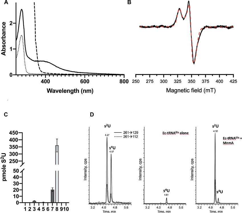
3998 Nucleic Acids Research, 2021, Vol. 49, No. 7
Downloaded from https://academic.oup.com/nar/article/49/7/3997/6179362 by guest on 23 October 2021
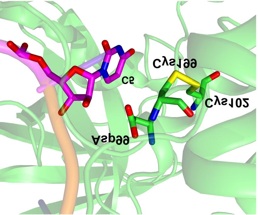
Figure 1. s2 U34-tRNA sulfuration and structure of Escherichia coli MnmA. (A) ATP-dependent reaction catalyzed by U34-tRNA thiolases through the
formation of an adenylated intermediate. Ad stands for ‘adenylate’. (B) Structure of the active site of tRNA-bound MnmA (PDB code: 2DET) with the
catalytic residues (green) and the U34 target (magenta) in stick representation. Cys102 and Cys199 form a disulfide bond. The Asp99 O2 atom is located
4.5 Å away from the Cys199 SG atom. (C) New mechanism proposed for [4Fe–4S]-dependent U34 sulfuration by E. coli MnmA.
ing residues in U34-tRNA sulfuration: Asp99, Cys102 and [Fe–S] clusters to most, if not all, [Fe–S] cellular proteins
Cys199 (making up the DXXC + C motif in the catalytic (19–21). Early work established the role of IscS in s2 U34-
domain; Supplementary Figure S1). In the currently ac- tRNA modification in E. coli and S. enterica (6,16,22,23)
cepted mechanism, Cys199 is proposed to carry the active but ruled out a role for the other ISC proteins in S. enter-
persulfide, Cys102, to assist sulfur transfer via formation of ica (24), leading to the current belief that MnmA is not an
a disulfide bond with Cys199, whereas Asp99 could act as [Fe–S] protein. This model was supported by the absence of
an acid/base catalyst to facilitate proton transfer (Supple- such a cluster within the crystal structures of as-purified E.
mentary Figure S2) (8). coli MnmA (8) and by the observation of a sulfuration ac-
IscS acts as a pleiotropic sulfur donor in the cell. In par- tivity by aerobically purified enzymes preparations under in
ticular, it forms, together with a series of protein partners vitro conditions, albeit very weak (6–8,25).
IscU, IscA, HscAB and Fdx, the well-conserved ISC [Fe– In contrast to the view presented above, we report
S] cluster biogenesis machinery, which builds and delivers here biochemical and spectroscopic results unambiguously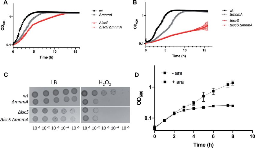
Nucleic Acids Research, 2021, Vol. 49, No. 7 3999
showing that E. coli MnmA can assemble a [4Fe–4S] cluster, plasmid. PCR analysis was used to check the absence of the
most likely chelated by Asp99, Cys102 and Cys199, and that iscUA genes and the suf operon. DNA sequence analysis of
this cluster is absolutely needed for in vitro activity. Intrigu- the isc region was carried out and MVA-dependent viabil-
ingly, we found that E. coli cells retain s2 U34 biosynthesis ity was controlled. For all constructions, transductants were
in the iscUA sufABCDSE strain, lacking functional ISC verified by PCR, using primers hybridizing upstream and
and SUF [Fe–S] cluster assembly machineries, thus suggest- downstream of the deleted gene. When necessary, antibiotic
ing an original and yet undescribed way of maturation of resistance cassettes were eliminated using plasmid pCP20 as
MnmA. described (31). Strains used in this study are listed in Sup-
plementary Table S1.
MATERIALS AND METHODS
Media and growth conditions Plasmid construction
Media used were Luria–Bertani (LB) rich medium or M9 To construct the pBADmnmA+ plasmid, the mnmA gene
Downloaded from https://academic.oup.com/nar/article/49/7/3997/6179362 by guest on 23 October 2021
minimal medium supplemented with glycerol (0.4%) and was first amplified from E. coli MG1655 chromosomal
MgSO4 (1 mM). L-Arabinose (Ara) (0.2%), casamino acids DNA using the ML40 primers, digested by EcoRI and
(0.05%), thiamine (50 g ml−1 ) and mevalonate ( MV), XhoI and cloned into the EcoRI and SalI sites of pBAD24.
1 mM were added when required. Solid media contained The mnmA gene was also subcloned into the pET28a vec-
1.5% agar. Antibiotics were used at the following concen- tor by PCR amplification from pBADmnmA+ to intro-
trations: chloramphenicol (Cam), 30 g ml−1 ; kanamycin duce the BamHI restriction site and the TEV nucleotide se-
(Kan), 30 g ml−1 ; spectinomycin (Spc), 100 g ml−1 ; and quence in 5 and the HindIII restriction site in 3 using the
ampicillin (Amp), 100 g ml−1 . Note that Kan was used ML52 primers. The PCR fragment was purified, digested
at 80 g ml−1 for transduction experiments involving the by BamHI and HindIII and cloned into the BamHI and
ΔiscS mutants as recipients. For growth at pH 7.0 or HindIII sites of pET28a. mnmA variants containing muta-
4.5, cultures grown in LB rich medium were diluted 1:100 tions were obtained by site-directed mutagenesis using the
into 200 l of LB or LB–HCl (pH 4.5) in 96-well plates. ML49–50-51 primers (NEB, E0554S). The sequence of the
Growth was monitored using a TECAN spectrophotome- expression plasmids was confirmed by sequencing. Primers
ter by recording OD600 every 10 min over 16 h at 37◦ C. and plasmids used in this study are listed in Supplementary
Tables S2 and S3, respectively.
Strains and plasmids
Synthetic lethality test
All strains used in this study are E. coli K-12 MG1655
derivatives. The mnmA mutant, FBE584, was obtained Cells were cultivated in M9 minimal medium supplemented
by in-frame deletion of mnmA and replaced with a with 0.05% casamino acids and 0.4% glycerol overnight at
kanamycin cassette (26). FBE597 was obtained from the 37◦ C. Saturated cultures were washed the next day twice
E. coli KEIO Knockout Collection (27) by P1 transduc- with PBS and then diluted 1:50 into LB rich medium with or
ing the mnmE::kan allele into MG1655 background. The without 0.2% L-arabinose. Cultures were incubated at 37◦ C
mnmE::cam mutant was constructed by deleting and re- for 8 h. The optical density at 600 nm was measured over
placing the mnmE gene with a chloramphenicol resistance time.
encoding cassette giving rise to the FBE707 strain (26).
The conditional RExBADmnmA mutant, FBE583, was
Hydrogen peroxide sensitivity test
obtained by amplifying a fragment carrying the aadA7I
(spectinomycin resistance gene) and araC genes by poly- Overnight cultures were diluted using serial dilution in ster-
merase chain reaction (PCR) from TG1 spec RExBAD ile PBS and 5 l was directly spotted onto LB plates con-
(FBE319) using the ML39 primers (28). The linear frag- taining 1 mM H2 O2 . The plates were incubated overnight
ment was then inserted upstream of the mnmA gene in at 37◦ C before growth was recorded.
E. coli MG1655 carrying the red expression plasmid
pKD46 (26). All mutants were P1-transduced into differ-
Overexpression of E. coli MnmA wild-type and the D99A-
ent E. coli K-12 backgrounds (Supplementary Table S1).
C102A and D99A-C102A-C199A variants
The CF8310 strain, a MG1655 strain derivative carry-
ing the T7 RNA polymerase encoding gene on a lambda The mnmA gene was sub-cloned into the pET15b plasmid
prophage (DE3), was used as recipient for P1 transduc- using a ligation independent cloning strategy by Eurofins to
tion of mnmA::kan, giving rise to the FBE598 strain. produce a 6His-MnmA protein construct whose 6His tag
Strain FBE605 (iscUA sufABCDSE MVA+ , kanR can be cleaved by the H3C protease. The mnmAD99A-C102A
piscR::lacZ Δlac) is a MG1655 strain derivative, which can and mnmAD99A-C102A-C199A genes were sub-cloned into the
synthesize isopentenyl diphosphate from the introduced eu- pET28a plasmid. The plasmids were transformed into E.
karyotic MVA-dependent pathway (29). Strain FBE605 was coli BL21(DE3) Star Codon Plus competent cells. One
derived from BR404 (30) as follows. First, the CamR cas- colony was used to inoculate 100–200 ml of LB medium
sette was removed from the suf locus, yielding to BR412 supplemented with ampicillin or kanamycin (50 g ml−1 )
that was used as a recipient for P1 transduction of the for MnmA wild-type and variants, respectively. 60 ml (20
iscUA::cat mutation, to generate FBE033. The Cat cas- ml) of this culture grown overnight at 37◦ C was used to
sette was removed from the iscUA locus by using the pCP20 inoculate 6 L (2 L) of LB medium supplemented with the4000 Nucleic Acids Research, 2021, Vol. 49, No. 7
same antibiotic. Cultures were grown at 37◦ C to an OD600 SEC-MALS
0.6–0.8, and overexpression was induced with 0.5 mM Iso-
Size exclusion chromatography coupled with multi-angle
propyl -D-1-thiogalactopyranoside. After 4 h incubation
light scattering (SEC-MALS) experiments were performed
at 37◦ C, cells were collected by centrifugation and stored at
using an HPLC-MALS system (Shimadzu) equipped with
−20◦ C.
light scattering detector (mini DAWN TREOS, Wyatt Tech-
nology), refractive index detector (Optilab T-rEX, Wy-
Purification of E. coli wild-type MnmA and variants att Technology) and UV detector (SPD-20A, Shimadzu).
Cells were resuspended in 50 mM Tris-HCl (pH 7.5), 500 Holo-MnmA (100 l at 2 mg ml−1 ) was injected on a Su-
mM NaCl, 10% glycerol, containing RNase A (2 g ml−1 ), perdex 200 10/300 GL increase column (GE Healthcare)
benzonase (1.6 U ml−1 , Sigma Aldrich), lysozyme (0.3 mg equilibrated in 50 mM Tris-HCl (pH 7.5), 200 mM NaCl
ml−1 ), 1 mM PMSF PhenylMethylSulfonyl Fluoride, 1 mM buffer, in the presence or absence of DTT at a flow rate of
-mercaptoethanol and disrupted by sonication. Cells de- 0.5 ml.min−1 . Molar masses of proteins were calculated us-
bris were removed by centrifugation at 25 000 rpm for 1 h ing the ASTRA 6.1 software (Wyatt Technology) using a
Downloaded from https://academic.oup.com/nar/article/49/7/3997/6179362 by guest on 23 October 2021
at 4◦ C. The supernatant was then loaded on an immobilized refractive index increment (dn/dc) value of 0.183 ml g−1 .
metal affinity Ni-NTA column (HisTrap 5 ml, GE Health-
care) equilibrated in 50 mM Tris-HCl (pH 7.5), 200 mM CD analysis
NaCl, 1 mM PMSF and eluted with a linear gradient of 0–
1 M imidazole. The proteins were collected, dialyzed twice Circular dichroism (CD) spectra were recorded on a
against 1 L of 50 mM Tris-HCl (pH 7.5), 200 mM NaCl, 1 Chirascan-plus CD Spectrometer (Applied Photophysics).
mM -mercaptoethanol in the presence of the PreScission The far ultraviolet spectra (195−260 nm) were measured at
Protease (25 g per mg wild-type MnmA) or TEV protease 20◦ C in quartz cells of 0.5 mm optical path length. The fi-
(43 g/mg MnmA variants). Wild-type MnmA was further nal concentration of MnmA proteins was 2 M in 25 mM
purified at 1 ml min−1 onto a gel filtration column (Hiload Tris-HCl (pH 7.5), 100 mM NaF. Spectra were acquired at
26/60 Superdex 200, GE Healthcare) equilibrated with 50 a resolution of 1 nm, with time per points set at 1 s and
mM Tris-HCl (pH 7.5), 200 mM NaCl, using an ÅKTA sys- a bandwidth of 1 nm. All spectra were corrected from the
tem. The as-purified proteins were concentrated to 24 mg contribution of the buffer and are an average of ten accu-
ml−1 (wild-type) with an Amicon Ultra filter device (30 kDa mulations.
cutoff, Millipore) or 4–5 mg.ml−1 (variant) with an Amicon
Ultra filter device (10 kDa cutoff, Millipore), frozen in liq- Preparation of bulk tRNA and in vitro transcribed Ec-
uid nitrogen and stored at −80◦ C. tRNAGlu
The GST–3C-protease (PreScission, a gift from S.
Mouilleron) was expressed using pGEX-2T recombinant Bulk tRNA from various E. coli strains was purified as re-
plasmids. After induction at 25◦ C with 0.1 mM IPTG for ported (35). The E. coli tRNAGlu UUC (Ec-tRNAGlu ) was
20 h, the protein was purified using glutathione–Sepharose synthesized in vitro by T7-RNA polymerase transcription
chromatography. as described in (36). Before use, the tRNA transcript was re-
folded by heating at 65◦ C for 15 min then 45◦ C for 15 min,
and finally cooling at 4◦ C for 30 min.
[Fe–S] cluster reconstitution and purification of holo-MnmA
wild-type and variants
In vitro enzyme assay
The reconstitution of the [4Fe–4S] cluster and purification
of holo-MnmA were performed in a glove box contain- Holo-MnmA (1 or 10 M) and Ec-tRNAGlu UUC (15 M)
ingNucleic Acids Research, 2021, Vol. 49, No. 7 4001
0.4 ml min−1 and at 35◦ C. A linear gradient of 0–15% upon further aerobic purification, strongly suggested the
acetonitrile in 0.1% formic acid over 7 min was used as the presence of an [Fe–S] cluster within the as-purified protein,
mobile phase. Mass spectrometry detection was carried which would be destroyed by air during purification, and
out in the multiple reactions monitoring mode to obtain prompted us to evaluate the potential of MnmA to assem-
high sensitivity and specificity. The transitions used to ble a well-defined cluster. For that purpose, in vitro reconsti-
quantify s2 U (or s4 U that has a similar fragmentation tution of the cluster was carried out by anaerobically treat-
pattern) were 261→129 and 261→112, corresponding ing the protein with ferrous iron and L-cysteine in the pres-
to the loss of ribose. Under the HPLC conditions used, ence of a cysteine desulfurase, as a source of sulfur. The
s2 U is eluted faster (4.3 min) than s4 U (4.6 min). Quan- protein was then purified by size-exclusion chromatogra-
tification was performed by external calibration. The phy, also under anaerobic conditions, leading to a homo-
5-methylaminomethyl-2-thiouridine (mnm5 s2 U) and 5- geneous brownish protein, indicative of the presence of an
carboxymethylaminomethyl-2-thiouridine (cmnm5 s2 U) [Fe–S] cluster in the protein that was subsequently called
standards were synthesized via nucleophilic substitution holo-MnmA. SEC-MALS analysis (Supplementary Figure
Downloaded from https://academic.oup.com/nar/article/49/7/3997/6179362 by guest on 23 October 2021
of 5-pivaloyloxymethyl-2-thiouridine with methylamine S3E) indicated that holo-MnmA is almost exclusively in the
or tetrabutylammonium salt of glycine, respectively, ac- monomeric form in solution, the molar mass of the protein
cording to the previously described procedures (37). The being 40.0 ± 2.6 kDa, close to the theoretical molar mass
structure and homogeneity of both nucleosides were of the monomer (41 kDa).
confirmed by 1 H and 13 C NMR, mass spectrometry and Quantification of the protein-bound iron and sulfur con-
reversed-phase HPLC analysis. tent gave 3.02 ± 0.2 Fe and 3.9 ± 0.3 S per MnmA
monomer, consistent with the presence of one [4Fe–4S] clus-
Characterization of the [4Fe–4S] cluster by UV-Visible and ter per monomer. Accordingly, the UV-visible spectrum of
EPR spectroscopies purified holo-MnmA displays a broad absorption band at
around 410 nm that is specifically characteristic for the pres-
UV-visible absorption spectra were recorded in quartz cu- ence of a [4Fe–4S] cluster (Figure 2A). Anaerobic addition
vettes (1 cm optic path) under anaerobic conditions in a of dithionite to holo-MnmA led to a rapid decrease in the
glove box on a Cary 100 UV-visible spectrophotometer intensity of the 410 nm absorption band, suggesting a fast
equipped with optical fibers. reduction of the cluster (Figure 2A). The frozen solutions
The EPR continuous wave measurements were per- of holo-MnmA, before and after reduction, were analyzed
formed on a spectrometer Bruker ELEXSYS-E500 operat- by continuous wave EPR spectroscopy at different tempera-
ing at 9.38 GHZ, equipped with SHQE cavity cooled by tures (Figure 2B and Supplementary Figure S3F). No EPR
a helium flow cryostat ESR 900 Oxford Instruments. The signal was observed for holo-MnmA solution, suggesting
EPR spectra of frozen solution of 220 M E. coli MmnA an S = 0 [4Fe–4S]2+ state, and excluding S = 1/2 param-
with reconstituted cluster, without treatment and reduced agnetic clusters such as [4Fe–4S]+ or [3Fe-4S]+ . Upon re-
with 5.5 mM dithionite in 50 mM Tris-HCl (pH 7.5), 200 duction, the EPR spectrum of the protein recorded at 15 K
mM NaCl was recorded at 10, 15 and 20 K, under non- exhibited a signal with a rhombic g tensor (gx = 1.88, gy =
saturating conditions and using the following parameters: 1.92, gz = 2.045) characteristic of the S = 1/2 [4Fe–4S]+
a microwave power of 2–10 mW, a modulation amplitude state (Figure 2B). Temperature variations allowed to dis-
of 0.4 mT, a modulation frequency of 100 kHz and an ac- criminate between a [2Fe-2S]+ cluster and a [4Fe–4S]+ clus-
cumulation of 4 scans. For the quantification of unpaired ter because of their different spin relaxation behaviors (38).
spins, a Cu-EDTA standard sample (200 M) was used. At 40 K, the EPR lines of the reduced holo-MnmA sam-
The simulation of the EPR spectrum was performed with ple broadened beyond recognition (Supplementary Figure
the Easyspin software (http://www.easyspin.org/). S3F), in agreement with the presence of a [4Fe–4S]+ cluster,
which usually cannot be detected above 35 K, in contrast
RESULTS to [2Fe–2S]+ clusters. Quantification with Cu-EDTA stan-
dard solution indicated the presence of around 0.5 spin per
Escherichia coli MnmA binds a [4Fe–4S] cluster
holo-MnmA monomer.
Escherichia coli MnmA with an N-terminal histidine tag
was purified under aerobic conditions using Ni-affinity
The [4Fe–4S] cluster is required for the tRNA sulfuration ac-
chromatography. The faint brownish color and the weak ab-
tivity of E. coli MnmA
sorbance at 410 nm suggested the presence of an [Fe–S] clus-
ter (Supplementary Figure S3A). The tag was then removed The sulfuration activity of E. coli MnmA was tested using
using the H3C protease and the protein was further purified in vitro transcribed Ec-tRNAGlu as a substrate in the pres-
by size-exclusion chromatography, which led to a mixture of ence of ATP, Mg2+ and inorganic sulfide, a sulfur donor
monomeric and dimeric species, as shown by SEC-MALS often used in enzyme sulfuration assays (39–41). The thi-
analysis (Supplementary Figure S3B and S3C). Because the olated nucleosides were quantified after digestion of the
monomer/dimer ratio increases with DTT concentration tRNA products by nuclease P1 and alkaline phosphatase.
(Supplementary Figure S3B), the dimer is formed by disul- The s2 U product was first identified by its elution position
fide bonds between two monomers. As-purified MnmA re- after HPLC-coupled mass spectrometry (MS), then quan-
tained a faint brownish color and its UV-visible spectrum tified by MS/MS using a synthetic s2 U standard (Figure
exhibited a band at around 410 nm (Supplementary Fig- 2C). The fragment spectrum corresponding to the loss of
ure S3D). These unexpected features, which disappeared sugar (-132) was monitored (Figure 2D). Holo-MnmA (14002 Nucleic Acids Research, 2021, Vol. 49, No. 7
Downloaded from https://academic.oup.com/nar/article/49/7/3997/6179362 by guest on 23 October 2021
Figure 2. Spectroscopic and enzymatic characterizations of E. coli MnmA. (A) UV-visible spectra of 40 M apo-MnmA (dotted line), 40 M holo-MnmA
(thick line) and reduced holo-MnmA after 20 min of incubation with 1 mM dithionite (dashed line). The spectra were recorded with 40 M protein in
50 mM Tris-HCl (pH 7.5), 200 mM NaCl, 5 mM DTT in 1 cm (apo-MnmA and reduced holo-MnmA) or 1 mm (holo-MnmA) pathlength cuvettes and
normalized. (B) X-band EPR spectrum (10 mW microwave power; modulation amplitude of 0.4 mT) of 220 M holo-MnmA reduced with 5.5 mM
dithionite at 15 K. The experimental (black solid line) and simulated (dashed line) spectra are superimposed. The cluster was simulated with the following
values of the g-tensor: gx = 1.880, gy = 1.920, gz = 2.045 and Gaussian distribution deviations (gx ) = 0.04, (gy ) = 0.02, (gz ) = 0.04. (C) In vitro
tRNA sulfuration activity tests of MnmA under anaerobic conditions. After tRNA digestion, s2 U was separated by HPLC-MS/MS and quantified using
a synthetic s2 U standard. The data shown are mean values based on three different experiments, with the standard error of the mean indicated as a bar. In
vitro transcribed Ec-tRNAGlu (15 M) was incubated for 1 h at 37◦ C in 50 mM Tris (pH 7.5), 200 mM NaCl with apo or holo-MnmA (wild-type or
mutant) in the presence or absence of 1 mM Na2 S, 2.5 mM MgCl2 , 0.25 mM ATP. Ec-tRNAGlu alone 15 M (1), apo-MnmA 1 M (2) or 10 M (3),
1 M holo-MnmA and no Na2 S (4), no MgCl2 (5) or no ATP (6), holo-MnmA 1 M (7) or 10 M (8), 1 M holo-MnmAD99A-C102A mutant (9) or 1
M holo-MnmAD99A-C102A-C199A mutant (10). (D) HPLC MS/MS detection of s2 U and s4 U (m/z 261). Samples were analyzed using the two transitions
261→129 and 261→112; the most intense one corresponds to the loss of the ribose moiety. Left: mixture of s2 U and s4 U synthetic standards (0.5 pmole
injected); middle: Ec-tRNAGlu alone after hydrolysis; right: Ec-tRNAGlu (15 M) after incubation with holo-MnmA (10 M) in the presence of Na2 S,
ATP, MgCl2 and hydrolysis.
M) was able to catalyze sulfuration of tRNA (15 M) in MnmA is necessary for E. coli to resist to stress
contrast to apo-MnmA (1 or 10 M) (Figure 2C). More-
It was previously shown that mnmA mutation in E. coli leads
over, the sulfur atom that was inserted into the nucleoside
to reduced growth rate (14). We reproduced this observation
is derived exclusively from the inorganic sulfide salt, and
(Figure 3A) and also noticed that the mutant exhibited a
not from the [4Fe–4S] cluster, since no sulfuration could
small colony phenotype when plated on rich medium (Fig-
be observed in the absence of sulfide. These control ex-
ure 3C). We constructed the mnmA strain and checked
periments indicated that the cluster and inorganic sulfide
by HPLC-MS that the bulk tRNA from this strain did not
were required for the tRNA sulfuration activity of MnmA.
contain mnm5 s2 U nor cmnm5 s2 U (Table 1). By monitoring
Moreover, the amount of product formed increased with
growth in liquid culture, we found that the mnmA strain
enzyme concentration (1 M versus 10 M in Figure 2C).
showed slower growth in the presence of a mild acid stress
Mg-ATP was also required, in agreement with its role in ac-
(i.e. growth medium at pH 4.5) (Figure 3B) and that it ex-
tivating the substrate via adenylation of the C2 oxygen atom
hibited hypersensitivity to H2 O2 (Figure 3C). Note that sur-
(Figure 1A and Supplementary Figure S2) (8).Nucleic Acids Research, 2021, Vol. 49, No. 7 4003
Downloaded from https://academic.oup.com/nar/article/49/7/3997/6179362 by guest on 23 October 2021
Figure 3. Phenotypes of mnmA and derivative strains. (A) Growth in LB pH 7.0 medium. Strains studied are wild-type (wt) (FBE051), ΔmnmA (FBE584),
ΔiscS (FBE653) and ΔiscS ΔmnmA (FBE703). Data are representative of three independent experiments (n = 3). (B) Growth in LB–HCl (pH 4.5). Strains
studied are wild-type (wt) (FBE051), ΔmnmA (FBE584), ΔiscS (FBE653) and ΔiscS ΔmnmA (FBE703), (n=3). (C) Hypersensitivity of ΔmnmA to H2 O2
(1 mM). Strains tested are wt, ΔmnmA, ΔiscS and ΔiscS ΔmnmA. Each spot represents a 10-fold serial dilution (n = 3). (D) The ΔmnmE and ΔmnmA
mutations are lethal synthetic in E. coli. The RExBADmnmA ΔmnmE strain (FBE601) was grown in LB rich medium without (- ara) or with (+ ara) 0.2%
L-arabinose, (n = 3).
Table 1. Analysis of s2 C, mnm5 s2 U and cmnm5 s2 U content (in pmoles) of the mnmA strain, and the combination of both dele-
of bulk tRNA (15 M) from various strains (mean of three measurements) tions failed to enhance the defect.
s2 C cmnm5 s2 U mnm5 s2 U
Besides hypersensitivity to H2 O2 , we wished to set-up
another MnmA-associated phenotypic test. For this, we
Wild-type 9.5 ± 1.7 107.6 ± 39.8 1080.7 ± 647.0 took advantage of the synthetic lethality between mnmA
mnmA 14.8 ± 2.2 0.0 ± 0.1 1.9 ± 1.0 and mnmE mutations. MnmE, along with MnmG, adds
iscUA 0.0 ± 0.0 64.5 ± 20.9 1421.9 ± 601.7
sufABCDSE the aminomethyl and carboxymethylaminomethyl groups
ttcA 0.4 ± 0.1 68.0 ± 44.4 1756.1 ± 1003.5 to the C5 position of U34 (42). We constructed a strain
named RExBADmnmA, in which expression of the mnmA
gene was under the control of an arabinose inducible pro-
vival plate assays run in parallel showed that the viability of moter and tested whether it could accommodate a mnmE
the mnmA strain was not impaired at pH 4.5. mutation. We observed that the mnmE mutation could
be introduced by transduction only when arabinose was
added to the medium (Figure 3D). This confirmed that a
U34-tRNA modification pathway in E. coli
strain lacking both mnmA and mnmE is not viable, as pre-
Biochemical studies together with a genetic approach cou- viously shown by using a strain, in which the mnmA gene
pled with mass spectrometry (ribonucleome analysis) have was deleted and the mnmE gene put under the control of an
shown that MnmA receives sulfur from the IscS cysteine arabinose inducible promoter (42).
desulfurase (6,22) and the TusABCDE sulfur relay path- Then, we reasoned that if iscS was required for MnmA
way (7). Thus, the prediction is that the ΔiscS strain should to function, then a strain carrying both ΔiscS and ΔmnmE
exhibit defects similar to those of the mnmA strain, as should not be viable. Therefore, we asked whether we could
shown in S. enterica (16). The ΔiscS mutation was found introduce a ΔiscS mutation into a mnmE mutant. The re-
to cause extreme sensitivity to pH 4.5, even to a larger ex- sult was negative, suggesting that a ΔiscS mutation recapit-
tent than that caused by the mnmA mutation (Figure 3B). ulated the mnmA mutation, in agreement with IscS pro-
Such a feature has never been described for IscS so far. viding the sulfur atom to MnmA.
Introducing the mnmA mutation in a ΔiscS background Taken together, these genetic analyses showed that the
did not worsen the defect (Figure 3B), in line with the idea mnmA gene is crucial for E. coli to grow under both bal-
that MnmA depends upon IscS for getting sulfur. Similarly, anced and stress conditions, and established its close func-
when challenging the strains with H2 O2 (Figure 3C), the tional interaction with iscS, in agreement with early studies
ΔiscS strain displayed hypersensitivity comparable to that in S. enterica (23).4004 Nucleic Acids Research, 2021, Vol. 49, No. 7
The ligands of the [4Fe–4S] cluster are most likely Asp99, but can be grown by introducing the eukaryotic [Fe–S]-
Cys102 and Cys199 independent mevalonate-dependent isoprenoid biosynthe-
sis pathway (29) to supply isopentenyl diphosphate, which
The D99A, C102S and C199A mutants of E. coli MnmA
is essential and normally synthesized by the [Fe–S] depen-
were reported to lack in vitro U34-tRNA sulfuration ac-
dent IspG/IspH proteins. First, we observed that s2 C was
tivity using cysteine as the sulfur donor, and the IscS and
absent in bulk tRNA from the mutated strain (Table 1), in
Tus proteins as sulfur carriers (8). We produced the dou-
full agreement with TtcA, the unique enzyme responsible
ble D99A-C102A and the triple D99A-C102A-C199A mu-
for s2 C formation, requiring a [4Fe–4S] cluster (39) assem-
tants (Supplementary Figure S4). Both mutants were cor-
bled by the ISC system (24). Then, we compared the content
rectly folded, as shown by their circular dichroism spec-
in mnm5 s2 U and cmnm5 s2 U in bulk tRNA from a wild-type
trum, which did not differ from that of the wild-type protein
and the ΔiscUA ΔsufABCDSE strains. Unexpectedly, we
(Supplementary Figure S4F). After cluster reconstitution,
found that the amounts of mnm5 s2 U and cmnm5 s2 U from
they both almost completely lost the absorption band at 410
bulk tRNA in the mutant strain were comparable to those
nm (Supplementary Figure S4G) and were shown to con-
Downloaded from https://academic.oup.com/nar/article/49/7/3997/6179362 by guest on 23 October 2021
in the wild-type strain. This suggests that [Fe–S] cluster bio-
tain very little Fe, namely 0.46 ± 0.03 and 0.25 ± 0.05 iron
genesis of the E. coli MnmA protein can occur in the ab-
per MnmA monomer, respectively. Finally, they did not ex-
sence of the ISC and SUF systems.
hibit any catalytic activity (Figure 2C, columns 9 and 10) in
standard in vitro assays with sulfide used as a sulfur donor.
Therefore, the mutagenesis results point out Asp99, Cys102 DISCUSSION
and Cys109 as being involved in cluster binding and catal-
We show here that MnmA, the only E. coli enzyme respon-
ysis (Figure 1B) (8). But one cannot unambiguously con-
sible for the sulfuration of U34 at C2 position in tRNAs
clude on the in vitro analysis of only the double and triple
(Figure 1A), assembles a [Fe–S] cluster. Moreover, the pres-
mutant variants that all three residues contribute to liga-
ence of this cluster proved essential for activity, under stan-
tion. Yet, we did not analyze the single variants because,
dard sulfuration assays previously used for other tRNA-
in many instances, a single mutation of a cluster ligand was
sulfurating [Fe–S] enzymes such as TtcA (39) and TtuA
shown to be not sufficient to disrupt cluster ligation (43,44).
(41). As a further confirmation of the functionality of the
Therefore, in addition, we tested the functional importance
cluster, no in vitro activity could be obtained using MmnA
of the Asp99, Cys102 and Cys109 residues by changing each
variants, in which the residues that presumably bind the
of them to alanine and testing the activity of the result-
cluster (Asp99, Cys102 and Cys199) were changed into ala-
ing single mutants in vivo using the series of phenotypic
nine. The occurrence of a [4Fe–4S] cluster was clearly es-
tests described above (Supplementary Figure S5). When ex-
tablished by Fe and S quantification and from UV-visible
pressed in trans from plasmids, all mutated variants, namely
and EPR spectroscopic characteristic features, after clus-
D99A, C102A and C109A failed to (i) suppress growth
ter reconstitution under anaerobic conditions. Interestingly,
rate defects of the mnmA recipient in rich medium (Sup-
residual cluster was also unambiguously observed within
plementary Figure S5A, top), (ii) suppress hypersensitivity
the as-purified protein before any chemical treatment, indi-
of the mnmA strain to acid (Supplementary Figure S5A,
cating that MnmA carried a cluster within the cell as well.
bottom) and oxidative stress (Supplementary Figure S5B)
Shigi et al. recently reported that MnmA from Ther-
and (iii) allow growth in the absence of arabinose in the
mus thermophilus also contains an active [4Fe–4S] cluster
RExBADmnmA mnmE containing background (Supple-
(43). However, the authors proposed that MnmA proteins
mentary Figure S5C). Note that expression of all three alle-
should be subdivided into [Fe–S]-containing and [Fe–S]-
les gave rise to large amounts of soluble protein so that the
independent types (43). In the first C-type class, the three
possibility of a destabilizing effect of the introduced muta-
cysteines from the CXXC + C motif would ligate the clus-
tions was ruled out (Supplementary Figure S5D). Hence,
ter, such as in the T. thermophilus enzyme. The second D-
based upon their inability to complement different defects
type class would harbor a DXXC + C motif instead, like
of the mnmA mutant, we concluded that substitution of
in the E. coli enzyme, which would be unable to bind a
Asp99, Cys102 or Cys109 by alanine yielded a nonfunc-
cluster. However, our results are strongly in agreement with
tional MnmA protein. Altogether, the simplest interpreta-
Asp99, Cys102 and Cys199 of the DXXC + C motif being
tion of the in vitro characterization of the mutants and the
the ligands of the cluster of E. coli MnmA. Indeed, both the
in vivo complementation assays is that Asp99, Cys102 and
D99A-C102A and D99A-C102A-C199A mutants were un-
Cys109 are the ligands of the [4Fe–4S] cluster.
able to assemble a [4Fe–4S] cluster in vitro, in agreement
with the observation that the D99A, C102A and C199A
mutants were not able to rescue the growth phenotype of the
Biosynthesis of mnm5 s2 U and cmnm5 s2 U in tRNAs occurs
mnmA strain in vivo. It is interesting to note that Asp99,
in the absence of the ISC and SUF [Fe–S] cluster biogenesis
Cys102 and Cys199 are in close proximity to each other in
systems
the crystal structure of apo-MnmA (8), the only 3D struc-
Since maturation of [Fe–S] proteins in E. coli relies on the ture available so far, and with the appropriate configuration
two ISC and SUF machineries, we analyzed the s2 U con- for binding a [Fe–S] cluster (Figure 1B). In fact, amino acids
tent in bulk tRNA from the ΔiscUA ΔsufABCDSE strain other than cysteine can act as [Fe–S] ligands, such as histi-
(29) after tRNA digestion and HPLC-MS analysis (Ta- dine, glutamate, arginine and threonine (45). There are also
ble 1) to check whether MnmA maturation also uses these precedents for aspartate as a ligand of a [4Fe–4S] or a [2Fe-
pathways. The ΔiscUA ΔsufABCDSE strain is not viable 2S] cluster, such as in ferrodoxin from hyperthermophilicNucleic Acids Research, 2021, Vol. 49, No. 7 4005
archaea (46,47), in a nitrogenase-like enzyme named pro- presence of a small fraction of holo form in the as-purified
tochlorophyllide reductase (48,49), in a transcriptional reg- MnmA, as we observed it here.
ulator (50) and in IscA, involved in [Fe–S] cluster assembly To date, maturation of all tested [Fe–S] proteins in E.
(51). coli has been found to depend upon ISC, SUF or both
Given that Ncs6-type proteins also use a cluster for s2 U34 systems. Therefore, after having shown that MnmA con-
formation in tRNAs (52) (PDB code: 6SCY), the biochem- tains an [Fe–S] cluster, we reinvestigated the contribution
ical findings presented here lead us to propose that this sul- of ISC and/or SUF to mature MnmA. Surprisingly, the
furation reaction depends on a [4Fe–4S] enzyme in all or- mnm5 s2 U/cmnm5 s2 U content in tRNAs of the ΔiscUA
ganisms and in mitochondria (9) (Supplementary Figure ΔsufABCDSE strain was comparable to that in wild-type
S1). More specifically, we propose that C-type and D-type cells, arguing against the involvement of either machinery
MnmA are all [Fe–S] enzymes. for maturation of MnmA. This result is consistent with pre-
In Figure 1C, we propose a tentative mechanism for the vious work by Björk’s (24) and Leimkühler’s groups (59),
holo-MnmA form, in which the cluster, adjacent to the who showed that ISC and SUF were dispensable for s2 U34
biosynthesis. Thus, to explain the s2 U content of tRNAs in
Downloaded from https://academic.oup.com/nar/article/49/7/3997/6179362 by guest on 23 October 2021
substrate, serves to bind and activate a hydrosulfide nucle-
ophilic substrate for subsequent attack on C2 from U34 the mutated strain, we are forced to entertain the possibility
to displace the AMP leaving group and form the final C– for MnmA to be targeted by an as yet unknown [Fe–S] clus-
S bond in the product. A cluster with only three protein- ter biogenesis pathway, which our current studies are aiming
bound ligands is appropriate for such a function since it pro- to identify.
vides a free coordination site on the fourth Fe atom where
hydrosulfide can bind (53). We and others previously pro-
DATA AVAILABILITY
posed a similar mechanism with the involvement of a [4Fe-
5S] intermediate for TtuA, another thiouridine synthetase All data are available in the manuscript; strains and con-
that targets position 54 in tRNA (41,54,55), and for TtcA structs are available on request.
responsible for the formation of s2 C32-tRNA (39). While
more studies are required to firmly establish such a mecha-
nism for tRNA thiolation enzymes, we have recently struc- SUPPLEMENTARY DATA
turally characterized a relevant [4Fe-5S] catalytic interme- Supplementary Data are available at NAR Online.
diate during the desulfuration of 4-thiouracil by thiouracil
desulfidase TudS (56), which thus provides a unique prece-
dent for this class of intermediate clusters. Obviously, this ACKNOWLEDGEMENTS
mechanism excludes persulfides as key reaction intermedi- We thank the French EPR CNRS Facility, Infrastructure
ates, as proposed previously, since the cysteines of the active de Recherche Renard (IR 3443); the Macromolecular Inter-
site proposed to carry the persulfide function are engaged as action Platform of I2BC for use of its facilities and exper-
ligands of the [4Fe–4S] cluster. tise; Ludovic Pecqueur for maintenance of the glove boxes;
As a consequence of the results reported here, we need Bruno Faivre for advice in the purification experiments;
to examine why an [Fe–S] cluster in E. coli MnmA was Djemel Hamdane for assistance in CD experiments, Simon
previously excluded, especially considering its essential cat- Arragain and Ornella Bimai for fruitful discussions that ini-
alytic role. First, the conclusion that MnmA could not be an tiated this project; and the SAMe Unit members for discus-
[Fe–S] enzyme came from an early study examining the s4 U sion, technical help and suggestions.
and mnm5 s2 U levels in ISC defective backgrounds; i.e. the
ΔiscU, ΔhscA, Δfdx and ΔiscA strains showed wild-type
like levels, while both s2 C and ms2 io6 A levels were found FUNDING
altered (24). It was concluded that downstream IscS, which Centre National de la recherche Scientifique and French
is the shared sulfur source for the four reactions, two dis- State Program ‘Investissements d’Avenir’ [LABEX DY-
tinct routes were likely to occur for biosynthesis of thiolated NAMO, ANR-11-LABX-0011, IBEID ANR-10-LABX-
nucleotides in tRNA: the [Fe–S]-dependent route, responsi- 62]; Institut Pasteur. Funding for open access charge:
ble for the formation of s2 C and ms2 io6 A, and the [Fe–S]- LABEX DYNAMO.
independent route, leading to s4 U and mnm5 s2 U. Therefore, Conflict of interest statement. None declared.
previous MnmA preparations, purified exclusively under
aerobic conditions, have not been analyzed for the presence
of small amounts of protein-bound clusters (6–8), as we did REFERENCES
here. It is well known that most clusters, and more specif- 1. El Yacoubi,B., Bailly,M. and de Crecy-Lagard,V. (2012) Biosynthesis
ically those with a labile coordination site, like in aconi- and function of posttranscriptional modifications of transfer RNAs.
tase (57) or radical-SAM enzymes (58), are very sensitive Annu. Rev. Genet., 46, 69–95.
2. Boccaletto,P., Machnicka,M.A., Purta,E., Piatkowski,P., Baginski,B.,
to air and degrade during purification, generally leading to Wirecki,T.K., de Crecy-Lagard,V., Ross,R., Limbach,P.A., Kotter,A.
colorless protein solutions. Second, while these seemingly et al. (2018) MODOMICS: a database of RNA modification
‘cluster-free’ enzyme preparations were active, the reported pathways. 2017 update. Nucleic Acids Res., 46, D303–D307.
activities were very weak, despite very large, far from cat- 3. Agris,P.F., Narendran,A., Sarachan,K., Vare,V.Y.P. and Eruysal,E.
(2017) The importance of being modified: the role of RNA
alytic, amounts of protein used (MnmA:tRNA ratios of 1:1 modifications in translational fidelity. Enzymes, 41, 1–50.
(6), 2.1:1 (7), 4:1 (8) or 6.4:1 (25). It is tempting to sug- 4. Ranjan,N. and Rodnina,M.V. (2016) tRNA wobble modifications and
gest that any measured activity might have been due to the protein homeostasis. Translation (Austin), 4, e1143076.4006 Nucleic Acids Research, 2021, Vol. 49, No. 7
5. Schaffrath,R. and Leidel,S.A. (2017) Wobble uridine 25. Black,K.A. and Dos Santos,P.C. (2015) Abbreviated pathway for
modifications––a reason to live, a reason to die?! RNA Biol., 14, biosynthesis of 2-thiouridine in Bacillus subtilis. J. Bacteriol., 197,
1209–1222. 1952–1962.
6. Kambampati,R. and Lauhon,C.T. (2003) MnmA and IscS are 26. Datsenko,K.A. and Wanner,B.L. (2000) One-step inactivation of
required for in vitro 2-thiouridine biosynthesis in Escherichia coli. chromosomal genes in Escherichia coli K-12 using PCR products.
Biochemistry, 42, 1109–1117. Proc. Natl. Acad. Sci. USA, 97, 6640–6645.
7. Ikeuchi,Y., Shigi,N., Kato,J., Nishimura,A. and Suzuki,T. (2006) 27. Baba,T., Ara,T., Hasegawa,M., Takai,Y., Okumura,Y., Baba,M.,
Mechanistic insights into sulfur relay by multiple sulfur mediators Datsenko,K.A., Tomita,M., Wanner,B.L. and Mori,H. (2006)
involved in thiouridine biosynthesis at tRNA wobble positions. Mol. Construction of Escherichia coli K-12 in-frame, single-gene knockout
Cell, 21, 97–108. mutants: the Keio collection. Mol. Syst. Biol., 2, 2006.0008.
8. Numata,T., Ikeuchi,Y., Fukai,S., Suzuki,T. and Nureki,O. (2006) 28. Roux,A., Beloin,C. and Ghigo,J.M. (2005) Combined inactivation
Snapshots of tRNA sulphuration via an adenylated intermediate. and expression strategy to study gene function under physiological
Nature, 442, 419–424. conditions: application to identification of new Escherichia coli
9. Umeda,N., Suzuki,T., Yukawa,M., Ohya,Y., Shindo,H., Watanabe,K. adhesins. J. Bacteriol., 187, 1001–1013.
and Suzuki,T. (2005) Mitochondria-specific RNA-modifying 29. Loiseau,L., Gerez,C., Bekker,M., Ollagnier-de-Choudens,S., Py,B.,
enzymes responsible for the biosynthesis of the wobble base in Sanakis,Y., Teixeira-de-Mattos,J., Fontecave,M. and Barras,F. (2007)
Downloaded from https://academic.oup.com/nar/article/49/7/3997/6179362 by guest on 23 October 2021
mitochondrial tRNAs. Implications for the molecular pathogenesis of ErpA, an iron–sulfur (Fe–S) protein of the A-type essential for
human mitochondrial diseases. J. Biol. Chem., 280, 1613–1624. respiratory metabolism in Escherichia coli. Proc. Natl. Acd. Sci. USA,
10. Dewez,M., Bauer,F., Dieu,M., Raes,M., Vandenhaute,J. and 104, 13626–13631.
Hermand,D. (2008) The conserved Wobble uridine tRNA thiolase 30. Roche,B., Huguenot,A., Barras,F. and Py,B. (2015) The iron-binding
Ctu1–Ctu2 is required to maintain genome integrity. Proc. Natl. CyaY and IscX proteins assist the ISC-catalyzed Fe-S biogenesis in
Acad. Sci. USA, 105, 5459–5464. Escherichia coli. Mol. Microbiol., 95, 605–623.
11. Noma,A., Sakaguchi,Y. and Suzuki,T. (2009) Mechanistic 31. Cherepanov,P.P. and Wackernagel,W. (1995) Gene disruption in
characterization of the sulfur-relay system for eukaryotic Escherichia coli: TcR and KmR cassettes with the option of
2-thiouridine biogenesis at tRNA wobble positions. Nucleic Acids Flp-catalyzed excision of the antibiotic-resistance determinant. Gene,
Res., 37, 1335–1352. 158, 9–14.
12. Nakai,Y., Nakai,M. and Hayashi,H. (2008) Thio-modification of 32. Bradford,M.M. (1976) A rapid and sensitive method for the
yeast cytosolic tRNA requires a ubiquitin-related system that quantitation of microgram quantities of protein utilizing the principle
resembles bacterial sulfur transfer systems. J. Biol. Chem., 283, of protein-dye binding. Anal. Biochem., 72, 248–254.
27469–27476. 33. Beinert,H. (1983) Semi-micro methods for analysis of labile sulfide
13. Grosjean,H., Breton,M., Sirand-Pugnet,P., Tardy,F., Thiaucourt,F., and of labile sulfide plus sulfane sulfur in unusually stable iron-sulfur
Citti,C., Barre,A., Yoshizawa,S., Fourmy,D., de Crecy-Lagard,V. proteins. Anal. Biochem., 131, 373–378.
et al. (2014) Predicting the minimal translation apparatus: lessons 34. Fish,W.W. (1988) Rapid colorimetric micromethod for the
from the reductive evolution of mollicutes. PLos Genet., 10, e1004363. quantitation of complexed iron in biological samples. Methods
14. Armengod,M.E., Meseguer,S., Villarroya,M., Prado,S., Enzymol., 158, 357–364.
Moukadiri,I., Ruiz-Partida,R., Garzón,M.J., Navarro-González,C. 35. Buck,M., Connick,M. and Ames,B.N. (1983) Complete analysis of
and Martı́nez-Zamora,A. (2014) Modification of the wobble uridine tRNA-modified nucleosides by high performance liquid
in bacterial and mitochondrial tRNAs reading NNA/NNG triplets chromatography: the 29 modified nucleosides of Salmonella
of 2-codon boxes. RNA Biol., 12, 1495–1507. typhimurium and Escherichia coli. Anal. Biochem., 129, 1–13.
15. Nilsson,K., Jager,G. and Björk,G.R. (2017) An unmodified wobble 36. Milligan,J.F., Groebe,D.R., Witherell,G.W. and Uhlenbeck,O.C.
uridine in tRNAs specific for glutamine, lysine, and glutamic acid (1987) Oligoribonucleotide synthesis using T7 RNA polymerase and
from Salmonella enterica serovar typhimurium results in synthetic DNA templates. Nucleic Acids Res., 15, 8783–8798.
nonviability-due to increased missense errors? PLoS One, 12, 37. Bartosik,K. and Leszczynska,G. (2015) Synthesis of various
e0175092. substituted 5-methyluridines (xm5U) and 2-thiouridines (xm5s2U)
16. Nilsson,K., Lundgren,H.K., Hagervall,T.G. and Bjork,G.R. (2002) via nucleophilic substitution of
The cysteine desulfurase IscS is required for synthesis of all five 5-pivaloyloxymethyluridine/2-thiouridine. Tetrahedron Lett., 56,
thiolated nucleosides present in tRNA from Salmonella enterica 6593–6597.
serovar typhimurium. J. Bacteriol., 184, 6830–6835. 38. Freibert,S.-A., Weiler,B.D., Bill,E., Pierik,A.J., Mühlenhoff,U. and
17. Kobayashi,K., Ehrlich,S.D., Albertini,A., Amati,G., Andersen,K.K., Lill,R. (2018) Biochemical reconstitution and spectroscopic analysis
Arnaud,M., Asai,K., Ashikaga,S., Aymerich,S., Bessieres,P. et al. of iron–sulfur proteins. Methods Enzymol., 599, 197–226.
(2003) Essential Bacillus subtilis genes. Proc. Natl. Acad. Sci. USA, 39. Bouvier,D., Labessan,N., Clemancey,M., Latour,J.M., Ravanat,J.L.,
100, 4678–4683. Fontecave,M. and Atta,M. (2014) TtcA a new tRNA-thioltransferase
18. Kotera,M., Bayashi,T., Hattori,M., Tokimatsu,T., Goto,S., with an Fe-S cluster. Nucleic Acids Res., 42, 7960–7970.
Mihara,H. and Kanehisa,M. (2010) Comprehensive genomic analysis 40. Chen,M., Asai,S.I., Narai,S., Nambu,S., Omura,N., Sakaguchi,Y.,
of sulfur-relay pathway genes. Genome Inform, 24, 104–115. Suzuki,T., Ikeda-Saito,M., Watanabe,K., Yao,M. et al. (2017)
19. Fontecave,M., Py,B., Ollagnier de Choudens,S. and Barras,F. (2008) Biochemical and structural characterization of oxygen-sensitive
From iron and cysteine to iron-sulfur clusters: the biogenesis protein 2-thiouridine synthesis catalyzed by an iron-sulfur protein TtuA.
machineries. EcoSal Plus, 3, doi:10.1128/ecosalplus.3.6.3.14. Proc. Natl. Acad. Sci. USA, 114, 4954–4959.
20. Roche,B., Aussel,L., Ezraty,B., Mandin,P., Py,B. and Barras,F. (2013) 41. Arragain,S., Bimai,O., Legrand,P., Caillat,S., Ravanat,J.L., Touati,N.,
Iron/sulfur proteins biogenesis in prokaryotes: formation, regulation Binet,L., Atta,M., Fontecave,M. and Golinelli-Pimpaneau,B. (2017)
and diversity. Biochim. Biophys. Acta, 1827, 455–469. Nonredox thiolation in tRNA occurring via sulfur activation by a
21. Blanc,B., Gerez,C. and Ollagnier de Choudens,S. (2015) Assembly of [4Fe-4S] cluster. Proc. Natl. Acad. Sci. USA, 114, 7355–7360.
Fe/S proteins in bacterial systems: biochemistry of the bacterial ISC 42. Armengod,M.E., Moukadiri,I., Prado,S., Ruiz-Partida,R.,
system. Biochim. Biophys. Acta, 1853, 1436–1447. Benitez-Paez,A., Villarroya,M., Lomas,R., Garzon,M.J.,
22. Lauhon,C.T. (2002) Requirement for IscS in biosynthesis of all Martinez-Zamora,A., Meseguer,S. et al. (2012) Enzymology of
thionucleosides in Escherichia coli. J. Bacteriol., 184, 6820–6829. tRNA modification in the bacterial MnmEG pathway. Biochimie, 94,
23. Lundgren,H.K. and Björk,G.R. (2006) Structural alterations of the 1510–1520.
cysteine desulfurase IscS of Salmonella enterica serovar typhimurium 43. Shigi,N., Horitani,M., Miyauchi,K., Suzuki,T. and Kuroki,M. (2020)
reveal substrate specificity of IscS in tRNA thiolation. J. Bacteriol., An ancient type of MnmA protein is an iron-sulfur cluster-dependent
188, 3052–3062. sulfurtransferase for tRNA anticodons. RNA, 26, 240–250.
24. Leipuviene,R., Qian,Q. and Björk,G.R. (2004) Formation of 44. Hewitson,K.S., Ollagnier-de Choudens,S., Sanakis,Y., Shaw,N.M.,
thiolated nucleosides present in tRNA from Salmonella enterica Baldwin,J.E., Münck,E., Roach,P.L. and M.,F. (2002) The iron-sulfur
serovar typhimurium occurs in two principally distinct pathways. J. center of biotin synthase: site-directed mutants. J. Biol. Inorg. Chem.,
Bacteriol., 186, 758–766. 1–2, 83–93.Nucleic Acids Research, 2021, Vol. 49, No. 7 4007
45. Bak,D.W. and Elliott,S.J. (2014) Alternative FeS cluster ligands: 52. Liu,Y., Vinyard,D.J., Reesbeck,M.E., Suzuki,T.,
tuning redox potentials and chemistry. Curr. Opin. Chem. Biol., 19, Manakongtreecheep,K., Holland,P.L., Brudvig,G.W. and Söll,D.
50–58. (2016) A [3Fe-4S] cluster is required for thiolation in archaea and
46. Calzolai,L., Gorst,C.M., Zhao,Z.H., Teng,Q., Adams,M.W. and La eukaryotes. Proc. Natl. Acad. Sci. USA, 113, 12703–12708.
Mar,G.N. (1995) 1H NMR investigation of the electronic and 53. Flint,D.H. and Allen,R.M. (1996) Iron-sulfur proteins with nonredox
molecular structure of the four-iron cluster ferredoxin from the functions. Chem. Rev., 96, 2315–2334.
hyperthermophile Pyrococcus furiosus. Identification of Asp 14 as a 54. Shigi,N. (2018) Recent advances in our understanding of the
cluster ligand in each of the four redox states. Biochemistry, 34, biosynthesis of sulfur modifications in tRNAs. Front. Microbiol, 9,
11373–11384. 2679.
47. Imai,T., Taguchi,K., Ogawara,Y., Ohmori,D., Yamakura,F., 55. Chen,M., Ishizaka,M., Narai,S., Horitani,M., Shigi,N., Yao,M. and
Ikezawa,H. and Urushiyama,A. (2001) Characterization and cloning Tanaka,Y. (2020) The [4Fe-4S] cluster of sulfurtransferase TtuA
of an extremely thermostable, Pyrococcus furiosus-type 4Fe desulfurizes TtuB during tRNA modification in Thermus
ferredoxin from Thermococcus profundus. J. Biochem., 130, 649–655. thermophilus. Commun Biol, 3, 168.
48. Muraki,N., Nomata,J., Ebata,K., Mizoguchi,T., Shiba,T., 56. Zhou,J., Pecqueur,L., Aučynaitė,A., Fuchs,J., Rutkienė,R.,
Tamiaki,H., Kurisu,G. and Fujita,Y. (2010) X-ray crystal structure of Vaitekūnas,J., Meškys,R., Boll,M., Fontecave,M., Urbonavičius,J.
the light-independent protochlorophyllide reductase. Nature, 465, et al. (2021) Structural evidence for a [4Fe-5S] intermediate in the
Downloaded from https://academic.oup.com/nar/article/49/7/3997/6179362 by guest on 23 October 2021
110–114. non-redox desulfuration of thiouracil. Angew. Chem. Int. Ed. Engl.,
49. Kondo,T., Nomata,J., Fujita,Y. and Itoh,S. (2011) EPR study of 60, 424–431.
1Asp-3Cys ligated 4Fe-4S iron-sulfur cluster in NB-protein 57. Beinert,H., Kennedy,M.C. and Stout,C.D. (1996) Aconitase as
(BchN-BchB) of a dark-operative protochlorophyllide reductase iron-sulfur protein, enzyme, and iron-regulatory protein. Chem. Rev.,
complex. FEBS Lett., 585, 214–218. 96, 2335–2374.
50. Gruner,I., Fradrich,C., Bottger,L.H., Trautwein,A.X., Jahn,D. and 58. Mulliez,E., Ollagnier-de Choudens,S., Meier,C., Cremonini,M.,
Hartig,E. (2011) Aspartate 141 is the fourth ligand of the Luchinat,C., Trautwein,A.X. and Fontecave,M. (1999) Iron-sulfur
oxygen-sensing [4Fe-4S]2+ cluster of Bacillus subtilis transcriptional interconversions in the anaerobic ribonucleotide reductase from
regulator Fnr. J. Biol. Chem., 286, 2017–2021. Escherichia coli. J. Biol. Inorg. Chem., 4, 614–620.
51. Jiang,H., Zhang,X., Ai,C., Liu,Y., Liu,J., Qiu,G. and Zeng,J. (2008) 59. Bühning,M., Valleriani,A. and Leimkühler,S. (2017) The role of SufS
Asp97 is a crucial residue involved in the ligation of the [Fe4S4] is restricted to Fe-S cluster biosynthesis in Escherichia coli.
cluster of IscA from Acidithiobacillus ferrooxidans. J. Microbiol. Biochemistry, 56, 1987–2000.
Biotechnol., 18, 1070–1075.You can also read