JBC Papers in Press. Published on January 9, 2020 as Manuscript RA119.011319
←
→
Page content transcription
If your browser does not render page correctly, please read the page content below
JBC Papers in Press. Published on January 9, 2020 as Manuscript RA119.011319
       The latest version is at http://www.jbc.org/cgi/doi/10.1074/jbc.RA119.011319

  A fluorescence activatable reporter of flavivirus NS2B-NS3 protease activity enables live imaging of
                                infection in single cells and viral plaques

  Jorge L. Arias-Arias1, Derek J. MacPherson2, Maureen E. Hill2, Jeanne A. Hardy2, and Rodrigo
                                       Mora-Rodríguez1*

From 1Centro de Investigación en Enfermedades Tropicales (CIET), Facultad de Microbiología,
Universidad de Costa Rica, San José 11501-2060, Costa Rica; 2Department of Chemistry, 104 LGRT, 710
N. Pleasant St., University of Massachusetts Amherst, MA 01003, USA

                  Running title: A cell-based fluorescent reporter for flavivirus infection

*corresponding author: Rodrigo Mora-Rodríguez: Centro de Investigación en Enfermedades Tropicales,
Facultad de Microbiología, Universidad de Costa Rica, Ciudad Universitaria Rodrigo Facio, San Pedro de
Montes de Oca, San José 11501-2060, Costa Rica; phone (506) 2511-8635;
rodrigo.morarodriguez@ucr.ac.cr

                                                                                                                 Downloaded from http://www.jbc.org/ by guest on October 21, 2020
Keywords: cell death, dengue virus (DENV), flavivirus, fluorescence, internal cleavage, NS2B-NS3,
protease, plaque assay, cell-based reporter, Zika virus (ZIKV)
_____________________________________________________________________________________
ABSTRACT                                                 kinetics of infection and cell death by DENV-2,
     The genus Flavivirus in the family Flaviviridae     ZIKV, and YFV. We anticipate that future studies
comprises many medically important viruses, such         of viral infection kinetics with this reporter system
as dengue virus (DENV), Zika virus (ZIKV), and           will enable basic investigations of virus–host
yellow fever virus (YFV). The quest for therapeutic      interactions and facilitate future applications in
targets to combat flavivirus infections requires a       antiviral drug research to manage flavivirus
better understanding of the kinetics of virus–host       infections.
interactions during infections with native viral
strains. However, this is precluded by limitations of        The genus Flavivirus in the family Flaviviridae
current cell-based systems for monitoring flavivirus     comprises more than 70 species of arthropod-borne
infection in living cells. In the present study, we      viruses (arboviruses) that are transmitted to
report the construction of fluorescence activatable      vertebrates by infected mosquitoes or ticks,
sensors to detect the activities of flavivirus NS2B-     producing diseases in animals and humans,
NS3 serine proteases in living cells. The system         including many medically important viruses like
consists of GFP-based reporters that become              West Nile virus (WNV), yellow fever virus (YFV),
fluorescent upon cleavage by recombinant DENV-           St. Louis encephalitis virus (SLEV), dengue virus
2/ZIKV proteases in vitro. A version of this sensor      (DENV), Japanese encephalitis virus (JEV), Zika
containing the flavivirus internal NS3 cleavage site     virus (ZIKV), and Tick-borne encephalitis virus
linker reported the highest fluorescence activation      (TBEV) (1).
in stably transduced mammalian cells upon DENV-
2/ZIKV infection. Moreover, the onset of                     The genome of flaviviruses is a positive sense
fluorescence correlated with viral protease activity.    RNA of ~11 kb that encodes three structural
A far-red version of this flavivirus sensor had the      proteins, i.e. capsid (C), membrane precursor
best signal-to-noise ratio in a fluorescent              (prM), and envelope (E), and seven nonstructural
Dulbecco’s plaque assay, leading to the                  proteins, i.e. NS1, NS2A, NS2B, NS3, NS4A,
construction of a multi-reporter platform                NS4B, and NS5. These proteins initially form a
combining the flavivirus sensor with reporter dyes       precursor polyprotein (NH2-C-prM-E-NS1-NS2A-
for detection of chromatin condensation and cell         NS2B-NS3-NS4A-NS4B-NS5-COOH) that is
death, enabling studies of viral plaque formation        cleaved by both cellular and viral proteases to
with single-cell resolution. Finally, the application    release the mature viral proteins (2). The flavivirus
of this platform enabled the study of cell-population    serine protease NS2B-NS3 consists of the N-

                                                      1
A cell-based fluorescent reporter for flavivirus infection

terminal domain of the NS3 protein, associated with                  Previously, we developed a series of caspase-
the membrane-resident NS2B cofactor to form an                  activatable reporters by fusing, via a linker
active complex. This viral protease cleaves the                 containing the caspase-3/-7 cleavage site DEVD, a
precursor polyprotein at the NS2A/NS2B,                         hydrophobic quenching peptide to the C-terminus
NS2B/NS3, NS3/NS4A, and NS4B/NS5 junctions,                     of a fluorescent protein (26–28). This quenching
as well as at internal sites within C, NS2A, NS3,               peptide inhibits the maturation of the chromophore
and NS4A (3–5).                                                 in the fluorescent protein until it is proteolytically
                                                                removed by an active caspase, fully restoring the
     Flaviviruses have continued to emerge in recent            fluorescence (26, 27). In the present study, we
years and together represent a global threat                    developed         genetically-encoded       flavivirus
responsible for pandemics associated with                       molecular reporters by inserting a flaviviral NS2B-
encephalitis and hemorrhagic fever diseases for                 NS3 cleavage site into our caspase-activatable GFP
which there are no specific treatments available                (CA-GFP) (26) or caspase-activatable mNeptune
other than supportive care upon hospitalization (2)             (CA-mNeptune) (28), giving rise to the flavivirus-
Moreover, the development of successful human                   activatable GFP (FlaviA-GFP) and flavivirus-
vaccines seems to be challenging for some                       activatable       mNeptune       (FlaviA-mNeptune)
flaviviruses. Although YFV, JEV, and TBEV                       reporters, respectively. To our knowledge, this is

                                                                                                                         Downloaded from http://www.jbc.org/ by guest on October 21, 2020
vaccines are highly effective, the development of               the first fluorescence activatable molecular reporter
vaccines for other flaviviruses like WNV and                    system for live-cell imaging of the infection by both
DENV have presented some drawbacks and safety                   reference and native strains of flaviviruses like
concerns (6–8) This situation partially arises from             DENV, ZIKV, and YFV.
the limitations of clinical studies and although there
are established animal models for flaviviruses, they            Results
do not faithfully reproduce all the clinical
manifestations observed in the human host (9, 10).              Fluorescence activatable GFP-based reporters of
Therefore, post-mortem studies and cell culture                 flavivirus NS2B-NS3 protease activity become
models still being an important approach to study               fluorescent upon cleavage by recombinant
flavivirus diseases (11–13), especially for the quest           DENV-2/ZIKV proteases in vitro
of novel therapeutic targets to combat these                        We based the design of a molecular sensor for
infections, either on the virus or on the host (14, 15).        flavivirus proteases on our previously reported CA-
                                                                GFP sensor that comprises GFP, a linker for
     Currently, the identification of flavivirus-               caspase cleavage and a C-terminal quenching
infected cells relies on either immunostaining of               peptide (26–28). However, we encountered several
viral proteins (12) the application of recombinant              limitations for the development of the new sensor,
reporter replicons or viral genomes (16–20), or the             mainly with the linker sequence for the reporter
use of cell-based molecular reporters of the NS2B-              function. This led us to envisage several alternative
NS3 activity (21–23). Antibody staining techniques              designs by changing the linker sequence. Indeed,
require both fixation and permeabilization due to               we generated several variants of the reporter that
the lack of flavivirus expressed proteins directly on           remained uncleaved and/or non-fluorescent upon
the cell surface of infected cells as a part of the viral       DENV-2 NS2B-NS3 protease treatment in vitro
replication cycle (2, 24, 25) which precludes their             (Table S1). Therefore, we designed a linker based
application for live-cell imaging. Reporter                     on previously characterized flavivirus polyprotein
replicons and viral genomes allow kinetic studies in            cleavage sites (29). After careful analysis and
living cells, but are limited to molecular clones and           avoiding the formation of cleavage sites for other
thus, not suitable to study clinical isolates or native         cellular proteases within the resulting protein
virus strains. In this respect, genetically-encoded             sequence            of          the            sensor
molecular reporters monitoring the flavivirus                   (http://web.expasy.org/peptide_cutter/),          we
NS2B-NS3 proteolytic activity upon infection are                selected the cleavage sequences that define the
an advantageous approach that is suitable for live-             linker. Three variants of this reporter were
cell imaging studies of native flavivirus strains.              constructed by changing the linker sequence:
                                                                ZIKVA-GFP (ZIKV polyprotein NS2B/NS3

                                                            2
A cell-based fluorescent reporter for flavivirus infection

cleavage site linker), DENV2A-GFP (DENV-2                     protease cleavage as designed, although with
polyprotein NS2B/NS3 cleavage site linker), and               different efficiencies and kinetics.
FlaviA-GFP with the internal NS3 cleavage site
present in many members of the Flavivirus genus                   In order to determine whether these cleavage
(3, 5, 30). Using these variants of the reporter, we          kinetics correlate with fluorescence activation of
verified the cleavage in vitro by Coomassie blue-             the reporters, we monitored the fluorescence signal
stained SDS-PAGE gels (Fig. 1A, 1B, and S1) and               of each reporter as a function of time and
the fluorescence activation (Fig. 1C) to evaluate at          normalized it to the background signal for each
the protein level the potential of these linkers to be        construct, obtaining thereby a time-resolved signal-
used within reporters of viral protease activity.             to-noise ratio for the fluorescence of the reporters.
                                                              All three reporters showed an increased in this
     Purified recombinant DENV-2 NS2B-NS3                     signal-to-noise ratio for both protease treatments,
protease (Fig. 1A, left panel) or ZIKV NS2B-NS3               indicating that the cleavage of the constructs
protease (Fig. 1A, right panel) were added to the             correlates with the fluorescence increase of the
three purified flavivirus activatable-GFP reporter            GFP. The ZIKVA-GFP reporter showed the highest
proteins. The DENV-2 NS2B-NS3 protease band                   increase in fluorescence for both protease
was observed at 25 kDa and the ZIKV NS2B-NS3                  treatments, followed by the FlaviA-GFP and the

                                                                                                                         Downloaded from http://www.jbc.org/ by guest on October 21, 2020
protease was located below 20 kDa, whereas all                DENV2A-GFP. These results indicate that the
three full-length reporter proteins appeared above            increase of the signal-to-noise ratio is a sensitive
30 kDa. To determine the location of the cleaved              marker of cleavage, especially for the ZIKVA-GFP
reporters, we generated a truncated variant of the            reporter (Fig. 1C).
FlaviA-GFP reporter protein (tRep/control) by
inserting a stop codon downstream of the cleavage             The FlaviA-GFP sensor reports the highest
site in the DNA sequence of the linker. The bands             fluorescence increase in stably-transduced
of the cleaved reporters appeared between the 25              mammalian cells upon DENV-2/ZIKV infection
kDa and the 30 kDa markers. The cleavage kinetics                  In order to validate our candidate GFP-based
of the reporters can be observed over time for the            reporters of flavivirus NS2B-NS3 proteases, we
three variants tested (Fig. 1A and 1B).                       generated three BHK-21 stable cell lines expressing
                                                              each reporter. Upon DENV-2 or ZIKV infection at
     The intensities of these bands were quantified           a low multiplicity of infection (MOI) of 0.25, we
and a ratio of the cleaved reporter to the total              monitored the cellular fluorescence as a function of
amount of reporter protein for each time point was            time using live imaging. A qualitative assessment
calculated. The results are displayed as time-                of the images suggested that the cell fluorescence
resolved cleavage efficiency (%) to compare among             started to increase significantly at 48 hours post-
the different variants of the reporter (Fig. 1B). The         infection (Fig. 2A and S2A). To quantify this
DENV-2 NS2B-NS3 protease has very similar                     increase, we constructed an image analysis pipeline
cleavage kinetics for the three variants with some            using CellProfiler 2.0 to identify single cells based
slight differences. Although the FlaviA-GFP                   on their nuclei, recognize their cytoplasms (white
reporter showed an earlier increase, the ZIKVA-               outlines), classify them as live (blue dots) or dead
GFP reporter also reached around 80% of cleavage              cells (red outlines and dots) and quantify the total
efficiency. The DENV2A-GFP reached only                       cell fluorescence (Fig. 2A and 2B). Our results
approximately 50% efficiency (Fig. 1B, left panel).           showed that the viral-induced cytotoxicity started
On the other hand, striking differences are observed          around 50-60 hours post-infection with DENV-2
for the cleavage efficiency of the three variants of          and around 40-50 hours post-ZIKV infection (red
the reporter by the ZIKV NS2B-NS3 protease. The               dots, Fig. 2B). However, the cellular fluorescence
ZIKVA-GFP reporter had a much earlier increase                started to increase in living cells (blue dots, Fig. 2B)
reaching almost 100% cleavage by 10 hours. In                 approximately at 48 hours post-infection and we
contrast, the FlaviA-GFP and the DENV2A-GFP                   could quantify living cells with increased
variants reached only 40% of cleavage efficiency              fluorescence until the end of this time course (96
after 20 hours (Fig. 1B, right panel). These results          hours). The population mean values for each
indicate that the reporters are sensitive to flavivirus       condition are represented by the green continuous

                                                          3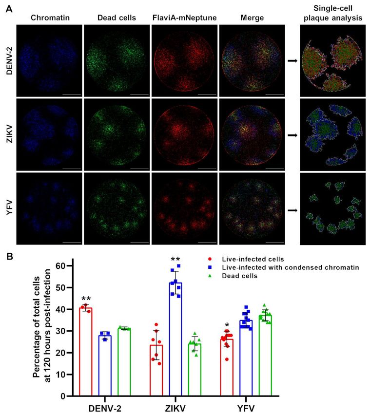
A cell-based fluorescent reporter for flavivirus infection

lines. In order to compare the different variants of        band corresponding to the cleaved NS3 protease
the reporter, we monitored the cell population              became visible at 48 hours post-infection, due to the
kinetics of the flavivirus-activatable GFP sensor’s         autoproteolytic activity of NS3. On the other hand,
fluorescence across multiple experiments and                the band of uncleaved FlaviA-GFP reporter was
confirmed that the flavivirus internal NS3 cleavage         visible in lysates of cells incubated with UV-
site linker (AAQRRGRIG) confers the highest                 inactivated DENV-2, at all the times tested (Fig.
signal-to-noise ratio for reporting the infection of        3C). In the presence of the infectious DENV-2 a
DENV-2 and ZIKV in stably-transduced BHK-21                 slight band of cleaved FlaviA-GFP reporter
cells (Fig. 2C). These results confirm that our GFP-        appeared by 48 hours, correlating with the onset of
based fluorescence activatable reporters of NS2B-           autoproteolytic activity of the viral NS3. This band
NS3 protease activity can be used in living cells and       showed a strong increase after 72 hours post-
that our variant harboring the flavivirus-conserved         infection similar to the increase of cleaved NS3
linker has the highest sensitivity to report viral          protease (Fig. 3C). This correlation was expected as
infection for both DENV-2 and ZIKV at the single-           both the FlaviA-GFP reporter and the viral NS3
cell level.                                                 protease contain the same flavivirus internal NS3
                                                            cleavage site (AAQRRGRIG). The cleavage of the
The fluorescence activation of the FlaviA-GFP               NS3 protease itself therefore represents an excellent

                                                                                                                     Downloaded from http://www.jbc.org/ by guest on October 21, 2020
reporter correlates with viral NS3 protease                 internal control for the NS3 protease activity in this
synthesis and activity in the cellular context              experiment. Together, these results demonstrate
    In order to validate the ability of the FlaviA-         that the fluorescence increase of the FlaviA-GFP
GFP construct to report the NS3 protease content in         reporter correlates with its proteolytic cleavage in
the cellular context, we first monitored the                the cellular context and this cleavage depends on
correlation of the reporter fluorescence with an            the presence of active viral NS3 protease.
anti-NS3 immunofluoresence staining. BHK-21
stable cells expressing the FlaviA-GFP reporter             The FlaviA-mNeptune, a far-red version of the
were infected with DENV-2 13538 and the NS3                 flavivirus sensor, reports the best signal-to-noise
protease was revealed by an immunofluorescence              ratio in a DENV-2/ZIKV fluorescent plaque assay
assay (IFA) with an anti-DENV NS3 protein                       To ascertain the ability of the FlaviA-GFP
antibody at 72 hours post-inoculation. High                 version of the sensor to report the cell population-
magnification images were obtained to study the             based kinetics of infection, we designed an
cellular patterns of both fluorescent signals. The          experimental protocol for a Dulbecco’s plaque
images showed a significant amount of colocalized           assay in 96-well plates. Briefly, confluent cell
fluorescence suggesting a correlation between               monolayers of BHK-21 cells stably expressing the
cellular NS3 amounts and the fluorescent form of            FlaviA-GFP reporter were infected with decimal
the FlaviA-GFP reporter (Fig. 3A). In order to              dilutions of a DENV-2 or ZIKV viral seed and
confirm this correlation, we obtained low                   incubated     with     a    medium        containing
magnification images and quantified the total               carboxymethylcellulose for 120 hours. The plaque-
cellular intensity for both fluorescent signals in          containing wells were completely imaged at 40X
many single cells using CellProfiler 2.0. This              magnification and the whole-well images of cell
quantification confirmed an important correlation           monolayers were generated with the stitching
(Fig. 3B), suggesting that the fluorescence of the          function of the Gen5 3.0 software (BioTek).
FlaviA-GFP reporter arises due to the viral-induced         However, as shown in Figure 4B, the raw images of
synthesis of NS3 protease in the cells. To confirm          the FlaviA-GFP stable cell monolayers presented
this hypothesis, we monitored for 72 hours the              high backgrounds at low magnification in the green
cleavage kinetics of the FlaviA-GFP reporter in             fluorescence channel, probably due to the
BHK-21 stable cells upon DENV-2 infection in                autofluorescence of the phenol-red and the fetal
parallel to the viral NS3 protease expression by            bovine serum of the culture medium. An alternative
western blot. Only the cells incubated with                 medium without phenol-red (Gibco, 11935-046)
infectious DENV-2 showed a band of uncleaved                was tested but showed an increase in cytotoxicity
NS3 protease. This signal was dim at 24 hours but           (data not shown). These images could be enhanced
became more evident at 48 and 72 hours, while a             by increasing the contrast but thereby lost a

                                                        4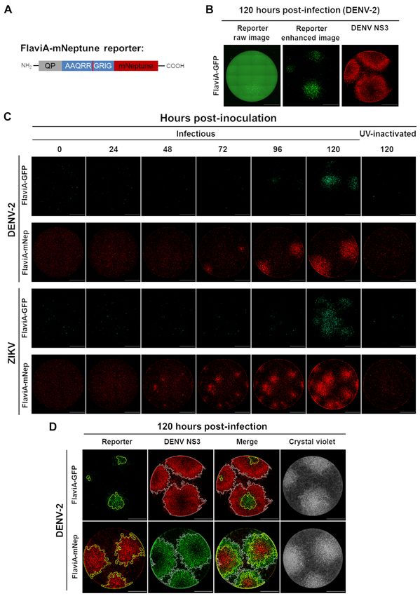
A cell-based fluorescent reporter for flavivirus infection

significant fraction of the cellular signal from the        A multi-reporter platform to study viral plaques
fluorescent viral plaques, as shown by the size             formation at a single-cell resolution reveals
comparison of the reporter plaques in the enhanced          differences in cell-population kinetics of infection
image to the plaques stained with an anti-NS3               and cell death induction by several flaviviruses
antibody at 120 hours post-infection (Fig. 4B).                  To further investigate the potential application
Therefore, we envisaged a far-red version of our            of our FlaviA-mNeptune reporter system to study
flavivirus reporter (FlaviA-mNeptune, Fig. 4A)              the cell population-based kinetics of viral infection
based on our previously published CA-mNeptune               and plaque formation, we designed an experimental
sensor (28). We generated a new stable cell line            protocol combining three types of reporters. The
with the far-red FlaviA-mNeptune reporter in order          FlaviA-mNeptune reporter was used as a surrogate
to compare the signal-to-noise ratio between the red        marker of viral replication, Hoechst 33342 to stain
fluorescent plaques and the green fluorescent               chromatin condensation as an early marker of an
plaques generated in FlaviA-GFP stable cells.               ongoing apoptosis and SYTOX green to label
                                                            nuclei of cells with permeabilized membranes as a
    A time-based comparison of the plaque                   late marker of cell death (Fig. 5A). We constructed
formation kinetics for both DENV-2 and ZIKV                 an image analysis protocol in CellProfiler 2.0 to
indicated that the FlaviA-mNeptune version of the           identify and characterize each individual plaque at

                                                                                                                      Downloaded from http://www.jbc.org/ by guest on October 21, 2020
reporter has a higher signal-to-noise ratio revealing       a single-cell level. Briefly, the pipeline adds the
earlier evidence of red fluorescent plaques 48-72           images of the three channels into one (ImageMath)
hours post-infection compared to the plaques                in order to identify all single cells with all possible
produced by the fluorescence increase of the                combinations of the reporters (Cell identification).
FlaviA-GFP reporter, which required 96-120 hours            These cells are characterized by the quantification
to become evident (Fig. 4C). To confirm the higher          of their neighbors within a specified distance
potential of the FlaviA-mNeptune reporter to reveal         (Neighbor count) and filtered based on a minimal
fluorescent viral plaques, we compared the size of          number of neighbors per cell (Filtered cells) to
the plaques for both types of reporters with the            identify the plaque-forming cells. Then, a series of
respective size of the same plaques stained with            image enhancing steps enable us to perform the
both an anti-NS3 antibody and crystal violet                identification of plaques as new primary objects
staining. We also identified the plaque outlines for        (Plaque identification). Finally, with the identified
both reporters using an image analysis protocol             plaques we were able to relate the single cells to
constructed in CellProfiler 2.0 based on cell-by-cell       their corresponding plaques and quantify thereby
neighbor counts and image thresholding (Fig. 5A).           the number of live-infected cells (red), live-infected
The protocol identified much larger plaques for             cells with chromatin condensation (blue), and dead
FlaviA-mNeptune infected cells compared to those            cells (green) (Fig. 5A). Moreover, the time-based
of infected FlaviA-GFP cells. In addition, the size         monitoring of these parameters for each fluorescent
of the plaques generated in the FlaviA-mNeptune             viral plaque enabled us to study the cell-population
infected cells was very similar to the size of the          kinetics of infection for ZIKV plaque formation
plaques revealed by the NS3 labeling and the                until 120 hours (Fig. 5B). The quantification of
crystal violet staining (Fig. 4D).                          these parameters indicated that the ratios of these
                                                            three cell subpopulations over time are very similar
    Together, these results demonstrate that the            among different plaques for this specific ZIKV
mNeptune version of the flavivirus sensor reports           strain, as depicted by the relatively low standard
the best signal-to-noise ratio in a Dulbecco’s plaque       deviations observed (Fig. 5C). These results
assay, indicating that this reporter is a good marker       suggest that with the combination of a FlaviA-
of viral replication to study cell population-based         mNeptune reporter as a marker of infection with a
kinetics of infection by this standard virological          reporter of chromatin condensation and a reporter
technique.                                                  for cell death, we can obtain a multi-reporter
                                                            platform to characterize the infection kinetics
                                                            induced by a specific viral strain.

                                                        5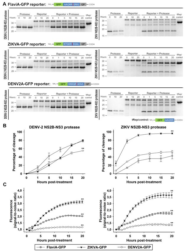
A cell-based fluorescent reporter for flavivirus infection

     In order to confirm the potential of this multi-          reporter to be used in combination with other
reporter platform to reveal virus-specific                     molecular sensors, in order to establish a multi-
differences in terms of viral replication, chromatin           parametric characterization of the infection
condensation, and cell death, we compared the                  produced by different flaviviruses.
fluorescent viral plaques generated upon the
infection with different flaviviruses including                Discussion
DENV-2, ZIKV, and YFV. YFV represents an
additional model to further validate the spectrum of               The present study reports the construction of
application of our flavivirus reporter (Fig. S2B and           fluorescent protein-based sensors of flavivirus
S3) and to explore the heterogeneity in replication            NS2B-NS3 serine proteases activity that become
kinetics and cell death induction across multiple              fluorescent upon cleavage by recombinant DENV-
flaviviruses using our multireporter platform. The             2/ZIKV proteases in vitro (Fig. 1). Moreover, the
acquired images at 120 hours post-infection with               variant of this sensor with the internal NS3 cleavage
DENV-2, ZIKV, and YFV show qualitative                         site linker (AAQRRGRIG) reported the highest
differences (Fig. 6A) that were examined using the             fluorescence increase in stably-transduced
image analysis protocol presented above (Fig. 5A).             mammalian cells upon DENV-2/ZIKV infection,
The artificial images generated by the image                   correlating with the viral induced NS3 protease

                                                                                                                         Downloaded from http://www.jbc.org/ by guest on October 21, 2020
analysis protocol highlighted those differences                activity in the cellular context (Fig. 2 and 3). A far-
(Single-cell plaque analysis, Fig. 6A). The                    red version of this flavivirus sensor reported the
identified plaques contained several subpopulations            best signal-to-noise ratio in a fluorescent
of infected cells: a central core of dead cells (green),       Dulbecco’s plaque assay enabling the construction
surrounded by a ring of cells presenting chromatin             of a multi-reporter platform to study plaque
condensation (blue) and another ring of live-                  formation with single-cell resolution (Fig. 4 and 5).
infected cells (red). Nevertheless, the proportions of         Finally, the application of this platform revealed
these cell subpopulations were very different for              important differences in cell-population kinetics of
DENV-2, ZIKV, and YFV. Finally, the                            infection and cell death induced by several
quantification of the percentage of cells                      flaviviruses (Fig. 6).
corresponding to each subpopulation within each
plaque confirmed the qualitative observations,                      As a starting point, we developed this
indicating differential proportions of infection and           genetically encoded flavivirus reporter system by
induced cell death among the three viral species               successfully engineering the linker of our
tested (Fig. 6B).                                              previously published caspase-activatable reporters,
                                                               CA-GFP and CA-mNeptune (26–28), in order to be
     Next, we asked if the presence of either type of          recognized and cleaved by the flavivirus NS2B-
reporters (FlaviA-GFP and FlaviA-mNeptune) and                 NS3 proteases, giving rise to the FlaviA-GFP and
dyes (Hoechst 33342 and SYTOX green) could                     FlaviA-mNeptune reporters, respectively. First, we
affect the viral replication by comparing the plaque-          focused on the validation of three proposed linkers
forming units determined with the standard crystal             in vitro as a proof of concept of the recognition and
violet staining on wild-type cells with those                  cleavage of our reporter upon treatment with
generated on both reporter cell lines at 96 hours              flavivirus proteases, without the possible effect of
post-infection with DENV-2, ZIKV, and YFV. A                   other proteases that are present in the cellular
representative experiment for DENV-2 is shown in               context. Our results showed that all three linkers
Figure S4A and the plaque count confirmed that                 tested were cleaved as soon as one hour post-
there is no difference in viral replication among all          treatment with both DENV-2 and ZIKV proteases
three cell lines infected with all three viral species         (Fig. 1A and 1B). This fast cleavage validated the
tested (Fig. S4B). This result validates that the              amino acid sequences of our linkers as well as the
behaviors observed for DENV-2, ZIKV, and YFV                   application of “unlinked” versions of both
arise from intrinsic differences in viral replication          recombinant DENV-2 and ZIKV proteases, in
kinetics and not from a differential effect of the             which the NS2B and NS3 regions were produced as
reporters and dyes in viral replication. Together,             independent polypeptides. This choice was based
these results demonstrate the applicability of our             on our own demonstration that the “unlinked”

                                                           6
A cell-based fluorescent reporter for flavivirus infection
version of DENV-2 protease rests predominantly in viral strains and clinical isolates. Among the latter,
a “closed”, active conformation, in contrast with the our system shows some advantages in that it is a
less active, relaxed conformation, adopted by the fluorescence activatable reporter, which allows an
frequently used “linked” construct, in which NS2B easier interpretation of the results and simplifies the
and NS3 polypeptides are attached by a nine-amino image analysis when compared with those systems
acid linker (31). based on the relocalization of the fluorescent signal
across cellular compartments (21, 23). Moreover,
Once the biochemical principle behind the the utility of our fluorescence activation approach
preliminary variants of our reporter was confirmed arises from its low fluorescent background and the
in vitro with recombinant viral proteases, we use of a single recombinant construct that codifies
decided to test its performance upon full flavivirus for a single fluorescent protein with intramolecular
particles infection in mammalian cells. The highest quenching, in contrast with other, difficult to
fluorescence increase to report the infection with optimize strategies, like the fluorescence resonance
DENV-2 and ZIKV in stably transduced BHK-21 energy transfer (FRET) probes (26, 33) and the
cells was obtained with the internal NS3 cleavage protease-triggered Cre-mediated reporter system,
site linker (Fig. 2) present in many members of the which rely on the cellular transfection of two
Flavivirus genus (3, 5, 30). Moreover, a BLASTp different recombinant constructs (22) and could be
Downloaded from http://www.jbc.org/ by guest on October 21, 2020
search of the NCBI reference proteins data base sensitive to early changes in gene expression.
(32) using the query sequence AAQRRGRIG, Another great advantage over other systems is that
revealed 100% coverage and 88.9%-100% identity our reporter has been validated with reference
to >60 members of the Flavivirus genus. The strains but also clinical isolates of different
alignment results included many medically flavivirus species like DENV, ZIKV, and YFV,
important flaviviruses like WNV, YFV, SEV, expanding its range of application to more than one
DENV, JEV, ZIKV, and TBEV (Fig. S5), among flavivirus.
others (1). This finding outlines the potential
application of our reporter system to other The fluorescence activation principle of our
flaviviruses and supports our hypothesis that the reporter also allowed us to envisage a cell-based
flavivirus internal NS3 cleavage site could fluorescent Dulbecco’s plaque assay for
represent a conserved viral target for the flaviviruses (Fig. 4). The plaque assay is one of the
development of a single antiviral therapy against “gold standard” virological techniques originally
the members of the Flavivirus genus, as previously developed by d’Hérelle for titration and isolation of
suggested for DENV (30). Nevertheless, further single clones of bacteriophages and later adapted by
work using other flaviviruses is needed to establish Dulbecco to animal viruses (34, 35). Viral plaques
the whole spectrum of utility and specificity of this arise after genome replication, transcription,
reporter system. translation, virus release from infected cells and
infection of surrounding cells (36), thus, plaque
Furthermore, our flavivirus reporter system development constitutes a hallmark of the infection
proved to be a useful method for monitoring the carried out by a specific viral clone. To date, the
kinetics of DENV-2 and ZIKV infection at a single- kinetics of flavivirus plaque formation could only
cell level by live-cell imaging (Fig. 2) and showed be monitored for transgenic fluorescent viruses
a positive correlation with standard virological made by modifying the genome of reference strains
approaches like detection of viral NS3 protease (16–20). However, we applied the far-red version
synthesis and activity by indirect of our flavivirus sensor to establish, for the first
immunofluorescence and western blot (Fig. 3). In time, a mammalian reporter cell line where the
this respect, flavivirus reporter replicons have been plaque assay can be monitored over time with
generated by engineering the viral genomes of unlabeled native flavivirus strains. This way, we
reference strains (16–20). However, in recent years were able to study the kinetics of viral plaque
special effort has been invested in the development development with the clinical isolate ZIKV CIET-
of cell-based molecular reporter systems for live- 01 (Fig. 4). Considering that viral plaques are clonal
cell imaging of flavivirus infections (21–23), lesions of infected cells formed by the cytopathic
mainly for their application on the study of native effect of replicating viruses (36), we decided to
7A cell-based fluorescent reporter for flavivirus infection
incorporate molecular sensors for chromatin by a specific viral strain. Nevertheless, our
condensation (Hoechst 33342) and cell death approach would be suitable for the screening of
(SYTOX green) to obtain multi-parametric data antiviral compounds by a plaque reduction assay,
from single viral plaques. This enabled us to with the advantage of having a readout of the
construct a multi-reporter platform to study possible cytotoxicity induced by those compounds
flavivirus plaque formation with single-cell in a single experiment.
resolution (Fig. 5), as a way to obtain a preliminary
characterization of the infection produced by a To our knowledge, this is the first fluorescence
particular flavivirus strain. activatable cell-based molecular reporter system for
live-cell imaging of flavivirus infection, suitable to
Following our approach, the quantification of be used in a plaque assay simultaneously with
the percentage of cells within each viral plaque molecular sensors of other cellular or viral-induced
corresponding to live-infected, live-infected with processes. Moreover, taking into account the
chromatin condensation, and dead cells, indicated enormous need for preventative and therapeutic
differential rates of infection and types of induced treatments for flavivirus infections like dengue and
cell death for DENV-2, ZIKV, and YFV (Fig. 6). Zika (47), our multi-reporter platform enables the
The infection with DENV-2 presents the highest decoding of viral-specific fingerprints of Dulbecco-
Downloaded from http://www.jbc.org/ by guest on October 21, 2020
proportion of live-infected cells, indicating that the plaques formation with a potential future
infected cells take longer time to die upon infection application for antiviral drug research and other
and suggesting that this behavior drives to an studies on viral replication/cell death induction for
increased viral production probably related to a both native flavivirus strains and clinical isolates.
faster infection kinetics leading to the formation of
larger plaques (37). The amount of DENV-infected Experimental procedures
cells with chromatin condensation is very similar to
that of dead cells, suggesting a fast type of cell Viruses
demise once the cell death program is engaged, Clinical isolate DENV-2 13538 (DENV-
probably necrosis (38, 39). A similar scenario is 2/CR/10066/2007) was provided by Instituto
shown by YFV that induces a fast type of cell death, Costarricense de Investigación y Enseñanza en
probably necrosis, but earlier during the infection, Nutrición y Salud, Cartago, Costa Rica (48).
which might limit viral spreading leading to a lower DENV-2 viruses were produced in C6/36 cells from
proportion of live-infected cells and smaller viral Aedes albopictus (ATCC, Manassas, VA) by
plaques (40). On the other hand, ZIKV has a lower inoculating a cellular monolayer at a MOI of 0.01
proportion of live-infected cells but a very high and incubating for 3 days with Roswell Park
number of infected cells with chromatin Memorial Institute medium (RPMI-1640, Gibco,
condensation, suggesting that this viral strain Gaithersburg, MD) supplemented with 2% fetal
induces a delayed type of cell death, probably bovine serum (FBS, Gibco) at 33 °C in an
apoptosis as previously reported (23, 41–43), atmosphere of 5% CO2. Then, culture supernatant
preserving for a longer time the cell integrity to was collected, and centrifuged at 3000 x g for 10
produce new viral particles and leading to larger min. Before storage at -80 °C, 23% newborn calf
plaques compared to YFV infection (44). However, serum (NBCS, Gibco) was added (49).
this could be also related to differences in viral
permissiveness of the BHK-21 cell line, since it is Clinical isolate ZIKV CIET-01
known that ZIKV virus tends to infect BHK-21 (ZIKV/CR/CIET-01/2016) was kindly provided by
cells poorly compared to YFV, as demonstrated by Claudio Soto-Garita from Universidad de Costa
envelope protein immunostaining (45, 46). Rica. Vaccine strain YFV 17D
Therefore, our multi-reporter platform serves only (YFV/US/17D/1937) was isolated from the
as a primary screening of the type of infection commercial vaccine YF-VAX® (Sanofi Pasteur,
exposed by flavivirus strains. A more detailed Lyon, France). ZIKV and YFV viruses were
exploration is needed using virological and cell produced in Vero cells (ATCC) by inoculating
biology methods to reach strong conclusions about cellular monolayers at a MOI of 0.1 and incubating
the viral fitness and cell death mechanism induced for 5 days with Minimum Essential Medium
8A cell-based fluorescent reporter for flavivirus infection
(MEM, Gibco) supplemented with 2% FBS at Campeau, Addgene plasmid #17448) (52) to
37 °C in an atmosphere of 5% CO2. Culture produce the plasmid pLenti-CMV-FlaviA-
supernatants were collected, centrifuged at 3000 x mNeptune-puro. All constructs were confirmed by
g for 10 min and stored at -80 °C. sequencing (Genewiz, South Plainfield, NJ).
Viruses were titrated by plaque assay in BHK- Protein expression and purification
21 cells (ATCC) as previously described (50). FlaviA-GFP, ZIKVA-GFP, DENV2A-GFP,
Briefly, 10-fold serial dilutions of viruses were and tRep/control expression constructs in pET21b
added to BHK-21 confluent monolayers. After 2 vectors were transformed into E. coli strain
hours of adsorption, cells were incubated at 37 °C BL21(DE3). Flasks containing 2 liters of 2X YT
in an atmosphere of 5% CO2 for 5 days with MEM media were inoculated with 8 ml of an overnight
supplemented with 2% FBS and 1% culture and grown at 37 °C to an OD600 of 0.6. The
carboxymethylcellulose (Sigma, St. Louis, MO). cultures were then induced using 1 mM Isopropyl
Plaque numbers were counted after staining with β-D-1-thiogalactopyranoside (IPTG, Sigma) and
crystal violet. Virus inactivation was carried out by incubated at 20 °C for 5 hours to allow protein
5 cycles of UV light (254nm) exposure at a energy expression. Cells were harvested by centrifugation
of 400 000 µJ/cm2 in the CL-100 Ultraviolet and disrupted by microfluidization. Clarified
Downloaded from http://www.jbc.org/ by guest on October 21, 2020
Crosslinker (UVP, Upland, CA). lysates were prepared by centrifugation at 15,000 x
g for 45 min. The reporter proteins were then
Reporter development and molecular cloning purified using Co2+-affinity liquid chromatography
FlaviA-GFP, ZIKVA-GFP, and DENV2A- with a 5 ml HiTrap Chelating HP column (GE
GFP reporters were developed by site-directed Healthcare, Chicago, IL). The column was washed
mutagenesis of the linker in the previously with a buffer composed of 50 mM imidazole, 300
described CA-GFP reporter (26–28) to a sequence mM NaCl, and 50 mM NaH2PO4 pH 8.0, and the
coding for the flavivirus NS3 internal cleavage site proteins were eluted using a similar buffer with 300
(AAQRRGRIG), the ZIKV polyprotein NS2B/NS3 mM imidazole and stored at 4 °C. Proteins purity
cleavage site (KTGKRSGAL), and the DENV-2 was assessed in SDS-PAGE gels stained with
polyprotein NS2B/NS3 cleavage site Coomassie blue.
(VKKQRAGVL), respectively, using the
QuikChange approach (Agilent, Santa Clara, CA). DENV-2 protease was expressed as an
The tRep/control, a truncated variant of the reporter “unlinked” construct by the co-transformation of
protein, was generated by inserting a stop codon BL21(DE3) cells with a pACYDuet plasmid
downstream the cleavage site in the linker of the encoding for residues 48-100 of the NS2B cofactor
FlaviA-GFP reporter. Then, the genes of the and a pETDuet plasmid comprising amino acids 1-
flavivirus-activatable GFP reporters in the plasmid 187 of the NS3 protease (a gift from Thomas
pET21b (Novagen, Madison, WI) were Keller) (53). ZIKV protease was expressed as an
independently amplified by PCR and ligated into “unlinked” construct by the transformation of
the SpeI and XhoI restriction sites of the pLenti- BL21(DE3) cells with pET15b vector encoding for
puro vector (a gift from Ie-Ming Shih, Addgene amino acids 48-100 of the NS2B cofactor and 1-178
plasmid #39481) (51) to generate the constructs of the NS3 protease (54). The cells harboring either
pLenti-ZIKVA-GFP-puro, pLenti-DENV2A-GFP- DENV-2 protease or ZIKV protease were grown in
puro, and pLenti-FlaviA-GFP-puro. Likewise and 2X YT media with antibiotics at 37 °C to an OD600
based on the previously described CA-mNeptune of 0.6. Protein expression was induced with 1 mM
reporter (28), we designed and commercially IPTG and proceeded for 3 hours at 25°C. The
synthetized (Atum, Newark, CA) the FlaviA- proteases were purified using Ni2+-affinity liquid
mNeptune reporter gene by changing the linker chromatography with a 5 ml HiTrap Chelating HP
sequence in order to codify for AAQRRGRIG. column and eluted using a step gradient with 300
Finally, the FlaviA-mNeptune gene in the plasmid mM imidazole. The eluted proteases were further
pD2109-CMV (Atum) was PCR amplified and purified using anion exchange with a HiTrap Q HP
ligated into the XbaI and SalI restriction sites of the column (GE Healthcare) with a linear gradient from
pLenti-CMV-GFP-Puro vector (a gift from Eric 10 mM to 500 mM NaCl and stored at -80 °C.
9A cell-based fluorescent reporter for flavivirus infection
Proteins purity was assessed in SDS-PAGE gels Flow Cytometer (BD Biosciences, Franklin Lakes,
stained with Coomassie blue. NJ), as previously described (56).
Reporter cleavage and fluorescence assay in vitro Reporter cell lines production and selection
Samples containing 10 µM of either FlaviA- BHK-21 cell monolayers at 80% confluency
GFP, ZIKVA-GFP or DENV2A-GFP reporters were stably transduced with lentiviral particles
with and without 10 µM of DENV-2 or ZIKV carrying a genetic construct codifying for either
proteases were prepared with digestion buffer FlaviA-GFP, ZIKVA-GFP, DENV2A-GFP or
(50mM Tris buffer pH 8.5, 0,1% CHAPS, 20% FlaviA-mNeptune reporters. For cell transduction,
glycerol) in a final volume of 120 µl and added to a lentiviral vector particles were added to the cells at
costar 96-well black plate. The fluorescence was a MOI of 1 and centrifuged for 2 hours at 300 x g,
measured every hour (Ex. 475 nm/Em. 512 nm) for 25 °C. At 72 hours post-transduction, cells were
20 hours at 27 °C. Another set of samples was selected with 8 μg/ml of puromycin (Sigma) in
incubated at 27 °C in 30 µl aliquots to which SDS MEM 10% FBS during 2 days and then cell
loading buffer was added at time points of 0, 1, 5, populations with homogeneous levels of expression
10, 15 and 20 hours. These samples were then run of the constructs were isolated by fluorescence-
on SDS-PAGE and stained with Coomassie blue to activated cell sorting with a BD FACSJazz™ Cell
Downloaded from http://www.jbc.org/ by guest on October 21, 2020
determine the cleavage kinetics of the flavivirus- Sorter (BD Biosciences) based on the fluorescent
activatable GFP reporters by DENV-2 and ZIKV basal signal of the reporter proteins. The selected
proteases. Gel images were acquired with a stable cell lines were grown and maintained in
ChemiDoc™ XRS+ System (BioRad, Hercules, MEM supplemented with 10% FBS and 0.5 μg/ml
CA) and analyzed with the Image J software (NIH, puromycin.
Bethesda, MD) (55) to determine the cleavage
efficiency. The fluorescence signal-to-noise ratio Infection kinetics in reporter cell lines by live-cell
was calculated by dividing the signal of the reporter imaging
treated with viral protease by the noise gave by the BHK-21 cells stably expressing the FlaviA-
untreated reporter at every time point. GFP, the ZIKVA-GFP or the DENV2A-GFP
reporters were seeded on µClear black 96-well
Lentiviral vectors assembly plates (Greiner Bio-One, Kremsmünster, Austria)
HEK293T cells (ATCC) were cultured in at a density of 15 000 cells per well with MEM
Dulbecco’s modified Eagle medium (DMEM, supplemented with 2% FBS. After 24 hours of
Gibco) supplemented with 10% FBS, 1X incubation at 37 °C - 5% CO2, reporter cells were
GlutaMAX (Gibco), 1 mM sodium pyruvate infected with either infectious or UV-inactivated
(Gibco), and 1X antibiotic-antimycotic solution DENV-2 13538 or ZIKV CIET-01 at a MOI of 0.25
(Gibco). Lentiviral particles were generated in 60% and allowed to adsorb for 2 hours at 37°C. After
confluent HEK293T cell monolayers by triple labeling with 1 µg/mL Hoechst 33342 (Invitrogen,
transfection with polyetilenimide (Polysciences, Carlsbad, CA) for 10 minutes, cells were washed
Warrington, PA) of either pLenti-FlaviA-GFP- with 1X PBS and incubated for 96 hours with
puro, pLenti-ZIKVA-GFP-puro, pLenti-DENV2A- FluoroBrite™ DMEM (Gibco) supplemented with
GFP-puro or pLenti-CMV-FlaviA-mNeptune-puro 2% FBS and containing 2,5 µg/mL of propidium
constructs and both packaging plasmids pMD2.G iodide (Invitrogen) at 37 °C in an atmosphere of 5%
and psPAX2 (a gift from Didier Trono, Addgene CO2. Images were acquired every 2 hours with a
plasmids #12259 and #12260). At 72 hours post- Cytation 3 Cell Imaging Multi-Mode Reader
transfection, virus-containing medium was (BioTek, Winooski, VT). The fluorescence signal-
collected, filtered through a 0.45 µm membrane, to-noise ratio was calculated by dividing the signal
supplemented with 5 µg/mL of polybrene (Sigma), of the reporter cells treated with infectious virus by
and stored at -80 °C. Finally, 10-fold serial dilutions the noise gave by the reporter cells treated with UV-
of each lentiviral seed were added to HEK293T inactivated virus at every time point.
cells and the biological titers in transducing units
(TU)/ml were determined by flow cytometry at 48
hours post-transduction using a BD Accuri™ C6
10A cell-based fluorescent reporter for flavivirus infection
Kinetic plaque assay on reporter cell lines by live- Mode Reader and cells were lysed with 2% SDS
cell imaging solution and stored at -20 °C. Later, samples were
BHK-21 cells stably expressing the FlaviA- run on a SDS-PAGE gel, transferred to a PVDF
mNeptune reporter were seeded on µClear black membrane (Millipore) and blotted in a single step
96-well plates at a density of 25 000 cells per well with a mixture of 1:100 dilution of rabbit polyclonal
with MEM supplemented with 10% FBS. After 24 anti-GFP antibody (Invitrogen, A-11122), 1:1000
hours of incubation at 37 °C - 5% CO2, reporter dilution of rabbit polyclonal anti-DENV NS3
cells were infected with 10-fold serial dilutions of antibody, and 1:500 dilution of rabbit monoclonal
either infectious or UV-inactivated DENV-2 anti-vinculin antibody (Invitrogen, 700062).
13538, ZIKV CIET-01, or YFV 17D. After 2 hours Finally, the membrane was treated with a 1:500
adsorption, cells were labeled with 1 µg/mL dilution of goat polyclonal anti-rabbit IgG HRP-
Hoechst 33342 for 10 minutes, washed once with conjugated antibody (Invitrogen, G-21234) and
1X PBS and incubated for 120 hours at 37 °C - 5% visualized using the Super Signal West Pico Plus
CO2 with MEM AutoMod™ (Sigma) supplemented Chemiluminescent Substrate (Thermo Fisher
with 2% FBS, 1% carboxymethylcellulose, and 500 Scientific, Waltham, MA). Images were adquired
nM SYTOX green (Invitrogen). Images of the with a ChemiDoc™ XRS+ System.
whole well were acquired every 24 hours with a
Downloaded from http://www.jbc.org/ by guest on October 21, 2020
Cytation 3 Cell Imaging Multi-Mode Reader. Image analysis and statistics
Finally, at 120 hours post-inoculation viral plaques Image analysis was performed with the
were confirmed by crystal violet staining. software CellProfiler 2.0
(http://www.cellprofiler.org; Broad Institute,
Indirect immunofluorescence Cambridge, MA). Data are expressed as mean ±
At 96 and 120 hours post-infection with either standard deviation (SD) of three independent
infectious or UV-inactivated DENV-2 13538, experiments. Statistical significance of the
medium was removed and cells were fixed for 1 differences between mean values was determined
hour with 1% paraformaldehyde (Sigma). Later, by using a one-way ANOVA followed by a Tukey’s
cells were permeabilized with 70% ethanol for 15 post hoc test with the software SigmaPlot 14 (Systat
minutes and 0,001% Triton X-100 (Sigma) for 10 Software Inc., San Jose, CA). The level of
minutes at rate temperature. Then, cells were significance is denoted in each figure.
incubated with a 1:800 dilution of rabbit polyclonal
anti-DENV NS3 antibody (GeneTex, Irvine, CA,
GTX124252) for 1 hour at 37 °C, washed twice
with 1X PBS and incubated with a 1:400 dilution of
goat polyclonal anti-rabbit IgG Alexa Fluor® 594-
conjugated antibody (Invitrogen, A11037) for 30
minutes at 37 °C. After three washes with 1X PBS,
FluoroBrite™ DMEM was added and images of the
whole well were acquired with a Cytation 3 Cell
Imaging Multi-Mode Reader.
Western blot
BHK-21 cells stably expressing the FlaviA-
GFP reporter were seeded on 12-well plates
(Greiner Bio-One) at a density of 350 000 cells per
well with MEM supplemented with 2% FBS. After
24 hours of incubation at 37 °C - 5% CO2, reporter
cells were infected with either infectious or UV-
inactivated DENV-2 13538 at a MOI of 0.25 and
allowed to adsorb for 2 hours at 37 °C. Every 24
hours during 96 hours of incubation, images were
acquired with the Cytation 3 Cell Imaging Multi-
11A cell-based fluorescent reporter for flavivirus infection
Acknowledgements: We thank Francisco Vega-Aguilar for his technical assistance with viral strains. We
thank Joseph P. Kennedy Jr. and Derrick P. Feuerstein for their support and suggestions during the
experimentation. We also thank Ralf Bartenschlager, Heidelberg University, for his scientific advice and
critical review of the manuscript. This work is dedicated with regard to Alina I. Arias-Barrantes and
Alexander Mora-Solano.
Conflict of interest: The authors declare that they have no conflicts of interest with the contents of this
article. The content is solely the responsibility of the authors and does not necessarily represent the official
views of the National Science Foundation.
Author contributions: J. L. A. and J. A. H. conceptualization; J. L. A. and R. M. methodology; J. L. A.,
D. J. M., and M. E. H. experimentation; J. L. A. and R. M. data curation and analysis; J. L. A. and R. M.
writing-original draft; J. L. A., J. A. H., and R. M. writing-review and editing; J. L. A., J. A. H. and R. M.
resources; J. A. H. and R. M. supervision; J. A. H. and R. M. funding acquisition; J. A. H. and R. M. project
administration.
Downloaded from http://www.jbc.org/ by guest on October 21, 2020
12A cell-based fluorescent reporter for flavivirus infection
References
1. Gould, E., and Solomon, T. (2008) Pathogenic flaviviruses. Lancet. 371, 500–509
2. Lindenbach, B., Murray, C. L., Thiel, H.-J., and Rice, C. M. (2013) Flaviviridae. in Fields Virology,
pp. 712–746
3. Teo, K. F., and Wright, P. J. (1997) Internal proteolysis of the NS3 protein specified by dengue virus
2. J. Gen. Virol. 78, 337–341
4. Lindenbach, B. D., and Rice, C. M. (2003) Molecular biology of flaviviruses. Adv. Virus Res. 59,
23–61
5. Bera, A. K., Kuhn, R. J., and Smith, J. L. (2007) Functional characterization of cis and trans activity
of the flavivirus NS2B-NS3 protease. J. Biol. Chem. 282, 12883–12892
6. Ishikawa, T., Yamanaka, A., and Konishi, E. (2014) A review of successful flavivirus vaccines and
the problems with those flaviviruses for which vaccines are not yet available. Vaccine. 32, 1326–
1337
Downloaded from http://www.jbc.org/ by guest on October 21, 2020
7. Scott, L. J. (2016) Tetravalent dengue vaccine: A review in the prevention of dengue disease. Drugs.
76, 1301–1312
8. Normile, D. (2017) Safety concerns derail dengue vaccination program. Science (80-. ). 358, 1514–
1515
9. Chan, K. W. K., Watanabe, S., Kavishna, R., Alonso, S., and Vasudevan, S. G. (2015) Animal
models for studying dengue pathogenesis and therapy. Antiviral Res. 123, 5–14
10. Reynolds, E. S., Hart, C. E., Hermance, M. E., Brining, D. L., and Thangamani, S. (2017) An
overview of animal models for arthropod-borne viruses. Comp. Med. 67, 232–241
11. Diamond, M. S., Edgil, D., Roberts, T. G., Lu, B., and Harris, E. (2000) Infection of human cells by
dengue virus is modulated by different cell types and viral strains. J. Virol. 74, 7814–23
12. Balsitis, S. J., Coloma, J., Castro, G., Alava, A., Flores, D., Beatty, R., and Harris, E. (2008) Tropism
of replicating dengue virus in mice and humans defined by viral nonstructural protein 3-specific
immunohistochemistry. Am. J. Trop. Med. Hyg. 79, 38
13. Arias-Arias, J. L., Vega-Aguilar, F., Corrales-Aguilar, E., Hun, L., Loría, G. D., and Mora-
Rodríguez, R. (2018) Dengue virus infection of primary human smooth muscle cells. Am. J. Trop.
Med. Hyg. 99, 1451–1457
14. Acosta, E. G., and Bartenschlager, R. (2016) The quest for host targets to combat dengue virus
infections. Curr. Opin. Virol. 20, 47–54
15. de Wispelaere, M., Lian, W., Potisopon, S., Li, P. C., Jang, J., Ficarro, S. B., Clark, M. J., Zhu, X.,
Kaplan, J. B., Pitts, J. D., Wales, T. E., Wang, J., Engen, J. R., Marto, J. A., Gray, N. S., and Yang,
P. L. (2018) Inhibition of flaviviruses by targeting a conserved pocket on the viral envelope protein.
Cell Chem. Biol. 25, 1006–1016
16. Li, S. H., Li, X. F., Zhao, H., Deng, Y. Q., Yu, X. D., Zhu, S. Y., Jiang, T., Ye, Q., Qin, E. De, and
Qin, C. F. (2013) Development and characterization of the replicon system of Japanese encephalitis
live vaccine virus SA14-14-2. Virol. J. 10, 1–6
17. Schmid, B., Rinas, M., Ruggieri, A., Acosta, E. G., Bartenschlager, M., Reuter, A., Fischl, W.,
Harder, N., Bergeest, J. P., Flossdorf, M., Rohr, K., Höfer, T., and Bartenschlager, R. (2015) Live
13A cell-based fluorescent reporter for flavivirus infection
cell analysis and mathematical modeling identify determinants of attenuation of dengue virus 2’-O-
methylation mutant. PLoS Pathog. 11, 1–36
18. Xie, X., Zou, J., Shan, C., Yang, Y., Kum, D. B., Dallmeier, K., Neyts, J., and Shi, P. Y. (2016)
Zika virus replicons for drug discovery. EBioMedicine. 12, 156–160
19. Tamura, T., Fukuhara, T., Uchida, T., Ono, C., Mori, H., Sato, A., Fauzyah, Y., Okamoto, T.,
Kurosu, T., Setoh, Y. X., Imamura, M., Tautz, N., Sakoda, Y., Khromykh, A. A., Chayama, K., and
Matsuura, Y. (2017) Characterization of recombinant Flaviviridae viruses possessing a small
reporter tag. J. Virol. 92, 1–19
20. Kümmerer, B. M. (2018) Establishment and application of flavivirus replicons. in Dengue and Zika:
Control and antiviral treatment strategies, pp. 165–173, 1062, 165–173
21. Medin, C. L., Valois, S., Patkar, C. G., and Rothman, A. L. (2015) A plasmid-based reporter system
for live cell imaging of dengue virus infected cells. J. Virol. Methods. 211, 55–62
22. Hsieh, M.-S., Chen, M.-Y., Hsieh, C.-H., Pan, C.-H., Yu, G.-Y., and Chen, H.-W. (2017) Detection
and quantification of dengue virus using a novel biosensor system based on dengue NS3 protease
Downloaded from http://www.jbc.org/ by guest on October 21, 2020
activity. PLoS One. 12, 1–19
23. McFadden, M. J., Mitchell-Dick, A., Vazquez, C., Roder, A. E., Labagnara, K. F., McMahon, J. J.,
Silver, D. L., and Horner, S. M. (2018) A fluorescent cell-based system for imaging zika virus
infection in real-time. Viruses. 10, 13–18
24. Winkler, G., Maxwell, S. E., Ruemmler, C., and Stollar, V. (1989) Newly synthesized dengue-2
virus nonstructural protein NS1 is a soluble protein but becomes partially hydrophobic and
membrane-associated after dimerization. Virology. 171, 302–305
25. Chung, K. M., Thompson, B. S., Fremont, D. H., and Diamond, M. S. (2007) Antibody Recognition
of Cell Surface-Associated NS1 Triggers Fc- Receptor-Mediated Phagocytosis and Clearance of
West Nile Virus-Infected Cells. J. Virol. 81, 9551–9555
26. Nicholls, S. B., Chu, J., Abbruzzese, G., Tremblay, K. D., and Hardy, J. A. (2011) Mechanism of a
genetically encoded dark-to-bright reporter for caspase activity. J. Biol. Chem. 286, 24977–24986
27. Nicholls, S. B., and Hardy, J. A. (2013) Structural basis of fluorescence quenching in caspase
activatable-GFP. Protein Sci. 22, 247–257
28. Wu, P., Nicholls, S. B., and Hardy, J. A. (2013) A Tunable, modular approach to fluorescent
protease-activated reporters. Biophys. J. 104, 1605–1614
29. Shiryaev, S. A., Kozlov, I. A., Ratnikov, B. I., Smith, J. W., Lebl, M., and Strongin, A. Y. (2007)
Cleavage preference distinguishes the two-component NS2B–NS3 serine proteinases of Dengue and
West Nile viruses. Biochem. J. 401, 743–752
30. Constant, D. A., Mateo, R., Nagamine, C. M., and Kirkegaard, K. (2018) Targeting intramolecular
proteinase NS2B/3 cleavages for trans -dominant inhibition of dengue virus . Proc. Natl. Acad. Sci.
115, 10136–10141
31. Hill, M. E., Yildiz, M., and Hardy, J. A. (2019) Cysteine disulfide traps reveal distinct
conformational ensembles in dengue virus NS2B-NS3 protease. Biochemistry. 58, 776–787
32. Altschul, S. F., Gish, W., Miller, W., Myers, E. W., and Lipman, D. J. (1990) Basic local alignment
search tool. J. Mol. Biol. 215, 403–410
33. Leavesley, S. J., and Rich, T. C. (2016) Overcoming limitations of FRET measurements. Cytom.
14You can also read