Book Chapter Active Crossfire between Cyanobacteria and Cyanophages in Phototrophic Mat Communities within Hot Springs - Vide Leaf
←
→
Page content transcription
If your browser does not render page correctly, please read the page content below
Prime Archives in Microbiology

Book Chapter

Active Crossfire between Cyanobacteria
and Cyanophages in Phototrophic Mat
Communities within Hot Springs
Sergio Guajardo-Leiva1, Carlos Pedrós-Alió2, Oscar Salgado1,
Fabián Pinto1 and Beatriz Díez1,3*
1
  Department of Molecular Genetics and Microbiology, Pontificia
Universidad Católica de Chile, Chile
2
  Programa de Biología de Sistemas, Centro Nacional de
Biotecnología – Consejo Superior de Investigaciones Científicas,
Spain
3
  Center for Climate and Resilience Research, Chile

*Corresponding Author: Beatriz Díez, Department of
Molecular Genetics and Microbiology, Pontificia Universidad
Católica de Chile, Santiago, Chile

Published March 27, 2020

This Book Chapter is a republication of an article published by
Beatriz Díez, et al. at Frontiers in Microbiology in September
2018. (Guajardo-Leiva S, Pedrós-Alió C, Salgado O, Pinto F and
Díez B (2018) Active Crossfire Between Cyanobacteria and
Cyanophages in Phototrophic Mat Communities Within Hot
Springs. Front. Microbiol. 9:2039. doi:
10.3389/fmicb.2018.02039)

How to cite this book chapter: Sergio Guajardo-Leiva, Carlos
Pedrós-Alió, Oscar Salgado, Fabián Pinto, Beatriz Díez. Active
Crossfire between Cyanobacteria and Cyanophages in
Phototrophic Mat Communities within Hot Springs. In:
Alexandre Morrot, editor. Prime Archives in Microbiology.
Hyderabad, India: Vide Leaf. 2020.

© The Author(s) 2020. This article is distributed under the terms
of the Creative Commons Attribution 4.0 International

1                                             www.videleaf.com
Prime Archives in Microbiology

License(http://creativecommons.org/licenses/by/4.0/), which
permits unrestricted use, distribution, and reproduction in any
medium, provided the original work is properly cited.

Data Availability Statement: The datasets generated for this
study can be found NCBI as follow: Access to raw data for
metagenomes and metatranscriptomes is available through NCBI
BioProject ID PRJNA382437.
https://www.ncbi.nlm.nih.gov/bioproject/?term=PRJNA382437.
The genome of TC-CHP58 has the GenBank accession number
KY888885. Contigs containing CRISPRs loci have been
submitted to NCBI with GenBank accession numbers
MG734911 to MG734917.

Author Contributions: SG-L and BD conceived and designed
the experiments. SG-L, OS, and FP performed the experiments.
SG-L, CP-A, OS, FP, and BD analyzed the data. SG-L, CP-A,
and BD wrote the paper.

Funding: This work was financially supported by Ph.D.
scholarships CONICYT N° 21130667 and 21172022, and
CONICYT grant FONDECYT N°1150171. Sequencing was
funded by Spanish grant CTM2013-48292-C3-1-R.

Conflict of Interest Statement: The authors declare that the
research was conducted in the absence of any commercial or
financial relationships that could be construed as a potential
conflict of interest.

Acknowledgments: We are grateful to Huinay Scientific Field
Station for making our work in the Porcelana hot spring possible.

Abstract
Cyanophages are viruses with a wide distribution in aquatic
ecosystems, that specifically infect Cyanobacteria. These viruses
can be readily isolated from marine and fresh waters
environments; however, their presence in cosmopolitan
thermophilic phototrophic mats remains largely unknown. This
study investigates the morphological diversity (TEM),

2                                              www.videleaf.com
Prime Archives in Microbiology

taxonomic composition (metagenomics), and active infectivity
(metatranscriptomics) of viral communities over a thermal
gradient in hot spring phototrophic mats from Northern
Patagonia (Chile). The mats were dominated (up to 53%) by
cosmopolitan      thermophilic     filamentous     true-branching
cyanobacteria from the genus Mastigocladus, the associated viral
community was predominantly composed of Caudovirales
(70%), with most of the active infections driven by cyanophages
(up to 90% of Caudovirales transcripts). Metagenomic assembly
lead to the first full genome description of a T7-like
Thermophilic Cyanophage recovered from a hot spring
(Porcelana Hot Spring, Chile), with a temperature of 58∘C (TC-
CHP58). This could potentially represent a world-wide
thermophilic lineage of podoviruses that infect cyanobacteria. In
the hot spring, TC-CHP58 was active over a temperature
gradient from 48 to 66∘C, showing a high population variability
represented by 1979 single nucleotide variants (SNVs). TC-
CHP58 was associated to the Mastigocladus spp. by CRISPR
spacers. Marked differences in metagenomic CRISPR loci
number and spacers diversity, as well as SNVs, in the TC-
CHP58 proto-spacers at different temperatures, reinforce the
theory of co-evolution between natural virus populations and
cyanobacterial hosts. Considering the importance of
cyanobacteria in hot spring biogeochemical cycles, the
description of this new cyanopodovirus lineage may have global
implications for the functioning of these extreme ecosystems.

Keywords
Hot-Springs; Cyanophages; Phototrophic         Microbial    Mat;
CRISPR; Thermophilic Cyanobacteria

Introduction
Hot springs host microbial communities dominated by a limited
variety of microorganisms that form well-defined mats [1,2].
Frequently, the uppermost layer of the mat is composed of
photoautotrophs; such as oxygenic phototrophic cyanobacteria,
including the unicellular cyanobacterium Synechococcus spp. [3-
6], the filamentous non-heterocystous Oscillatoria spp., the

3                                             www.videleaf.com

Prime Archives in Microbiology

filamentous heterocystous Mastigocladus spp. [7-10], as well as
filamentous anoxygenic phototrophs (FAPs), such as Roseiflexus
sp. and Chloroflexus sp. [6,11,12]. These primary producers
interact with heterotrophic prokaryotes through element and
energy cycling [13]. Heterocystous cyanobacteria are a key
component in hot springs, since these systems are commonly N-
limited due to the rapid assimilation and turnover of inorganic
nitrogen forms [10,14]. Thus, N2-fixation by cyanobacteria is
identified to be a key biological process in neutral hot spring
microbial mats [10].

These simplified but highly cooperative communities have been
historically used as models for understanding the composition,
structure, and function of microbial consortia [2,6]. The role of a
variety of abiotic factors, such as pH, sulfide concentration, and
temperature, in determining microbial assemblages and life
cycles in these ecosystems have been investigated [2,15].
However, there is a lack of investigation into biotic factors, such
as viruses, on thermophilic photoautotrophic mats, with existing
studies only reporting short or partial viral sequences [16,17].
Currently, viral communities from thermal mats have been
characterized through indirect approaches, indicating the
hypothetical presence of viruses [16,17]. Heidelberg et al. [16]
used CRISPR spacer sequences extracted from the genomes of
two thermophilic Synechococcus isolates, from a phototrophic
mat in Octopus Spring. Subsequently, they searched for viral
contigs from previously published water metaviromes from the
Octopus and Bear Paw Springs in Yellowstone National Park
(United States) [18]. Furthermore, Davison et al. used CRISPR
spacers and nucleotide motive frequencies to link viral contigs to
known hosts using a metavirome obtained by Multiple
Displacement Amplification (MDA) of VLPs from a mat in
Octopus Spring [17], as well as reference genomes from
dominant species (Synechococcus sp., Roseiflexus sp., and
Chloroflexus sp.) previously described in the same microbial
mat. A key finding from these studies was the link between
viruses and their hosts, indicating their co-evolution and an
effective “arms race” within hot spring phototrophic mats.

4                                              www.videleaf.com
Prime Archives in Microbiology

Unlike thermophilic mat studies, most viral investigation carried
out in hot springs occur within the source waters [18-23]. In
these waters, virus abundances range between 104 and 109 virus
like particles (VLPs) mL-1 [18,24,25]. They play an important
role in both the structuring of host populations and as drivers of
organic and inorganic nutrient recycling [24]. The majority of
the viruses were dsDNA, with new and complex viral
morphotypes, distinct to the typical head and tail morphologies
[18,19,25-27]. Furthermore, the few metaviromes obtained in
thermal waters indicate that natural thermophilic virus
communities differ from those obtained in culture, given that
there was only a 20–50% similarity between the sequences
obtained and those in the databases [18,22,28,29]. Thus far, the
genomes that have been isolated and sequenced from
thermophilic viruses (57 genomes, of which 37 infected archaea
and 20 infected Bacteria) generally yielded few significant
matches to sequences in public databases [1]. More recently, a
water metaviromic study from Brandvlei hot spring (BHS),
South Africa [23] reported the presence of two partial genomes
(10 kb and 27 kb), the first related to Podoviridae and the second
to lambda-like Siphoviridae families. Both Caudovirales
genomes did not have a confirmed host, but the presence of
green microbial mat-patches around the contours of the hot
spring, implied that filamentous Cyanobacteria and unclassified
Gemmata species were the potential hosts, respectively. The last,
based on the proximity of some viral predicted proteins with
bacteria from well characterized microbial mats present in a
nearby hot spring [30,31].

Given the lack of knowledge of viral communities within hot
spring phototrophic microbial mats, the present study used the
mats of Porcelana hot spring (Northern Patagonia, Chile), as a
pH neutral model, to better understand the associated
thermophilic viral communities within these mats. This pristine
spring is covered by microbial mats that grow along a thermal
gradient between 70 and 46°C, dominated by bacterial
phototrophs, such as filamentous cyanobacteria from the genus
Mastigocladus [9,10]. This is the dominant and most active
cyanobacterial genus in the Porcelana mat environment, carrying
out important biological processes such as carbon- and N2-

5                                              www.videleaf.com
Prime Archives in Microbiology

fixation [10,32]. Thus, this study proposes that the mats in
Porcelana hot spring are dominated by viral communities of the
Order Caudovirales, which is able to infect Cyanobacteria,
preferably Mastigocladus spp.

The viral diversity in Porcelana was determined through the
detection of viral signals in microbial mat omics data, and by
TEM along the thermal gradient. The results demonstrate that the
viral community was dominated by Caudovirales, which actively
infect Cyanobacteria. Furthermore, the first complete genome
description of a thermophilic cyanobacterial T7-like podovirus,
Thermophilic Cyanophage Chile Porcelana 58°C (from now on
TC-CHP58) is realized. The host is the dominant phototroph
Mastigocladus spp, based on CRISPR spacers. Finally, the
presence of different populations of this new podovirus are
identified through single nucleotide variants (SNVs) analyses,
and the co-evolution of Mastigocladus spp. and particular
populations of TC-CHP58 at different temperatures is described
through association of specific SNVs to different CRISPR
spacers.

Materials and Methods
Sampling Site

Porcelana hot spring is located in Chilean Patagonia (42° 27′
29.1′′S – 72° 27′ 39.3′′W). It has a neutral pH range between 7.1
and 6.8 and temperatures ranging from 70 to 46°C, when
sampled on March 2013. Phototrophic microbial mats growing at
66, 58, and 48°C were sampled using a cork borer of 7 mm
diameter. Cores of 1 cm thick were collected in triplicate at noon
(12:00 PM), transported in liquid nitrogen and kept at -80°C
until DNA and RNA extraction.

Transmission Electron Microscopy

Five liters of interstitial fluid was squeezed using 150 μm
sterilized polyester net SEFAR PET 1000 (Sefar, Heiden,
Switzerland) and filtered through 0.8 μm pore-size
polycarbonate filters (Isopore ATTP, 47 mm diameter, Millipore,
Millford, MA, United States) and 0.2 μm pore-size (Isopore

6                                              www.videleaf.com
Prime Archives in Microbiology

GTTP, 47 mm diameter, Millipore) using a Swinex filter holder
(Millipore). Particles in the 0.2 μm filtrate were concentrated to a
final volume of approximately 35 ml using a tangential-flow
filtration cartridge (Vivaflow 200, 30 kDa pore size,
Vivascience, Lincoln, United Kingdom). Viral concentrates (15
μL) were spotted onto Carbon Type-B, 200 mesh, Copper
microscopy grids (Ted Pella, Redding, California, United
States), stained with 1% uranyl acetate and imaged on an FEI
Tecnai T12 electron microscope at 80 kV (FEI Corporate,
Hillsboro, OR, United States) with attached Megaview G2 CCD
camera (Olympus SIS, Münster, Germany). Imaging analysis
was done at the Advanced Microscopy Unit, School of
Biological Sciences at Pontificia Universidad Católica de Chile
(Santiago, Chile).

Nucleic Acid Extractions and High Throughput Sequencing

Nucleic acids (DNA and RNA) were extracted as previously
described [10]. For RNA, Trizol (Invitrogen, Carlsbad, CA,
United States) was added to the mat sample, and homogenized
by bead beating, two pulses of 20 s. Quality and quantity of the
extracted nucleic acids were checked and kept at -80°C.

Samples were sequenced by Illumina Hi-seq technology
(Research and Testing Laboratory, Texas, United States).
Briefly, for metagenomes, DNA was fragmented using NEBNext
dsFragmentase (New England Biolabs, Ipswich, MA, United
States), followed by DNA clean up using column purification,
and a NEBUltra DNA Library Prep Kit for Illumina (New
England Biolabs, Ipswich, MA, United States) was used for
library construction.

For metatranscriptomes, DNase treated total RNA was cleaned
up of rRNA by a Ribo-Zero rRNA Removal Kit Bacteria
(Illumina, San Diego, CA, United States), followed by
purification using an Agencourt RNAClean XP Kit (Beckman
Coulter, Indianapolis, IN, United States), and a NEXTflexTM
Illumina Small RNA Sequencing Kit v3 (Bio Scientific, Austin,
TX, United States) was used for library construction.

7                                               www.videleaf.com
Prime Archives in Microbiology

For quality filtering, the following filters were applied using
Cutadapt [33], leaving only mappable sequences longer than 30
bp (-m 30), with a 3′ end trimming for bases with a quality below
28 (-q 28), a hard clipping of the first five leftmost bases (-u 5),
and finally a perfect match of at least 10 bp (-O 10) against the
standard Illumina adaptor. Finally, the removal of sequences
representing simple repetitions that are usually due to sequencing
errors was applied using PRINSEQ [34] DUST threshold 7 (-
lc_method dust, -lc_threshold 7). Details of the number of
sequences obtained are shown in Supplementary Table S1.

Identification of rRNA-Like Sequences and Viral
Mining From Metagenomes and Metatranscriptomes

Metagenomic Illumina TAGs (miTAGs) [35] that are small
subunit (SSU) 16S and 18S rRNA gene sequences in the
metagenomes were identified and annotated using the
Ribopicker tool [36] with the Silva 123 SSU database [37].

For viral mining, bacterial, archaeal and eukaryotic sequences
were removed through end-to-end mapping, allowing a 5% of
mismatch (-N 1 -L 20) against the NCBI non-redundant (NR)
database (Nov-2015) using bowtie2 [38]. Viral sequences were
then recruited against modified NCBI RefSeq (Release 75) viral
proteins, where only amino acid sequences from viruses that do
not infect animals (NAV) were considered to build the database,
using the UBLAST algorithm (-strand both -accel 0.9) through
the USEARCH sequence analysis tool [39]. Recruitment was
made for sequences with over 65% of coverage and an E-value <
1 × 10⁻³ (-query_cov 0.65 -evalue 1e-3). For taxonomic
assignment, recruited sequences were aligned against the NAV
database using BLASTX [40] and parsed using the lowest
common ancestor algorithm trough MEGAN 6 [41] (LCA score
= 30). The latter displays a graphical representation of
abundance for each taxonomic group identified at the family and
species levels. Species classification of viral reads was used to
infer the phyla of the putative hosts based on viral RefSeq host
information or through a manual search of the publication
associated with each viral genome.

8                                               www.videleaf.com
Prime Archives in Microbiology

To extract putative viral genomes, all metagenomes (48, 58, and
66°C) were assembled using De Bruijn graphs as implemented in
the Spades assembler [42], followed by gene prediction using
Prodigal software [43] and the recovery of circular contigs over
5 kb using a Python script [44]. Only sequences over 5 kb were
used in the subsequent analysis because all dsDNA viruses in the
databases have genomes over that size. A homology search of
the viral predicted proteins by Prokka [45] was done using
BLASTX against the NAV protein database and NCBI nr as
described before. Additionally, all contigs over 5 kb were
analyzed using VirSorter [46] against the virome database
option.

To quantify the abundance and activity of the retrieved viral
genome, reads recruitment from each metagenome and
metatranscriptome was performed using BWA-MEM (-M),
resulting SAM file was parsed by BBmap pileup script (Bushnell
B.)1.

Phylogenetic Analysis

The protein inferred sequences of DNA polymerase and major
capsid were aligned by Muscle [47] and MAFFT [48],
respectively, using the amino acid substitution model determined
by ProtTest 3 (Blosum62+G+F) [49] and modelFinder
(LG+F+G4), respectively. The Bayesian Markov chain Monte
Carlo method was implemented with MrBayes 3.6 [50] and
MCMC results were summarized with Tracer 1.62. MrBayes was
run using two independent runs, four chains, 1,500,000
generations and a sampling frequency of 100 with a burn-in
value of 33% until the standard deviations of split frequencies
remained below 0.01.

The maximum likelihood method was implemented with IQtree
(-bb 10000 -nm 10000 -bcor 1 -numstop 1000) [51] using 100
standard bootstrap and 10,000 ultrafast bootstrap to evaluate
branch supports. The details of the sequences used for
phylogenetic analyses are listed in Supplementary Table S2.

9                                            www.videleaf.com
Prime Archives in Microbiology

CRISPR/Cas Virotopes

Assemblies for each temperature, were taxonomically grouped
(bins) using the Expectation–Maximization (EM) algorithm
implemented in MaxBin 2.0 [52]. In order to asses the
completeness and contamination of each bin, CheckM [53]
analyses were performed. Finally, the closest genome of each bin
was searched using the Tetra Correlation Search (TCS) analysis
implemented in Jspecies tool [54] with selection criteria of Z
score greater than 0.999 and ANI over 95% [55].

CRISPR/Cas loci were identified in contigs assigned to
Mastigocladus spp. from 48, 58, and 66°C assembled
metagenomes using CRISPRFinder tool [56]. To quantify the
activity of the CRISPR loci, reads recruitment from
metatranscriptomes for the same temperatures was performed
using BWA-MEM (-M), and the resulting SAM file was parsed
by BBmap pileup script (Bushnell B.) (see footnote 1) and
normalized by total number of reads and length of each loci.

Spacers from CRISPR containing contigs were mapped to viral
contigs using bowtie2 [38] parameters (-end-to-end -very
sensitive -N 1). Mapped spacers were manually annotated to the
viral predicted proteins in viral contig.

Single Nucleotide Variants (SNVs)

To call variants occurring in TC-CHP58 populations at the three
different metagenome temperatures, LoFreq method [57] was
used. SNVs frequencies were quantified in ORFs from TC-
CHP58 genome using Bedtools suite [58]. The alleles of SNVs
present in proto-spacers were visualized in IGV tools for each
virotope at each temperature.

Results
Morphological and Genetic Composition of VLPs

Transmission electron microscopy (TEM) was applied to
identify the VLPs present in the interstitial fluid from microbial
mats in Porcelana hot spring. Caudovirus-like particles

10                                              www.videleaf.com
Prime Archives in Microbiology

belonging to Myoviridae, Podoviridae and Siphoviridae families,
typically infecting bacteria (Figures 1A–G) were identified.
Additionally, filamentous and rod shaped VLPs were detected,
that could be associated with Lipothrixviridae and Clavaviridae
families, usually infecting archaea (Figures 1H–K). Viral read
counts ranged between 0.47 and 0.78% of the total metagenome
reads, and between 0.35 and 3.71% in the metatranscriptomes
(Supplementary Table S1). At all temperatures, viral
metagenomic sequences (Figure 2) revealed the dominance of
the Order Caudovirales, followed by the Order Megavirales, with
∼70% and ∼23% of the total viral reads, respectively.
Metatranscriptomic analysis results (Figure 2) showed a slightly
different pattern, with a reduction in Caudovirales with
increasing temperature (from ∼78% at 48°C to ∼57% at 66°C),
whereas Megavirales did the opposite (from ∼7% at 48°C to
∼36% at 66°C).

Figure 1: Transmission electronic micrographs of VLPs obtained from the
interstitial fluid of phototrophic microbial mats growing between 62°C and
42°C in Porcelana hot spring. Scale bar: 100 nm. (A–G) Caudovirus-like
particles belonging to Myoviridae, Podoviridae, and Siphoviridae families. (H–
K) Filamentous and rod shaped VLPs that could be associated with
Lipothrixviridae and Clavaviridae families.

11                                                      www.videleaf.com
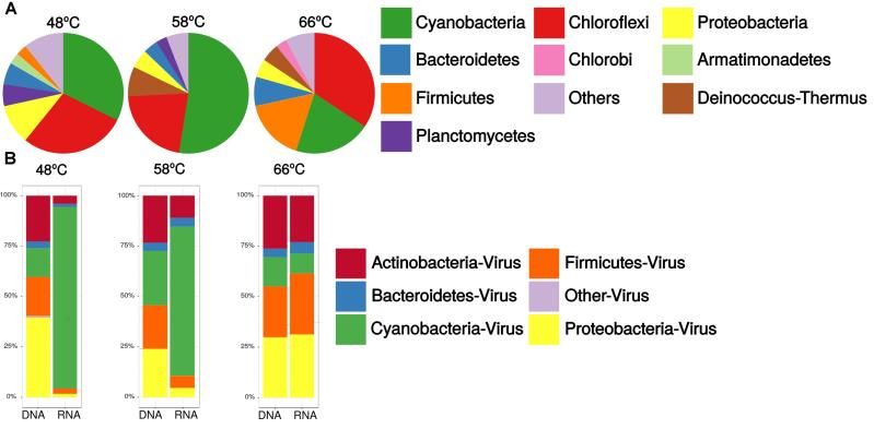
Prime Archives in Microbiology

Figure 2: Relative abundances of viral Families in microbial mats from
Porcelana hot spring; standardized by the total number metagenomic reads
(DNA), and metatranscriptome (RNA) from each temperature samples.

In the metagenomes, Siphoviridae was the most abundant family
of Caudovirales, with maximum abundance at 48°C. Myoviridae
members were also well represented with a maximum of ∼31%
at 58°C and a minimum (∼25%) at 48°C. Meanwhile,
Podoviridae accounted for just ∼8% at all temperatures (Figure
2). In metatranscriptomes, Siphoviridae increased sixfold with
temperature, while Podoviridae and Myoviridae decreased with
temperature (fivefold and twofold, respectively).

The Megavirales order was also present, however, at a lower
abundance compared to Caudovirales. Megavirales were
represented by Phycodnaviridae (∼13%), Mimiviridae (∼8%),
and Marseilleviridae (∼2%) families, remaining constant through
all temperatures. Metatranscriptomics showed an increase in
abundance of these three virus families with temperature.

Caudovirales Host Assignments

Porcelana mat communities based on miTAGs were dominated
by bacteria (∼96%), with low abundances of eukarya (∼3%) and
archaea (∼1%) (Supplementary Table S1). At the phylum level
(Figure 3A), bacterial communities were mostly composed of
Cyanobacteria oxygenic phototrophs (33, 53, and 21% of total
rRNA SSU sequences at 48, 58, and 66°C, respectively) and
Chloroflexi anoxygenic phototrophs (higher than Cyanobacteria
only at 66°C, with 35% of total rRNA SSU sequences). Other
representative members of the community were Proteobacteria
(5–11%), Deinococcus–Thermus (2–7%), Firmicutes (1–17%),
and Bacteroidetes (4–8%) (Figure 3A).

12                                                  www.videleaf.com
Prime Archives in Microbiology

Figure 3: Relative abundances of (A) Bacterial community, to Phylum level, in
the microbial mats obtained from 16S miTAGs, standardized by the total
number of metagenomic reads (DNA) for each temperature sample, and (B)
Caudovirales community at the host Phylum level, obtained from shotgun
sequences in metagenomes (DNA) and metatranscriptomes (RNA),
standardized by the total number of reads from each temperature sample.

The host assignment, based on taxonomy from viral reads of the
most representative Caudovirales (Figure 3B), showed that
viruses putatively infected members of the bacterial phyla
Proteobacteria, Cyanobacteria, Actinobacteria, and Firmicutes.
Metagenomic data showed that increases in temperature led to an
increase in viruses from Actinobacteria and Firmicutes.
Additionally, an increase in Cyanobacteria viruses was observed
at 58°C. Viruses from Proteobacteria, Actinobacteria, and
Firmicutes were represented by the three Caudovirales families,
while viruses from Cyanobacteria were represented by
Podoviridae and Myoviridae families only (Supplementary Table
S3), where cyanopodovirus and cyanomyovirus reads increase
from 31 to 50% at 48°C and from 30 to 45% at 58°C, then
decrease to 23 to 28% at 66°C, respectively.

Metatranscriptomic sequences from Caudovirales potentially
infecting Cyanobacteria, were predominant at 48°C and 58°C,
with ∼90% and ∼74% of the total viral sequences, respectively.
However, cyanophage transcripts abruptly decrease at 66°C.
Cyanophages were exclusively related to the Myoviridae and
Podoviridae families (Supplementary Table S3). Reads
associated with cyanopodoviruses and cyanomyoviruses

13                                                      www.videleaf.com
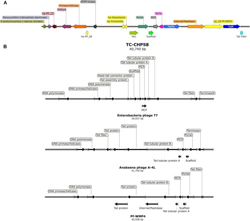
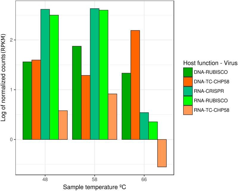
Prime Archives in Microbiology

gradually decreased with temperature; between 48 and 58°C,
virus reads declined from 95% and 96% to 84% and 89%,
respectively. On the other hand, at 66°C a more severe decline
was observed, to 15% and 20%, respectively. Conversely, with
the reduced representation of Cyanobacteria at 66°C, other
caudovirales transcripts increased, including those that infect
Proteobacteria (∼31%), Firmicutes (∼30%), and Actinobacteria
(∼23%).

Thermophilic Cyanophage Genome Recovery

The metagenome assembly recovered 3,912; 2,697; and 2,758
contigs, at 48°C, 58°C, and 66°C, respectively. A script search
[44] resulted in 11 circular contigs, possibly indicating complete
genomes. Subsequent BLASTP analysis [40] of predicted
proteins indicated that only one circular contig had viral
hallmark genes, meanwhile nine contigs had genes associated
with bacterial mobile genetic elements and one contig remain
completely unknown. These hallmark genes are shared by many
viruses but are absents from cellular genomes [59]. VirSorter
tool analysis [46] confirmed these results, obtaining the same
complete putative viral contig from the 58°C assembly, 40,740
bp long and 43.9% of GC content. This contig, TC-CHP58
(Figure 4A), was associated with a Cyanobacterial host. TC-
CHP58 was present (reads recruitment) over all temperatures in
Porcelana hot spring (Figure 5 and Supplementary Figure S1). At
66°C, TC-CHP58 was sevenfold more abundant than their
putative host (measured as Mastigocladus RUBISCO gene
abundance); at 48°C, the virus-host ratio was 1:1, and at 58°C
the host was fourfold more abundant than TC-CHP58.
Metatrancriptomic reads also show that TC-CHP58 was active
over all temperatures (Figure 5 and Supplementary Figure S2),
but with lower transcription levels than the putative host
(measured as Mastigocladus RUBISCO gene activity), ranging
between 80- and 8-fold lower (Supplementary Table S4). TC-
CHP58 viral DNA:RNA ratio indicated similar proportions (2.4)
at 58°C, least similar (552.9) at 66°C; while at 48°C the ratio
was 10.4 (Supplementary Table S4).

14                                              www.videleaf.com
Prime Archives in Microbiology

Figure 4: (A) Genomic organization of the thermophilic cyanophage CHP58
(TC-CHP58). Arrows indicate the size, position, and orientation of annotated
ORFs, with predicted functions or homologs (e.g., DNApol, DNA polymerase;
TailY a/b, tail tubular protein a/b; MCP, major capsid protein; TerL, large
terminase subunit; TailP, Tail protein; hp-PP_08/23, homologous to
hypothetical proteins 08/23 from cyanophage PP; hp-Fischerella/Gloeobacter,
homologous to hypothetical proteins in Fischerella sp. PCC 9605/Gloeobacter
kilaueensis). (B) Genomic organization of Enterobacteria phage T7, Anabaena
phage A4L and Pf-WMP4. Arrows indicate the size, position, and orientation
of viral core ORFs.

15                                                     www.videleaf.com
Prime Archives in Microbiology

Figure 5: Relative abundance and transcriptomic expression of Mastigocladus
spp. CRISPR systems and TC-CHP58. Abundance and expression of
Mastigocladus RUBISCO was used as reference of the cyanobacterial presence
and metabolic activity. Only specific CRISPR loci with proto-spacers in TC-
CHP58 were fully quantified for each temperature. For improved visualization,
counts are represented as Log of reads per kilobase million (RPKM).

Genomic Features and Organization of TC-CHP58

Complete protein prediction and annotation of TC-CHP58 using
Prokka [45] and BLASTP revealed 39 putative ORFs, 10 of
which were viral core proteins (i.e., capsid and tail-related
proteins, DNA polymerase, Terminase, etc.), 22 had no
significant similarities in NCBI nr database, and 4 were present
in the database but with unknown function (Table 1).

16                                                      www.videleaf.com
Prime Archives in Microbiology

Table 1: Blastp analysis of predicted CDS from TC-CHP58 of known function
against NCBI RefSeq (Release 75) and NR databases.

Blast analysis of the viral genes in TC-CHP58, revealed 25 to
48% identity (amino acidic level) with proteins from
Cyanophage PP, PF-WMP3 and Anabaena phage A-4L, that
infect freshwater filamentous Cyanobacteria such as
Phormidium, Plectonema, and Anabaena (Table 1). At the
nucleotide level, there was almost no similarity to any known
sequence except for a short segment of 40 nucleotides, which
showed 93% similarity to a Portal protein gene sequence of
Plectonema and Phormidium cyanopodoviruses (Cyanophage
PP; NC_022751 and PF-WMP3; NC_009551).

Gene prediction by Prodigal indicated that the TC-CHP58
genome might be structured into two clusters, based on the
transcriptional direction and putative gene functions (Figure 4A).
The predicted ORFs (Table 1) in the sense strand encode
proteins involved in DNA replication and modification, such as
DNA polymerase and DNA primase/helicase. Conversely, the
ORFs in the antisense strand (Table 1) encode proteins necessary
for virion assembly, such as major capsid protein (MCP), tail
fiber proteins, internal protein/peptidase, tail tubular proteins,
scaffold protein, and portal protein. Moreover, two ORFs in the
antisense strand had the best hits to the cyanobacterial
hypothetical proteins found in the filamentous cyanobacterium
Fischerella (WP_026731322. 1) and the unicellular Gloeobacter
(WP_023172199.1).

17                                                   www.videleaf.com
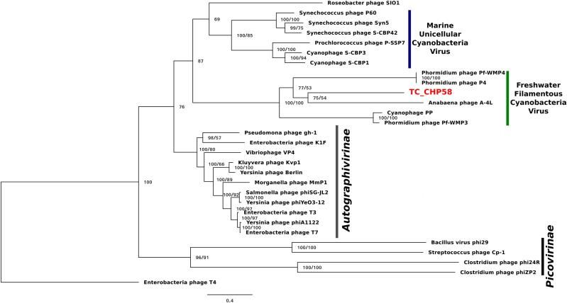
Prime Archives in Microbiology

Additionally, VIRFAM [60] was used to classify TC-CHP58
according to their neck organization (Supplementary Figure S3),
being assigned to the Podoviridae Type 3 category with neck
structural organization similar to the Enterobacteria phage P22
[60]. Hierarchical clustering of neck proteins grouped TC-
CHP58 together with the freshwater cyanophages Pf-WMP3 and
Pf-WMP4, separating them from marine cyanophages such as
P60 and Syn5.

Even when a large number of viral reads were assigned to
cyanophages of Myoviridae family, it was not possible to
recover any genome of this type. Most of the Myoviridae related
contigs only had non-structural genes or hypothetical proteins of
unknown function which align with proteins of known
cyanomyoviruses. Here, the absence of hallmark genes from
Cyanobacteria related viruses makes their accurate classification
as cyanomyoviruses impossible.

Phylogenetic Analysis of Phage TC-CHP58

To investigate the relationship of the phage TC-CHP58 within
the Podoviridae family, the DNApol gene was selected for
comparison, using published viral genomes. The analysis
included representatives of Picovirinae and Autographivirinae
subfamilies, plus all the available DNApol genes from known
freshwater podoviruses (Pf-WMP3, PP, Pf-WMP4 and A-4L)
infecting filamentous heterocystous cyanobacteria from the order
Nostocales and non-heterocystous from order Oscillatoriales,
plus those infecting marine Synechococcus spp. and
Prochlorococcus spp. The DNApol tree (Figure 6) showed the
phage TC-CHP58 as part of a monophyletic clade with all
cyanopodoviruses described as infecting freshwater filamentous
cyanobacteria, and more distantly, with the marine
cyanopodovirus clade that infects Synechococcus spp. and
Prochlorococcus spp. Both cyanophage subgroups are closely
related with podoviruses from the Autographivirinae subfamily,
which includes all T7 relatives. Furthermore, the phylogeny of
the MCP was constructed for freshwater and marine
representatives of the Autographivirinae subfamily. The
available MCP gene from BHS3 Cyanophage partial genome,

18                                             www.videleaf.com
Prime Archives in Microbiology

that is the only known thermophilic representative within the
Podoviridae family, was also included [23]. The MCP tree
(Supplementary Figure S4) showed similar results to the
DNApol tree (Figure 6), with a monophyletic origin for all
freshwater cyanophages infecting filamentous cyanobacteria,
emphasizing the division between freshwater and marine
cyanobacterial viruses, and their affiliation with T7 phage. The
thermophilic representatives of Podoviridae family were located
in different branches inside the freshwater clade, with BHS3
more basal than TC-CHP58.

Figure 6: Bayesian inference phylogenetic reconstruction of DNA polymerase
I protein of TC-CHP58. Numbers indicate Bayesian posterior probabilities as
percentage/ultra-fast bootstrap values. Only UFBoot values over 80 and
Bayesian PP over 50 are shown. The sequence characterized in the present
study is reported in bold letters. Scale bar: 0.4 amino acid substitutions per site.

CRISPR Arrays on TC-CHP58 Host

Given the high abundance [9,10] and activity [10] of
cyanobacteria, such as Mastigocladus spp., in Porcelana hot
spring (Figure 3A), and in order to confirm the putative host of
phage TC-CHP58, CRISPR spacer arrays were identified using
the CRISPRFinder tool [56] for seven Mastigocladus spp.
Contigs, obtained from metagenome assemblies at 48°C, 58°C,
and 66°C. Three CRISPR loci were common between all
temperatures      (48_CRISPR_2,        58_CRISPR_5,         and
66_CRISPR_2), while four loci were specific to higher

19                                                           www.videleaf.com
Prime Archives in Microbiology

temperatures (58–66°C) (Table 2). In total, the seven CRISPR
loci contain 562 spacers, of which 25 of them had a proto-spacer
sequence in the TC-CHP58 genome (Table 2). From the 25
spacers, 19 have a target ORF of known function, such as DNA
polymerase, dTMP, portal protein, M23-petidase, tail protein,
tail fiber, and deoxycytidine triphosphate deaminase. In general,
each CRISPR loci contained spacers against different ORFs on
TC-CHP58, or even against different locations on the same ORF.
For the 25 spacers, searching the nt/nr database, using BLASTN
and BLASTX, showed no similarity to any know sequence.
Finally, in order to check if CRISPR systems were active,
expression of the seven loci was directly quantified in the three
metatranscriptomes. For all temperatures, slightly lower
transcript levels were found compared to the Mastigocladus
RUBISCO gene (Figure 5).

Identifying Single Nucleotide Variants in TC-CHP58
Genome

To assess if mismatches between the CRISPR spacer and proto-
spacer sequences in TC-CHP58 genome were concealing
potential variations in TC-CHP58 populations, a SNV calling
was conducted. For this task LoFreq tool was used, as it is high
sensitivity and has low false positive rates, lower as
Prime Archives in Microbiology

A detailed analysis of SNVs in CRISPRs proto-spacer sites
revealed the presence of these polymorphisms in 14 of the 25
spacer targets, with 13 mismatches and 4 perfect matches (Table
2). The total number of polymorphic sites was 22, with 13 SNVs
causing a synonymous substitution and 7 causing a non-
synonymous substitution (Table 2).
Table 2:CRISPR loci at each temperature detailed information and SNVs
analysis for TC-CHP58 proto-spacer including alleles frequency and SNV
coding effect.

Discussion
The study of viruses from thermophilic phototrophic microbial
mat communities remains largely unexplored except for a few
cases providing limited information on viral presence within
these communities [16,17]. Thus far, no study has characterized

21                                                 www.videleaf.com
Prime Archives in Microbiology

viral composition and activity, or the identity of any complete
viral genome. Here, using metagenomic and metatranscriptomic
approaches, the composition of the most abundant and active
viruses associated with the dominant members of the
thermophilic bacterial community have been characterized,
describing for the first time a full genome from a thermophilic
cyanopodovirus (TC-CHP58). Moreover, the active cross-fire
between this new cyanophage and its host is demonstrated,
through TC-CHP58 population diversification (SNV), and
Mastigocladus spp. CRISPR heterogeneity, as a response to
selective pressure from the host defense system and viral
predation, respectively.

Active and Ubiquitous Cyanophage-Type Caudovirales
in Phototrophic Microbial Mats

The taxonomic classification of small subunit rRNA
(Supplementary Table S1) indicates that the phototrophic mats in
Porcelana hot spring are dominated by Bacteria (96% on
average) as commonly observed in other thermophilic
phototrophic microbial mats [2,62].

Porcelana microbial mats are mainly built by filamentous
representatives of two phototrophic phyla, Cyanobacteria
(oxygenic) and Chloroflexi (anoxygenic), with Mastigocladus,
Chloroflexus, and Roseiflexus as the main genera, respectively.
This is verified by previous surveys carried out by the authors
[9,10], as well as investigations from the White Creek,
Mushroom, and Octopus hot springs in Yellowstone
[2,13,62,63], presenting similar pH, thermal gradient and low
sulfide concentrations.

Porcelana dominant viruses (∼70% and ∼68% of metagenomic
and metatranscriptomic reads) are from the families Myoviridae,
Podoviridae, and Siphoviridae within the Caudovirales Order
(Figure 2), which typically infect Bacteria and some non-
hyperthermophilic Archaea [64]. These results were also
supported by TEM images (Figure 1). The small decrease in
transcripts associated to caudovirales with the increase in
temperature is due to the reduction of sequences related to

22                                            www.videleaf.com
Prime Archives in Microbiology

Podovirus and Myovirus families. A plausible explanation, is
that at high temperatures some representatives of these families
might have a lysogenic lifestyle, then a fraction of them will
remain inactive as prophages.

Dominance by Caudovirales was only reported recently from the
Brandvlei hot spring, South Africa, a slightly acidic (pH 5.7) hot
spring with moderate temperature (60°C) and green microbial
mat patches [23]. Previously, the presence of this viral order had
only been suggested in moderate thermophilic phototrophic mats
from Yellowstone hot springs, through indirect genomic
approximations, such as spacers in CRISPR loci, from dominant
bacterial members [16,17] or classifications based on nucleotide
motives in metaviromic data [17,28].

Contributions from megavirus sequences were also identified in
Porcelana hot spring (Figure 2), with an average of ∼24% viral
metagenomic reads, associated with unicellular eukaryotic hosts
such as those from Phycodnaviridae and Mimiviridae families,
and also the family Marseilleviridae, but to a lesser extent. The
presence of VLPs from these three viral families could not be
corroborated through TEM, using the limited available viral
fraction (
Prime Archives in Microbiology

difficult to accurately estimate the relative abundances or activity
of specific phages at low ranks of taxonomy tree, such as the
species level [67]. To avoid this problem, our strategy focused
on the use of the LCA algorithm at higher taxonomic levels
(Order and Family) to classify the viral reads, as well as for the
inferred hosts, we use the phylum level.

Virus-host inference in Porcelana phototrophic mats (Figure 3B),
demonstrated that the most frequent targets for viral infections
were the most dominant and active components of the bacterial
communities. Similarly, this is the case in other environments,
such as in the human microbiome [68] and marine communities
[69,70]. In Porcelana, it is demonstrated that within microbial
mats at 48°C and 58°C, cyanophages were among the most
active viruses (Figure 3B), as were Cyanobacteria, such as
Mastigocladus spp., as exemplified in terms of primary
production and nitrogen fixation [10]. The presence of
cyanophages has been previously suggested in Yellowstone hot
spring phototrophic mats [16,17], and more recently in the
Brandvlei hot spring, South Africa [23]. Heidelberg et al. [16]
found that CRISPR spacers in unicellular cyanobacteria
Synechococcus isolates (Syn OS-A and Syn OS-B9) from
Octopus Hot Spring, might have 23 known viral targets
(lysozyme-related reads, PFAM DUF847) on an independently
published metavirome from the same hot spring. More recently,
171 viral contigs associated with the host genus Synechococcus,
based on tetranucleotide frequencies, were identified from a
microbial mat (60°C) metavirome from Octopus Spring. The
majority of the annotated ORFs on the viral contigs coded for
glycoside hydrolases, with lysozyme activity, identifying six
CRISPR proto-spacers in those genes [17]. Even though a
taxonomic relationship with cyanophages was not confirmed for
those proto-spacers containing contigs [16,17], it provides
evidence toward the presence of cyanophages related sequences
within these thermophilic mats. The work by Zablocki et al. [23]
reconstructed a 10 kb partial genome of a new cyanophage
(BHS3) from Brandvlei hot spring metavirome, stating that
cyanophages appear to be the dominant viruses in the hot spring.
The BHS3 contig (MF098555) contains nine ORFs, with the
majority of the identified proteins having a close relation to the

24                                               www.videleaf.com
Prime Archives in Microbiology

Cyanophage PP and Phormidium phage Pf-WMP3, which infect
freshwater filamentous cyanobacteria Phormidium and
Plectonema.

The presence of cyanophages related sequences in thermophilic
phototrophic mats is significant, since these viruses are known to
play an important role in the evolution of cyanobacteria [71].
Cyanophages affect the rate and direction of cyanobacterial
evolutionary processes, through the regulation of abundance,
population dynamics, and natural community structure. This has
been extensively studied and demonstrated for marine
environments [72,73]. These cyanophages are proven to play a
relevant role in the marine biogeochemical cycles, through the
infection and lysis of Cyanobacteria, affecting carbon and
nitrogen fixation [74]. Moreover, cyanophages act as a global
reservoir of genetic information, as they are vectors for gene
transfer, meaning that cyanobacteria can acquire novel attributes
within aquatic environments [75,76].

Caudoviruses were prevalent at 66°C in Porcelana, and
potentially    infecting    Firmicutes,    Proteobacteria,     and
Actinobacteria. These phila have also been previously identified
in other hot springs at temperatures above 76°C, such as in
Octopus and Bear Paw [28]. At high temperatures in Porcelana
also the phylum Chloroflexi was dominant in the phototrophic
mat (Figure 3A). However, viral sequences related to this taxon
could not be retrieved, as neither viruses nor viral sequences
have been confirmed to infect members of this phylum in any
environment. Davison et al. [17], described viral contigs
associated with Roseiflexus sp. from a metavirome from Octopus
Spring, but only raw reads are publicly available, without
taxonomic assignation. Finally, the recently released IMG/VR
database [77] contains three contigs associated by CRISPR
spacers to Chloroflexus sp. Here, a BLASTP analyses against
RefSeq viral proteins revealed that six of these proteins have a
best hit in Mycobacterium phage proteins and one which best hit
was a Clavibacter phage protein. These findings, suggest that
some of the viral reads classified as Actinobacteria viruses could
be instead from unknown Chloroflexi viruses.

25                                              www.videleaf.com
Prime Archives in Microbiology

Viral Mining Reveals a New Infective Thermophilic
Cyanopodovirus Lineage

Metagenomic surveys of viral genomes are an effective way to
detect unknown viruses [46,66,78,79]. In metagenomics, two key
elements for virus detection are the presence of viral hallmark
genes and the circularity of viral contigs [46,78]. Based on these
two principles, a complete genome (TC-CHP58) was identified.
The genome was represented by a viral contig of 50 kb, which is
a typical size for Caudovirales members from the Podoviridae
family. The genome size and viral core proteins affiliated with
the Podovirus seems to make TC-CHP58 the first report of a full
genome of a thermophilic cyanopodovirus. Moreover, the
genome organization (Figure 4B) shows a consistent synteny
with other cyanopodoviruses, which also lack RNA polymerase
inside the T7 supergroup, as described for the viruses Pf-WMP4,
Pf-WMP3, Cyanophage PP, Anabaena phage A-4L [80-83], and
the recently reported partial genome of the thermophilic BHS3
cyanophage [23]. Initially, the presence of a single-subunit RNA
polymerase that binds phage specific promoters was considered
to be a major, and unique characteristic of the T7 supergroup
[84]. However, more recently, it has been proposed that
podoviruses that share extensive homology with T7, but lack the
phage RNA polymerase, are still part of the T7 supergroup, as
distant and probably ancient branches [85].

TC-CHP58 presented a genome organization that can be divided
into two portions (Figure 4A); with ORFs in the sense strand
related to DNA replication and modification, and genes encoded
in the antisense strand related to virion assembly. This genome
organization is also present in other freshwater T7-related
podoviruses that infect filamentous cyanobacteria [80,82,83],
including the thermophilic BHS3 cyanophage [23]. This setup is
also similar to the class II and III organization genes in T7-like
viruses, where class II genes are responsible for DNA replication
and metabolism, and class III genes include structural and
maturation genes [84]. The VIRFAM analysis of neck protein
organization verifies the classification of TC-CHP58 within the
Podoviridae family (Supplementary Figure S3), where the Type
3     podovirus      encompasses       T7-like    phages     from

26                                              www.videleaf.com
Prime Archives in Microbiology

Autographivirinae subfamilies and several other genera [60]. The
T7-like classification for TC-CHP58, and other podoviruses that
infect freshwater filamentous cyanobacteria, is supported by the
organization of the genome into two portions as well as the
organization of the neck proteins.

The phylogenetic position of TC-CHP58, based on DNA
polymerase I (DNApol) (Figure 6) and MCP (Supplementary
Figure S4) predicted proteins, confirm the affiliation of this new
virus within the family Podoviridae. Both phylogenetic markers
verify the separation between the marine from the freshwater
cyanopodoviruses within the T7 family, as previously proposed
[80,83]. These results also support the connection between the
T7 phages and marine and freshwater cyanopodoviruses
[80,83,85,86], including TC-CHP58 and BHS3 as
representatives of a novel, and potentially globally distributed
thermophilic cyanophage lineage. Moreover, this data
demonstrates that marine and freshwater cyanopodoviruses,
including the thermophilic TC-CHP58, are part of the
Autographivirinae subfamily as previously suggested for
Cyanophage P60 and Roseophage SIO1 [87], both included in
this analysis.

In Porcelana, the virus host ratio relating to TC-CHP58 presence
was lower than the typical values observed in freshwater
environments [88], being more similar to other geothermal
environments where viral density is typically lower, with 10- to
100-fold less viruses than host cells [89]. This is expected,
considering that there are abundant cyanobacteria in
phototrophic mats in Porcelana in comparison with the 104 mL-1
VLPs observed in the water of hot springs [24]. It is also
demonstrated that TC-CHP58 presented higher infection
efficiency, as revealed by the viral DNA to RNA ratios at lower
temperatures (58°C, then 48°C) with cyanobacteria dominating,
while at 66°C most of the TC-CHP58 remained inactive (Figure
5). Infection inefficiency is multidimensional, as it initiates from
reduced phage adsorption, RNA, DNA, and protein production
[90]. Thus, the high copy number of TC-CHP58 DNA at 66°C
may be due to the persistence of viral DNA [91] encapsidated
extracellularly and intermixed in the microbial mat were the host

27                                               www.videleaf.com
Prime Archives in Microbiology

(Mastigocladus spp.) has a low activity as evidenced by the low
expression of the RUBISCO gene and the CRISPR loci. An
alternative explanation is the absence, or the diminished
presence, of the specific host due to intraspecific diversification
as evidenced by the existence of different CRISPR loci at
different temperatures. This theory has been proposed for other
cyanobacteria, such as Prochlorococcus and Phormidium, where
slight differences in fitness, niche, and selective phage predation,
explain the coexistence of different populations [79,92]. The last
explanation acquires special importance in light of recent
evidence that variations in the structure and function of the
heterocyst and differential CRISPR loci are fundamental to
diversification of Mastigocladus laminosus (also known as
Fischerella     thermalis),      a    cosmopolitan     thermophilic
cyanobacterium, reinforcing the importance of viral predation
[93].

CRISPR Spacers Assign Mastigocladus spp. as Putative Hosts for
TC-CHP58

It was possible to verify Mastigocladus spp. as putative hosts for
the new cyanopodovirus (TC-CHP58), via the analysis of
CRISPR spacers found in the cyanobacteria, recovered from
contigs obtained in the same metagenomic datasets. This
methodology has been previously used for the identification of
novel viruses in hot springs [16,17,94], as well as in other
environments such as acid mines [95] the human microbiome
[96], as well as sea ice and soils [97].

Observations from the CRISPR loci over all temperatures (Table
2) indicated that, in general, proto-spacers in the TC-CHP58
genome were distributed on coding, and therefore more
conserved regions. The expression of seven CRISPR loci (Figure
5), demonstrated the activity of the Mastigocladus spp. defense
system against TC-CHP58 over all temperatures. CRISPR arrays
are transcribed into a long precursor, containing spacers and
repeats, that are processed into small CRISPR RNAs (crRNAs)
by dedicated CRISPR-associated (Cas) endoribonucleases [98].
Although it is not possible to measure mature crRNAs, as due to
their small size they are likely to be filtered out in RNA-seq

28                                               www.videleaf.com
Prime Archives in Microbiology

libraries, this approximation has been validated using large
datasets [99].

Despite variations in the number of CRISPR loci observed at
each temperature, with 60% of the total CRISPR loci found in
Mastigocladus contigs at 58°C, the abundance of reads agreed
with the abundance of other genes required by these
cyanobacteria, such as the RUBISCO gene (Figure 5). This
further verified that the loci are from Mastigocladus populations.
The different CRISPR loci found over the different temperatures
in Porcelana (Table 2), also reinforces the notion that
diversification of Mastigocladus is partly due to selective
pressure exerted by the predation of viruses, such as TC-CHP58.
This theory has been previously put forward for Mastigocladus
laminosus in Yellowstone [93], and proposed for marine
cyanobacteria [92,100].

Furthermore, each CRISPR loci contains spacers that
corresponds to different proto-spacers in the TC-CHP58 genome.
Increases in spacer number and diversity against the same virus
may explain the increase in interference, whilst decreasing the
selection of escape mutants [10]. Priming mechanisms are the
most efficient form of obtaining new spacers [101], using a
partial match between a pre-existing spacer and the genome of
an invading phage to rapidly acquire a new “primed” spacer
[102]. Then, over-representation of spacer sequences in some
regions of the TC-CHP58 genome may be related to a site that
has already been sampled by the CRISPR-Cas machinery or by
other biases such as the secondary structure of phage ssDNA,
GC content, and transcriptional patterns [103].

The selection pressure of multiple spacers in Mastigocladus
CRISPR loci leads to the emergence of SNVs in the TC-CHP58
viral populations (Table 2), which cause mismatches between
spacers and proto-spacers, resulting in the attenuation or evasion
of the host immune response [104]. It is still possible to utilize
mismatched spacers for interference and/or primed adaptation,
however, the degree of tolerance to mismatches for interference
among the CRISPR-Cas, varies substantially between different
CRISPR-Cas type systems [104]. The variable frequency (0.6–

29                                              www.videleaf.com
Prime Archives in Microbiology

0.02) of the corresponding spacer SNVs alleles on TC-CHP58
proto-spacers, suggests that some variants are more prevalent
throughout the population, regardless of whether the SNV causes
a silent mutation. Based on this evidence, it has been proposed
that, for other microbial communities, only the most recently
acquired spacer can exactly match the virus. This suggests that
community stability is driven by compensatory shifts in host
resistance levels and virus population structure [95].

The present study describes the underlying viral community
structure and activity of thermophilic phototrophic mats.
Moreover, abundant virus populations are linked to dominant
bacteria, demonstrating the effectiveness of omics approaches in
estimating the importance and activity of a viral community, in
this case with thermophilic cyanophages.

Additionally, the first full genome of a new T7-related virus that
infects thermophilic representatives of the cyanobacterium
Mastigocladus spp. was here retrieved. This genome may
represent a novel, globally present, freshwater thermophilic virus
from a new lineage from the Podoviridae family. The latter was
strongly suggested by the significant phylogenetic relationship
and shared gene organization with the BHS3 cyanophage partial
genome (South Africa). Even more, TC-CHP58 proteins also
matches several contigs that include common viral hallmarks
genes in the IMG/VR database. However, further work is
necessary to fully understand the global representation and
relevance of this virus, which complete genome is presented here
as first reference available.

Finally, the evolutionary arms race between a specific
cyanobacteria-cyanophage in the natural environment is
exposed, where a there exist a variety of potential scenarios. For
instance, host resistance may increase over time forcing the
decrease of viral populations, or a specific virus population may
occasionally become extremely virulent and cause the crash of
the host population as proposed by the “kill the winner” model
[95]. Alternatively, if CRISPR systems and the diversification of
the viral population remain in balance through time, a relatively
stable virus and host community may result.

30                                              www.videleaf.com
Prime Archives in Microbiology

Footnotes

     1. ^sourceforge.net/projects/bbmap/
     2. ^http://beast.bio.ed.ac.uk/Tracer

References
1. Uldahl K, Peng X. Biology, biodiversity and application of
   thermophilic viruses. In: T Satyanarayana, KY Littlechild
   Jennifer, editors. Thermophilic Microbes in Environmental
   and Industrial Biotechnology. Berlin: Springer. 2013; 271–
   306.
2. Inskeep WP, Jay ZJ, Tringe SG, Herrgård MJ, Rusch DB.
   The YNP metagenome project: environmental parameters
   responsible for microbial distribution in the yellowstone
   geothermal ecosystem. Front. Microbiol. 2013; 4: 67.
3. Steunou AS, Bhaya D, Bateson MM, Melendrez MC, Ward
   DM, et al. In situ analysis of nitrogen fixation and metabolic
   switching in unicellular thermophilic cyanobacteria
   inhabiting hot spring microbial mats. Proc. Natl. Acad. Sci.
   U.S.A. 2006; 103: 2398–2403.
4. Steunou AS, Jensen SI, Brecht E, Becraft ED, Bateson MM,
   et al. Regulation of nif gene expression and the energetics of
   N2 fixation over the diel cycle in a hot spring microbial mat.
   ISME J. 2008; 2: 364–378.
5. Bhaya D, Grossman AR, Steunou AS, Khuri N, Cohan FM,
   et al. Population level functional diversity in a microbial
   community revealed by comparative genomic and
   metagenomic analyses. ISME J. 2007; 1: 703–713.
6. Klatt CG, Wood JM, Rusch DB, Bateson MM, Hamamura
   N, et al. Community ecology of hot spring cyanobacterial
   mats: predominant populations and their functional potential.
   ISME J. 2011; 5: 1262–1278.
7. Stewart W. Nitrogen fixation by blue-green algae in
   Yellowstone thermal areas. Phycologia. 1970; 9: 261–268.
8. Miller SR, Purugganan M, Curtis SE. Molecular population
   genetics and phenotypic diversification of two populations of
   the thermophilic cyanobacterium Mastigocladus laminosus.
   Appl. Environ. Microbiol. 2006; 72: 2793–2800.

31                                             www.videleaf.com
You can also read