Cellular oxidative stress stimulated by microcystin: review
←
→
Page content transcription
If your browser does not render page correctly, please read the page content below
Research, Society and Development, v. 10, n. 11, e422101119765, 2021
            (CC BY 4.0) | ISSN 2525-3409 | DOI: http://dx.doi.org/10.33448/rsd-v10i11.19765

Cellular oxidative stress stimulated by microcystin: review
Estresse oxidativo celular estimulado por microcistina: revisão
Estrés oxidativo celular estimulado por microcistina: revisión

                                                    Received: 08/23/2021 | Reviewed: 08/29/2021 | Accept: 09/03/2021 | Published: 09/05/2021

                                                                                                            Iara Bezerra de Oliveira
                                                                                             ORCID: https://orcid.org/0000-0003-1382-7296
                                                                                              Federal University of Campina Grande, Brazil
                                                                                                            E-mail: iara_bio@yahoo.com.br
                                                                                                              Hanndson Araújo Silva
                                                                                             ORCID: https://orcid.org/0000-0001-8914-9589
                                                                                              Federal University of Campina Grande, Brazil
                                                                                                             E-mail: hanndson@gmail.com

      Abstract
      Introduction: Cyanobacteria are organisms capable of producing a high number of bioactive molecules, known as
      cyanotoxins. Among the cyanotoxins, microcystins stand out, compounds with hepatotoxic potential. Studies claim
      that the most common and most toxic isoform among microcystins is microcystin-LR. One of the most frequently
      detected properties of microcystins is their ability to generate cellular oxidative stress. Thus, the present study is a
      bibliographic research about the biochemical mechanism of free radical generation caused by Microcystin LR.
      Methodology: for the preparation of this review, a survey was carried out in the national and international literature.
      The inclusion criteria for the construction of this work were original and review articles that addressed the ability of
      microcystin LR to generate oxidative damage. Results: Once they enter the body, microcystins accumulate in the liver,
      so that toxicity is associated with specific inhibition of protein phosphatase 1 and 2A (PP1 and PP2A), leading to
      disruption of cell integrity. Studies prove that MCs produce oxidative stress in vitro and in vivo and that they can act
      as tumor promoters. Conclusion: there is a possible relationship between cellular oxidative stress caused by
      microcystin. Thus, cyanobacterial blooms represent a threat to the health of several animals, including man, however,
      further studies on the topic addressed are needed.
      Keywords: Free radicals; Cyanotoxins; Toxicity.

      Resumo
      Introdução: Cianobactérias são organismo capazes de produzir um alto número de moléculas bioativas, conhecidas
      como cianotoxinas. Dentre as cianotoxinas destaca-se as microcistinas, compostos com potencial hepatotóxico.
      Estudos afirmam que a isoforma mais comum e mais tóxica entre as microcistinas é a microcistina-LR. Uma das
      propriedades das microcistinas mais frequentemente detectadas é a sua capacidade de gerar estresse oxidativo celular.
      Desse modo, o presente estudo trata-se de uma pesquisa bibliográfica acerca do mecanismo bioquímico de geração de
      radicais livres ocasionado pela Microcistina LR. Metodologia: para a elaboração desta revisão foi realizado um
      levantamento na literatura nacional e internacional. Os critérios de inclusão para construção desse trabalho foram
      artigos originais e de revisão que abordavam a capacidade da microcistina LR gerar o dano oxidativo. Resultados: As
      microcistinas, uma vez que entram no organismo, se acumulam no fígado, de modo que a toxicidade está associada à
      inibição específica da proteína fosfatase 1 e 2A (PP1 e PP2A), levando a interrupção da integridade celular. Estudos
      comprovam que as MCs produzem estresse oxidativo in vitro e in vivo e que podem atuar como promotores tumorais.
      Conclusão: existe uma possível relação entre o estresse oxidativo celular ocasionados pela microcistina. Assim, as
      florações de cianobactérias representam uma ameaça à saúde de diversos animais, inclusive o homem, no entanto, se
      faz necessário mais estudos acerca do tema abordado.
      Palavras-chave: Radicais livres; Cianotoxinas; Toxicidade.

      Resumen
      Introducción: Las cianobacterias son organismos capaces de producir un elevado número de moléculas bioactivas,
      conocidas como cianotoxinas. Entre las cianotoxinas destacan las microcistinas, compuestos con potencial
      hepatotóxico. Los estudios afirman que la isoforma más común y más tóxica entre las microcistinas es la microcistina-
      LR. Una de las propiedades de las microcistinas detectadas con mayor frecuencia es su capacidad para generar estrés
      oxidativo celular. Así, el presente estudio es una investigación bibliográfica sobre el mecanismo bioquímico de
      generación de radicales libres causado por Microcystin LR. Metodología: para la elaboración de esta revisión se
      realizó un relevamiento en la literatura nacional e internacional. Los criterios de inclusión para la construcción de este
      trabajo fueron artículos originales y de revisión que abordaron la capacidad de la microcistina LR para generar daño
      oxidativo. Resultados: una vez que ingresan al cuerpo, las microcistinas se acumulan en el hígado, por lo que la
                                                               1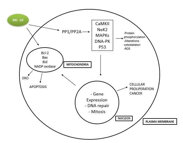
Research, Society and Development, v. 10, n. 11, e422101119765, 2021
               (CC BY 4.0) | ISSN 2525-3409 | DOI: http://dx.doi.org/10.33448/rsd-v10i11.19765

         toxicidad se asocia con la inhibición específica de la proteína fosfatasa 1 y 2A (PP1 y PP2A), lo que conduce a la
         alteración de la integridad celular. Los estudios demuestran que los MC producen estrés oxidativo in vitro e in vivo y
         que pueden actuar como promotores de tumores. Conclusión: existe una posible relación entre el estrés oxidativo
         celular causado por microcistina. Por lo tanto, las floraciones de cianobacterias representan una amenaza para la salud
         de varios animales, incluido el hombre, sin embargo, se necesitan más estudios sobre el tema abordado.
         Palabras clave: Radicales libres; Cianotoxinas; Toxicidad.

1. Introduction
         Climate change is transforming ecosystems and their composition across the planet. In recent years, studies have
indicated eutrophication, increased CO2 levels and global warming as responsible for the frequency, intensity and duration of
cyanobacterial proliferation in different ecosystems worldwide (Rastogi, et al, 2014).
         Eutrophication is considered one of the main events that affect water bodies and, consequently, water parameters. The
eutrophication process interferes with the physical and chemical characteristics of the water and can mediate profound changes
in the qualitative and quantitative conditions of aquatic communities. In this way, several problems are induced, such as the
proliferation of toxic algae, known as cyanobacteria (Carpenter, 2005; Dhanam, et al, 2016).
         Cyanobacteria are prokaryotic and photosynthetic microorganisms present in the most diverse terrestrial
environments. Planktonic representatives are of special interest, as a result of ecological success and competitive strategies,
they are among the pioneer organisms of terrestrial life and present themselves to the present day, producing a range of
secondary metabolites that increasingly arouse scientific interest (Carmichael, 1994; Carmichael & Boyer, 2016).
Cyanobacteria were pioneering organisms on primitive Earth, and the oxygen produced through photosynthesis for the
cyanobacteria contributed to the formation of the ozone layer. However, these microorganisms are currently known for their
potentially toxic flowering, causing problems for water treatment and being of potential risk to human health (Szlag, et al,
2015).
         Known as cyanotoxins, the metabolites produced by cyanobacteria in natural concentrations are toxic to plants,
invertebrates and vertebrates, including humans. Those, in turn, comprise several classes, with different mechanisms of action
and their own characteristics (Chorus, 2000).
         The accelerated growth of cyanobacteria and the formation of blooms lead to a potential increase in the concentration
of toxins in the water, which represents a serious risk to public health. Toxins, produced by cyanobacteria, can cause changes
in the taste, odor of water, and cause toxicity in aquatic biota, which can lead animals to death from liver failure and the
development of cancerous tumors. They can also accumulate in the tissues of fish intended for human consumption. In
addition, the long-term effects of cyanotoxins in humans are still unclear (Carmichael & Boyer, 2016; Janssen, 2019; Metcalf
& Codd, 2020).
         In the case of cyanotoxins, liver toxins, such as microcystins (MCs), are the most abundant and widely studied natural
toxins (Teneva, et al., 2016). More than 90 variants of CM have been detected (Pearson, et al, 2010), among which
microcystin-LR (MC-LR) is the most widely distributed and toxic (Gupta, et al., 2003; Rastogi, et al., 2014). It is known that
MC-LR is a hepatotoxin that acts in the intense inhibition of intracellular serine / threonine phosphatases 1 and 2A (PP1 and
PP2A) (MacKintosh, et., 1995). Because of this inhibition, an imbalance of cell phosphorylation occurs, culminating in a cell
signaling disorder (Humpage & Falconer, 2003; Sotton, et al., 2012). In addition, it can cause oxidative stress due to the
intracellular excess of reactive oxygen species (ROS) considered, therefore, another important mechanism of hepatotoxicity
MC-LR (Ma, et al., 2017). Studies reveal that apoptosis induced by MC-LR is possibly mediated by the mitochondrial pathway
where ROS and transcription factor such as NF-kB and p53 may be involved (Fu, et al., 2005; Feng, et al., 2011; Ji, et., 2011).
         In these terms, the present study aimed to collect and summarize detailed and available data on the occurrence of

                                                               2
Research, Society and Development, v. 10, n. 11, e422101119765, 2021
(CC BY 4.0) | ISSN 2525-3409 | DOI: http://dx.doi.org/10.33448/rsd-v10i11.19765
cyanotoxins worldwide and the human and animal intoxications associated with cyanotoxins. Seeking to provide an in-depth
analysis, which this information providers of its toxic potential, as well as providing a discussion of its toxicity in the activity
related to the generation of reactive oxygen species and consequently generation of cellular oxidative damage, in order to
understand the risks to human health caused by MCs.results.
2. Methodology
This research can be considered of the conceptual-theoretical type because it focuses on conducting a systematic
literature review, followed by a structured analysis of the contents published on the subject. An inductive analysis method is
adopted because, based on the information collected in the publications, their analysis and classification, the logical structure
on the topics that involve the theme presented follows an inference criterion. Systematic literature review is a reliable research
approach due to its comprehensiveness and explicit presentation of the means and results obtained. Compared to the traditional
review, it includes a clear statement of the purpose of the review, a thorough search for publications, a critical evaluation of the
main publications and the possibility of replicating the research method. (Pai, et al., 2004).
In the present study, was carried out with a search for articles in the Pubmed, Science Direct, SciELO databases,
between the months of April 2020 and June 2021, using the descriptors according to the theme. Thus, the research of articles
was divided into 2 parts, of which the objective would be to list the information more precisely for each survey, with the
keywords:1) “Oxidative Stress”; “Cyanotoxin” and “Microcystin”: to conduct a literature survey that addressed the
biochemical mechanisms of cellular oxidative stress and the toxicology of cyanotoxins. 2) “Cyanotoxin and Oxidative Stress”
and “Microcystin and Oxidative Stress”: to obtain studies that addressed the relationship of cyanotoxins with cellular oxidative
stress.
The articles were selected after reading the title and abstract, and the inclusion criteria were: articles published any
year dealing with cyanotoxins and oxidative stress, and from 2010 to 2021 on the relationship of oxidative stress and
microcystins, available in full and that dealt with the theme.
3. Cyanobacteria
Cyanotoxins
Cyanotoxins are toxins produced by some species of cyanobacteria in fresh or salt water. These toxic substances can
be stored in the cell's cytosol and are released if cell lysis occurs, they are classified as hepatotoxins (microcystin and
nodularin), neurotoxins (anatoxin-a, homoanatoxin-a and saxitoxin), cytotoxins (cylinderspermopsin) and dermatoxins
(lingbiatoxin) (Carmichael & Boyer, 2016) (See Table 1). Some of the most important roles that cyanotoxins can play for the
algae that produce them are to prevent the herbivore, as they can function as allopathic substances and even as signaling
molecules between species and individuals (Carmichael, 1994; Svirčev, et al., 2017). Some of these toxins, which are
characterized by their rapid action, causing death by respiratory arrest after a few minutes of exposure, have been identified as
neurotoxic alkaloids or organophosphates. Others act less quickly and are identified as hepatotoxic peptides or alkaloids. These
are the two main groups of cyanotoxins that can be classified as: neurotoxins and hepatotoxins (Azevedo, 1998; Andrinolo, et
al., 1998).
3Research, Society and Development, v. 10, n. 11, e422101119765, 2021
(CC BY 4.0) | ISSN 2525-3409 | DOI: http://dx.doi.org/10.33448/rsd-v10i11.19765
Table 1: Classification as to the pharmacological action and chemical structure of the main cyanotoxins.
ACTION STRUCTURE TYPE
Hepatotoxins Cyclic Peptides Microcystin
Nodularin
Alkaloid Cylinderspermopsin
Dermatotoxins Lipid and Carbohydrate Lipopolisacaride
Neurotoxins Alkaloid Saxitoxin
Anatoxin-a
Homoanatoxin-a
Anatoxin-a (S)
Source: Authors.
Hepatoxoxins
The main hepatotoxins characterized so far are cyclic hepatapeptides known as microcystins and pentapeptides
designated as nodularins. The general structure of microcystins is D-Ala-X-D-MeAsp-Z-Adda-D-Glu-Mdha, where X and Z
are the two variable L amino acids, D-MeAsp is D-erythro metilaspartic acid and Mdha is N-methyl -hydroalanine (See Figure
1 - A) (Carmichael, 1994; Meriluoto, et al., 2016; Bouaïcha, et al., 2019).
Nodularins (NODs) are cyclic pentapeptide hepatotoxins with the overall ciclo structure (-d - erythro - β - methylAsp
(iso - bond) –l - Z - Adda - d - Glu (iso - bond) -2 - (methylamino) -2 Acid (Z) - dehydrobutyric) (See Figure 1 - B) (Meriluoto,
et al., 2016). NODs share many structural and functional properties with MCs, the primary without two aminoalkanoic acid
residues and showing a substitution of the N-methyl-hydroalanine residue with N-methyl-hydrobutyrine. The second
aminoalkanoic acid residue (Z) is l-Arg within the frequently occurring nodularin-R (also referred to as “nodularin” without
suffix) (Meriluoto, et al., 2016).
The qualitative variations observed in the two L amino acids in microcystins are used to designate their different
variants, for example, microcystin-LR (leucine-arginine); -RR (arginine-arginine); -YA (tyrosine-alanine). Adda, is 3-amino-9-
methoxy-2,6,8-trimethyl-10-phenyl-deca-4,6-dienoic acid, which is also present in nodularins and has been determined to be
responsible for the biological activity of these hepatotoxins (Meriluoto, et al.,2016; Bouaïcha, et al., 2019 ).
Hepatotoxins reach hepatocytes through bile acid receptors and promote a disorganization of the intermediate
filaments and actin filaments, which are polymers of proteins that are part of the cytoskeleton (Yan, et al., 2020). This
disorganization leads to a retraction of the hepatocytes, causing loss of contact between them and the cells that form the
sinusoidal capillaries. Consequently, the liver loses its architecture and develops serious internal injuries. The loss of contact
between the cells creates internal spaces that are filled by the blood that begins to flow from capillaries to these locations
(Carmichael, 1994; Yan, et al., 2020).
Through the study of the mechanisms of action of these hepatotoxins, it was demonstrated that several microcystins
and nodularins are potent inhibitors of type 1 and 2A eukaryotic cell protein phosphatases. These toxins are recognized as
potent liver tumor promoters (Yan, et al., 2020; Takai, et al., 2018).
Nodularins have stronger tumor initiation and promotion activities compared to MCs. this will stem from its smaller
ring structure, allowing easier ingestion by hepatocytes (Carmichael, et al., 1994).
Like MCs, NODs also induce the assembly of reactive intracellular oxygen species, causing peroxidation of lipids,
proteins and DNA (Žegura, et al., 2011).
4Research, Society and Development, v. 10, n. 11, e422101119765, 2021
(CC BY 4.0) | ISSN 2525-3409 | DOI: http://dx.doi.org/10.33448/rsd-v10i11.19765
Figure 1: Structural formula of hepatotoxins, A - Microcystin and B - Nodularine.
(A)
(B)
Source: Meriluoto, et al. (2017).
Cylindrospermopsins (CYNs) also are considered hepatotoxins, but they're guanidine alkaloids, produced by a series
of cyanobacterial genera (Li, 2012). Currently, five CYN analogs are known, namely, CYN, 7 - epi - CYN and seven - deoxy -
CYN (See Figure 2) and therefore the two recently characterized congeners, 7 - deoxidesulfo - CYN and seven - deoxidesulfo -
12 - acetyl - CYN (Wimmer, et al., 2014). The structures of CYN and 7-epi‐ CYN have repeatedly undergone name changes,
with each at some point being called CYN. at the present, 7‐ (S) -CYN is considered the most toxin produced by
Cylindrospermopsis raciborskii. supported the reassessment of absolutely the C-7 configuration of the isolated CYN, revised
synthesis, and asymmetric synthesis, absolutely the configuration of all asymmetric CYN centers, 7 - epi - CYN and seven -
deoxy - CYN were established. it had been concluded that the names remain an equivalent as they were assigned (Meriluoto, et
al., 2016).
CYN is a strong inhibitor of protein synthesis. The liver is its main target, with four successive stages of pathological
alterations in: inhibition of protein synthesis, membrane proliferation, accumulation of fat droplets and cell death (Moreira, et
al., 2013).
Studies show that exposure to CYN led to a decrease in the decreased glutathione (GSH) content in rat hepatocytes.
This reduction has been attributed to the inhibition of GSH synthesis (Moreira, et al., 2013; Runnegar, et al. 2002).
Norris et al., 2002, suggested that the activation of CYN by cytochrome P450 is of fundamental importance in its
mechanism of action. Initiation of CYN by cyt-p450 results in increased toxicity; therefore, it is regarded as a progenotoxic
substance (Kinnear, 2010). In addition, oxidative stress can play a significant role in CYN toxicity in vitro (Gutiérrez-Praena,
et al., 2011a), and in vivo Gutiérrez-Praena, et al., 2011b).
In general, CYN appears as a toxic molecule with wide reach. It disturbs various organs and metabolic pathways, both
directly and after some metabolic alteration. The absence of a particular objective for CYN hinders further efforts to
comprehend its potent toxicity and classify appropriate exposure limits (Meriluoto, et al., 2016).
5Research, Society and Development, v. 10, n. 11, e422101119765, 2021
(CC BY 4.0) | ISSN 2525-3409 | DOI: http://dx.doi.org/10.33448/rsd-v10i11.19765
Figure 2: Structural formula of cylindropemopsin.
Source: Meriluoto (2007).
Among all cyanotoxins, the most common type of poisoning involving cyanobacteria is related to the presence of
hepatotoxins in water bodies. Hepatotoxins act mainly on liver cells, where, by different mechanisms, they mediate the
destruction of liver cells, leading all tissues to necrosis and intrahepatic hemorrhage, which results in the animal's death in a
few hours in acute doses. In addition to acting on the liver, hepatotoxins can accumulate in other organs, such as kidneys,
stomach, lung, brain, gonads and heart (Mohamed, et al., 2020).
Neurotoxins
Neurotoxins belong to three chemically distinct families, anatoxin-a and homoanatoxin-a, which mimics the effect of
acetylcholine; anatoxin-a (s), which is an anticholinesterase, and saxitoxins, which block the sodium channels of nerve cells
(Andrinolo, et al., 2008). Neurotoxins are produced by species and lineages included in the genera: Anabaena,
Aphanizomenon, Oscillatoria, Trichodesmium and Cylindrospermopsis (Buratti, et al., 2017; Svirčev, et al., 2017).
Anatoxin-a (ANTX) and homoanatoxin-a (HANTX) are effective neurotoxins, created by several of cyanobacterial
species. ANTXs contaminate the lakes and rivers and have been linked to several animal deaths. ANTX is characterized by a
very fast death factor for animals that have consumed toxic cyanobacteria.
More ANTX derivatives have additionally been identified in samples at different concentrations. ANTX and HANTX
undergo quick chemical decay in nature; the main degradation products constitute dihydro and epoxy analogs (Stevens &
Krieger, 1991).
Anatoxin-a was the first cyanobacterial toxin to be chemically and functionally defined, it is a low molecular weight
secondary amine, (1 - [(1R, 6R) -9-azabicyclo [4.2.1] non-4- en-5-yl] ethanone) (See Figure 3 - A). This neurotoxic alkaloid is
a potent postsynaptic neuromuscular blocker and cholinergic receptors. This action occurs because anatoxin-a binds
irreversibly to acetylcholine receptors, as it is not degraded by acetylcholinesterase (Devlin, et al., 1997; Andrinolo & Sedan,
2015; Chen & Blatchley, 2020).
In this way, it acts on cholinergic synapses, activating the nicotinic receptors of the postsynaptic cell. The
postsynaptic cell may be another neuron that will respond with the start of a nerve impulse or a muscle or effective glandular
cell that will respond to the presence of contraction or anatoxin-inducing discharge, depending on the case. Unlike
acetylcholine, anatoxins-a will not be deactivated by acetylcholinesterase, so that the activation signal remains "on". According
to their action at the synaptic level, mammalian poisonings are characterized by intense muscle contractions and abundant
salivation. Muscle cells continue to be stimulated, causing muscle contractions, fatigue and paralysis (Devlin, et al., 1977;
Andrinolo & Sedan). Signs of poisoning by this toxin in wild and domestic animals include imbalance, muscle fasciculation,
wheezing and seizures. Death occurs due to respiratory arrest and occurs from a few minutes to a few hours, depending on the
dosage and previous food consumption. Clinical signs of intoxication show progression of muscle fasciculation, decreased
movement, exaggerated abdominal breathing, cyanosis, convulsion and culmination with death (Azevedo, 1998; Kubickova, et
al., 2019).
6Research, Society and Development, v. 10, n. 11, e422101119765, 2021
(CC BY 4.0) | ISSN 2525-3409 | DOI: http://dx.doi.org/10.33448/rsd-v10i11.19765
Another neurotoxin, later characterized, which shows the same signs of anatoxin-a poisoning, plus intense salivation,
was designated as anatoxin-a (s). This neurotoxin has an action mechanism similar to anatoxin-a, as it inhibits the action of
acetylcholinesterase, preventing the degradation of acetylcholine bound to receptors. Anatoxin-a (s) (See Figure 3 - B) binds to
the enzyme and does not allow its interaction with acetylcholine. As acetylcholine is not deactivated, it remains in the synaptic
space for a longer time, acting on nicotinic receptors. Thus, the result is similar to that described for anatoxin-a (Andrinolo &
Sedan, 2015). Another main group of toxins, the most common intoxications involving cyanobacteria are hepatotoxins, which
are slower in action, causing death between a few hours and a few days, due to intrahepatic hemorrhage and hypovolemic
shock. The signs observed after ingesting these hepatotoxins are prostration, anorexia, vomiting, abdominal pain and diarrhea
(Azevedo, 1998; Andrinolo & Sedan, 2015; Svirčev, et al., 2017). The species already identified as producing these
hepatotoxins are included in the genera Microcystis, Anabaena, Nodularia, Oscillatoria, Nostoc and Cylindrospermopsis
(Carmichael, 1994; Köker, et al., 2017; Kubickova, et al., 2019).
Another type is the saxitoxins (STXs), which are powerful neurotoxic alkaloids, manufactured by marine eukaryotic
dinoflagellates and prokaryotic freshwater cyanobacteria. They constitute a notable example of toxins that are manufactured by
organisms belonging to various kingdoms of life (Dittmann, et al., 2012). In freshwater conditions, STXs are mostly linked to
the filamentous cyanobacteria of several genera, including Aphanizomenon, Dolichospermum (Anabaena), Lyngbya,
Cylindrospermopsis, Raphidiopsis, Scytonema, Geitlerinema, Cylindrospermum and Phormidium (Borges, et al., 2015).
Saxitoxin (STX) was the first analog to be identified (Schantz, 1975). Saxitoxin and its analogs (STXs) are composed
of a 3,4-peridropurine tricyclic system and have two guanidinium groups. The STX molecule can be replaced in multiple
positions (See Figure 3 - C).
Currently, 57 STXs are described. Dependent about their chemical forms, STXs can be categorized into several
groups, including carbamoyl toxins (C), descarbamoyl toxins (dc), N-sulfocarbamoyl toxins (G), gonyautoxins (GTX) and
deoxidecarbamoyl toxins (LW) (Wiese, et al., 2010; Meriluoto, et al., 2016).
STXs are powerful neurotoxins. They are blocking voltage-gated sodium ion channels in neuronal cells, working on
the extracellular sides by interacting with a neurotoxin receptor referred to as site 1 (Nakagawa, et al., 2019). Recently, it was
discovered that STX also binds to the potassium and calcium channels (Llewellyn, 2006; Catterall, 2015).
Consumption of STXs by humans is leading to the syndromes known as paralytic mollusk poisoning (PMP). In
serious cases, PMP causes death from lung failure. From the marine environment, about 2,000 cases of human poisoning from
shellfish and fish use are reported annually with a mortality rate of roughly 15%. An antidote or detox route is still not clear
(Meriluoto, et al., 2016).
STXs accumulate in all aquatic food webs in marine environments. Usual vectors for STXs are filter mollusks like
mussels and oysters, but are also transported by non-traditional vectors such as fish, crabs or snails to the terrestrial biota,
including human beings, is caused by case PMP (Deeds, 2008).
7Research, Society and Development, v. 10, n. 11, e422101119765, 2021
(CC BY 4.0) | ISSN 2525-3409 | DOI: http://dx.doi.org/10.33448/rsd-v10i11.19765
Figure 3: Structural formula of Neurotoxins: A - Anatoxin-a; B- Anatoxin-a (S) and C- Saxitoxin.
(A) (B) (C)
Source: Meriluoto (2007).
4. Cyanotoxins And Health
The increase in the population of cyanobacteria when it occurs in aquatic communities, is called flowering. The
consequence is that higher concentrations of toxic cyanobacteria are responsible for producing high concentrations of
cyanotoxins that can induce and mediate the death of fish and other animals, including man, through the consumption of
contaminated water or organisms. Therefore, the study of cyanotoxins is important for public health (Carmichael, 1994;
Melaram, 2020).
These toxins, when present in water used for domestic supply, fishing or leisure, can reach human populations and
cause adverse effects such as gastroenteritis, hepato-enteritis and other diseases of the liver and kidney, cancer, skin irritations,
allergies, conjunctivitis, problems with vision, muscle weakness, breathing problems, choking, convulsions and death,
depending on the type of toxin, concentration and contact route. (Carmichael, 1994; Huisman, et al., 2018; Melaram, 2020)
(See Table 2).
In recent years, cyanotoxins have been the subject of many studies, because they are soluble in water and pass through
the conventional system that use common treatment adsorbents and, therefore, this type of treatment is unlikely to provide an
efficient elimination of cyanotoxins. It is commonly understood that in most species, most toxins remain within healthy cells
and are only released in lysis, which can be a consequence of natural senescence, changes in environmental conditions or
during corrective practices, such as the use of algaecide in the process treatment (Teixeira, et al., 2020).
Many countries have defined their health recommendations according to the provisional guidance of the World Health
Organization, which was set at 1.0 µg / L for microcystin-LR (Jang, et al., 2003). Therefore, the main concern with
cyanotoxins is related to the presence of these toxins in reservoirs destined for public supply (Wood, 2016; Abbas, et al.,
2020).
Human exposure and possible cyanotoxin poisoning are causally related to cyanobacterial cell proliferation and lysis.
So, intoxication can occur in different ways: when drinking contaminated water, during the recreational use of water,
especially in the presence of blooms, through contact with the skin and also when drinking water unintentionally; inhaling
aerosol particles, potentially possible during showers, when playing water sports; consumption of exposed food and by
hemodialysis, if the water is not treated properly (Carmichael & Boyer, 2016; Huisman, et al., 2018).
Thus, cyanotoxins can have a toxic effect on humans and other animals, even in small concentrations. Among the
most known types of toxins, they can be classified according to their mechanism of action into hepatotoxic, which are
microcystins and nodularins; neurotoxic, represented by anatoxin-a, homoanatoxin-a, anatoxin-a (s) and a large group called
saxitoxins; cytotoxic, cylinderspermopsin; and dermatotoxins, which are lipopolysaccharide toxins, common to several species
of cyanobacteria. There are still other toxins not common: aplysiatoxin, debromoaplysiatoxin and lyngbiatoxin-a, with distinct
actions, such as dermatotoxic, tumor-promoting and gastric irritants, but not yet fully elucidated (Malik, et al., 2020).
8Research, Society and Development, v. 10, n. 11, e422101119765, 2021
(CC BY 4.0) | ISSN 2525-3409 | DOI: http://dx.doi.org/10.33448/rsd-v10i11.19765
Table 2: Cyanotoxins, genera and symptoms after exposure.
Toxin Producing Bodies Symptoms of acute exposure Referências
Microcystins Anabaenopsis, Anabaena, Prostration, pilo erection, (Falconer et al., 1983; Rinehart
Aphanizomenon, anorexia, nausea, vomiting, et al., 1988;
Aphanocapsa, abdominal pain, diarrhea, renal Turner et al. 1990;
Arthrospira, damage; hepatotoxic, tumour Lawton and Codd, 1991;
Fischerella, promoters, hypovolemic shock Sivonen et al., 1991;
and intrahepatic hemorrhage. Fawell et al., 1993;
Gloeotrichia,
Death (in
Hapalosiphon, Yu, 1994;
some cases)/immediate
Merismopedia, Yoshida et al., 1997;
up to 24 h.
Microcystis, Jochimsen et al., 1998;
Oscillatoria, Mahakhant et al., 1998;
Pleurocapsalean, dos Vieira et al., 2003; Ballot,
Phormidium, 2004;
Planktothrix, Botha et al., 2004;
Radiocystis, Ballot et al., 2005;
Synechococcus, Carey et al., 2007;
Snowella, Fiore et al., 2009;
Synechocystis, Chen et al., 2015;
Woronichinia Spoof and Catherine, 2017).
Nodularins Nodularia Prostration, pilo erection, (Sivonen et al., 1989;
Nostoc anorexia, vomiting, abdominal Soong et al., 1992;
pain, diarrhea, hypovolemic Ressom et al., 1994;
shock and intrahepatic Carmichael and Boyer, 2016;
hemorrhage. Spoof and Catherine, 2017).
Hepatotoxic,
tumour promoters;
carcinogenic..
Anatoxin – a Anabaena, Progressive paralysis, strong (Lippy and Erb, 1976;
Aphanizomenon, abdominal breathing, cyanosis, Devlin et al., 1977;
Arthrospira, convulsion, Tingling in fingers Carmichael and Gorham, 1981;
Cylindrospermum, and Sykora and Keleti, 1981;
Microcystis, toes; dizziness, convulsions; Carmichael et al.,1985;
Oscillatoria, paralysis, muscle Fawell and James, 1994;
Planktothrix, fasciculation, gasping death Fitzgeorge et al., 1994; Ressom
Raphidiopsis, from asphyxiation. Death (in et al., 1994;
Tychonema(bourrellyi). some cases)/ Sivonen and Jones, 1999;
immediate up to 1–2 h. Ballot, 2004;
Ballot et al., 2005;
Van Apeldoorn et al., 2007;
Shams et al., 2015; Carmichael
and Boyer, 2016).
Anatoxin-a (S) Anabaena lemmermannii, Progressive paralysis, muscle (Dillenberg and Dehnel 1960;
A. flos-aquae, weakness, decreased respiratory Mahmood and Carmichael,
A. spiroides. rate, Hyper salivation, diarrhea, 1987;
exaggerated Matsunaga et al., 1989;
abdominal breathing, Carmichael et al., 1990;
cyanosis, convulsions. Death El Saadi and Cameron 1993;
and survival time of 10–30 min. Stewart, 2004;
Van Apeldoorn et al., 2007;
Aráoz et al., 2010).
Cylinderspermopsin Anabaena Nausea, vomiting, bloody (Byth, 1980;
Aphanizomenon, diarrhoea, kidney Bourke et al., 1983; Hawkins et
Chrysosporum, damage, headache, al., 1985;
Cylindrospermopsis dehydration, convulsions, Ohtani et al., 1992;
raciborskii, intense salivation. Death occurs Li et al., 2001;
Lyngbya, from respiratory failure. Schembri et al., 2001;
Oscillatoria, Raphidiopsis, Svrcek and Smith, 2004; Spoof
Sphaerospermopsis, et al., 2006;
Umezakia. Seifert et al., 2007).
Source: Authors.
9Research, Society and Development, v. 10, n. 11, e422101119765, 2021
(CC BY 4.0) | ISSN 2525-3409 | DOI: http://dx.doi.org/10.33448/rsd-v10i11.19765
5. Microcystin And Oxidative Stress
Microcystins are hepatotoxins composed of a cyclic heptapeptide with more than 100 isoforms with different
toxicities (Puddick, et al., 2014). Being produced by several genera of cyanobacteria, including Microcystis and Anabaena
(Schreidah, et al., 2020). These toxins are considered to be very potent and widely distributed cyanotoxins, occurring
worldwide. Microcystins have a strong affinity with serine / threonine protein phosphatases due to their cyclic heptapeptide
structure, thus acting as potent inhibitors of the eukaryotic protein phosphatase families PP1 and PP2A, leading to cell
apoptosis. Through this mechanism, microcystins induce oxidative stress in the cell. (Carmichael, 1994; Campos &
Vasconcelos, 2010; Greer, et al., 2017).
Since microcystins are recognized for their ability to cause acute poisoning, which can lead vertebrate animals,
including humans, to severe liver changes, causing damage and culminating in tissue failure (Carmichael, 1994; Svirčev, et al.,
2017).
Microcystin-LR (MC-LR) is the most common and most toxic isoform (See Figure 4) among microcystins, with
leucine and arginine in positions 2 and 4, respectively (Ding & Nam Ong., 2003). The lethal dose for 50% of the exposed
individuals (LD50) for mice is quite low, approximately 60 μg L -1, therefore considered highly toxic (Jang, et al., 2003).
Figure 4: General structure of microcystins (MCs) and an overview of their structure, observing diversity. R1 = H or CH3; R2
= H or CH3; R3 = H, CH3 or C3H6OH; R4 = H, CH3 or COCH3; X and Z = variable L-amino acids.
Source: Bouaicha (2019).
One of the characteristics of microcystins is the presence of the β-amino acid Adda. This amino acid has the ability to
increase the overall hydrophobicity of the microcystin molecule and another unsaturated amino acid, Mdha, acts as an
additional receptor (Bouaïcha, et al., 2019). Adda plays an important role in hepatotoxicity, since the removal or saturation of
Adda dramatically reduces the toxicity of microcystin-LR. Adda C-7 geometric isomers in microcystins -LR and RR proved to
be essentially non-toxic, but the substitution of the Adda C-9 methoxy group with acetoxy or hydroxy groups has been studied
and is not able to reduce toxicity (Harada, et al., 2004).
A biochemical characteristic of MC toxicity is the production of ROS. The generation of ROS has been reported in in
vitro systems, in human liver cells (Nong, et al., 2007), in fish cells (Pichardo, et al., 2007), lymphocytes (Zhang, et al., 2008)
10Research, Society and Development, v. 10, n. 11, e422101119765, 2021
(CC BY 4.0) | ISSN 2525-3409 | DOI: http://dx.doi.org/10.33448/rsd-v10i11.19765
and human erythrocytes (Sicińska, 2006), as well as in several in vivo studies in mice and rat liver, heart and reproductive
system (Ding, et al., 2001; Weng, et al., 2007; Wei, et al., 2008; Li, et al., 2008; Qiu, et al., 2009).
The generation of ROS is intrinsically related to mitochondrial metabolism and can lead to cell death due to necrosis
or apoptosis. Likewise, it is believed to be involved in a number of pathologies, such as impaired heart blood pumping function
and diseases in the liver and kidneys. (Weng, et al., 2007; Wei, et al., 2008; Qiu, et al., 2009).
The mechanisms of ROS production mediated by MC and cell injury are poorly understood (Wei, et al., 2008).
Studies have reported for the first time the increase in Ca 2 + in the mitochondria of rat hepatocytes in culture, as the first event
that precedes apoptosis induced by MC-LR (Ding & Ong, 2001).
After the beginning of the permeability of the carrier membrane, three important cellular events may occur (i)
elevation of the ROS formation, (ii) loss of the mitochondrial membrane (MMP) potential and (iii) release of mitochondrial
apoptotic factors, such as cytochrome c, triggering the execution of apoptosis (Lemasters, et al., 1998).
Another plausible mechanism for the generation of ROS is the increase in the activity of NADPH oxidase. Thus,
positive regulation of CYP2E1 mRNA, an isoform of cytochrome P450 that exhibits NADPH oxidase activity, concomitant
with oxidative stress in HepG2 cells induced by MC-LR, was verified. In addition, other molecules have been reported to be
related to MC-LR-mediated mitochondrial dysfunction and oxidative stress (Nong, et al 2007).
The pro-apoptotic proteins of the BCL-2 family, including Bax and Bak, normally act on the mitochondrial membrane
to promote the permeabilization and release of cytochrome C and ROS, important signals in the cascade of the apoptosis
mechanism. Thus studies indicate that the pro-apoptotic proteins Bax and Bid were positively regulated in hepatocytes in vivo
of mouse liver after oxidative stress induced by MC-LR. This positive regulation of proteins was concomitant with the loss of
mitochondrial membrane potential and cell apoptosis. Pro-apoptotic proteins are known to associate to create pores in the
mitochondrial membrane, therefore, capable of inducing permeabilization of the outer membrane. In addition to Ca 2 + and
CYP2E1 (membrane protein expressed at high levels in the liver), these proteins are important in oxidative stress induced by
MC-LR and cellular apoptosis (Campos & Vasconcelos, 2010). (See Figure 5).
Figure 5: Suggested pathways for MC absorption, toxicity, biotransformation and excretion in animal cells.
Source: Campos and Vasconcelos (2010 - Adapted).
11Research, Society and Development, v. 10, n. 11, e422101119765, 2021
(CC BY 4.0) | ISSN 2525-3409 | DOI: http://dx.doi.org/10.33448/rsd-v10i11.19765
Likewise, Wei et al., 2008 found that the activation of the JNK protein affects some crucial enzymes of energy
metabolism and leads to MC-LR-induced mitochondrial dysfunction. MC-LR has also been shown to interact directly with
isolated rat kidney mitochondria. The toxin led to a strong decrease in the transmembrane potential as a result of the inhibition
of redox complexes (Zareba, et al., 2007).
Microcystins can also increase oxidative stress by changing the antioxidant concentrations. It is known that reduced
glutathione (GSH) is the main intracellular antioxidant with various biological purposes. GSH can participate as an indirect
antioxidant or directly. In the first instance, GSH serves as a substrate for GSH peroxidase to minimize hydrogen peroxide.
Additionally, GSH acts directly as a free radical hunter to react with OH, HOCl, peroxinitrito, ER and carbon-centered
radicals. Therefore, the depletion of GSH often accompanies the generation of ROSs (Halliwell & Gutteridge, 1989).
In the second situation, GSH can be combined with xenobiotics and plays a significant role in the metabolic pathway
that leads to detoxification. Cellular GSH are also known to be important for the regulation of the cytoskeleton organization.
Disturbance of cellular redox status by depletion of intracellular GSH has been shown to interrupt microfilament structures in
human fibroblasts (Kletsas, et al., 1998). Cellular GSH was also found to be rapidly depleted within 30 minutes of exposure to
microcystin (Runnegar, et al., 1987). Preprocessing of mice with GSH shielded from the lethality of microcystin (Hermansky,
et al., 1991).
It was later shown that microcystin can conjugate solution with GSH and cysteine in cell-systems that and below in
vivo circumstances through the Mdha portion of the microcystins (Kondo, et al., 1992). This conjugation reaction can occur
under enzymatic activity by glutathione S-transferase (GST) (Pflugmacher, et al, 1998). In line with the observation above, it is
also possible to verify that there has been an early reduction in intracellular GSH amounts following exposure to microcystin in
cultured rat hepatocytes. Astoundingly, the GSH level increased significantly later, possibly due to the cell's identity-protection
mechanisms. A precursor of GSH, significantly increased intracellular levels of GSH and decreased cytotoxicity induced by
microcystin, and changes in the cytoskeleton. In contrast, butionin-sulfoximine (BSO), a specific inhibitor of GSH synthesis,
improved cellular susceptibility to microcystin-induced cytotoxicity and cytoskeletal changes (Ding, et al., 2000).
Consequently, all the evidence above suggests that GSH plays a crucial role in detoxifying cyanobacterial toxins.
Thus, the proliferation of cyanobacteria poses a serious threat to the aquatic environment. Because microcystin may
be causing cell death for at least three routes. First, microcystin can change the antioxidant balance through early depletion of
GSH, subsequently intracellular oxidative stress and oxidative injury and cell death. Second, microcystin can disrupt
mitochondrial ETC, followed by ROS production and membrane permeabilization. After membrane permeabilization,
mitochondrial cytochrome c and Ca2 + are released. Then, calpain and Ca2-dependent protein kinase II + calmodulin are
activated, which ultimately leads to cell death. Third, microcystin causes cell protein phosphorylation and leads to cell death in
a less explicit mechanism. (See Figure 6).
12Research, Society and Development, v. 10, n. 11, e422101119765, 2021
(CC BY 4.0) | ISSN 2525-3409 | DOI: http://dx.doi.org/10.33448/rsd-v10i11.19765
Figure 6: A proposed model of cell events induced by microcystin.
Source: Ding and Ond (2003-Adaptad).
As shown in the image above, it is possible to understand that when the production of free radicals exceeds the
antioxidant defense capacity of organisms, there is an imbalance between the production of reactive species and the biological
system in its ability to immediately detoxify reactive oxygen intermediates or repair the resulting damage, which can cause
toxic effects through the production of peroxides and free radicals that damage all components of the cell, including proteins,
lipids and DNA.
Due to the relevance of oxidative stress in the toxicity of MC, antioxidant compounds can be considered in the
prevention of animals exposed to the toxin. The compounds have been shown to extinguish free radicals, decrease oxidative
stress and reduce histological damage to organisms exposed to MC (Prieto, et al., 2008).
6. Conclusion
MC are potent hepatotoxins with genotoxic properties. They are strong inhibitors of the serine / threonine
phosphatases PP1 and PP2A. This is probably the main mechanism of action of these toxins through which it alters cellular
metabolism and triggers a cascade of events that lead to necrosis or apoptosis of animal cells. MC regulates the activity of
protein kinases by directly inhibiting PP1 and PP2A. This can have a strong impact on the activity of phosphoproteins, DNA
repair systems and gene expression. On the other hand, the cyclic peptide appears to interact with the mitochondria of animal
cells, triggering oxidative stress and apoptosis. In addition, the molecule can be inactivated in cells in a process that involves
conjugation with glutathione. MC toxicity is a multi-path process and, regardless of recent achievements, the molecular
mechanisms underlying MC toxicity remain elusive and a lack of knowledge persists regarding specific interaction proteins
and the target of MC with the signaling pathways that trigger the cell response and the toxicity pathways of the cell injury.
Therefore, it is necessary to further study able to bring more information related to the capacity of microcystin
generate cellular oxidative damage.
13Research, Society and Development, v. 10, n. 11, e422101119765, 2021
(CC BY 4.0) | ISSN 2525-3409 | DOI: http://dx.doi.org/10.33448/rsd-v10i11.19765
References
Abbas, T., Kajjumba, G. W., Ejjada, M., Masrura, S. U., Marti, E. J., Khan, E., & Jones‐lepp, T. L. (2020). Recent advancements in the removal of
cyanotoxins from water using conventional and modified adsorbents—a contemporary review. Water (Switzerland), 12(10).
https://doi.org/10.3390/w12102756
Andrinolo, D., & Sedan, D. (2015). Cianotoxinas. Farmacología y efectos de las principales toxinas Cylindrospermopsinas , Lipopolisacáridos. Cianobacterias
Como Determinates Ambientales de La Salud, 49–66.
Andrinolo, D., Sedan, D., Telese, L., Aura, C., Masera, S., Giannuzzi, L., Marra, C. A., & de Alaniz, M. J. T. (2008). Hepatic recovery after damage produced
by sub-chronic intoxication with the cyanotoxin microcystin LR. Toxicon, 51(3), 457–467. https://doi.org/10.1016/j.toxicon.2007.11.012
Aráoz R, Molgó J, & Tandeau de Marsac N (2010) Neurotoxic cyanobacterial toxins. Toxicon 56:813–828
Azevedo, S. M. F. O. (1998). Toxinas de cianobactérias : causas e conseqüências para a saúde pública. Med On Line, 1, 1–16.
Ballot, A. (2004) Cyanobacteria and cyanobacterial toxins in three alkaline Rift Valley lakes of Kenya-Lakes Bogoria, Nakuru and Elmenteita. J Plankton Res
26:925–935. https ://doi.org/10.1093/ plank t/fbh08 4
Ballot, A, Krienitz, L, Kotut, K, Wiegand, C, & Pflugmacher, S (2005) Cyanobacteria and cyanobacterial toxins in the alkaline crater lakes Sonachi and Simbi,
Kenya. Harmful Algae 4:139–150. https ://doi.org/10.1016/j.hal.2004.01.001
Borges, H. L. F., Branco, L. H. Z., Martins, M. D., Lima, C. S., Barbosa, P. T., Lira, G. A. S. T., Bittencourt-Oliveira, M. C., & Molica, R. J. R. (2015).
Cyanotoxin production and phylogeny of benthic cyanobacterial strains isolated from the northeast of Brazil. Harmful Algae, 43, 46–57.
https://doi.org/10.1016/j.hal.2015.01.003
Botha, N., Gehringer, M. M., Downing, T. G., van de Venter, M., & Shephard, E G. (2004) The role of microcystin-LR in the induction of apoptosis and
oxidative stress in CaCO2 cells. Toxicon 43:85–92
Bouaïcha, N., Miles, C. O., Beach, D. G., Labidi, Z., Djabri, A., Benayache, N. Y., & Nguyen-Quang, T. (2019). Structural diversity, characterization and
toxicology of microcystins. Toxins, 11(12), 1–40. https://doi.org/10.3390/toxins11120714
Bourke, A. T. C., Hawes, R. B., Neilson, A., & Stallman, N D. (1983) An outbreak ofhepato-enteritis (the Palm Island mysterious disease) possibly caused by
algal intoxication. Toxicon Suppl3:45-48.
Buratti, F. M., Manganelli, M., Vichi, S., Stefanelli, M., Scardala, S., Testai, E., & Funari, E. (2017). Cyanotoxins: producing organisms, occurrence, toxicity,
mechanism of action and human health toxicological risk evaluation. Archives of Toxicology, 91(3), 1049–1130. https://doi.org/10.1007/s00204-016-1913-6
Byth S (1980) Palm Island mystery disease. Med J Aust 2:40-42.
Campos, A., & Vasconcelos, V. (2010). Molecular mechanisms of microcystin toxicity in animal cells. International Journal of Molecular Sciences, 11(1),
268–287. https://doi.org/10.3390/ijms11010268
Cardona, T., Sánchez-Baracaldo, P., Rutherford, A. W., & Larkum, A. W. (2019). Early Archean origin of Photosystem II. Geobiology, 17(2), 127–150.
https://doi.org/10.1111/gbi.12322
Carey. C. C., Haney, J. F., & Cottingham, K. L. (2007) First report of microcystin-LR in the cyanobacterium Gloeotrichia echinulata. Environ Toxicol
22:337–339. https ://doi.org/10.1002/tox.20245
Carmichael, W. W., Gorham, P. R. (1981) The mosaic nature of toxic blooms of cyanobacteria. In: Carmichael WW (ed) The Water Environment: Algal
Toxins and Health. Plenum Press, New York, pp 161-172.
Carmichael, W. W., Jones, C. L. A., Mahmood, N. A., & Theiss, W. C. (1985) Algal toxins and waterbased diseases. CRC Crit Rev Environ
Control15(3):275-303.
Carmichael, W. W., Mahmood, N. A., & Hyde, E. G. (1990) Natural toxins from cyanobacteria (blue-green) algae. In: Hall S, Strichartz G (eds) Marine
Toxins: Origins, Structure and Molecular Pharmacology. American Chemical Society, 87- 106.
Carmichael, W. W. (1994). The toxins of cyanobacteria. Scientific American, 270(1), 78–86. https://doi.org/10.1038/scientificamerican0194-78
Carmichael, W. W., & Boyer, G. L. (2016). Health impacts from cyanobacteria harmful algae blooms: Implications for the North American Great Lakes.
Harmful Algae, 54(April), 194–212. https://doi.org/10.1016/j.hal.2016.02.002
Carmichael, W. W., Sueoka, E., Iida, N., Komori, A., Suganuma, M., Nishiwaki, R., & Fujiki, H. (1994). Nodularin, a Potent Inhibitor of Protein Phosphatases
1 and 2A, Is a New Environmental Carcinogen in Male F344 Rat Liver. Cancer Research, 54(24), 6402–6406.
Carpenter, S. R. (2005). Eutrophication of aquatic ecosystems: Bistability and soil phosphorus. Proceedings of the National Academy of Sciences of the United
States of America, 102(29), 10002–10005. https://doi.org/10.1073/pnas.0503959102
Catterall, W. A. (2015). Finding channels. Journal of Biological Chemistry, 290(47), 28357–28373. https://doi.org/10.1074/jbc.X115.683383
Chen, L, Chen J, Zhang X, & Xie P (2015) A review of reproductive toxicity of microcystins. J Hazard Mater 301:381–399
Chen, M., & Blatchley, E. R. (2020). Chlorine/UV treatment of anatoxin-a by activation of the secondary amine functional group. Environmental Science:
Water Research and Technology, 6(5), 1412–1420. https://doi.org/10.1039/c9ew01112a
14Research, Society and Development, v. 10, n. 11, e422101119765, 2021
(CC BY 4.0) | ISSN 2525-3409 | DOI: http://dx.doi.org/10.33448/rsd-v10i11.19765
Chorus, I., Falconer, I. R., Salas, H. J., & Bartram, J. (2000). Health risks caused by freshwater cyanobacteria in recreational waters. Journal of Toxicology
and Environmental Health - Part B: Critical Reviews, 3(4), 323–347. https://doi.org/10.1080/109374000436364
Chorus, I., Falconer, I. R., Salas, H. J., & Bartram, J. (2000). Health risks caused by freshwater cyanobacteria in recreational waters. Journal of Toxicology
and Environmental Health - Part B: Critical Reviews, 3(4), 323–347. https://doi.org/10.1080/109374000436364
Deeds, J. R. (2008). Non-Traditional Vectors for Paralytic Shellfish Poisoning. Marine Drugs, 6(2), 308–348. https://doi.org/10.3390/md20080015
Devlin, J. P., Edwards, O. E., Gorham, P. R., Hunter, N. R., Pike, R. K., & Stavric, B. (1977). Anatoxin- a, a toxic alkaloid from Anabaena flos-aquae NRC-
44h. Canadian Journal of Chemistry, 55(8), 1367–1371. https://doi.org/10.1139/v77-189
Dhanam, S., Sathya, A., & Elayaraj, B. (2016). Study of physico-chemical parameters and phytoplankton diversity of Ousteri lake in Puducherry. World
Scientific News, 54, 153–164.
Dillenberg, H. O., & Dehne! M. K. (1960) Toxic waterbloom in Saskatchewan, 1959. Can Med Assoc J 83:1151-1154.
Ding, W. X., Shen, H. M., & Ong, C. N. (2000). Microcystic cyanobacteria extract induces cytoskeletal disruption and intracellular glutathione alteration in
hepatocytes. Environmental Health Perspectives, 108(7), 605–609. https://doi.org/10.1289/ehp.00108605
Ding, W. X., Shen, H. M., & Ong, C. N. (2001). Pivotal role of mitochondrial Ca2+ in microcystin-induced mitochondrial permeability transition in rat
hepatocytes. Biochemical and Biophysical Research Communications, 285(5), 1155–1161. https://doi.org/10.1006/bbrc.2001.5309
Ding, W.-X., & Nam Ong, C. (2003). Role of oxidative stress and mitochondrial changes in cyanobacteria-induced apoptosis and hepatotoxicity. FEMS
Microbiology Letters, 220(1), 1–7. https://doi.org/10.1016/S0378-1097(03)00100-9
Dittmann, E., Fewer, D. P., & Neilan, B. A. (2012). Cyanobacterial toxins: biosynthetic routes and evolutionary roots. FEMS Microbiology Reviews, n/a-n/a.
https://doi.org/10.1111/1574-6976.12000
dos Vieira, J. M. S., de P Azevedo, M. T., de Oliveira Azevedo, S. M. F., Honda, R. Y., Corrêa, B. (2003) Microcystin production by Radiocystis fernandoi
(Chroococcales, Cyanobacteria) isolated from a drinking water reservoir in the city of Belém, PA, Brazilian Amazonia region. Toxicon 42:709–713
El Saadi, & Cameron, A. S. (1993) Illness associated with blue-green algae. Med J Aust 158:792-793.
Evans, D. M., Hughes, J., Jones, L. F., Murphy, P. J., Falfushynska, H., Horyn, O., Sokolova, I. M., Christensen, J., Coles, S. J., & Rzymski, P. (2019).
Elucidating cylindrospermopsin toxicity via synthetic analogues: An in vitro approach. Chemosphere, 234, 139–147.
https://doi.org/10.1016/j.chemosphere.2019.06.021
Falconer, I. R., Beresford, A. M., & Runnegar, M. T. C. (1983). Evidence of liver damage by toxin from a bloom of the blue-green alga, Microcystis
aeruginosa. Medical Journal of Australia, 1(11), 511–514. https://doi.org/10.5694/j.1326-5377.1983.tb136192.x
Fawell, J. K., James, .C P., & James, H. A. (1993) Toxins from blue-green algae: toxicological assessment of microcystin-LR and a method for its
determination in water Foundation for Water Research, Marlow, Bucks.
Feng, G., Abdalla, M., Li, Y., & Bai, Y. (2011). NF-κB mediates the induction of Fas receptor and Fas ligand by microcystin-LR in HepG2 cells. Molecular
and Cellular Biochemistry, 352(1–2), 209–219. https://doi.org/10.1007/s11010-011-0756-y
Fiore, M. F., Genuário, D. B., da Silva, C. S. P., Shishido, T. K., Moraes, L. A. B., Cantúsio Neto, R., & Silva-Stenico, M. E. (2009) Microcystin production
by a freshwater spring cyanobacterium of the genus Fischerella. Toxicon 53(7–8):754–761. https ://doi.org/10.1016/j. toxic on.2009.02.010
Fitzgeorge, R. B., Clark, S. A., Keevil, C. W. (1994) Routes of intoxication. In: Codd GA, Jefferies TM, Keevil CW, Potter C (eds) Detection Methods for
Cyanobacterial Toxins. Royal Society of Chemistry, London, pp 69-74.
Fu, W. Y., Chen, J. P., Wang, X. M., & Xu, L. H. (2005). Altered expression of p53, Bcl-2 and Bax induced by microcystin-LR in vivo and in vitro. Toxicon,
46(2), 171–177. https://doi.org/10.1016/j.toxicon.2005.03.021
Greer, B., Maul, R., Campbell, K., & Elliott, C. T. (2017). Detection of freshwater cyanotoxins and measurement of masked microcystins in tilapia from
Southeast Asian aquaculture farms. Analytical and Bioanalytical Chemistry, 409(16), 4057–4069. https://doi.org/10.1007/s00216-017-0352-4
Gupta, N., Pant, S. C., Vijayaraghavan, R., & Rao, P. V. L. (2003). Comparative toxicity evaluation of cyanobacterial cyclic peptide toxin microcystin variants
(LR, RR, YR) in mice. Toxicology, 188(2–3), 285–296. https://doi.org/10.1016/S0300-483X(03)00112-4
Gutiérrez-Praena, D., Jos, A., Pichardo, S., & Cameán, A. M. (2011). Oxidative stress responses in tilapia (Oreochromis niloticus) exposed to a single dose of
pure cylindrospermopsin under laboratory conditions: Influence of exposure route and time of sacrifice. Aquatic Toxicology, 105(1–2), 100–106.
https://doi.org/10.1016/j.aquatox.2011.05.015
Gutiérrez-Praena, D., Pichardo, S., Jos, Á., & María Cameán, A. (2011). Toxicity and glutathione implication in the effects observed by exposure of the liver
fish cell line PLHC-1 to pure cylindrospermopsin. Ecotoxicology and Environmental Safety, 74(6), 1567–1572. https://doi.org/10.1016/j.ecoenv.2011.04.030
Halliwell, B., Gutteridge, J. M. C., 1989. Free Radicals in Biology and Medicine, second ed. Claredon Press, Oxford
Harada, K. I., Imanishi, S., Kato, H., Mizuno, M., Ito, E., & Tsuji, K. (2004). Isolation of Adda from microcystin-LR by microbial degradation. Toxicon,
44(1), 107–109. https://doi.org/10.1016/j.toxicon.2004.04.003.
Hawkins, P. R., Runnergar, M. T. C., Jackson, A. R. B., Falconer, I. R (1985) Severe hepatotoxicity caused by the tropical cyanobacterium (blue-green alga)
Cylindromopsis raciborskii (Woloszynska) Seenaya and Subba Raju isolated from a domestic water supply reservoir. Appl Environ Microbiol50(50): 1292-
1295.
15You can also read