Developmental Cognitive Neuroscience
←
→
Page content transcription
If your browser does not render page correctly, please read the page content below
Developmental Cognitive Neuroscience 47 (2021) 100909
Contents lists available at ScienceDirect
Developmental Cognitive Neuroscience
journal homepage: www.elsevier.com/locate/dcn
Early childhood stress is associated with blunted development of ventral
tegmental area functional connectivity
Anne T. Park a, Ursula A. Tooley a, b, Julia A. Leonard a, Austin L. Boroshok a,
Cassidy L. McDermott a, M. Dylan Tisdall c, Allyson P. Mackey a, *
a
Department of Psychology, School of Arts and Sciences, University of Pennsylvania, United States
b
Neuroscience Graduate Group, Perelman School of Medicine, University of Pennsylvania, United States
c
Department of Radiology, Perelman School of Medicine, University of Pennsylvania, United States
A R T I C L E I N F O A B S T R A C T
Keywords: Early life stress increases risk for later psychopathology, due in part to changes in dopaminergic brain systems
Adversity that support reward processing and motivation. Work in animals has shown that early life stress has a profound
Childhood impact on the ventral tegmental area (VTA), which provides dopamine to regions including nucleus accumbens
Reward
(NAcc), anterior hippocampus, and medial prefrontal cortex (mPFC), with cascading effects over the course of
Resting-state fMRI
development. However, little is known about how early stress exposure shifts the developmental trajectory of
Socioeconomic status
mesocorticolimbic circuitry in humans. In the current study, 88 four- to nine-year-old children participated in
resting-state fMRI. Parents completed questionnaires on their children’s chronic stress exposure, including so
cioeconomic status (SES) and adverse childhood experiences (ACEs). We found an age x SES interaction on VTA
connectivity, such that children from higher SES backgrounds showed a positive relationship between age and
VTA-mPFC connectivity. Similarly, we found an age x ACEs exposure interaction on VTA connectivity, such that
children with no ACEs exposure showed a positive relationship between age and VTA-mPFC connectivity. Our
findings suggest that early stress exposure relates to the blunted maturation of VTA connectivity in young
children, which may lead to disrupted reward processing later in childhood and beyond.
1. Introduction Miczek, 2016). The VTA is the primary source of dopamine projections
to other reward-related regions, including the nucleus accumbens
Early stress exposure is associated with heightened risk for poor (NAcc), anterior hippocampus (aHipp), and medial prefrontal cortex
mental health later in life (Green et al., 2010; McLaughlin et al., 2012). (mPFC), which collectively form the mesocorticolimbic pathway
One mechanism by which early life stress leads to mental health (Yamaguchi et al., 2011). Recent work in rodent models has suggested
vulnerability is via alterations in dopaminergic neurocircuitry (Hollon that the adverse sequelae of early life stress may be critically mediated
et al., 2015; Ironside et al., 2018; Russo and Nestler, 2013), which by long-lasting alterations in the VTA. Specifically, work by Peña et al.
supports reward processing, cognitive flexibility, and goal-directed (2017) found that exposure to early life stress (during a period roughly
behavior (Lloyd and Dayan, 2016; Salamone and Correa, 2012). Dis comparable to human infancy) induced long-lasting changes in the
ruptions to this key neural circuitry have been associated with symp expression of Otx2, a developmental transcription factor implicated in
toms that frequently underlie adult psychopathology, like anhedonia, dopamine neuron development (Di Salvio et al., 2010; Omodei et al.,
impulsivity, and reduced motivation (Belujon and Grace, 2017; Novick 2008) and experience-dependent plasticity (Beurdeley et al., 2012; Lee
et al., 2018). However, little is known about how stressful experiences et al., 2017), in the VTA. These mice were primed to be less resilient
impact dopaminergic circuitry in early childhood, a critical time point when they encountered additional stressors later in adulthood, i.e., they
for understanding how stress causes long-term biological change. developed depression-like behavior like decreases in exploration and
The ventral tegmental area (VTA) in the midbrain has emerged as a social approach (Peña et al., 2017). Indeed, experimentally suppressing
key potential target for examining how stressors can have cascading Otx2 in the VTA increased the likelihood of poor response to later stress,
effects on reward circuitry (Douma and de Kloet, 2019; Holly and while overexpressing Otx2 reversed the effects of early life stress,
* Corresponding author at: 425 S. University Ave, Philadelphia, PA, 19104, United States.
E-mail address: mackeya@upenn.edu (A.P. Mackey).
https://doi.org/10.1016/j.dcn.2020.100909
Received 29 May 2020; Received in revised form 10 October 2020; Accepted 22 December 2020
Available online 25 December 2020
1878-9293/© 2020 Published by Elsevier Ltd. This is an open access article under the CC BY-NC-ND license (http://creativecommons.org/licenses/by-nc-nd/4.0/).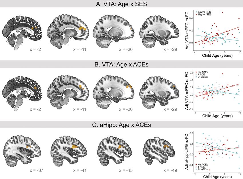
A.T. Park et al. Developmental Cognitive Neuroscience 47 (2021) 100909
highlighting the pivotal role of the VTA as a mediator for downstream 2. Methods
behavior problems. Complementary work finds that stress exposure in
early adolescent rats results in aberrant activity in VTA and anterior 2.1. Participants
hippocampus, with evidence for a possible critical period in the
vulnerability of this circuit (Gomes et al., 2019). Taken together, early The Institutional Review Board at the University of Pennsylvania
life stress causes enduring alterations in the VTA that create latent approved this study. All parents provided informed, written consent.
vulnerability for dysregulated reward-related behaviors, perhaps via Children younger than age 8 provided verbal assent, and children ages 8
changes in the connectivity between VTA and other parts of the dopa and older provided written assent. Participants were recruited from
minergic reward circuitry. Philadelphia and the surrounding regions through advertisements on
Additional work in rodents shows that VTA dopamine neurons have public transportation, partnerships with local schools, outreach pro
a protracted developmental trajectory, continuing to grow and inner grams, community family events, and social media ads.
vate mPFC throughout adolescence (Hoops and Flores, 2017). This Resting-state scans were acquired for 137 participants. Eighty-eight
protracted development plays an important role in shaping the matu participants were included in the final sample (see exclusion criteria
ration of mPFC (Reynolds et al., 2018), but also leaves dopamine neu below). Children were between the ages of 4 and 9 (M = 6.80, SD = 1.38,
rons vulnerable to environmental insult for an extended period of time. range = 4.1 to 9.9). The racial and ethnic makeup of the sample was as
Throughout life, rodents exposed to chronic stress show loss of dopa follows: 56 % Black, 41 % White, 11 % Asian, 10 % Other, and 10 %
mine neurons in the VTA, and functional changes in the synaptic con Hispanic/Latino. Percentages sum to greater than 100 % because par
nections of VTA dopamine neurons to other projection targets in the ents could endorse multiple races. For comparison, Philadelphia is 42.9
mesocorticolimbic circuitry (reviewed in (Burke and Miczek, 2014; % Black, 35.3 % White, 6.9 % Asian, 0.5 % Other, and 12.4 % Hispanic,
Douma and de Kloet, 2019)). Thus, chronic stress exposure has signifi and the United States overall is 12.6 % Black, 62.0 % White, 5.2 % Asian,
cant effects on how VTA dopamine neurons communicate with other 1 % Other, and 16.9 % Hispanic (2010 US Census, StatisticalAtlas.com).
brain regions, and may impair the mPFC’s top-down regulation of the We controlled for race and ethnicity in all analyses. Because the most
VTA. However, it is unclear how stress exposure interacts with the numerous racial group was Black, we treated Black as the reference
timing of reward circuitry development in humans. category, and included dummy variables for White, Asian, Other, and
In humans, early life stress is associated with differences in the Hispanic/Latino.
activation and connectivity of reward processing regions, as measured Participants were excluded for: missing data on both measures of SES
by functional magnetic resonance imaging (fMRI), with the bulk of prior (parental education and income) (n = 1), race/ethnicity (n = 4), or the
work focusing on the NAcc and mPFC (Hanson et al., 2016, 2017; Adverse Childhood Experiences questionnaire (n = 8); not completing
Herzberg and Gunnar, 2020; Ironside et al., 2018). Several studies have the resting-state scan (e.g., due to falling asleep or wanting to end the
found associations between early stress exposure and alterations in NAcc scan early, n = 21); an artifact in the resting-state data (due to hair
functional connectivity with medial prefrontal cortex (Fareri et al., glitter, n = 1); incorrect registration of the participant at the scanner
2017; Hanson et al., 2017; Marshall et al., 2018). For example, Hanson (n = 1); or parent-reported diagnosis of Attention-Deficit/Hyperactivity
et al. (2017) found that childhood maltreatment was related to alter Disorder during the visit, despite not reporting a diagnosis during
ations in NAcc-mPFC task-dependent functional connectivity in young screening (n = 2). Participants were also excluded for having no usable
adults, but that this depended on also experiencing greater levels of resting-state data due to average head displacement (3 translations, 3
recent life stress (Hanson et al., 2017), consistent with the work by Peña rotations) of greater than 1.2 mm (n = 10), as well as for having an
et al. (2017) in mice showing that early stress may sensitize the VTA to unusable structural scan (n = 1).
later stress. Early life stress has also been linked to reduced functional
connectivity between the VTA and the hippocampus in late childhood 2.2. Questionnaires
and adolescence (Marusak et al., 2017). However, little is known about
how early stress exposure impacts the developmental trajectory of VTA Questionnaire response distributions are shown in Fig. 1. The
connectivity during childhood. parent/guardian filling out the questionnaires was as follows: 80 %
Here, we examined whether early life stress was associated with VTA Mother, 17 % Father, 1 % Guardian, 2 % Grandmother. Parental edu
resting-state functional connectivity development between the ages of 4 cation and family income were assessed using the MacArthur Founda
and 9. We additionally examined three other mesocorticolimbic regions, tion Research Network on Socioeconomic Status and Health
NAcc, aHipp, and mPFC, to test for the specificity of the effects on VTA. sociodemographic questionnaire (Operario et al., 2004). Parents were
Resting-state is thought to reflect the history of co-activation between asked to report their highest education level (possible responses ranged
brain regions, making it a useful measure for probing individual dif from “less than high school” to “professional degree (J.D., M.D., Ph.
ferences in functional anatomy (Gabard-Durnam et al., 2016; Guerra- D.)”), as well as the highest education level of their partner if applicable
Carrillo et al., 2014). Its stimulus-free nature also helps mitigate the (85 % of parents reported the education level of their partner). Average
possibility that individuals from different backgrounds may differ in parental education ranged from 11 to 20 years (Mdn = 14, SD = 2.61,
their familiarity with, and interpretation of, task stimuli. We concep n = 88). Thirty-four percent had a high school diploma or less education,
tualized early life stress in two ways. First, we examined childhood so compared to 58 % of adults over the age of 25 in Philadelphia, and 61.5
cioeconomic status (SES). Low SES increases risk for chronic stress % in the United States (2010 US Census, StatisticalAtlas.com). Total
because it is associated with experiences like reduced access to re family income was assessed by asking “Which of these categories best
sources, increased chaos in the home, and more exposure to violence describes your total combined family income for the past 12 months?
(Evans, 2004; McLaughlin and Sheridan, 2016). Second, we examined This should include income (before taxes) from all sources, wages, rent
exposure to Adverse Childhood Experiences, or ACEs, which capture from properties, social security, disability and/or veteran’s benefits,
children’s cumulative exposure to household dysfunction, abuse, and/or unemployment benefits, workman’s compensation, help from relatives
neglect, in order to test whether these types of stressful home experi (including child payments and alimony), and so on.” Possible responses
ences uniquely explain reward circuitry differences above and beyond included: Less than $5,000, $5,000 through $11,999, $12,000 through
other negative impacts of lower SES. We investigated whether these $15,999, $16,000 through $24,999, $25,000 through $34,999, $35,000
measures of early life stress were associated with differences in the through $49,999, $50,000 through $74,999, $75,000 through $99,999,
functional connectivity of VTA, NAcc, aHipp, and mPFC, and whether $100,000 through $149,999, $150,000 through $199,999, $200,000
connectivity patterns with age differed as a function of stress exposure. and greater, and Unsure. Annual family income was estimated as the
median value of the selected income bracket (Mdn = $62.5 K, SD =
2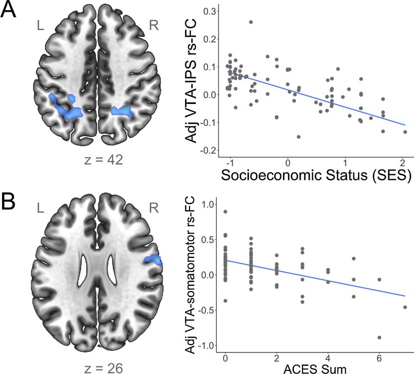
A.T. Park et al. Developmental Cognitive Neuroscience 47 (2021) 100909
Fig. 1. Histograms of demographic information (n = 88; for income, n = 83). The Socioeconomic Status composite is the average of Z-scored income and parent
education. ACEs, Adverse Childhood Experiences.
$63 K, n = 83). For comparison, the median household income in Phil 2+ ACEs. For comparison, a nationally representative survey
adelphia in 2010 was $64 K, and in the United States was $55 K. So (2011–2014) of youth under the age of 18, using a similar ACEs mea
cioeconomic status (SES) was defined as the average of Z-scored income sure, found that 38.5 % reported 0 ACEs, 23.5 % reported 1 ACE, and 38
and Z-scored years of parental education (parental education was % reported 2+ ACEs (Merrick et al., 2018).
averaged across parents if available for both parents).
Parents completed the child version of the Adverse Childhood Ex 2.3. Neuroimaging data acquisition
periences (ACEs) questionnaire (Murphy et al., 2016), which asked
parents about their child’s lifetime experiences with broader household Prior to the scanning session, participants were acclimated to the
dysfunction and parent separation, abuse, and neglect. An ACEs score scanning environment with a mock scanner that simulates typical MRI
was calculated by summing the binary ratings for these ten types of noises. Participants practiced keeping still in the mock scanner for about
experiences (Table 1). ACEs sum ranged from 0 to 7 (Mdn = 1, 10 min, by watching a movie that would pause each time they moved
SD = 1.62). Number of ACEs was as follows: 38.6 % reported that their their heads more than 1 mm. During the actual MRI session, a researcher
child experienced 0 ACEs, 28.4 % reported 1 ACE, and 33.9 % reported stayed in the scanner room with the participant to reassure the child and
to gently squeeze the child’s foot if the child moved.
Table 1 Imaging was performed at the Center for Advanced Magnetic Reso
Associations between specific Adverse Childhood Experiences (ACEs) and So nance Imaging and Spectroscopy (CAMRIS) at the University of Penn
cioeconomic Status (SES). SES was compared between families who endorsed sylvania. Scanning was conducted using a Siemens MAGNETOM Prisma
each item and those who did not. Negative t-statistics indicate lower SES for 3 T MRI scanner with the vendor’s 32-channel coil. A whole-brain, high-
families that endorsed an item as compared to those that did not. Dashed lines resolution, T1-weighted 3D-encoded multi-echo structural scan
indicate ACEs with too few instances to test for SES differences. (MPRAGE) was collected (acquisition parameters: TR = 2530 ms,
Percentage t p TEs = 1.69 ms/3.55 ms/5.41 ms/7.27 ms, BW =650 Hz/px, 3x GRAPPA,
endorsing flip angle = 7◦ , voxel size =1 mm isotropic, matrix size = 256 × 256,
Parental separation/divorce 31 % − 4.69
A.T. Park et al. Developmental Cognitive Neuroscience 47 (2021) 100909
of usable data, in two ways: 1) monitoring head motion in real-time defined from the Harvard-Oxford subcortical atlas provided through
using the Framewise Integrated Real-time MRI Monitor (FIRMM) soft FSL. Anterior hippocampus was defined from a probabilistic atlas of the
ware (Dosenbach et al., 2017), and 2) collecting two resting-state scans medial temporal lobe (Hindy and Turk-Browne, 2016), divided into
when possible. This resulted in 54 participants who continued to be anterior and posterior sections at Y = -21, and masked by the
scanned until our movement criterion was achieved (at least 5 min of Harvard-Oxford hippocampus in order to limit the seed from extending
functional data with head motion less than 1 mm, maximum 8 min, per into surrounding white matter. Medial prefrontal cortex was defined as
resting-state scan). Runs were dropped from subsequent analyses if they FreeSurfer’s individual parcellation of bilateral rostral anterior cingu
exceeded 1.2 mm average motion (see Summary of motion consider late cortex (rACC) based on the Desikan-Killiany atlas (Desikan et al.,
ations), resulting in a final breakdown of 51 participants with one usable 2006). The seed for mPFC will be referred to throughout as rACC. The
resting-state scan and 37 participants with two usable resting-state average temporal signal-to-noise ratio (tSNR) within the seed regions
scans. All analyses controlled for the total amount of data collected were as follows: VTA (M = 24.02, SD = 4.39), NAcc (M = 41.79,
(total number of resting-state volumes), as well as average motion, SD = 9.05), aHipp (M = 33.26, SD = 5.10), rACC (M = 48.65,
weighted by run length. We controlled for the amount of data provided SD = 13.43) (whole-brain tSNR maps are provided in Supplemental
by each child so that children with more available data would not be Fig. 1). We extracted the average time series within each ROI from un
overrepresented in the models. Participants had an average head smoothed data, to ensure that signal was not blurred outside of the ROI.
displacement of 0.37 mm (SD =0.28 mm, significantly non-normally We used the average time series within each seed to generate whole-
distributed, W = 0.86, p < .001). brain subject-level functional connectivity maps for the VTA, NAcc,
aHipp, and rACC, using FSL’s fsl_glm tool. Because motion and physio
2.4. Preprocessing logical noise were already filtered out of the functional data during
earlier preprocessing steps, subject-level GLMs only contained the seed
The functional imaging data were preprocessed using Nipype, a time series as a regressor. For participants with two resting-state runs,
Python-based framework for flexibly integrating neuroimaging analysis seed connectivity maps were generated separately for each run, and then
tools (Gorgolewski et al., 2011). The software packages used in this the runs were averaged together using a Nipype implementation of a
preprocessing pipeline included FMRIB Software Library (FSL v5.0.8; fixed-effects model (using FSL’s flameo) to produce a single connectivity
Jenkinson et al., 2012), FreeSurfer (v6.0.0; Dale et al., 1999), Advanced map per participant to be entered into group-level analyses.
Normalization Tools (ANTs v2.1.0; Avants et al., 2011)), and Nipype’s Whole-brain group-level analyses were performed with FMRIB’s
implementation of Artifact Detection Tools (ART; http://www.nitrc.or Local Analysis of Mixed Effects tool (FSL’s FLAME 1). Only voxels that
g/projects/artifact_detect/). had unanimous coverage across subjects were tested, resulting in a mask
Simultaneous realignment and slice timing correction was conducted that covered the entire brain. We ran the following GLMs (testing for
using an algorithm implemented in Nipy (Roche, 2011). Outlier volumes both positive and negative associations): 1) group average; 2) main ef
in the functional data were defined using ART based on composite fects of age, 3) SES, and 4) ACEs exposure; and the 5) age x SES inter
motion (greater than 1 mm of head displacement between volumes) and action and 6) age x ACEs interaction. All group GLMs included the
global signal intensity (greater than 3 standard deviations from the following covariates: age (except when age was the main effect being
mean). tested), gender, average head motion (in millimeters), number of
The following confounds were regressed out of the functional data: 6 resting-state volumes, and race/ethnicity. To examine the robustness of
realignment parameters (3 translations, 3 rotations) and their first-order the results, we additionally ran GLMs for SES and the age x SES inter
derivatives, outlier volumes flagged by ART (one nuisance regressor per action (GLMs 3 and 5) that controlled for the main effect of ACEs
outlier), composite motion, and linear and quadratic polynomials to exposure, and GLMs for ACEs exposure and the age X ACEs interaction
detrend the data. Five principal components were also derived from (GLMs 4 and 6) that controlled for the main effect of SES. We did not
segmentations of both cerebrospinal fluid (CSF) and white matter (WM), include both age x SES and age x ACEs in the same model because the
and regressed from the data, to correct for physiological noise like heart regressors were collinear.
rate and respiration (aCompCor; Behzadi et al., 2007). The CSF and WM Z-statistic maps were corrected for multiple comparisons with
segmentations were derived from Freesurfer’s individual parcellations parametric clusterwise inference using FSL’s cluster tool (relies on
of the lateral ventricles and total white matter, respectively; these seg Gaussian Random Field Theory) at a cluster-defining threshold of z = 3.1
mentations were transformed into functional space. Confound regres (p < .001), neighborhood size of 26, and an FWE-corrected threshold of
sion occurred within a skull-stripped functional mask which was created p < .05, based on evidence from Eklund et al. that false positives are not
using FSL’s Brain Extraction Tool (BET; Smith, 2002); BET’s fractional well controlled at a less stringent threshold (Eklund et al., 2016).
intensity threshold was set at 0.4. Following recommendations for neuroimaging reporting (Poldrack,
The functional data were bandpass filtered (0.01-0.1 Hz), spatially 2017), uncorrected statistical maps are available on NeuroVault (https
smoothed with an isotropic 6 mm Gaussian kernel (FWHM), and ://neurovault.org/collections/ZSVLTNSF/) (Gorgolewski et al., 2016).
normalized to the OASIS-30 Atropos template (in MNI152 2 mm space) All statistical analyses were conducted in R.
in a two-step process. First, the median functional image was coregis
tered to the reconstructed surfaces using FreeSurfer’s bbregister (Greve 2.6. Summary of motion considerations
and Fischl, 2009); next, the structural image was registered to the
OASIS-30 Atropos MNI152 template using ANTs. The transformation Studying brain development in early childhood requires addressing
matrices generated by these two steps were concatenated, allowing higher levels of motion than typically seen at later ages. The effect of
images to be transformed directly from functional to MNI space in a motion during our structural scans was minimized using prospective
single interpolation step. motion correction, which provided high-quality data for our structural
processing (Tisdall et al., 2012). We dealt with motion during our fMRI
2.5. Seed-based analyses scans in four ways, each designed to mitigate the specific effects of
motion on fMRI studies. First, we implemented real-time motion
We examined the functional connectivity of the ventral tegmental monitoring at the scanner (FIRMM) in order to more systematically
area (VTA), nucleus accumbens (NAcc), anterior hippocampus (aHipp), track participants’ motion during the scans and maximize the amount of
and medial prefrontal cortex (mPFC) (seeds shown in Fig. 2A). The VTA data that met our motion thresholds (Dosenbach et al., 2017). Second,
was defined using a probabilistic atlas that was constructed from hand- we set a motion exclusion threshold based on our data quality, as data
drawn ROIs of the VTA (Murty et al., 2014). Nucleus accumbens was quality differs by features of the acquisition, and therefore there is no
4
A.T. Park et al.                                                                                                     Developmental Cognitive Neuroscience 47 (2021) 100909

Fig. 2. (A) Positive group average functional connectivity for the ventral tegmental area (VTA), nucleus accumbens (NAcc), anterior hippocampus (aHipp), and
rostral anterior cingulate cortex (rACC). Seed regions are shown on the left (VTA = yellow, NAcc = green, aHipp = purple, rACC = blue). (B) Positive relationships
between age and functional connectivity. (C) Negative relationships between age and functional connectivity. No regions showed negative relationships between age
and VTA functional connectivity, or between age and aHipp functional connectivity. Models control for age (in the group average), gender, average head motion,
number of resting-state volumes, and race/ethnicity, and are corrected for multiple comparisons at z = 3.1, p < 0.05, N = 88. Scatterplots show the relationship
between age and extracted parameter estimates (adjusted for covariates) (For interpretation of the references to color in this figure legend, the reader is referred to
the web version of this article.).

                                                                                   5
A.T. Park et al. Developmental Cognitive Neuroscience 47 (2021) 100909
universal motion cutoff that balances participant inclusion with data 3.3. Main effects of age
quality (i.e., multiband factor, head coil selection). The motion
threshold was set at 1.2 mm based on qualitative evaluations of con VTA functional connectivity showed a positive association with age
nectivity maps of a left motor cortex seed, as the anatomy of motor in the right caudate (Fig. 2B; peak voxel coordinates (MNI): 14, 2, 20,
cortex is well-known and established by early childhood (Gao et al., maximum z-statistic = 4.09, cluster volume = 171 voxels). No regions
2015; Grayson and Fair, 2017). Participants with connectivity maps that showed a negative age-related association with VTA functional con
did not look as expected all had composite motion over 1.2 mm. Eval nectivity. NAcc, rACC, and aHipp functional connectivity showed posi
uations were done by an experimenter blind to any demographic in tive associations with age in dorsal medial prefrontal cortex (dmPFC)
formation about the participants. For participants with two usable (Fig. 2B, NAcc peak voxel coordinates (MNI): 6, 62, 18, maximum z-
resting-state runs, head motion was averaged between the two runs, statistic = 4.42, cluster volume = 372 voxels; aHipp peak voxel co
weighted by run length. The participants who were excluded for motion ordinates (MNI): -2, 52, 10, maximum z-statistic = 4.44, cluster vol
(n = 10) were slightly younger than included participants (Exc. median: ume = 475 voxels; rACC peak voxel coordinates (MNI): 8, 48, 46,
5.5 years, Inc. median: 6.6 years, U = 277.0, p = .06). Excluded par maximum z-statistic = 5.38, cluster volume = 1788 voxels). aHipp
ticipants did not differ from included participants on parental education functional connectivity also showed a positive association with age in
(Exc. median: 15.5, Inc. median: 14, U = 483.0, p = .61), family income two additional clusters (Fig S2): right cerebellum (peak voxel co
(Exc. median: $42.5K, Inc. median: $62.5K, U = 444.5, p = .35), or ordinates (MNI): 42, -62, -44, maximum z-statistic = 4.72, cluster vol
ACEs (Exc. median: 1, Inc. median: 1, U = 399.5, p = .62). Third, within ume = 349 voxels); and left dorsal PFC (peak voxel coordinates (MNI):
the included sample, we tested how motion was related to our variables -20, 24, 60, maximum z-statistic = 4.52, cluster volume = 521 voxels).
of interest: age, SES, and ACEs. Higher average head motion was NAcc and rACC connectivity showed similar negative associations
significantly related to younger age (rs = -0.31, p = .003) and to lower with age (Fig. 2C). NAcc connectivity showed a negative relationship
SES (rs = -0.23, p = .03), but there was no age x SES interaction on head with age in right parahippocampal gyrus (PHG; peak voxel coordinates
motion (t(84) = -0.11, p = .91). Average head motion was not signifi (MNI): 28, -48, -2, maximum z-statistic = 4.43, cluster volume = 388
cantly related to ACEs sum (rs = -0.12, p = .23), and there was no age x voxels), and rACC connectivity showed a negative relationship with age
ACEs interaction on head motion (t(84) = -1.06, p = .29). Fourth, in left PHG (peak voxel coordinates (MNI): -18, -42, -2, maximum z-
because motion was related to age and SES, we included average motion statistic = 4.28, cluster volume = 436 voxels). No negative relationship
(across runs) in all models. with age was found for aHipp.
3. Results 3.4. Main effects of stress measures
3.1. Demographics and stress measures Higher SES was related to lower VTA functional connectivity with
bilateral intraparietal sulcus (Fig. 3A; right IPS: peak voxel coordinates
Lower SES was associated with greater exposure to ACEs (rs = -0.32, (MNI): 30, -54, 42, maximum z-statistic = 4.70, cluster volume = 242
p = .002). Specifically, SES was lower among children who experienced voxels; left IPS: peak voxel coordinates (MNI): -22, -56, 38, maximum z-
parental separation (t(86) = -4.69, p < .001), family incarceration (t(86) statistic = 4.50, cluster volume = 553 voxels). The main effect of SES
= -8.08, p < .001), and family drug abuse (t(86) = -3.35, p = .002) was similar when additionally controlling for ACEs exposure, although
(Table 1). ACEs exposure was lower for White children than for Black limited only to left IPS (Fig S3A). Higher ACEs exposure was related to
children (t(83) = -2.63, p = .01). SES was higher for White (t lower VTA functional connectivity with right somatomotor cortex
(83) = 4.74, p < .001) and Asian (t(83) = 4.75, p < .001) children than (Fig. 3B; peak voxel coordinates (MNI): 64, -2, 26, maximum z-statis
for Black children. ACEs exposure was slightly, but not significantly, tic = 4.55, cluster volume = 213 voxels). The main effect of ACEs
higher in Hispanic children as compared to Non-Hispanic children (t exposure was highly similar when additionally controlling for SES (Fig
(83) = 1.43, p = .16). SES was slightly, but not significantly, lower in S3B). No regions showed a positive association with VTA functional
Hispanic children as compared to Non-Hispanic children (t(83) = -1.77, connectivity for SES or ACEs exposure. There were no significant posi
p = .08). Age was not significantly related to SES (rs = -.17, p = .12), but tive or negative main effects of SES or ACEs exposure on NAcc, aHipp, or
it was significantly positively related to ACEs (rs = 0.26, p = .01). SES rACC functional connectivity.
and ACEs exposure did not differ by gender.
3.5. Interactions between age and stress measures
3.2. Group average results
There was an age x SES interaction on connectivity between VTA and
Across the entire group, VTA was positively functionally connected left dorsal mPFC (dmPFC) (Fig. 4A; peak voxel coordinates (MNI): -10,
to subcortical regions including nucleus accumbens, hippocampus, 40, 24, maximum z-statistic = 4.38, cluster volume = 509 voxels). VTA-
amygdala, and cerebellum, as well as the striatum and thalamus dmPFC connectivity showed a stronger positive association with age in
(Fig. 2A). In cortex, VTA was most strongly correlated with medial children from higher SES backgrounds, compared to children from lower
prefrontal cortex, precuneus, and visual cortex, broadly consistent with SES backgrounds. The age x SES interaction was nearly identical after
D1 receptor binding sites in the human brain (Palomero-Gallagher et al., additionally controlling for the main effect of exposure to ACEs (Fig
2015). Notably, we did not observe strong connectivity with lateral S4A). To examine whether the relationship between age and VTA con
prefrontal and parietal regions in this age range, consistent with nectivity is significantly different from zero within SES groups, partici
developmental work on VTA connectivity (Tomasi and Volkow, 2014). pants were divided into 2 groups by median split: higher SES (n = 46)
The positive group averages for NAcc, aHipp, and rACC were similar, and lower SES (n = 42). VTA-dmPFC connectivity was positively related
showing broad connections to subcortical regions and to the default to age in children from a higher SES background (t(77) = 4.56, p < .001,
mode network. NAcc and aHipp additionally showed connectivity with 95 % CI [0.02, 0.06]), and not significantly related to age in children
somatomotor cortex. There were no regions that showed negative from a lower SES background (t(77) = -1.52, p = .13, 95 % CI [-0.04,
functional connectivity in the group average for the VTA, NAcc, aHipp, 0.005]; Fig. 4A).
or rACC. We found a similar age x ACEs interaction on connectivity between
VTA and left dorsal mPFC (Fig. 4B; peak voxel coordinates (MNI): -14,
36, 36, maximum z-statistic = 4.43, cluster volume = 564 voxels). VTA-
dmPFC connectivity showed a stronger positive association with age in
6A.T. Park et al. Developmental Cognitive Neuroscience 47 (2021) 100909
Fig. 3. (A) Higher Socioeconomic Status (SES) is
associated with lower ventral tegmental area (VTA)
functional connectivity with bilateral intraparietal sul
cus (IPS). (B) Higher exposure to Adverse Childhood
Experiences (ACEs) is associated with lower VTA
functional connectivity with right somatomotor cortex.
Models control for age, gender, average head motion,
number of resting-state volumes, and race/ethnicity.
Results are corrected for multiple comparisons at z =
3.1, p < 0.05. Scatterplots show the relationship be
tween the independent variables (SES, ACEs) and
extracted parameter estimates (adjusted for
covariates).
children with lower exposure to ACEs, compared to children with higher 4. Discussion
exposure to ACEs. The dmPFC cluster overlapped with the age x SES
result but additionally extended to the lateral surface of PFC. The age x Resting-state functional connectivity of the ventral tegmental area
ACEs interaction was almost identical after additionally controlling for (VTA) showed different patterns with age as a function of stress exposure
the main effect of SES (Fig S4B). To test whether the relationship be in 4- to 9-year-old children. Specifically, children from higher socio
tween age and VTA connectivity is significantly different from zero economic status (SES) backgrounds showed age-related increases in VTA
within ACEs exposure levels, participants were divided into 3 groups: functional connectivity with dorsal medial prefrontal cortex (dmPFC),
exposure to 0 ACEs (n = 34), 1 ACE (n = 25), or 2 or more ACEs (n = 29). while children from lower SES backgrounds did not show this pattern.
The 2+ ACEs group reflects greatest cumulative risk, while the 1 ACE Similarly, only children with low exposure to Adverse Childhood Ex
group is heterogeneous in its experiences (32 % parental separation/ periences (ACEs) showed age-related increases in VTA functional con
divorce, 24 % living with someone with mental illness, 20 % witnessing nectivity with dmPFC. Our results suggest that stress-related differences
adults treated violently, 12 % physical neglect, 4 % living with someone in VTA connectivity begin to emerge during childhood. It is possible that
who abuses substances, 4 % living with someone who has been incar children from lower SES backgrounds or with high ACEs exposure
cerated, 4 % sexual abuse). VTA-dmPFC connectivity was positively accumulate negative experiences as they grow up, or that they do not
related to age in children with no ACEs exposure (t(74) = 3.45, p = .001, accumulate positive experiences in the same way as their peers from
95 % CI [0.02, 0.09]), positively related to age in children with exposure lower stress backgrounds. It is also possible that early experiences,
to 1 ACE (t(74) = 2.19, p = .03, 95 % CI [0.004, 0.08], and unrelated to before age 4, set children on different developmental trajectories. Lon
age in children with 2+ ACEs (t(74) = -1.95, p = .055, 95 % CI [-0.06, gitudinal work beginning in infancy or even at conception, with detailed
0.0006]); Fig. 4B). information on the specific timing of stressors, is needed to better un
There was an age x ACEs interaction on connectivity between aHipp derstand the causes of the cross-sectional relationships presented here.
and left inferior frontal gyrus (IFG) (Fig. 4C; peak voxel coordinates Our findings are consistent with recent work in rodents showing that
(MNI): -40, 4, 22, maximum z-statistic = 4.23, cluster volume = 352 exposure to early life stress causes changes in the VTA that create sus
voxels). aHipp-LIFG connectivity showed a stronger negative association ceptibility to later stressors ((Peña et al., 2017)). There is evidence that
with age in children with higher exposure to ACEs, compared to children chronic stress results in the loss of dopaminergic neurons in the VTA
with lower exposure to ACEs. The age x ACEs interaction was nearly (Sugama and Kakinuma, 2016), as well as altered dopamine activity in
identical after additionally controlling for the main effect of exposure to VTA projection targets like NAcc and mPFC (Holly and Miczek, 2016).
SES (Fig S4C). aHipp-LIFG connectivity was unrelated to age in children Stress also has been shown to alter mPFC neuron morphology (Cook and
with no ACEs exposure (t(74) = 0.84, p = .40, 95 % CI [-0.04, 0.11]), Wellman, 2004; Liston et al., 2006). Communication between VTA and
unrelated to age in children with exposure to 1 ACE (t(74) = -0.35, mPFC is complex and bidirectional, with both excitatory and inhibitory
p = .73, 95 % CI [-0.10, 0.07], and negatively related to age in children connections determining dopamine activity (Beier et al., 2015). Studies
with 2+ ACEs (t(74) = -2.33, p = .02, 95 % CI [-0.14, -0.01]); Fig. 4C). in animal models have not yet determined whether early life stress im
There was no significant age x SES interaction for the aHipp seed, and no pacts projections from VTA, or projections to VTA from mPFC. Unfor
significant age x SES or age x ACEs exposure interactions for the NAcc or tunately, resting-state fMRI cannot differentiate between excitatory and
rACC seeds. inhibitory connections, and analytical approaches that can shed light
7A.T. Park et al. Developmental Cognitive Neuroscience 47 (2021) 100909
Fig. 4. (A) Age x Socioeconomic Status (SES) interaction on ventral tegmental area (VTA) functional connectivity. Scatterplot shows the relationship between child
age and extracted parameter estimates, plotted by median split on SES for visualization purposes. (B) Age x Adverse Childhood Experiences (ACEs) interaction on
VTA functional connectivity. (C) Age x ACEs interaction on anterior hippocampus (aHipp) functional connectivity. Scatterplots show the relationship between age
and extracted parameter estimates (adjusted for covariates). For visualization purposes, participants were grouped by having exposure to 0, 1, or 2+ ACEs. Results
are corrected for multiple comparisons at z = 3.1, p < 0.05. All models control for gender, average head motion, number of resting-state volumes, and race/ethnicity.
onto directionality, i.e., bottom-up vs. top-down, require more data than support of goal-oriented behavior and decision making (Murty et al.,
can be acquired in young children (Mitra et al., 2015). Our work is also 2016).
consistent with cross-species evidence that the development of VTA Across our entire sample of children, irrespective of stress exposure,
projections in rodents (Hoops and Flores, 2017), and the maturation of we found that the VTA showed increased connectivity with the caudate
dopamine availability in humans (Larsen et al., 2020), continues with age. This finding is consistent with prior work showing greater
through childhood and adolescence. VTA-caudate connectivity in adults (ages 15–46) relative to youth (ages
The age x ACEs and age x SES interactions on VTA connectivity were 7–22, but note overlapping age ranges) (Tomasi and Volkow, 2014). The
observed in a region of mPFC that is often called dorsal anterior caudate plays an important role in behaviors like performance moni
cingulate cortex (dACC). The dACC has been hypothesized to play an toring and cognitive control (Brovelli et al., 2011). Indeed, recent work
important role in appraising environmental states and integrating in in young adults has shown that the structural connectivity between VTA
ternal and external motivational factors, in order to guide behavior via and caudate is related to conflict monitoring (Mamiya et al., 2019).
top-down modulation of the VTA (Beier et al., 2015; Haber and Behrens, Thus, increased functional coupling between the VTA and caudate may
2014; Heilbronner and Hayden, 2016). Blunted age-related changes in support the rapid development of cognitive control between the ages of
VTA-mPFC functional coupling in childhood may thus reflect alterations 4 and 9 (Akshoomoff et al., 2018; Fjell et al., 2012). We also found that
in effective dACC top-down regulation of dopamine activity. Recent NAcc, aHipp, and rACC show very similar positive relationships between
work also suggests that the dopaminergic midbrain drives dorsal mPFC’s age and connectivity with dmPFC. Past work found different develop
role in learning about the amount of effort needed to perform a task, mental changes in NAcc resting-state connectivity, i.e., decreased con
highlighting the bottom-up influences that additionally support moti nectivity between NAcc and subgenual ACC, but the age range was much
vated behavior (Hauser et al., 2017). We did not observe any age x stress broader, from 4 and 23 (Fareri et al., 2015). Another study found no
interactions on nucleus accumbens (NAcc) or rostral anterior cingulate developmental changes in VTA-NAcc connectivity at rest in adolescence
cortex (rACC) connectivity, but we did observe that connectivity be and young adulthood (ages 10–30), but found that VTA-NAcc connec
tween anterior hippocampus and left inferior frontal gyrus (LIFG) tivity during a motivational task decreased with age (Murty et al., 2018).
differed by ACEs exposure. aHipp-LIFG connectivity showed a stronger Reward system development is likely nonlinear through adolescence
negative association with age in children with higher exposure to ACEs, and differs by task state (Hartley and Somerville, 2015; Somerville et al.,
compared to children with lower exposure to ACEs. It has been sug 2010; Somerville and Casey, 2010). We also observed age-related de
gested that dopaminergic neuromodulation may play a critical role in creases in connectivity between rACC and NAcc with the para
refining hippocampal-prefrontal circuitry during adolescence, in hippocampal gyrus (PHG), a region involved in spatial processing and
8A.T. Park et al. Developmental Cognitive Neuroscience 47 (2021) 100909
memory. PHG is at the border of visual and default mode networks in to the pivotal impact of a stressor experienced during an early sensitive
adults, depending on the parcellation (Yeo et al., 2011), but little is window. Fourth, it is possible that certain VTA connectivity relation
known about how its network membership changes with development. ships may only emerge during task-dependent states (Ballard et al.,
Fareri and colleagues found a positive relationship between age and 2011; Murty et al., 2018; Salehi et al., 2020). Future work should
NAcc-PHG connectivity in their broad age range of 4–23 (Fareri et al., examine developmental patterns in VTA connectivity during motiva
2015), suggesting that changes early in childhood may not extend lin tional contexts. Fifth, we are limited by the spatial resolution of our fMRI
early through adolescence and early adulthood. data, as the VTA is a small brain region flanked closely by the substantia
We observed a negative main effect of SES on VTA connectivity with nigra (part of the nigrostriatal dopamine pathway). Thus, it is possible
bilateral intraparietal sulcus (IPS), a task-positive region spanning the that some of our findings reflect connectivity of the midbrain more
frontoparietal network (FPN) and the dorsal attention network (DAN; broadly rather than VTA alone. We also note that the signal-to-noise
Yeo et al., 2011)), and implicated in processing number and space ratio (SNR) in VTA was lower than that of the other seed regions.
(Hubbard et al., 2005). Parietal cortex more broadly is rich in dopamine Fifth, resting-state connectivity relies on blood oxygen-level dependent
receptor expression (Palomero-Gallagher et al., 2015), but few parietal (BOLD) signal and therefore can be influenced by individual differences
neurons project back down to VTA (Watabe-Uchida et al., 2012), sug in vasculature and physiology (K. Murphy et al., 2013; Shmueli et al.,
gesting that our result reflects differences in bottom-up innervation of 2007), though these relationships may be functionally important and
IPS by VTA neurons. The cognitive implications of lower VTA-IPS con not just confounds (Tak et al., 2015). Cardiovascular function is
nectivity are unclear. We additionally observed a main effect of ACEs impacted by SES and ACEs in adolescents and adults (Lagraauw et al.,
exposure on VTA connectivity, such that higher ACEs exposure was 2015; Low et al., 2009; Monnat and Chandler, 2015), but the links are
associated with lower connectivity between VTA and right ventral less well-studied in children, and it is unclear how physiological dif
somatomotor cortex, a region that does directly project to VTA (Wata ferences would result in the specific pattern of findings presented here.
be-Uchida et al., 2012), and has been linked to action-related learning In sum, our findings suggest that early stress exposure may disrupt
(Coddington and Dudman, 2019). Further research is needed to repli the typical developmental trajectory of the dopamine system, which is
cate this unexpected finding and determine the behavioral relevance of essential for reward processing and goal-directed behavior. Future work
adversity effects on VTA-somatomotor connectivity. In contrast with should examine how early life stress impacts the interplay between VTA,
prior studies in older children (Marusak et al., 2017; Richter et al., a broader set of subcortical regions, and cortical networks, as well as its
2019), we did not observe a main effect of stress on hippocampal con consequences for a variety of behaviors, like impulse control (Zisner and
nectivity, nor did we find stress effects on connectivity of NAcc or rACC. Beauchaine, 2015), effortful behavior (Assadi et al., 2009; Westbrook
SES and ACEs exposure were moderately correlated at about .3. The et al., 2020), exploration (Laureiro-Martínez et al., 2015), and curiosity
main effects of ACEs and SES on VTA connectivity were distinct, and (Gruber and Ranganath, 2019). We hypothesize that a blunted
held even when controlling for the other stress measure. The in VTA-mPFC developmental trajectory could serve as a biomarker for
teractions between stress measures and age were highly similar, sug reduced resilience to stress: these children may be more at risk for the
gesting that both measures have similar effects on the maturation of VTA development of psychopathology following stressors experienced later
connectivity with mPFC. It will be critical in future work to examine in life. In order to help buffer against the long-lasting and potentially
how a broader set of stressors have unique impacts on the early orga compounding effects of early disruptions to reward neurocircuitry, it
nization of dopaminergic circuitry (Palacios-Barrios and Hanson, 2019). will be critical to design early childhood interventions that help children
This would include experiences such as negative parenting behaviors, develop effective strategies for coping with chronic stress, and that focus
lack of social support, a chaotic and/or dangerous home environment, on positive development of motivated behavior.
broader community disadvantage, and racial discrimination. In this
study, as in many others, race was associated with SES and ACEs Declaration of Competing Interest
exposure. We controlled for race, rather than examine its impacts
directly, because our samples within racial groups were small, and The authors report no declarations of interest.
because we did not collect data on individuals’ experiences with racism,
which likely constitute major stressors for families of color. Acknowledgments
This study has a number of limitations. First, our data are cross-
sectional, so although we have demonstrated a possible age-related We would like to first thank all of the families who participated in
blunting of reward circuitry development, we have not yet followed this research. We would like to thank Jasmine Forde, Katrina Simon,
children as they grow up. Second, our measures of early stress exposure Sophie Sharp, Yoojin Hahn, Stephanie Bugden, Jamie Bogert, Alexis
rely on parents accurately and completely reporting on their children’s Broussard, Ava Cruz, Samantha Ferleger, Destiny Frazier, Jessica
experiences. It is possible that some parents under-report on child ACEs George, Abigail Katz, Sun Min Kim, Hunter Liu, Dominique Martinez,
due to a lack of complete knowledge about their child’s experiences, Ortal Nakash, Emily Orengo, Christina Recto, Leah Sorcher, and Alexis
poor memory for retrospective events, or the sensitive nature of some of Uria for their help with data acquisition. Finally, we would like to thank
the items (e.g., abuse and neglect). Our definition of SES, which com Catherine Jensen Peña for guidance on the study’s hypotheses and
bines parent education and income, is also limited since it is a snapshot feedback on initial results. This study was supported by the Jacobs
measure that may not capture potentially important household dy Foundation Early Career Award (A.P.M.), NIDA (1R34DA050297-01 to
namics, like changes in partner or changes in income. Future work M.D.T. and A.P.M.), Behavioral and Cognitive Neuroscience Training
should also examine whether similar findings are found in population Grant (NIH T32-MH017168 to A.T.P.), the MindCORE postdoctoral
representative samples. Third, our measures of SES and ACEs exposure research fellowship from the University of Pennsylvania to J.A.L., and
reflect cumulative and chronic stressors, and thus we cannot draw National Science Foundation Graduate Research Fellowships to U.A.T.,
conclusions about the nature of the stressors impacting VTA connec A.L.B., and C.L.M under Grant No. DGE-1845298.
tivity. Future work could take a more dimensional approach, to see if
particular types of stressors are driving these effects (McLaughlin and Appendix A. Supplementary data
Sheridan, 2016), and to explore what other environmental experiences
associated with lower SES are impacting VTA-mPFC connectivity in Supplementary material related to this article can be found, in the
early childhood. It will also be important to further investigate the online version, at doi:https://doi.org/10.1016/j.dcn.2020.100909.
timing of adversity, i.e., whether alterations in VTA connectivity emerge
due to the cumulative impact of stressors encountered over time, or due
9A.T. Park et al. Developmental Cognitive Neuroscience 47 (2021) 100909
References connectivity influences resting-state connectivity years later in human development:
a prospective study. J. Neurosci. 36 (17), 4771–4784. https://doi.org/10.1523/
JNEUROSCI.0598-16.2016.
Akshoomoff, N., Brown, T.T., Bakeman, R., Hagler, D.J., 2018. Developmental
Gao, W., Alcauter, S., Elton, A., Hernandez-Castillo, C.R., Smith, J.K., Ramirez, J.,
differentiation of executive functions on the NIH Toolbox Cognition Battery.
Lin, W., 2015. Functional Network Development During the First Year: Relative
Neuropsychology 32 (7), 777–783. https://doi.org/10.1037/neu0000476.
Sequence and Socioeconomic Correlations. Cereb. Cortex 25 (9), 2919–2928.
Assadi, S.M., Yücel, M., Pantelis, C., 2009. Dopamine modulates neural networks
https://doi.org/10.1093/cercor/bhu088.
involved in effort-based decision-making. Neurosci. Biobehav. Rev. 33 (3), 383–393.
Gomes, F.V., Zhu, X., Grace, A.A., 2019. The pathophysiological impact of stress on the
https://doi.org/10.1016/j.neubiorev.2008.10.010.
dopamine system is dependent on the state of the critical period of vulnerability.
Avants, B.B., Tustison, N.J., Song, G., Cook, P.A., Klein, A., Gee, J.C., 2011.
Mol. Psychiatry. https://doi.org/10.1038/s41380-019-0514-1.
A reproducible evaluation of ANTs similarity metric performance in brain image
Gorgolewski, K., Burns, C.D., Madison, C., Clark, D., Halchenko, Y.O., Waskom, M.L.,
registration. NeuroImage 54 (3), 2033–2044. https://doi.org/10.1016/j.
Ghosh, S.S., 2011. Nipype: a flexible, lightweight and extensible neuroimaging data
neuroimage.2010.09.025.
processing framework in python. Front. Neuroinform. 5, 13. https://doi.org/
Ballard, I.C., Murty, V.P., Carter, R.M., MacInnes, J.J., Huettel, S.A., Adcock, R.A., 2011.
10.3389/fninf.2011.00013.
Dorsolateral prefrontal cortex drives mesolimbic dopaminergic regions to initiate
Gorgolewski, K.J., Varoquaux, G., Rivera, G., Schwartz, Y., Sochat, V.V., Ghosh, S.S.,
motivated behavior. The Journal of Neuroscience: The Official Journal of the Society
Maumet, C., Nichols, T.E., Poline, J.-B., Yarkoni, T., Margulies, D.S., Poldrack, R.A.,
for Neuroscience 31 (28), 10340–10346. https://doi.org/10.1523/
2016. NeuroVault.org: a repository for sharing unthresholded statistical maps,
JNEUROSCI.0895-11.2011.
parcellations, and atlases of the human brain. NeuroImage 124 (Pt B), 1242–1244.
Behzadi, Y., Restom, K., Liau, J., Liu, T.T., 2007. A component based noise correction
https://doi.org/10.1016/j.neuroimage.2015.04.016.
method (CompCor) for BOLD and perfusion based fMRI. NeuroImage 37 (1), 90–101.
Grayson, D.S., Fair, D.A., 2017. Development of large-scale functional networks from
https://doi.org/10.1016/j.neuroimage.2007.04.042.
birth to adulthood: a guide to the neuroimaging literature. NeuroImage 160, 15–31.
Beier, K.T., Steinberg, E.E., DeLoach, K.E., Xie, S., Miyamichi, K., Schwarz, L., Gao, X.J.,
https://doi.org/10.1016/j.neuroimage.2017.01.079.
Kremer, E.J., Malenka, R.C., Luo, L., 2015. Circuit architecture of VTA dopamine
Green, J.G., McLaughlin, K.A., Berglund, P.A., Gruber, M.J., Sampson, N.A.,
neurons revealed by systematic input-output mapping. Cell 162 (3), 622–634.
Zaslavsky, A.M., Kessler, R.C., 2010. Childhood adversities and adult psychiatric
https://doi.org/10.1016/j.cell.2015.07.015.
disorders in the national comorbidity survey replication I: associations with first
Belujon, P., Grace, A.A., 2017. Dopamine system dysregulation in major depressive
onset of DSM-IV disorders. Arch. Gen. Psychiatry 67 (2), 113–123. https://doi.org/
disorders. The International Journal of Neuropsychopharmacology / Official
10.1001/archgenpsychiatry.2009.186.
Scientific Journal of the Collegium Internationale Neuropsychopharmacologicum 20
Greve, D.N., Fischl, B., 2009. Accurate and robust brain image alignment using
(12), 1036–1046. https://doi.org/10.1093/ijnp/pyx056.
boundary-based registration. NeuroImage 48 (1), 63–72. https://doi.org/10.1016/j.
Beurdeley, M., Spatazza, J., Lee, H.H.C., Sugiyama, S., Bernard, C., Di Nardo, A.A.,
neuroimage.2009.06.060.
Hensch, T.K., Prochiantz, A., 2012. Otx2 binding to perineuronal nets persistently
Gruber, M.J., Ranganath, C., 2019. How curiosity enhances hippocampus-dependent
regulates plasticity in the mature visual cortex. J. Neurosci. 32 (27), 9429–9437.
memory: the prediction, appraisal, curiosity, and exploration (PACE) framework.
https://doi.org/10.1523/JNEUROSCI.0394-12.2012.
Trends Cogn. Sci. 23 (12), 1014–1025. https://doi.org/10.1016/j.tics.2019.10.003.
Brovelli, A., Nazarian, B., Meunier, M., Boussaoud, D., 2011. Differential roles of caudate
Guerra-Carrillo, B., Mackey, A.P., Bunge, S.A., 2014. Resting-state fMRI: a window into
nucleus and putamen during instrumental learning. NeuroImage 57 (4), 1580–1590.
human brain plasticity. The Neuroscientist: A Review Journal Bringing
https://doi.org/10.1016/j.neuroimage.2011.05.059.
Neurobiology, Neurology and Psychiatry 20 (5), 522–533. https://doi.org/10.1177/
Burke, A.R., Miczek, K.A., 2014. Stress in adolescence and drugs of abuse in rodent
1073858414524442.
models: role of dopamine, CRF, and HPA axis. Psychopharmacology 231 (8),
Haber, S.N., Behrens, T.E.J., 2014. The neural network underlying incentive-based
1557–1580. https://doi.org/10.1007/s00213-013-3369-1.
learning: implications for interpreting circuit disruptions in psychiatric disorders.
Coddington, L.T., Dudman, J.T., 2019. Learning from action: reconsidering movement
Neuron 83 (5), 1019–1039. https://doi.org/10.1016/j.neuron.2014.08.031.
signaling in midbrain dopamine neuron activity. Neuron 104 (1), 63–77. https://doi.
Hanson, J.L., Albert, D., Iselin, A.-M.R., Carré, J.M., Dodge, K.A., Hariri, A.R., 2016.
org/10.1016/j.neuron.2019.08.036.
Cumulative stress in childhood is associated with blunted reward-related brain
Cook, S.C., Wellman, C.L., 2004. Chronic stress alters dendritic morphology in rat medial
activity in adulthood. Soc. Cogn. Affect. Neurosci. 11 (3), 405–412. https://doi.org/
prefrontal cortex. J. Neurobiol. 60 (2), 236–248. https://doi.org/10.1002/
10.1093/scan/nsv124.
neu.20025.
Hanson, J.L., Knodt, A.R., Brigidi, B.D., Hariri, A.R., 2017. Heightened connectivity
Dale, A.M., Fischl, B., Sereno, M.I., 1999. Cortical surface-based analysis. I. Segmentation
between the ventral striatum and medial prefrontal cortex as a biomarker for stress-
and surface reconstruction. NeuroImage 9 (2), 179–194. https://doi.org/10.1006/
related psychopathology: understanding interactive effects of early and more recent
nimg.1998.0395.
stress. Psychol. Med. 1–9. https://doi.org/10.1017/S0033291717003348.
Desikan, R.S., Ségonne, F., Fischl, B., Quinn, B.T., Dickerson, B.C., Blacker, D.,
Hartley, C.A., Somerville, L.H., 2015. The neuroscience of adolescent decision-making.
Buckner, R.L., Dale, A.M., Maguire, R.P., Hyman, B.T., Albert, M.S., Killiany, R.J.,
Curr. Opin. Behav. Sci. 5, 108–115. https://doi.org/10.1016/j.cobeha.2015.09.004.
2006. An automated labeling system for subdividing the human cerebral cortex on
Hauser, T.U., Eldar, E., Dolan, R.J., 2017. Separate mesocortical and mesolimbic
MRI scans into gyral based regions of interest. NeuroImage 31 (3), 968–980. https://
pathways encode effort and reward learning signals. Proc. Natl. Acad. Sci. U.S.A. 114
doi.org/10.1016/j.neuroimage.2006.01.021.
(35), E7395–E7404. https://doi.org/10.1073/pnas.1705643114.
Di Salvio, M., Di Giovannantonio, L.G., Omodei, D., Acampora, D., Simeone, A., 2010.
Heilbronner, S.R., Hayden, B.Y., 2016. Dorsal anterior cingulate cortex: a bottom-up
Otx2 expression is restricted to dopaminergic neurons of the ventral tegmental area
view. Annu. Rev. Neurosci. 39, 149–170. https://doi.org/10.1146/annurev-neuro-
in the adult brain. Int. J. Dev. Biol. 54 (5), 939–945. https://doi.org/10.1387/
070815-013952.
ijdb.092974ms.
Herzberg, M.P., Gunnar, M.R., 2020. Early life stress and brain function: activity and
Dosenbach, N.U.F., Koller, J.M., Earl, E.A., Miranda-Dominguez, O., Klein, R.L., Van, A.
connectivity associated with processing emotion and reward. NeuroImage 209,
N., Snyder, A.Z., Nagel, B.J., Nigg, J.T., Nguyen, A.L., Wesevich, V., Greene, D.J.,
116493. https://doi.org/10.1016/j.neuroimage.2019.116493.
Fair, D.A., 2017. Real-time motion analytics during brain MRI improve data quality
Hindy, N.C., Turk-Browne, N.B., 2016. Action-based learning of multistate objects in the
and reduce costs. NeuroImage 161, 80–93. https://doi.org/10.1016/j.
medial temporal lobe. Cereb. Cortex 26 (5), 1853–1865. https://doi.org/10.1093/
neuroimage.2017.08.025.
cercor/bhv030.
Douma, E.H., de Kloet, E.R., 2019. Stress-induced plasticity and functioning of ventral
Hollon, N.G., Burgeno, L.M., Phillips, P.E.M., 2015. Stress effects on the neural substrates
tegmental dopamine neurons. Neurosci. Biobehav. Rev. 108, 48–77. https://doi.org/
of motivated behavior. Nat. Neurosci. 18 (10), 1405–1412. https://doi.org/
10.1016/j.neubiorev.2019.10.015.
10.1038/nn.4114.
Eklund, A., Nichols, T.E., Knutsson, H., 2016. Cluster failure: why fMRI inferences for
Holly, E.N., Miczek, K.A., 2016. Ventral tegmental area dopamine revisited: effects of
spatial extent have inflated false-positive rates. Proc. Natl. Acad. Sci. U.S.A. 113
acute and repeated stress. Psychopharmacology 233 (2), 163–186. https://doi.org/
(28), 7900–7905. https://doi.org/10.1073/pnas.1602413113.
10.1007/s00213-015-4151-3.
Evans, G.W., 2004. The environment of childhood poverty. Am. Psychol. 59 (2), 77–92.
Hoops, D., Flores, C., 2017. Making dopamine connections in adolescence. Trends
https://doi.org/10.1037/0003-066X.59.2.77.
Neurosci. 40 (12), 709–719. https://doi.org/10.1016/j.tins.2017.09.004.
Fareri, D.S., Gabard-Durnam, L., Goff, B., Flannery, J., Gee, D.G., Lumian, D.S.,
Hubbard, E.M., Piazza, M., Pinel, P., Dehaene, S., 2005. Interactions between number
Caldera, C., Tottenham, N., 2015. Normative development of ventral striatal resting
and space in parietal cortex. Nat. Rev. Neurosci. 6 (6), 435–448. https://doi.org/
state connectivity in humans. NeuroImage 118, 422–437. https://doi.org/10.1016/j.
10.1038/nrn1684.
neuroimage.2015.06.022.
Ironside, M., Kumar, P., Kang, M.-S., Pizzagalli, D.A., 2018. Brain mechanisms mediating
Fareri, D.S., Gabard-Durnam, L., Goff, B., Flannery, J., Gee, D.G., Lumian, D.S.,
effects of stress on reward sensitivity. Curr. Opin. Behav. Sci. 22, 106–113. https://
Caldera, C., Tottenham, N., 2017. Altered ventral striatal–medial prefrontal cortex
doi.org/10.1016/j.cobeha.2018.01.016.
resting-state connectivity mediates adolescent social problems after early
Jenkinson, M., Beckmann, C.F., Behrens, T.E.J., Woolrich, M.W., Smith, S.M., 2012. FSL.
institutional care. Dev. Psychopathol. 29 (5), 1865–1876. https://doi.org/10.1017/
NeuroImage 62 (2), 782–790. https://doi.org/10.1016/j.neuroimage.2011.09.015.
S0954579417001456.
Lagraauw, H.M., Kuiper, J., Bot, I., 2015. Acute and chronic psychological stress as risk
Fjell, A.M., Walhovd, K.B., Brown, T.T., Kuperman, J.M., Chung, Y., Hagler Jr, D.J.,
factors for cardiovascular disease: insights gained from epidemiological, clinical and
Venkatraman, V., Roddey, J.C., Erhart, M., McCabe, C., Akshoomoff, N., Amaral, D.
experimental studies. Brain Behav. Immun. 50, 18–30. https://doi.org/10.1016/j.
G., Bloss, C.S., Libiger, O., Darst, B.F., Schork, N.J., Casey, B.J., Chang, L., Ernst, T.
bbi.2015.08.007.
M., et al., 2012. Multimodal imaging of the self-regulating developing brain. Proc.
Larsen, B., Olafsson, V., Calabro, F., Laymon, C., Tervo-Clemmens, B., Campbell, E.,
Natl. Acad. Sci. U.S.A. 109 (48), 19620–19625. https://doi.org/10.1073/
Minhas, D., Montez, D., Price, J., Luna, B., 2020. Maturation of the human striatal
pnas.1208243109.
dopamine system revealed by PET and quantitative MRI. Nat. Commun. 11 (1), 846.
Gabard-Durnam, L.J., Gee, D.G., Goff, B., Flannery, J., Telzer, E., Humphreys, K.L.,
https://doi.org/10.1038/s41467-020-14693-3.
Lumian, D.S., Fareri, D.S., Caldera, C., Tottenham, N., 2016. Stimulus-elicited
10You can also read