Nitric oxide synthase mediates cerebellar dysfunction in mice exposed to repetitive blast-induced mild traumatic brain injury - Nature
←
→
Page content transcription
If your browser does not render page correctly, please read the page content below
www.nature.com/scientificreports

            OPEN           Nitric oxide synthase mediates
                           cerebellar dysfunction in mice
                           exposed to repetitive blast-induced
                           mild traumatic brain injury
                           Aric F. Logsdon1,2, Abigail G. Schindler1,4, James S. Meabon3,4, Mayumi Yagi1,
                           Melanie J. Herbert1, William A. Banks1,2, Murray A. Raskind3,4, Desiree A. Marshall5,
                           C. Dirk Keene5, Daniel P. Perl6, Elaine R. Peskind3,4 & David G. Cook1,2 ✉
                           We investigated the role of nitric oxide synthase (NOS) in mediating blood-brain barrier (BBB)
                           disruption and peripheral immune cell infiltration in the cerebellum following blast exposure.
                           Repetitive, but not single blast exposure, induced delayed-onset BBB disruption (72 hours post-
                           blast) in cerebellum. The NOS inhibitor N(G)-nitro-L-arginine methyl ester (L-NAME) administered
                           after blast blocked BBB disruption and prevented CD4+ T-cell infiltration into cerebellum. L-NAME
                           also blocked blast-induced increases in intercellular adhesion molecule-1 (ICAM-1), a molecule that
                           plays a critical role in regulating blood-to-brain immune cell trafficking. Blocking NOS-mediated
                           BBB dysfunction during this acute/subacute post-blast interval (24–71 hours after the last blast)
                           also prevented sensorimotor impairment on a rotarod task 30 days later, long after L-NAME cleared
                           the body. In postmortem brains from Veterans/military Servicemembers with blast-related TBI,
                           we found marked Purkinje cell dendritic arbor structural abnormalities, which were comparable to
                           neuropathologic findings in the blast-exposed mice. Taken collectively, these results indicate that blast
                           provokes delayed-onset of NOS-dependent pathogenic cascades that can later emerge as behavioral
                           dysfunction. These results also further implicate the cerebellum as a brain region vulnerable to blast-
                           induced mTBI.

                           Blast-induced mild traumatic brain injury (mTBI) is prevalent among military personnel and Veterans who have
                           served in Operation Enduring Freedom/Operation Iraqi Freedom/Operation New Dawn (OEF/OIF/OND).
                           Between 2000–2018, more than 380,000 OEF/OIF/OND Veterans have been diagnosed with TBI, which is con-
                           sidered the signature injury of these wars1.
                              The risk of dementia more than doubles among military Veterans with TBI, but the mechanisms mediat-
                           ing this increased risk remain poorly understood2. Neurovascular dysfunction, including blood-brain barrier
                           (BBB) disruption, may be an important initiating mechanism by which mTBI sets in motion pathogenic cascades
                           leading to chronic neurodegenerative pathophysiology3–19. BBB disruption has been examined in a number of
                           pre-clinical models of blast mTBI10,11,20–25. We and others have shown that blast exposure causes transient BBB
                           disruption8,10,26, suggesting that blast also stimulates short-term neurovascular repair processes that interact with
                           more protracted, co-occurring, pathological cascades that undermine BBB integrity. In this regard, blast has been
                           shown to provoke delayed-onset BBB disruption, days after an initial BBB opening was restored to normal8. This
                           delayed-onset BBB dysintegrity occurs in specific brain regions and is prominent in the hippocampus and the
                           cerebellum8,10,27.

                           1
                             Geriatric Research Education and Clinical Center (GRECC), Veterans Affairs Puget Sound Health Care System,
                           Seattle, WA, 98108, USA. 2Division of Gerontology and Geriatric Medicine, Department of Medicine, University
                           of Washington School of Medicine, Seattle, WA, 98195, USA. 3VA Northwest Mental Illness Research Education
                           and Clinical Center, Veterans Affairs Puget Sound Health Care System, Seattle, WA, 98108, USA. 4Department
                           of Psychiatry and Behavioral Sciences, University of Washington School of Medicine, Seattle, WA, 98195, USA.
                           5
                             Department of Pathology, University of Washington, Seattle, WA, 98195, USA. 6Department of Pathology, Center
                           for Neuroscience and Regenerative Medicine, School of Medicine, Uniformed Services University, Bethesda, MD,
                           20814, USA. ✉e-mail: dgcook@u.washington.edu

Scientific Reports |   (2020) 10:9420 | https://doi.org/10.1038/s41598-020-66113-7                                                            1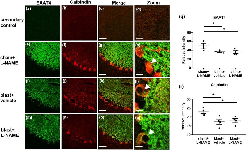
www.nature.com/scientificreports/                                                              www.nature.com/scientificreports

                           Figure 1. Blast overpressures generated with a well-established pneumatic shock tube simulate a Friedlander
                           waveform expected from high explosives. Red trace denotes the mean waveform of 24 blasts sampled
                           throughout the experiments comprising this report. Black trace shows an estimated Friedlander waveform
                           expected from detonation of approximately 21 kg TNT detonated at a distance of 8 m in the open field. Error
                           bars denote SEM.

                               Multiple clinical studies show the cerebellum is vulnerable to the effects of blast mTBI11,28–32. Moreover, bio-
                           physical simulations suggest a specific vulnerability of the human cerebellum to blast exposure33,34. Pre-clinical
                           models of blast-induced mTBI have revealed persistent cerebellar white matter injury11,35,36. The cerebellum is
                           classically appreciated for its role in sensorimotor integration, which is affected by blast and other forms of neu-
                           rotrauma11,37–41. However, it is increasingly clear that the cerebellum also plays important roles in modulating
                           higher order cognitive and behavioral functions that are prominently affected in individuals with blast-related
                           mTBI30,42,43. The translational significance of the cerebellum, coupled with its vulnerability to blast-induced mTBI,
                           prompted us to focus on better understanding the mechanisms of BBB disruption in the cerebellum.
                               In this study, we sought to determine whether cerebellar blast-induced BBB disruption is NOS-dependent
                           and to address whether delayed onset BBB disruption and co-occurring leukocyte infiltration are associated with
                           cerebellar pathophysiology and sensorimotor function.

                           Results
                           Repetitive blast exposure increases BBB permeability to radiolabeled albumin.                          Using estab-
                           lished methods8,10,11,14,44–46, male C57BL/6 mice were exposed to one or three blast overpressures (BOPs) using
                           a pneumatic shock tube delivering a peak static pressure of 20.19 psi (±0.29), positive phase duration of 5.86 ms
                           (±0.065), and impulse of 0.038 psi*ms (±0.0016) (Fig. 1).
                               Under normal conditions, only very low levels of blood-borne albumin cross the intact BBB into the CNS47–49.
                           We have previously reported that repetitive (2X), but not single (1X) blast causes delayed-onset (72 h) BBB per-
                           meability to albumin in whole brain8. For the experiments in this report, we used a 3X blast paradigm (one per
                           day for three days) because it better corresponds with commonly used preclinical exposure regimens and is in
                           keeping with our previous translational mTBI findings11. The results in Fig. 2 confirmed that in whole brain
                           3X, but not 1X blast, significantly increased delayed-onset 99mTc-albumin BBB disruption (Fig. 2; F(2,25) = 6.94;
                           p ≤ 0.01). We have previously shown that this same 3X exposure regimen caused acute BBB disruption, which
                           was closely associated with later emerging chronic neuron loss in the cerebellum11. Thus, for this study we focused
                           attention on the effects of 3X blast exposure on BBB disruption in the cerebellum.

                           Nitric oxide inhibition blocks albumin permeability in the cerebellum following repetitive
                           blast. Nitric oxide (NO) signaling is known to regulate BBB permeability50–52. In keeping with this, we have
                           reported that nitric oxide synthase (NOS) inhibition attenuates single and double blast-induced delayed-onset
                           BBB disruption8. To address whether 3X blast-induced BBB disruption in the cerebellum is similarly regulated by
                           NOS, we measured uptake of blood-borne 99mTc-albumin 72 h after the final exposure in mice injected with the
                           pan-specific NOS inhibitor, N(G)-nitro-L-arginine methyl ester (L-NAME). We found that 3X blast significantly
                           increased delayed-onset BBB disruption in cerebellum (Fig. 3a; F(2,30) = 4.23; p ≤ 0.05), which was significantly
                           ameliorated by L-NAME treatment (Fig. 3a; p ≤ 0.05). In contrast to the cerebellum, 3X blast under these specific
                           experimental conditions did not increase BBB permeability to blood-borne radiolabeled albumin in forebrain
                           structures that excluded the hindbrain (i.e., posterior midbrain, pons, medulla, and cerebellum) (Fig. 3b; F(2, 30)
                           = 0.073; n.s.).

                           Nitric oxide synthase inhibition blocks blast-induced CD4+ T-cell infiltration in the cerebel-
                           lum. The results above further support the idea that the cerebellum is particularly vulnerable to blast-induced
                           BBB dysfunction and that delayed-onset BBB disruption is mediated (at least in part) by NOS-dependent sig-
                           naling cascades. In addition, there is evidence that NOS activity underlies T-cell infiltration into the CNS53,54,
                           corresponds with BBB breakdown, and occurs within a temporal window consistent with the delayed-onset
                           BBB disruption we observed following blast8,55,56. This prompted us to ask: (i) whether blast increases immune
                           cell infiltration in cerebellum, (ii) whether this occurs in a NOS-dependent fashion, and (iii) if blast-induced

Scientific Reports |   (2020) 10:9420 | https://doi.org/10.1038/s41598-020-66113-7                                                            2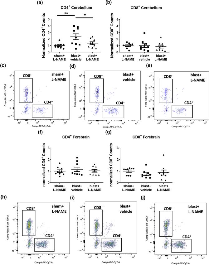
www.nature.com/scientificreports/                                                            www.nature.com/scientificreports

                           Figure 2. Repetitive blast exposure increases BBB permeability to radiolabeled albumin. A significant increase
                           (**p ≤ 0.01) in brain/serum (uμl/g) ratios of 99mTc-albumin was observed in whole brain at 72 h after repetitive
                           mTBI (3X blast; n = 12) compared to 1X blast (n = 12), and sham control mice (n = 12). One-way ANOVA
                           post-hoc Newman-Keuls. Values represent means ± SEM and are expressed as microliters per gram of brain
                           tissue. Brain/serum ratios were calculated by dividing the cpm per brain by the cpm per microliter in the
                           corresponding serum and then by the weight of the brain.

                           Figure 3. Nitric oxide synthase inhibition blocks blast-induced delayed-onset BBB disruption in cerebellum.
                           (a) 3X blast significantly increased brain/serum ratios of 99mTc-albumin in the cerebellum (*p ≤ 0.05), which
                           was significantly attenuated by L-NAME administration (10 mg/kg; ip at 48, 54, and 71 h after the last blast)
                           (*p ≤ 0.05). (b) No differences were observed in brain/serum ratios of 99mTc-albumin in the forebrain after
                           repetitive mTBI (p > 0.05). One-way ANOVA post-hoc Newman-Keuls. Values represent means ± SEM.
                           Timeline portrays the mTBI exposure, L-NAME treatment paradigm, and measurement of BBB permeability.

                           immune cell infiltration follows the same brain region-specific pattern (i.e., cerebellum versus forebrain) as BBB
                           disruption.
                              To test these questions, we employed flow cytometry to quantify CD4+ T-cell (CD45+/CD3+/CD8−) and
                           CD8+ T-cell (CD45+/CD3+/CD4−) infiltration into cerebellum 72 h after 3X blast exposure (see Methods, Fig. 4,
                           and Supplemental Fig. 1). Blast significantly increased CD4+ infiltration into the cerebellum (Fig. 4a; F(2,24) =
                           5.56; p ≤ 0.01; scatter plots Fig. 4c-e). Neuman-Keuls post-hoc analyses adjusting for multiple comparisons con-
                           firmed that L-NAME treatment blocked blast-induced CD4+ T-cell infiltration (blast + vehicle versus blast +

Scientific Reports |   (2020) 10:9420 | https://doi.org/10.1038/s41598-020-66113-7                                                            3
www.nature.com/scientificreports/                                                                www.nature.com/scientificreports

                           Figure 4. Nitric oxide inhibition attenuates CD4+ T-cell infiltration in the cerebellum following repetitive
                           blast. (a) Flow cytometry revealed that repetitive TBI significantly increased CD4+ counts in the cerebellum
                           (**p ≤ 0.01), which was significantly attenuated by L-NAME administration (*p ≤ 0.05). (b) No differences
                           were measured in cerebellar CD8+ counts after repetitive TBI (p > 0.05). (c) Scatter plots for cerebellum
                           of shams, (d) blast vehicle-treated, and (e) blast L-NAME-treated mice. (f) No differences in CD4+ counts
                           (p > 0.05), or (g) CD8+ counts were measured in forebrain after repetitive mTBI (p > 0.05). (h) Scatter plots for
                           forebrain of shams, (i) blast + vehicle-treated, and (j) blast + L-NAME-treated mice. One-way ANOVA post-
                           hoc Newman-Keuls. Values represent means ± SEM.

                           L-NAME, p ≤ 0.027), reducing infiltration to levels comparable to sham + L-NAME controls. An additional a pri-
                           ori planned comparison Helmert analysis further confirmed that CD4+ T-cell infiltration into the cerebellum was
                           significantly greater in the blast + vehicle treated group than in the blast + L-NAME and sham control groups
                           (p ≤ 0.004); and that the blast + L-NAME group was statistically equivalent to shams (n.s.). In contrast to cere-
                           bellum, blast exposure did not increase CD4+ T-cell infiltration into the forebrain of the same animals (Fig. 4f;
                           F(2,24) = 0.331, n.s.; scatter plots Fig. 4h-j). Blast exposure did not affect infiltration of CD8+ T-cells into either
                           the cerebellum (Fig. 4b; F(2,24) = 0.16, n.s.) or forebrain (Fig. 4g; F(2,24) = 0.69, n.s.). We also measured peripheral

Scientific Reports |   (2020) 10:9420 | https://doi.org/10.1038/s41598-020-66113-7                                                               4
www.nature.com/scientificreports/ www.nature.com/scientificreports
Figure 5. Repetitive blast significantly increased ICAM-1, but not VCAM-1 expression in cerebellum. (a)
Western blot analysis revealed that repetitive mTBI significantly increased ICAM-1 expression in cerebellum at
72 h (**p ≤ 0.01), and that L-NAME significantly attenuated this effect (*p ≤ 0.05). (b) No significant differences
were observed in VCAM-1 expression in cerebellum at 72 h after repetitive mTBI (p > 0.05). One-way ANOVA
post-hoc Newman-Keuls. Values represent means ± SEM.
monocyte (CD11b+, CD45 high) infiltration, which was not significantly affected by blast exposure in either the
cerebellum (F(2,24) = 0.081, n.s.) or forebrain (F(2,24) = 0.902, n.s.).
These results demonstrate that repetitive blast exposure induces brain region-specific, NOS-dependent, CD4+
T-cell infiltration that corresponds to the differential effects of blast on BBB integrity in the cerebellum versus
forebrain regions. These results, coupled with reports that NOS signaling regulates expression of Intercellular
Adhesion Molecule-1 (ICAM-1)66, which is known to play a critical role in T-cell transit across the BBB57,58, raised
the question whether blast increases cerebellar ICAM-1 expression in a NOS-dependent fashion.
Repetitive blast exposure specifically increases cerebellar ICAM-1, but not VCAM-1 expression
in a NOS-dependent fashion. Brain endothelial cell-expressed ICAM-1 plays a critical role in mediating
T-cell infiltration into the brain57. Thus, we investigated whether blast-induced changes in ICAM-1 could play a
role mediating the T-cell results above. Western blot analysis revealed a significant difference in ICAM-1 protein
expression in the cerebellum at 72 h after 3X TBI (Fig. 5; F(2,15) = 7.66; p ≤ 0.01). Moreover, L-NAME adminis-
tration after blast significantly attenuated ICAM-1 protein expression (Fig. 5a; p ≤ 0.05). VCAM-1 is another
endothelial membrane protein involved in T-cell tethering59. In contrast to the ICAM-1 findings, blast did not
alter cerebellar VCAM-1 levels, neither was it affected by L-NAME treatment. We found no significant differences
in VCAM-1 protein expression after repetitive blast (Fig. 5b; F(2,15) = 0.93; p > 0.05).
The increase in ICAM-1 expression corresponds with the observed T-cell infiltration in the cerebellum.
Complementing this finding and in keeping with the functional BBB results in Figs. 2 and 3, we found that
L-NAME attenuated blast-induced ICAM-1 co-localized with the endothelial cell marker, glucose transporter
1 (GLUT1)60 on microvessels in the cerebellum (Supplementary Fig. 4; F(2,9) = 10.93; p ≤ 0.01). Taken together,
these data suggest that repetitive TBI increases cerebellar ICAM-1 expression in a NOS-dependent fashion to
regulate endothelial T-cell entry into the CNS.
L-NAME prevents blast-induced sensorimotor impairment. In cerebella of mice exposed to repet-
itive TBI, L-NAME prevented BBB disruption (Fig. 3a) and CD4+ T-cell infiltration 72 h post-blast (Fig. 4a).
To test whether this intervention during the acute/subacute post-blast interval prevents sensorimotor perfor-
mance deficits, we examined rotarod performance 30 days later. Sensorimotor performance was measured by
latency to fall (Fig. 6). Sham and 3X blasted mice received L-NAME or vehicle injections 48, 54, and 71 h after
the last blast. We first confirmed that there were no significant effects of L-NAME treatment among sham con-
trols (F(1,19) = 3.05, n.s.). Thus, vehicle- and L-NAME-treated shams were pooled for comparisons to 3X blast
vehicle- and 3X blast L-NAME-treated animals. A two-way mixed (between-within) repeated measures ANOVA
revealed a significant overall difference between the sham, blast-vehicle, and blast-L-NAME groups (F(2,34) = 3.31;
p ≤ 0.045). Further post-hoc paired comparison tests confirmed that blast L-NAME-treated mice performed sig-
nificantly better than blast vehicle-treated (p ≤ 0.02) and sham controls (p ≤ 0.03). Because L-NAME improved
Scientific Reports | (2020) 10:9420 | https://doi.org/10.1038/s41598-020-66113-7 5www.nature.com/scientificreports/ www.nature.com/scientificreports
Figure 6. L-NAME improves sensorimotor impairment in mice exposed to repetitive blast. 3X blast and sham
mice received L-NAME mice. In shams, L-NAME treatment had no significant effect on performance and were
thus pooled (sham-vehicle n = 10, and sham L-NAME-treated n = 11). There was a significant overall difference
(p ≤ 0.045) between shams, blast vehicle-treated (n = 14), and blast L-NAME-treated (n = 14) mice. Mice
exposed to repetitive mTBI one month prior exhibited impaired sensorimotor performance on a rotarod task
(p ≤ 0.05) compared to sham controls (pooled mice, mice administered L-NAME). Post-hoc analysis revealed
blast L-NAME-treated mice performed significantly better than blast vehicle-treated mice (p ≤ 0.02). Two-way
ANOVA Helmert’s test. Values represent means ± SEM.
sensorimotor function in mice exposed to TBI, we investigated whether L-NAME treatment influenced the
degree of blast-induced neuropathology.
Repetitive blast causes dystrophic Purkinje cell arbor EAAT4 expression. Using the same blast
exposure regimen employed in this report, we have previously found significant Purkinje cell loss occurring in
patchy domains consistent with anatomical sub-domains of transient BBB disruption11. Following up on this, we
examined the effects L-NAME treatment on Purkinje cell arbor structure following 3X blast exposure. To accom-
plish this, we performed confocal microscopy on cerebella obtained 30 days post-3X blast and immunostained
them for EAAT4. EAAT4 is a neuronal glutamate transporter expressed exclusively in the plasma membranes
of mature Purkinje cell bodies and their dense dendritic arbors that ramify throughout the cerebellar molecular
layer61,62.
Secondary only fluorescent-labeled antibodies were included to validate and support EAAT4 (Fig. 7a), and
calbindin (Fig. 7b) morphological specificity (merged Fig. 7c; zoomed Fig. 7d). EAAT4 immunostaining in sham
control mice revealed normal-appearing, dense Purkinje cell arbors (Fig. 7e). Normal Purkinje cell morphol-
ogy was also observed in sham control mice, as evidenced by calbindin immunostaining (Fig. 7f) in lobule IX
(merged Fig. 7g; zoomed Fig. 7h). One month after 3X blast exposure, reduced EAAT4 (Fig. 7i) and calbindin
(Fig. 7j) immunoreactivity was observed in lobule IX (merged Fig. 7k; zoomed Fig. 7l). This is in keeping with our
previous reports of persistent Purkinje cell body pathology, particularly in lobule IX11. Of note, L-NAME admin-
istration did not rescue the blast-induced decreases in EAAT4 (Fig. 7m) or calbindin immunoreactivity (Fig. 7n)
in lobule IX (merged Fig. 7o; zoomed Fig. 7p).
Quantification revealed that repetitive blast mTBI significantly reduced the expression of EAAT4 (Fig. 7q; F(2,9)
= 5.25; p ≤ 0.05), and calbindin (Fig. 7r; F(2,9) = 6.93; p ≤ 0.05). These results confirm that repetitive TBI causes
persistent cerebellar pathology, which may be associated with persistent sensorimotor impairment. Although
L-NAME prevented blast-induced sensorimotor deficits, this treatment failed to prevent overt blast-induced cer-
ebellar Purkinje cell neuropathology. This suggests NOS inhibition may possess beneficial effects on persistent
functional outcomes after repetitive TBI that are not reflected by gross Purkinje cell loss under these experimental
conditions. Nonetheless, this does not rule out the possibility that properly timed NOS inhibition could still be
beneficial, or at least delay development of persistent neuropathology in the cerebellum.
Cerebella from Veterans with blast-related TBI display Purkinje cell dysmorphology. We
and others have shown a number of functional and structural neuroimaging abnormalities in Veterans with
blast-related mTBI11,28,32,63–68. However, currently there is little neuropathological information regarding
blast-related mTBI in the cerebellum in humans. In order to gain initial insights into the possible translational
significance of the cerebellar pathology in blast exposed mice, we examined cerebella from a rare set of four
postmortem brains from US military Servicemembers and Veterans with blast-related mTBI and two comparable
non-TBI veteran controls.
Scientific Reports | (2020) 10:9420 | https://doi.org/10.1038/s41598-020-66113-7 6www.nature.com/scientificreports/ www.nature.com/scientificreports
Figure 7. Reduced EAAT4 expression on cerebellar Purkinje cells was observed in mice exposed to repetitive
blast injury. (a) Shows representative images of secondary only controls (no primary antibodies) for Alexa
488, (b) Alexa Cy3, (c) merged, and (d) zoomed (40×). (e) Shows representative immunofluorescent images
of EAAT4 (green), (f) calbindin (red), (g) merged, and (h) zoomed images in lobule IX of the cerebellum
in control mice at one month after 3X sham and L-NAME administration. (i) Shows representative images
of EAAT4, (j) calbindin, (k) merged, and (l) zoomed images at one month after repetitive mTBI. (m) Shows
representative images of EAAT4, (n) calbindin, (o) merged, and (p) zoomed images at one month after
repetitive mTBI with L-NAME. A significant decrease in (q) EAAT4 (*p ≤ 0.05), and (r) calbindin (*p ≤ 0.05)
immunofluorescence was observed in lobule IX of the cerebellum of mice exposed to both repetitive mTBI
plus vehicle (*p ≤ 0.05), and repetitive mTBI plus L-NAME (*p ≤ 0.05). One-way ANOVA post hoc Newman-
Keuls. Values represent means ± SEM. Arrowheads highlight EAAT4+/calbindin+ Purkinje cell bodies. Scale
bars = 30 µm, 20 µm (zoomed).
Control1 was a male Navy officer with no known TBIs who died at 43 due to cardiac arrest (likely myocardial
infarction). Medical history included hypertension, hypercholesterolemia, and chronic obstructive pulmonary
disease. There were no significant neuropathological abnormalities, including no evidence of tauopathy or neu-
rodegeneration. Control2 was a male Army veteran who died at age 57 due to cardiac arrest. He had no lifetime
history of TBI. However, he reported falling down the stairs and bumping his head twice at the ages of 4 and 7
years without loss of consciousness or requiring any urgent care or emergency room evaluation. Medical/psychi-
atric diagnoses included headaches and remote history of a “mental breakdown.” TBI1 was a male who died at age
31 of undetermined causes. He had known exposure to high explosives and had a history of PTSD and reported
memory and concentration impairment, headaches, anxiety, and depression. Neuropatholgical findings were
interface astroglial scarring (IAS), and two tau foci. TBI2 was a 46-year-old male Navy SEAL who died from a
self-inflicted gunshot wound. Military blast exposure included multiple improvised explosive devices (IEDs) and
other explosive ordnance. Contact sports history consisted of high school, college, semi-professional football, and
mixed martial arts. Medical/psychiatric history included diagnoses of PTSD, alcohol use disorder, hearing loss,
and chronic pain. Neuropathologic findings were CTE (Stage 1) and IAS. TBI3 was a male Navy SEAL who died
at age 35 by drowning. Military blast exposure included multiple IEDs and other explosive ordnance. There was
no history of participation in contact sports. Medical/psychiatric diagnoses included PTSD, alcohol/substance
use disorder, chronic pain, paranoid ideation, and manic episodes. Neuropathological findings were IAS without
tau pathology. TBI4 was a male Army veteran who died at age 46 following complications of elective back surgery.
This case had previously participated in a study of blast mTBI and had extensive pre-mortem clinical characteri-
zation. Military blast exposure consisted of >50 blast mTBIs due to IEDs and other explosive ordnance with acute
symptoms consistent with VA/DoD criteria for mTBI and no history of contact sports or non-blast TBIs. Medical/
psychiatric diagnoses included PTSD, alcohol use disorder, migraine headaches, chronic back pain, hearing loss,
and tinnitus. Neuropathologic diagnoses included CTE (Stage 1) and meningitis.
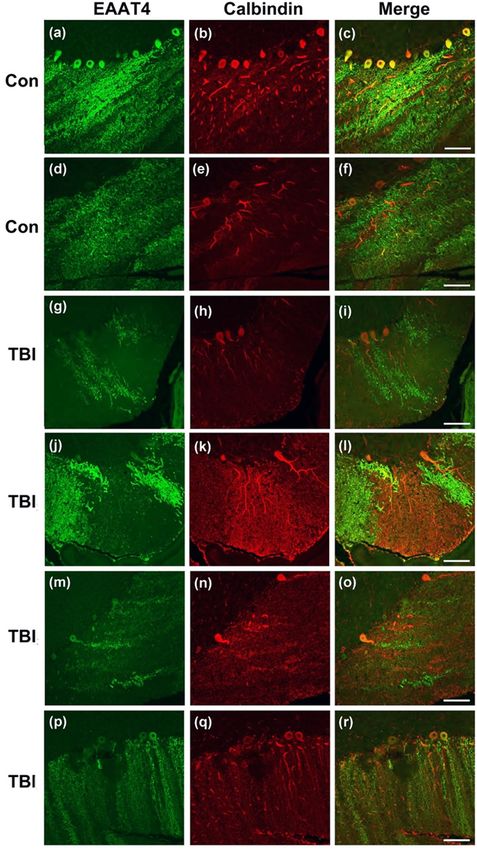
In keeping with the mouse neuropathologic findings (Fig. 8) and our previous results in 3X blast exposed mice
also examined 30 days post treatment11, we observed patchy domains of cerebellar Purkinje cell loss (Fig. 8) in the
cases with blast-related mTBI (red calbindin staining). Accompanying this, we observed striking patchy domains
of EAAT4+ arbor loss in the molecular layer (green). In some instances, these arbors were not lost (evidenced by
the calbindin immunostaining), yet still displayed a marked decrease in EAAT4, a molecule that is critical in reg-
ulating glutamate uptake and glutamatergic signaling in these cells. Overall, these results support the translational
Scientific Reports | (2020) 10:9420 | https://doi.org/10.1038/s41598-020-66113-7 7www.nature.com/scientificreports/ www.nature.com/scientificreports
Figure 8. Dystrophic EAAT4 expression in cerebellar Purkinje cells in human with blast-related mTBI.
Confocal microscopy shows representative immunofluorescent images of EAAT4 (green), and calbindin (red)
in the cerebella of Veteran/military Servicemember controls with no blast TBI (a–c and d–f, Controls1–2,
respectively described in Results), and in the cerebellum of Veteran/military Servicemembers with blast-related
mTBI (g–i, j–l, m–o, and p–r, TBIs1–4, respectively as described in Results) Scale bars = 50 µm.
significance of the neuropathology we and others have reported in blast-exposed animals11,14,69,70, and suggest that
the vast network of highly arborized Purkinje cells, which are the sole output of the cerebellum, may be chroni-
cally disrupted by blast-related mTBI.
Scientific Reports | (2020) 10:9420 | https://doi.org/10.1038/s41598-020-66113-7 8www.nature.com/scientificreports/ www.nature.com/scientificreports
Discussion
We have found that repetitive mTBI causes delayed-onset BBB disruption in the cerebellum and was markedly
more robust than in forebrain structures of blast-exposed mice. Biphasic BBB disruption has been observed in
other rodent models of neurotrauma71,72. This dynamic property of BBB injury is potentially important because
latent BBB disruption has been proposed as a target for post-injury TBI intervention8. In this report, we found
that the cerebellum was more susceptible to BBB dysfunction than the forebrain. This finding is well in keeping
with our previous results showing that the cerebellum is vulnerable to blast-induced BBB disruption8, subsequent
Purkinje cell loss11, and is also in keeping with pioneering findings in other neurotrauma animal models that
show cerebellum is prone to injury even when the initiating insult is focused elsewhere in the brain40,41.
Previously it has been thought that the CNS and the peripheral immune system are functionally independent
of one another. This view has more recently undergone important revisions based on studies that demonstrated
active interactions between immune cells in the periphery and in the CNS73. In this study, CD4+ T-cell infiltra-
tion was observed in the cerebellum. Leukocyte infiltration has been considered a functional surrogate for BBB
dysfunction, which has been observed in the injured CNS after more severe impact TBI74. Recently there has been
a report that blast can induce CCL2+ monocytes to cross the BBB9. The results herein demonstrate that CD4+
T-cells infiltrate the cerebellum following repetitive blast-induced mTBI.
A growing body of evidence indicates that neurotrauma can provoke peripheral immune cells to infiltrate
the brain56. While there is gross histological evidence indicating that leukocytes enter the CNS after blast expo-
sure75,76, the effects of blast on immune cell brain infiltration have thus far received limited attention and there
has been no information regarding potential mechanisms or functional consequences. One mechanism by which
leukocytes and, in particular, T-cells infiltrate the brain involve changes in ICAM-1 expression57. We found that
ICAM-1 expression was both increased in cerebellum after blast mTBI and was reduced by NOS inhibition.
Increased expression of ICAM-1 has also been observed in human endothelial cells exposed to inflammatory
cytokines77. In addition, NOS signaling modulates ICAM-1 expression and function53. Excessive inflammation
can cause microvascular hyper-permeability through NOS-dependent mechanisms78. Interestingly however, inhi-
bition of constitutive NOS signaling promotes leukocyte adhesion79. Blast exposed mice exhibit excessive NOS
signaling80 and NOS-dependent BBB dysfunction8. The data in this report shed new light on the NOS-dependent
mechanisms of delayed leukocyte infiltration associated with BBB dysfunction.
In keeping with our previous work on delayed-onset BBB disruption8, these results speak further to the poten-
tial significance of the acute/subacute post-injury interval in shaping long term outcomes. Even though L-NAME
and it’s active NOS inhibiting metabolite L-NG-nitro arginine (LNOARG) have short half-lives on the order of
20 minutes to 22 hours, respectively81, we found that L-NAME treatment from 48–71 hours post-blast prevented
impaired sensorimotor performance tested 30 days after the last blast exposure.
Changes in NOS signaling have been observed in other pre-clinical brain injury models82,83 and have been
implicated in disease progression after TBI84,85. A current clinical trial using inhaled nitric oxide is underway as
a means to improve TBI outcome (NCT03260569 clinicaltrails.gov). The potential relationships between periph-
eral NO administration, the timing of such treatment post injury, and other factors make it very difficult to
draw precise correspondences between the findings in this report, Nonetheless, this effort in the clinical arena
lends additional support to the idea NO/NOS signaling pathways are relevant targets for further investigation in
treating mTBI. NOS inhibition has also been shown to improve sensorimotor function38 and reduce neuronal
cell death86 in preclinical models of TBI. Our results suggest that acute/subacute NOS inhibition does not block
the full spectrum of cerebellar neuropathology. It is possible that this specific intervention may only delay the
sequelae of blast mTBI. Nonetheless, it is possible that long-term strategies to modulate NOS function could offer
additional neuroprotection; or at least even further delay onset and progression of mTBI-related neurotrauma.
Currently there is very little information from postmortem human cerebellum after blast exposure, primar-
ily because available tissue from such cases is extremely rare. In the cerebella from four Veterans and military
Servicemembers with histories of blast-related TBI, we found marked dystrophic Purkinje cell arbor morphology
and loss of EAAT4 expression, which strengthens the potential translational significance of the neuropathologic
findings we and others have observed in animal models of blast mTBI.
The vulnerability of the cerebellum to mTBI is not well understood. It is likely due to a number of interacting
factors that include: (i) high metabolic demand due to the very large number of neurons (69 billion of the 86
billion total neurons in adult human brain are in cerebellum); (ii) a distinct microvascular structure, and (iii) its
anatomical location with respect to the skull87–89. In considering these factors, BBB disruption and/or immune
cell infiltration may still represent key interacting pathologic processes that drive persistent cerebellar dysfunc-
tion and neuropathology.
Veterans with mild TBI exhibit aberrant cerebellar function30,31,90,91. BBB dysfunction in the cerebellum could
set in motion chronic low-grade inflammation associated with NOS signaling that could eventually result in
long-term neurodegeneration. Overall, our results argue that NOS plays an important role in mediating cerebellar
responses to mild blast injury and suggest that properly timed interventions blocking NO signaling delay patho-
physiological progression. Not only do these complex interactions play important roles in mediating delayed BBB
dysfunction, but they may also play a critical role in mTBI neurodegenerative progression.
Methods
Animals. Male C57BL/6 (n = 148) mice (Jackson Laboratories) 8–12 weeks of age were studied. Mice had ad
libitum access to food, water, and were group housed. Animals were randomly assigned to sham, drug, or TBI
groups. All studies were approved by the Veterans Affairs Puget Sound Health Care System’s Institutional Animal
Care and Use Committee. Experiments were conducted in accordance with the National Institutes of Health
Guide for the Care and Use of Laboratory Animals and reported in compliance with the ARRIVE guidelines.
Scientific Reports | (2020) 10:9420 | https://doi.org/10.1038/s41598-020-66113-7 9www.nature.com/scientificreports/ www.nature.com/scientificreports
Blast exposure. Mice were acclimatized to the animal facility for at least one week prior to experimen-
tation. For blast exposure, animals were first anesthetized with 5% isoflurane, followed by maintenance with
2% isoflurane (1 L/min oxygen). Mice were mounted in a restraint harness in the shock tube with their ventral
body surface facing the oncoming shock wave in a pneumatic shock tube as previously described10,11,44. Sham
control animals were mounted in the restraint harness and held under anesthesia for the same amount of time as
the blast-exposed mice. Mice that survived blast exposure (146/148) all displayed normal ambulation, visually
guided grasping responses when elevated by the tail near a foothold, and grooming behavior within 1 hour after
exposure.
Radiolabeled tracer preparation. Following established procedures50, albumin (Sigma, St. Louis MO)
was labeled with Tc (GE Healthcare, Piscataway, NJ). A mixture of 240 mg/ml stannous tartrate and 1 mg/ml
99m
albumin was adjusted to pH 3.0 with HCl. One millicurie of 99mTc-NaOH4 was added to this mixture and allowed
to incubate for 20 min. The 99mTc-albumin was purified on a column of G-10 Sephadex (GE Healthcare) in 0.1 ml
fractions of phosphate buffer (0.25 M). Radioactivity in the purified 99mTc-albumin peak was more than 90% acid
precipitable in an equal volume of 1% bovine serum albumin (BSA) and trichloroacetic acid (30%). 5 × 10⁶ cpm/
mouse of purified 99mTc-albumin fraction was prepared in a final volume (0.2 ml/mouse) of lactated Ringer’s
solution containing 1% BSA.
Radiolabeled tracer injections. Following established procedures50, at 72 h after the final blast/sham treat-
ment, mice were anesthetized with urethane (4 g/kg; 0.2 ml; ip); jugular veins were exposed, and injected with
99m
Tc-albumin (5 × 10⁶ Counts per minute (cpm)) in 0.2 ml of lactated Ringer’s solution with 1% BSA for 10 min.
Blood was collected from a cut in the descending abdominal aorta. Vascular space of the brain was then washed
free of blood by opening the thorax, clamping the descending thoracic aorta, severing both jugular veins, and
perfusing 20 ml of lactated Ringer’s solution through the left ventricle of the heart in less than 1 min. After wash-
out, brain was removed. The cerebellum and forebrain structures (remaining brain regions excluding posterior
midbrain, pons, medulla, and cerebellum) were dissected and individually weighed. Brains with visible blood
after washout were excluded from analysis (n = 2). Serum was obtained by centrifuging the carotid artery blood
for 10 min at 3,200 × g at 4 °C. Levels of 99mTc radioactivity in the serum and brain regions were determined in a
gamma counter. Brain tissue radioactivity was calculated by dividing the cpm in the brain region by the weight
of the brain region to yield cpm/g. Serum radioactivity was calculated by dividing the cpm in the serum by the
microliters of serum counted to yield cpm/microliter. The brain tissue radioactivity was then divided by the cor-
responding serum radioactivity and the results given in units of microliters/gram of brain tissue.
N(G)-nitro-L-arginine methyl ester treatment. For all studies in this report, L-NAME treatment indi-
cates that the mice received three intraperitoneal injections of N(G)-nitro-L-arginine methyl ester (L-NAME;
Sigma; 10 mg/kg; 100 µl 7% NaHCO3; ip) at 48, 54, and 71 h after the last blast. For these experiments, shams
received identical L-NAME injections along with their respective blast cohorts. We employed this 3X dosing
paradigm based on the plasma half-life of L-NAME81. Vehicle controls received three 100 µl injections of 7%
NaHCO3.
Rotarod. A rotarod apparatus (San Diego Instruments, San Diego, CA) was used to measure motor coordina-
tion and balance. Five trials are used, each with a different speed of rotation (8, 16, 24, 32, and 40 rpm). For each
trial the rod was accelerated to its final speed over 120 s, remained at the final speed for an additional 30 s, and
finally decreased back to 0 rpm over 30 s. 2 min intertrial intervals were used. The latency to fall of the rotarod was
recorded for each trial.
Confocal microscopy. Using previously established methods8, brains were post-fixed in 4% paraformalde-
hyde in PBS at 4° C for ten 99mTc-albumin half-lives (~60 h). Hemisections were equilibrated in 30% sucrose in
PBS for 24 h at 4 °C and embedded in OCT (Tissue-Tek, Torrance, CA). Antigen retrieval was performed with
50 mM sodium citrate (pH 9.0) and then heating at 80 °C for 30 min. Immunostaining was performed as pre-
viously described14. Floating tissue sections were cover slipped with a drop of Prolong Gold Antifade Reagent.
The following antibodies were used: rabbit polyclonal anti-EAAT4 (Abcam, Cambridge, MA; 1:1,000), mouse
monoclonal calbindin (Abcam; 1:1,000), rabbit polyclonal anti-GLUT1 (Millipore, Billerica, MA; 1:500), and goat
polyclonal anti-ICAM-1 (Novus Biologics, Centennial, CO; 1:1,000). Corresponding secondary antibodies labe-
led with Alexa 488, and Cy3 were applied for 2 h (Jackson Immunoresearch, West Grove, PA; 1:1,000). Confocal
microscopy was performed with a Leica TCS SP5 II microscope. Microscopic images were acquired with the Leica
Application Suite and processed using image adjustments limited only to linear contrast and brightness adjust-
ments applied identically to data from blast- and sham-treated animals in each experiment.
Flow cytometry. At 72 h after last blast or after time-matched sham procedures, mice were anesthetized with
urethane (4 g/kg; 0.2 ml; ip), and transcardially perfused with ice-cold PBS. Brains were extracted and dissected
into cerebellum and forebrain. Brain regions were mashed on ice in PBS. Homogenates were centrifuged at 300
× g for 5 min at 4o C. Pellets were resuspended in enzyme dissociation buffer (Tumor Dissociation kit, Miltenyi
Biotec, Auburn, CA), triturated with a 22-gauge needle, and rocked at 37 oC. Digested brain suspension were then
passed through a 70 μm SmartStrainer and rinsed thoroughly with 5 ml HBSS/FBS/EDTA. The collected sus-
pension was centrifuged at 300 × g. Pellets were resuspended in 5 ml of 40% Percoll (GE Healthcare, Pittsburgh,
PA, USA) and centrifuged at 400 × g for 20 min at 25 oC. The myelin layer (top) and Percoll supernatant were
aspirated and the pellets were washed with 5 ml HBSS/FBS/EDTA and centrifuged at 300 × g for 10 min at 25 oC.
Supernatant was aspirated and pellet was resuspended in 100 μl PBS and transferred to 1.5 ml centrifuge tubes.
Scientific Reports | (2020) 10:9420 | https://doi.org/10.1038/s41598-020-66113-7 10www.nature.com/scientificreports/ www.nature.com/scientificreports
Aliquots of each cell suspension were pooled for use as controls for staining. One aliquot from the pool was
reserved as an unstained control; a second was heat-treated for 10 min at 55 °C. All cell suspensions, including the
pool and the heat-killed aliquot (but not the unstained control) were incubated with an equal volume of viability
dye (Ghost Dye510, Tonbo Biosciences, San Diego, CA) diluted 1:500 in PBS for 30 min at 4 °C. Labeling was ter-
minated by the addition of 5 volumes HBSS/FBS/EDTA, and centrifuging at 300 × g for 5 min. Cells were resus-
pended in HBSS/FBS/EDTA containing 5 μg/ml mouse Fc Block (ThermoFisher Grand Island, NY) for 15 min
on ice before staining with a panel of fluorophore-conjugated primary antibodies (Supplementary Table 1). Cells
were incubated with antibodies on ice for 30 min, covered, then washed and resuspended in 250 μl 1% paraform-
aldehyde in PBS, and stored at 4 °C until analyzed. Data were acquired on an LSRFortessa SORP using FACSDiva
8.0 (BD Biosciences, San Jose, CA, USA) and analyzed using FlowJo (Tree Star, San Carlos, CA, USA). A stand-
ardized gating strategy was used for infiltrating T-cells (Supplementary Fig. 1). Fluorescence-minus-one (FMO)
controls were used to determine antibody positivity, and compensation controls were performed by adding 1 drop
of UltraComp eBeads (ThermoFisher Invitrogen).
Immunoblotting. Using previously established methods8, brains were flash frozen in liquid nitrogen and
stored at −80 °C. Dissected tissue was sonicated in radioimmunoprecipitation assay (RIPA) buffer with protease/
phosphatase inhibitors (Thermo; Rockford, IL 1:100), centrifuged for 15 min at 12,000 × g, supernatant col-
lected, and protein determined with a bicinchoninic acid (BCA) assay. Protein lysates were resolved by sodium
dodecyl sulfate-polyacrylamide gel electrophoresis (SDS-PAGE) (20 µg total protein/lane) under reducing con-
ditions, transferred to nitrocellulose, and probed with goat polyclonal anti-ICAM-1 (Novus Biologics, 1:1,000),
rabbit monoclonal anti-VCAM-1 (Abcam, 1:2000), and rabbit monoclonal β-actin (Cell signaling, Danvers, MA;
1:10,000) as loading controls. Blots were probed with anti-goat and anti-rabbit horseradish peroxidase-conjugated
secondary antibodies (Jackson ImmunoResearch; 1:5,000) and visualized by enhanced chemiluminescence
(GE Healthcare, Piscataway, NJ). Optical densities of the immunoreactive protein bands were quantified using
Image-Quant with background subtraction and normalization to loading control (β-actin). Full immunoblots can
be found in Supplementary Fig. 2 (ICAM-1), and Supplementary Fig. 3 (VCAM-1).
Human tissue. Collection of all brain specimens and case characteristics information were conducted in
accordance with approved University of Washington and Uniformed Services University Institutional Review
Board (IRB) procedures allowing for use of de-identified tissue samples. Tissue samples were obtained from
autopsies of blast-exposed Veterans and age, sex-matched, non-exposed controls. Brain sections were stained
using a commercially available kit (Opal Manual IHC Kit, Akoya Bioscience). The following antibodies were
applied overnight at 4 °C: rabbit polyclonal anti-EAAT4 (Abcam; 1:1,000), and mouse monoclonal calbindin
(Millipore; 1:1,000). Heat-mediated antigen retrieval was performed in AR6 buffer. Stained slides were mounted
with ProLong Diamond antifade mountant (ThermoFisher). Confocal microscopy was performed as described
above.
Statistical analysis. Statistical analyses used GraphPad Prism 8.0 8 (GraphPad Software, Inc., La Jolla, CA)
and SPSS software (IBM Corp, Armonk, NY). Error bars represent standard error of the mean (SEM). One-way
analysis of variance (ANOVA) were followed by Newman-Keuls, post-hoc tests. Two-way ANOVAs were followed
by a Helmert test92,93.
Notification. The opinions expressed herein are those of the authors and are not necessarily representative of
those of the Uniformed Services University, the United States Department of Defense or the United States Army,
Navy, or Air Force.
Data availability
Authors will provide published data and relevant protocols upon request to the Principal Investigator by phone
or email.
Received: 15 October 2019; Accepted: 16 March 2020;
Published: xx xx xxxx
References
1. AFHSB. DoD Worldwide Numbers for TBI https:/dvbic.dcoe.mil/dod-worldwide-numbers-tbi (2019).
2. Barnes, D. E. et al. Association of Mild Traumatic Brain Injury With and Without Loss of Consciousness With Dementia in US
Military Veterans. JAMA Neurol. 75, 1055–1061, https://doi.org/10.1001/jamaneurol.2018.0815 (2018).
3. Janelidze, S. et al. Increased blood-brain barrier permeability is associated with dementia and diabetes but not amyloid pathology or
APOE genotype. Neurobiol. aging 51, 104–112, https://doi.org/10.1016/j.neurobiolaging.2016.11.017 (2017).
4. Nation, D. A. et al. Blood-brain barrier breakdown is an early biomarker of human cognitive dysfunction. Nat. Med. 25, 270–276,
https://doi.org/10.1038/s41591-018-0297-y (2019).
5. Nelson, A. R., Sweeney, M. D., Sagare, A. P. & Zlokovic, B. V. Neurovascular dysfunction and neurodegeneration in dementia and
Alzheimer’s disease. Biochim. Biophys. Acta 1862, 887–900, https://doi.org/10.1016/j.bbadis.2015.12.016 (2016).
6. Elder, G. A. et al. Vascular and inflammatory factors in the pathophysiology of blast-induced brain injury. Front. Neurol. 6, 48,
https://doi.org/10.3389/fneur.2015.00048 (2015).
7. Glushakova, O. Y., Johnson, D. & Hayes, R. L. Delayed increases in microvascular pathology after experimental traumatic brain
injury are associated with prolonged inflammation, blood-brain barrier disruption, and progressive white matter damage. J.
neurotrauma 31, 1180–1193, https://doi.org/10.1089/neu.2013.3080 (2014).
8. Logsdon, A. F. et al. Blast exposure elicits blood-brain barrier disruption and repair mediated by tight junction integrity and nitric
oxide dependent processes. Sci. Rep. 8, 11344, https://doi.org/10.1038/s41598-018-29341-6 (2018).
9. Kuriakose, M., Rama Rao, K. V., Younger, D. & Chandra, N. Temporal and Spatial Effects of Blast Overpressure on Blood-Brain
Barrier Permeability in Traumatic Brain Injury. Sci. Rep. 8, 8681, https://doi.org/10.1038/s41598-018-26813-7 (2018).
Scientific Reports | (2020) 10:9420 | https://doi.org/10.1038/s41598-020-66113-7 11www.nature.com/scientificreports/ www.nature.com/scientificreports
10. Huber, B. R. et al. Blast exposure causes dynamic microglial/macrophage responses and microdomains of brain microvessel
dysfunction. Neuroscience 319, 206–220, https://doi.org/10.1016/j.neuroscience.2016.01.022 (2016).
11. Meabon, J. S. et al. Repetitive blast exposure in mice and combat veterans causes persistent cerebellar dysfunction. Sci. Transl. Med.
8, 321ra326, https://doi.org/10.1126/scitranslmed.aaa9585 (2016).
12. Gama Sosa, M. A. et al. Low-level blast exposure disrupts gliovascular and neurovascular connections and induces a chronic
vascular pathology in rat brain. Acta neuropathologica Commun. 7, 6, https://doi.org/10.1186/s40478-018-0647-5 (2019).
13. Abutarboush, R. et al. Exposure to Blast Overpressure Impairs Cerebral Microvascular Responses and Alters Vascular and Astrocytic
Structure. Journal of neurotrauma, https://doi.org/10.1089/neu.2019.6423 (2019).
14. Huber, B. R. et al. Blast exposure causes early and persistent aberrant phospho- and cleaved-tau expression in a murine model of
mild blast-induced traumatic brain injury. J. Alzheimers Dis. 37, 309–323, https://doi.org/10.3233/JAD-130182 (2013).
15. Kondo, A. et al. Antibody against early driver of neurodegeneration cis P-tau blocks brain injury and tauopathy. Nature 523,
431–436, https://doi.org/10.1038/nature14658 (2015).
16. Goldstein, L. E. et al. Chronic traumatic encephalopathy in blast-exposed military veterans and a blast neurotrauma mouse model.
Sci. Transl. Med. 4, 134ra160, https://doi.org/10.1126/scitranslmed.3003716 (2012).
17. Mez, J. et al. Clinicopathological Evaluation of Chronic Traumatic Encephalopathy in Players of American Football. Jama 318,
360–370, https://doi.org/10.1001/jama.2017.8334 (2017).
18. Lucke-Wold, B. P. et al. Linking traumatic brain injury to chronic traumatic encephalopathy: identification of potential mechanisms
leading to neurofibrillary tangle development. J. neurotrauma 31, 1129–1138, https://doi.org/10.1089/neu.2013.3303 (2014).
19. Omalu, B. I. et al. Chronic traumatic encephalopathy in a National Football League player. Neurosurgery 57, 128-134; discussion
128–134 (2005).
20. Shetty, A. K., Mishra, V., Kodali, M. & Hattiangady, B. Blood brain barrier dysfunction and delayed neurological deficits in mild
traumatic brain injury induced by blast shock waves. Front. Cell. Neurosci. 8, 232, https://doi.org/10.3389/fncel.2014.00232 (2014).
21. Logsdon, A. F. et al. Altering endoplasmic reticulum stress in a model of blast-induced traumatic brain injury controls cellular fate
and ameliorates neuropsychiatric symptoms. Front. Cell. Neurosci. 8, 421, https://doi.org/10.3389/fncel.2014.00421 (2014).
22. Lucke-Wold, B. P. et al. Bryostatin-1 Restores Blood Brain Barrier Integrity following Blast-Induced Traumatic Brain Injury. Mol.
Neurobiol. 52, 1119–1134, https://doi.org/10.1007/s12035-014-8902-7 (2015).
23. Hue, C. D. et al. Time Course and Size of Blood-Brain Barrier Opening in a Mouse Model of Blast-Induced Traumatic Brain Injury.
J. neurotrauma 33, 1202–1211, https://doi.org/10.1089/neu.2015.4067 (2016).
24. Garman, R. H. et al. Blast exposure in rats with body shielding is characterized primarily by diffuse axonal injury. J. neurotrauma 28,
947–959, https://doi.org/10.1089/neu.2010.1540 (2011).
25. Yeoh, S., Bell, E. D. & Monson, K. L. Distribution of blood-brain barrier disruption in primary blast injury. Ann. Biomed. Eng. 41,
2206–2214, https://doi.org/10.1007/s10439-013-0805-7 (2013).
26. Readnower, R. D. et al. Increase in blood-brain barrier permeability, oxidative stress, and activated microglia in a rat model of blast-
induced traumatic brain injury. J. Neurosci. Res. 88, 3530–3539, https://doi.org/10.1002/jnr.22510 (2010).
27. Hall, A. A. et al. Repeated Low Intensity Blast Exposure Is Associated with Damaged Endothelial Glycocalyx and Downstream
Behavioral Deficits. Front. Behav. Neurosci. 11, 104, https://doi.org/10.3389/fnbeh.2017.00104 (2017).
28. Peskind, E. R. et al. Cerebrocerebellar hypometabolism associated with repetitive blast exposure mild traumatic brain injury in 12
Iraq war Veterans with persistent post-concussive symptoms. Neuroimage 54(Suppl 1), S76–82, https://doi.org/10.1016/j.
neuroimage.2010.04.008 (2011).
29. Warden, D. L. et al. Case report of a soldier with primary blast brain injury. Neuroimage 47(Suppl 2), T152–153, https://doi.
org/10.1016/j.neuroimage.2009.01.060 (2009).
30. Petrie, E. C. et al. Neuroimaging, behavioral, and psychological sequelae of repetitive combined blast/impact mild traumatic brain
injury in Iraq and Afghanistan war veterans. J. neurotrauma 31, 425–436, https://doi.org/10.1089/neu.2013.2952 (2014).
31. Jorge, R. E. et al. White matter abnormalities in veterans with mild traumatic brain injury. Am. J. Psychiatry 169, 1284–1291, https://
doi.org/10.1176/appi.ajp.2012.12050600 (2012).
32. Mac Donald, C. L. et al. Detection of blast-related traumatic brain injury in U.S. military personnel. N. Engl. J. Med. 364, 2091–2100,
https://doi.org/10.1056/NEJMoa1008069 (2011).
33. Taylor, P. A. & Ford, C. C. Simulation of blast-induced early-time intracranial wave physics leading to traumatic brain injury. J.
Biomech. Eng. 131, 061007, https://doi.org/10.1115/1.3118765 (2009).
34. Sundaramurthy, A. et al. Blast-induced biomechanical loading of the rat: an experimental and anatomically accurate computational
blast injury model. J. neurotrauma 29, 2352–2364, https://doi.org/10.1089/neu.2012.2413 (2012).
35. Bauman, R. A. et al. An introductory characterization of a combat-casualty-care relevant swine model of closed head injury resulting
from exposure to explosive blast. J. neurotrauma 26, 841–860, https://doi.org/10.1089/neu.2009-0898 (2009).
36. Badea, A. et al. Repeated mild blast exposure in young adult rats results in dynamic and persistent microstructural changes in the
brain. Neuroimage Clin. 18, 60–73, https://doi.org/10.1016/j.nicl.2018.01.007 (2018).
37. Peterson, T. C., Maass, W. R., Anderson, J. R., Anderson, G. D. & Hoane, M. R. A behavioral and histological comparison of fluid
percussion injury and controlled cortical impact injury to the rat sensorimotor cortex. Behav. Brain Res. 294, 254–263, https://doi.
org/10.1016/j.bbr.2015.08.007 (2015).
38. Wada, K., Chatzipanteli, K., Busto, R. & Dietrich, W. D. Effects of L-NAME and 7-NI on NOS catalytic activity and behavioral
outcome after traumatic brain injury in the rat. J. neurotrauma 16, 203–212, https://doi.org/10.1089/neu.1999.16.203 (1999).
39. Bhowmick, S., D’Mello, V., Ponery, N. & Abdul-Muneer, P. M. Neurodegeneration and Sensorimotor Deficits in the Mouse Model
of Traumatic Brain Injury. Brain Sci 8, https://doi.org/10.3390/brainsci8010011 (2018).
40. Potts, M. B., Adwanikar, H. & Noble-Haeusslein, L. J. Models of traumatic cerebellar injury. Cerebellum 8, 211–221, https://doi.
org/10.1007/s12311-009-0114-8 (2009).
41. Fukuda, K. et al. Purkinje cell vulnerability to mild traumatic brain injury. J. neurotrauma 13, 255–266, https://doi.org/10.1089/
neu.1996.13.255 (1996).
42. Stoodley, C. J. The cerebellum and cognition: evidence from functional imaging studies. Cerebellum 11, 352–365, https://doi.
org/10.1007/s12311-011-0260-7 (2012).
43. Koziol, L. F. et al. Consensus paper: the cerebellum’s role in movement and cognition. Cerebellum 13, 151–177, https://doi.
org/10.1007/s12311-013-0511-x (2014).
44. Schindler, A. G. et al. Blast-related disinhibition and risk seeking in mice and combat Veterans: Potential role for dysfunctional
phasic dopamine release. Neurobiol. Dis. 106, 23–34, https://doi.org/10.1016/j.nbd.2017.06.004 (2017).
45. Cao, J. et al. ApoE4-associated phospholipid dysregulation contributes to development of Tau hyper-phosphorylation after traumatic
brain injury. Sci. Rep. 7, 11372, https://doi.org/10.1038/s41598-017-11654-7 (2017).
46. Del Razo, M. J. et al. Computational and in vitro studies of blast-induced blood-brain barrier disruption. Soc. Ind. Appl. Mathematics:
J. Sci. Computing. 38, B347–374 (2016).
47. Levin, V. A., Fenstermacher, J. D. & Patlak, C. S. Sucrose and inulin space measurements of cerebral cortex in four mammalian
species. Am. J. Physiol. 219, 1528–1533, https://doi.org/10.1152/ajplegacy.1970.219.5.1528 (1970).
48. Blasberg, R. G., Fenstermacher, J. D. & Patlak, C. S. Transport of alpha-aminoisobutyric acid across brain capillary and cellular
membranes. J. Cereb. blood flow. metabolism: Off. J. Int. Soc. Cereb. Blood Flow. Metab. 3, 8–32, https://doi.org/10.1038/jcbfm.1983.2
(1983).
Scientific Reports | (2020) 10:9420 | https://doi.org/10.1038/s41598-020-66113-7 12www.nature.com/scientificreports/ www.nature.com/scientificreports
49. Patlak, C. S., Blasberg, R. G. & Fenstermacher, J. D. Graphical evaluation of blood-to-brain transfer constants from multiple-time
uptake data. J. Cereb. blood flow. metabolism: Off. J. Int. Soc. Cereb. Blood Flow. Metab. 3, 1–7, https://doi.org/10.1038/jcbfm.1983.1
(1983).
50. Banks, W. A. et al. Lipopolysaccharide-induced blood-brain barrier disruption: roles of cyclooxygenase, oxidative stress,
neuroinflammation, and elements of the neurovascular unit. J. neuroinflammation 12, 223, https://doi.org/10.1186/s12974-015-
0434-1 (2015).
51. Banks, W. A. et al. Nitric oxide isoenzymes regulate lipopolysaccharide-enhanced insulin transport across the blood-brain barrier.
Endocrinology 149, 1514–1523, https://doi.org/10.1210/en.2007-1091 (2008).
52. Xaio, H., Banks, W. A., Niehoff, M. L. & Morley, J. E. Effect of LPS on the permeability of the blood-brain barrier to insulin. Brain
Res. 896, 36–42 (2001).
53. Xu, S., Zhou, X., Yuan, D., Xu, Y. & He, P. Caveolin-1 scaffolding domain promotes leukocyte adhesion by reduced basal endothelial
nitric oxide-mediated ICAM-1 phosphorylation in rat mesenteric venules. Am. J. Physiol. Heart Circ. Physiol 305, H1484–1493,
https://doi.org/10.1152/ajpheart.00382.2013 (2013).
54. Gao, F. et al. Reduction of Endothelial Nitric Oxide Increases the Adhesiveness of Constitutive Endothelial Membrane ICAM-1
through Src-Mediated Phosphorylation. Front. Physiol. 8, 1124, https://doi.org/10.3389/fphys.2017.01124 (2017).
55. Wu, M. & Tsirka, S. E. Endothelial NOS-deficient mice reveal dual roles for nitric oxide during experimental autoimmune
encephalomyelitis. Glia 57, 1204–1215, https://doi.org/10.1002/glia.20842 (2009).
56. McKee, C. A. & Lukens, J. R. Emerging Roles for the Immune System in Traumatic. BraInjury. Front. immunology 7, 556, https://doi.
org/10.3389/fimmu.2016.00556 (2016).
57. Engelhardt, B., Conley, F. K. & Butcher, E. C. Cell adhesion molecules on vessels during inflammation in the mouse central nervous
system. J. Neuroimmunol. 51, 199–208 (1994).
58. Wong, D., Prameya, R., Wu, V., Dorovini-Zis, K. & Vincent, S. R. Nitric oxide reduces T lymphocyte adhesion to human brain
microvessel endothelial cells via a cGMP-dependent pathway. Eur. J. pharmacology 514, 91–98, https://doi.org/10.1016/j.
ejphar.2005.03.025 (2005).
59. Steiner, O. et al. Differential roles for endothelial ICAM-1, ICAM-2, and VCAM-1 in shear-resistant T cell arrest, polarization, and
directed crawling on blood-brain barrier endothelium. J. Immunol. 185, 4846–4855, https://doi.org/10.4049/jimmunol.0903732
(2010).
60. Rahner-Welsch, S., Vogel, J. & Kuschinsky, W. Regional congruence and divergence of glucose transporters (GLUT1) and capillaries
in rat brains. J. Cereb. blood flow. metabolism: Off. J. Int. Soc. Cereb. Blood Flow. Metab. 15, 681–686, https://doi.org/10.1038/
jcbfm.1995.84 (1995).
61. Dehnes, Y. et al. The glutamate transporter EAAT4 in rat cerebellar Purkinje cells: a glutamate-gated chloride channel concentrated
near the synapse in parts of the dendritic membrane facing astroglia. J. Neurosci. 18, 3606–3619 (1998).
62. Fairman, W. A., Vandenberg, R. J., Arriza, J. L., Kavanaugh, M. P. & Amara, S. G. An excitatory amino-acid transporter with
properties of a ligand-gated chloride channel. Nature 375, 599–603 (1995).
63. Liu, W. et al. Perfusion deficits in patients with mild traumatic brain injury characterized by dynamic susceptibility contrast MRI.
NMR biomedicine 26, 651–663, https://doi.org/10.1002/nbm.2910 (2013).
64. Fischer, B. L. et al. Neural activation during response inhibition differentiates blast from mechanical causes of mild to moderate
traumatic brain injury. J. neurotrauma 31, 169–179, https://doi.org/10.1089/neu.2013.2877 (2014).
65. Yeh, P. H. et al. Postconcussional disorder and PTSD symptoms of military-related traumatic brain injury associated with
compromised neurocircuitry. Hum. brain Mapp. 35, 2652–2673, https://doi.org/10.1002/hbm.22358 (2014).
66. Petrie, E. C. et al. Neuroimaging, Behavioral, and Psychological Sequelae of Repetitive Combined Blast/Impact Mild Traumatic
Brain Injury in Iraq and Afghanistan War Veterans. J. neurotrauma 31, 425–436, https://doi.org/10.1089/neu.2013.2952 (2014).
67. Mac Donald, C. et al. Cerebellar white matter abnormalities following primary blast injury in US military personnel. PLoS one 8,
e55823, https://doi.org/10.1371/journal.pone.0055823 (2013).
68. Robinson, M. E. et al. Positron emission tomography of tau in Iraq and Afghanistan Veterans with blast neurotrauma. Neuroimage
Clin. 21, 101651, https://doi.org/10.1016/j.nicl.2019.101651 (2019).
69. Lu, J. et al. Effect of blast exposure on the brain structure and cognition in Macaca fascicularis. J. neurotrauma 29, 1434–1454,
https://doi.org/10.1089/neu.2010.1591 (2012).
70. Koliatsos, V. E. et al. A mouse model of blast injury to brain: initial pathological, neuropathological, and behavioral characterization.
J. neuropathology Exp. Neurol. 70, 399–416, https://doi.org/10.1097/NEN.0b013e3182189f06 (2011).
71. Baldwin, S. A., Fugaccia, I., Brown, D. R., Brown, L. V. & Scheff, S. W. Blood-brain barrier breach following cortical contusion in the
rat. J. Neurosurg. 85, 476–481, https://doi.org/10.3171/jns.1996.85.3.0476 (1996).
72. Baskaya, M. K., Rao, A. M., Dogan, A., Donaldson, D. & Dempsey, R. J. The biphasic opening of the blood-brain barrier in the cortex
and hippocampus after traumatic brain injury in rats. Neurosci. Lett. 226, 33–36 (1997).
73. Louveau, A. et al. Structural and functional features of central nervous system lymphatic vessels. Nature 523, 337–341, https://doi.
org/10.1038/nature14432 (2015).
74. Braun, M. et al. Activation of Myeloid TLR4 Mediates T Lymphocyte Polarization after Traumatic Brain Injury. J. Immunol. 198,
3615–3626, https://doi.org/10.4049/jimmunol.1601948 (2017).
75. Tompkins, P. et al. Brain injury: neuro-inflammation, cognitive deficit, and magnetic resonance imaging in a model of blast induced
traumatic brain injury. J. neurotrauma 30, 1888–1897, https://doi.org/10.1089/neu.2012.2674 (2013).
76. Braun, M. et al. White matter damage after traumatic brain injury: A role for damage associated molecular patterns. Biochimica et
biophysica acta. Mol. basis Dis. 1863, 2614–2626, https://doi.org/10.1016/j.bbadis.2017.05.020 (2017).
77. Wertheimer, S. J., Myers, C. L., Wallace, R. W. & Parks, T. P. Intercellular adhesion molecule-1 gene expression in human endothelial
cells. Differential regulation by tumor necrosis factor-alpha and phorbol myristate acetate. J. Biol. Chem. 267, 12030–12035 (1992).
78. Hatakeyama, T. et al. Endothelial nitric oxide synthase regulates microvascular hyperpermeability in vivo. J. Physiol. 574, 275–281,
https://doi.org/10.1113/jphysiol.2006.108175 (2006).
79. Kubes, P., Suzuki, M. & Granger, D. N. Nitric oxide: an endogenous modulator of leukocyte adhesion. Proc. Natl Acad. Sci. USA 88,
4651–4655 (1991).
80. Abdul-Muneer, P. M. et al. Induction of oxidative and nitrosative damage leads to cerebrovascular inflammation in an animal model
of mild traumatic brain injury induced by primary blast. Free. Radic. Biol. Med. 60, 282–291, https://doi.org/10.1016/j.
freeradbiomed.2013.02.029 (2013).
81. Avontuur, J. A., Buijk, S. L. & Bruining, H. A. Distribution and metabolism of N(G)-nitro-L-arginine methyl ester in patients with
septic shock. Eur. J. Clin. Pharmacol. 54, 627–631, https://doi.org/10.1007/s002280050525 (1998).
82. Khaldi, A., Chiueh, C. C., Bullock, M. R. & Woodward, J. J. The significance of nitric oxide production in the brain after injury. Ann.
N. Y. Acad. Sci. 962, 53–59 (2002).
83. Mishra, V. et al. Primary blast causes mild, moderate, severe and lethal TBI with increasing blast overpressures: Experimental rat
injury model. Sci. Rep. 6, 26992, https://doi.org/10.1038/srep26992 (2016).
84. Rao, V. L., Dogan, A., Bowen, K. K. & Dempsey, R. J. Traumatic injury to rat brain upregulates neuronal nitric oxide synthase
expression and L-[3H]nitroarginine binding. J. neurotrauma 16, 865–877, https://doi.org/10.1089/neu.1999.16.865 (1999).
85. Tweedie, D. et al. Changes in mouse cognition and hippocampal gene expression observed in a mild physical- and blast-traumatic
brain injury. Neurobiol. Dis. 54, 1–11, https://doi.org/10.1016/j.nbd.2013.02.006 (2013).
Scientific Reports | (2020) 10:9420 | https://doi.org/10.1038/s41598-020-66113-7 13You can also read