Hedonic Hot Spots in the Brain
←
→
Page content transcription
If your browser does not render page correctly, please read the page content below
REVIEW
Hedonic Hot Spots in the Brain
SUSANA PECIÑA, KYLE S. SMITH, and KENT C. BERRIDGE
Department of Psychology
University of Michigan, Ann Arbor
Hedonic “liking” for sensory pleasures is an important aspect of reward, and excessive ‘liking’ of particular
rewards might contribute to excessive consumption and to disorders such as obesity. The present review
aims to summarize recent advances in the identification of brain substrates for food ‘liking’ with a focus on
opioid hot spots in the nucleus accumbens and ventral pallidum. Drug microinjection studies have shown
that opioids in both areas amplify the ‘liking’ of sweet taste rewards. Modern neuroscience tools such as
Fos plume mapping have further identified hedonic hot spots within the accumbens and pallidum, where opi-
oids are especially tuned to magnify ‘liking’ of food rewards. Hedonic hot spots in different brain structures
may interact with each other within the larger functional circuitry that interconnects them. Better understand-
ing of how brain hedonic hot spots increase the positive affective impact of natural sensory pleasures will help
characterize the neural mechanisms potentially involved in ‘liking’ for many rewards. NEUROSCIENTIST
12(6):500–511, 2006. DOI: 10.1177/1073858406293154
KEY WORDS Hedonic, Reward, Liking, Pleasure, Fos plume, Taste reactivity, Opioid, GABA, Nucleus accumbens, Ventral pallidum
Hedonic impact, or ‘liking,’ is a critical aspect of reward. Levine 1983; Doyle and others 1993; Peciña and Berridge
Food and sex, for example, are potent sensory pleasures 1995; Rideout and Parker 1996; Peciña and Berridge
with liked hedonic impact, and it is widely acknowledged 2000; Kelley and others 2002). Injections of drugs that
that the ‘liking’ of food and sex carries important survival boost µ-opioid neurotransmission can dramatically increase
and reproductive benefits for humans and animals. consumption of palatable food, and opioid drugs also
However, sensory pleasure may not always be beneficial. increase taste hedonic reactions to palatable sucrose in
Rewards with large hedonic impact (e.g., junk food) may humans and rodents (Parker and others 1992; Doyle and
often be consumed more than those with low hedonic others 1993; Cooper and Higgs 1994; Peciña and Berridge
impact (e.g., vegetables), and in this way, hedonics may 1995; Rideout and Parker 1996; Peciña and Berridge 2000).
contribute to overeating and obesity and also to abuse of Conversely, manipulations that block or attenuate µ-opioid
drugs. Although the ‘wanting’ and ‘liking’ of rewards are activity reduce the consumption and incentive qualities of
separable, pleasure ‘liking’ may be a major contributor to sweet tastes and other rewards (Parker and others 1992;
normal and excessive reward consumption. Thus, it is Cooper and Higgs 1994).
important that behavioral neuroscience gains an under- Affective neuroscientists have begun to pinpoint par-
standing of how the brain causes reward hedonic impact. ticular brain systems responsible for opioid effects on
So how does it? How does the brain transform a mere hedonic ‘liking’ using behavioral techniques that reflect
sensory stimulus into a pleasurable and liked stimulus? the affective value of tastes and novel mapping proce-
For example, taste sweetness by itself is merely a sensa- dures. Two brain structures have emerged as likely candi-
tion, so its pleasure must arise within the brain, where dates to contain opioid hot spots that mediate hedonic
neural systems actively paint pleasure onto the gustatory impact, based in part on work conducted in our labora-
sensation to generate a ‘liking’ reaction, as a sort of tory: the nucleus accumbens and the ventral pallidum.
“pleasure gloss” (Berridge 2004). The question of how These structures are located within the ventral forebrain,
sensations are painted with pleasure to become liked can share reciprocal projections with one another, and are
be answered in part by identifying which particular neu- embedded within larger mesocorticolimbic reward sys-
ral systems are able to amplify objective indicators of the tems (Heimer and Wilson 1975; Mogenson and others
hedonic impact of natural rewards. 1983; Churchill and Kalivas 1994; Zahm 2000). Not only
Parts of the brain opioid system may be especially does opioid neurotransmission in these structures con-
important in painting a pleasure gloss onto sensation in tribute generally to reward motivation (Majeed and others
both humans and animals (Cooper 1983; Morley and 1986; Mucha and Iversen 1986; Bakshi and Kelley 1993;
Peciña and Berridge 2000; Peciña and Berridge 2005;
This work was supported by National Institutes of Health grants Smith and Berridge 2005b), but also each structure con-
DAO15188 and MH63649 (KCB) and DC00011 (KSS). tains an anatomical subregion in which opioids are par-
Address correspondence to: Susana Peciña, Department of
ticularly able to amplify the hedonic impact of sensory
Psychology, University of Michigan, 530 Church Street, Ann Arbor, MI pleasure (Peciña and Berridge 2005; Smith and Berridge
48109-1109 (e-mail: pesu@umich.edu). 2005b). This is what we refer to as a hedonic hot spot.
500 THE NEUROSCIENTIST Hedonic Hot Spots
Volume 12, Number 6, 2006
Copyright © 2006 Sage Publications
ISSN 1073-8584
In the following sections, we describe advances in
functional mapping of hedonic hot spots. We begin by
describing a behavioral measure to assess ‘liking’ of nat-
ural sensory pleasure in animals and techniques for map-
ping hedonic hot spots, proceed to review hedonic hot
spots within the nucleus accumbens and ventral pal-
lidum, and then speculate about circuitry dynamics and
additional hedonic transmitters and structures.

How Can We Measure
Hedonic ‘Liking’ in Rats?
Traditional studies of pleasure ‘liking’ have focused on
human adult subjects who can describe their feelings
(Cabanac 1971). But how can we measure ‘liking’ in
nonverbal animals such as rats for detailed neurobiolog-
ical research? The premise that underlies our research is
that hedonic impact or ‘liking’ is a basic evaluative reac-
tion of the brain, with objective neural and behavioral
indicators that can be quantified by appropriate methods.                Fig. 1. Taste ‘liking’ reactions across species. The top
One method involves measuring affective reactions to                     row shows an example of positive ‘liking’ reactions to a
rewards as an objective measure of hedonic stimulus                      pleasant sweet taste in a rat, primate, and human infant
impact (Fig. 1; Movie 1*). Given a sweet taste of sugar,                 (homologous rhythmic tongue protrusions). The bottom
rats emit distinctive facial and body affective expres-                  row shows an example of aversive ‘disliking’ reactions to
sions that mirror human affective reactions to tastes                    an unpleasant bitter taste (homologous gapes). Orofacial
(Grill and Norgren 1978; Grill and Berridge 1985).                       expressions such as these provide an objective index of
Sweet tastes elicit a positive hedonic pattern of reactions              ‘liking’ and ‘disliking’ reactions to the hedonic impact of
such as tongue protrusions (licking of lips), paw licking,               tastes.
and related movements. Bitter tastes elicit an aversive
pattern of different expressions such as gapes, head
shakes, and frantic wiping of the mouth. These affective                 structure and allow comparison of subregions. To meas-
reactions are homologous across rodents, primates, and                   ure the impact of drug/site manipulations on hedonic
human infants (Steiner and others 2001), share some                      ‘liking’, taste solutions are orally infused and ‘liking’ or
basic movement patterns with equivalent human expres-                    ‘disliking’ reaction patterns are quantified and compared
sions of hedonics, and fluctuate in similar ways as                      to normal vehicle levels.
human subjective pleasure when circumstances change                          For precision mapping of hedonic hot spots, however,
(e.g., in hunger or satiety states; Berridge 1996). Readers              it is not enough to know where a drug has been injected.
are referred to Grill and Berridge (1985) and Berridge                   Drugs can diffuse from the site of injection, which
(2000) for a more detailed review of taste reactivity and                makes pinpointing functional ‘liking’ effects rather ten-
‘liking’ analysis.                                                       uous and imprecise unless one knows exactly how far
                                                                         the impact spreads. Recently, we have developed a
Neuroscience Tools for Identifying Hedonic                               microinjection Fos plume tool based on local Fos protein
Hot Spots: Microinjections and Fos Plumes                                expression measurement for mapping of drug effects
                                                                         (Peciña and Berridge 2000; Peciña and Berridge 2005;
Finding brain systems responsible for painting a pleas-
                                                                         Smith and Berridge 2005b; Peciña and others 2006). Fos
ure gloss onto sensation requires brain manipulation
                                                                         plumes are elevations of Fos protein expression 2 times
experiments that can ethically be done only in animals.
                                                                         to >10 times around the microinjection tip (Fig. 2).
Pharmacological microinjections and analysis of behav-
                                                                         Many drugs microinjected into the brain activate partic-
ioral consequences have been the principal means to
                                                                         ular immediate early genes in surrounding neurons that
identify hedonic hot spots in brain structures. In these
                                                                         begin producing proteins, such as Fos, the spread of
studies, microinjections of neurotransmitter receptor
                                                                         which can be seen later as a dark plume of stained neu-
agonists or antagonists are made into a brain structure of
                                                                         rons on a slice of brain tissue. Measurement of Fos acti-
interest and are staggered in placement to fill the entire
                                                                         vation around the site of microinjection has proven to be
                                                                         a useful technique for identifying zones of local neu-
* A video showing affective taste reactivity is available online at
http://nro.sagepub.com/supplemental/. Video examples of affective        ronal activation (Peciña and Berridge 2000; Peciña and
‘liking’ and ‘disliking’ reactions to tastes are shown. The babies       Berridge 2005; Smith and Berridge 2005b; Peciña and
receive a taste solution from a dropper. The rats are viewed from        others 2006). Quantifying intense and moderate zones of
underneath a transparent floor as tastant solutions are painlessly       Fos activation within these plumes compared to control
infused into its mouth through a previously-implanted oral cannula.
Sucrose taste elicits positive hedonic or ‘liking’ reactions from rats
                                                                         tissue from vehicle-microinjected or uninjected rats
(e.g., rhythmic tongue protrusions). Quinine taste elicits negative      reveals the zones in which drugs are acting to elevate
aversive or ‘disliking’ reactions (e.g., gapes).                         ‘liking’ in behavioral experiments. By assigning the

Volume 12, Number 6, 2006                                                                              THE NEUROSCIENTIST        501
Nucleus Accumbens Hedonic Hot Spot
ateral Intense elevation
Moderate elevation
No change
Moderate suppression
Intense suppression
Fig. 2. Opioid hedonic hot spot in nucleus accumbens. The nucleus accumbens hedonic hot spot is localized to the ros-
trocaudal quarter of medial shell, represented in orange and red in sagittal, horizontal, and coronal views. The colors denote
the intensity of µ-opioid amplification of ‘liking’ reactions elicited by sucrose taste, compared to control vehicle levels in
the same rats, and the symbol size shows the diameter of Fos plumes surrounding DAMGO microinjections. A nucleus
accumbens affective cold spot is represented in blue and purple in the caudal half of the nucleus accumbens, where
DAMGO suppressed ‘liking’ reactions to sweetness. Modified from Peciña and Berridge (2005). Reprinted with permission.
behavioral ‘liking’ enhancements caused by microinjec- Interestingly, DAMGO does not increase hedonic
tions at particular sites to Fos plume–sized spreads of reactions at other sites of the nucleus accumbens tested
activation around those sites, mapping of the ‘liking’ so far, such as the caudal or ventral subregions of medial
consequences of Fos plumes in particular locations shell, even though DAMGO in these sites still stimulates
allows for objective and precise plots of hedonic hot a ‘wanting’ for food as reflected in increased intake. In
spots in the brain. fact, DAMGO microinjections in a small cold spot in the
caudal half of the medial shell appear to suppress ‘lik-
Hedonic Hot Spot in Nucleus ing’ reactions below vehicle control levels (while still
Accumbens Shell stimulating intake). DAMGO microinjections also
simultaneously decreased aversive ‘disliking’ reactions
Evidence for an Accumbens Opioid to quinine to less than 25% of control levels, sometimes
Hedonic Hot Spot nearly abolishing aversive reactions entirely, both in the
Using the experimental techniques described above, we hedonic hot spot and in surrounding regions in which
have identified a hedonic hot spot of an approximate DAMGO selectively stimulated intake (Fig. 3).
1-mm3 size within the nucleus accumbens and in particu- One way in which opioids might modulate taste hedo-
lar within its medial shell subregion. Although the nucleus nics in the nucleus accumbens shell is by modulating
accumbens has long been linked to reward processes, the neuronal firing patterns there. Neurons in the dorsome-
location within it of specialized opioid circuits for ampli- dial hot spot of the medial shell respond electrophysio-
fying hedonic impact was not previously known. Within logically to intraoral sucrose taste infusion, which is
this 1-mm3 rostral and dorsal hedonic hot spot in the correlated at least with mouth movements (Roitman
nucleus accumbens shell, the opioid agonist DAMGO and others 2005). Accumbens shell firing is also sensi-
robustly elevates hedonic ‘liking’ reactions to a sucrose tive to the concentration of sucrose, which influences
taste. Specifically, a DAMGO microinjection in the hot its palatability, and to other rewards, such as cocaine or
spot causes sucrose taste infusions into the rat’s mouth to heroin (Chang and others 1994; Peoples and West 1996;
elicit up to quadruple the usual number of positive ‘liking’ Carelli and Deadwyler 1997; Cromwell and others 2005;
reactions (Peciña and Berridge 2005). This hedonic sub- Taha and Fields 2005). In humans, neuroimaging studies
region or ‘liking’ hot spot appears to be located in the ros- report accumbens activation during consumption of
tral half of the medial shell and slightly dorsal within it, foods and juices that are rated as highly pleasant (Berns
just anterior to the caudal edge of the islands of Calleja and others 2001). Although it is unknown whether
but posterior to the caudal edge of the dorsal tenia tecta reward stimuli particularly activate opioid neurons in the
and the lateral septum and at or rostral to the level of the shell hedonic hot spot for ‘liking’ reactions to sweetness,
anterior commissure (Fig. 2). it may be relevant that accumbens opioid activity has
502 THE NEUROSCIENTIST Hedonic Hot Spotsbasal amygdaloid complex, caudal prelimbic area, and
brain stem norepinephrine projections (Phillipson and
Griffiths 1985; Groenewegen and others 1987). Regarding
dorsoventral differences, the dorsal half of the medial
shell that contains the hot spot receives more fibers than
the ventral shell from the parvicellular basal nucleus,
medial amygdale nucleus, caudal periamygdaloid cortex,
and basal parvicellular amygdaloid nucleus and receives
special convergence of inputs from the medial and central
amygdala, at least in primates (Fudge and others 2002). In
efferent projections, the dorsal shell sends more outputs to
the medioventral tegmental area (Voorn and others 1986;
Berendse and others 1992), whereas the ventral shell
sends more to the lateroventral tegmental area (Berendse
and others 1992). However, which (if any) of these fea-
tures are actually important in generating the hot spot’s
capacity to amplify hedonic impact remains unclear and
will need to be resolved by future research.
Food-Wanting Roles of Accumbens Opioids
Fig. 3. Contrast map for opioid ‘liking’ amplification,
‘disliking’ suppression, and eating stimulation functions in The localization of the opioid hedonic hot spot for
nucleus accumbens. Summary map shows hedonic enhancing sensory ‘liking’ reactions contrasts dramati-
enhancement effects of DAMGO microinjections on posi- cally with the widespread distribution of substrates able
tive hedonic ‘liking’ reactions to sucrose (shown in red/ to stimulate eating (a reflection of food ‘wanting’) in
orange), reduction of negative aversive ‘disliking’ reactions
the nucleus accumbens. Microinjections of DAMGO
to quinine (shown in purple), and stimulation of food 3
intake (shown in green; green eating sites also extend throughout the entire medial shell (roughly 2.87 mm )
beneath red/orange and purple sites; purple aversion dramatically increase chow intake, including in the
suppression sites also extend beneath the red/orange hedonic cold spots, whereas the hedonic hot spot is only
hedonic hot spot). The localized hedonic hot spot con- 1 mm3 in size (Peciña and Berridge 2005). µ-Opioid ago-
trasts sharply with the moderately larger and overlapping nists/antagonists also elevate/suppress consumption of
aversive suppression spot, and both contrast with the sucrose solution or palatable food in the wider 2.87-mm3
massively large and overlapping stimulation zone for vol- accumbens region (Peciña and Berridge 2000; Kelley and
untary food intake. Modified from Peciña and Berridge others 2002; Ward and others 2006). Thus, µ-opioid ‘lik-
(2005). Reprinted with permission. ing’ functions are much more anatomically restricted than
intake or ‘wanting’ functions in the nucleus accumbens. In
other words, food intake can be stimulated throughout all
been linked to both positive heroin reward (Greenwald parts of the medial shell of the nucleus accumbens, but the
and others 2003) and affective relief from negative pain opioid driving force behind eating behavior may vary
(Zubieta and others 2005) and that systemic opioids depending on the subregion (Fig. 3). This suggests that
modulate the pleasantness of foods in humans (Yeomans individuals with excessive µ-opioid activity in the hedonic
and Gray 2002). hot spot may eat, at least partly, because food tastes nicer,
whereas individuals with opioid activation in surrounding
Neurobiological Support of the Nucleus areas of the accumbens shell (but not the hot spot) may eat
Accumbens Shell Hedonic Hot Spot because of nonhedonic incentive motivational reasons
(‘wanting’ food more, even if not ‘liking’ it any more than
Neurobiological and neuroanatomical studies have usual). This opioid subregional difference highlights the
revealed details about the accumbens opioid hedonic hot distinction between ‘wanting’ and ‘liking’ the same
spot that might be relevant to its special ability to enhance reward, which previously has been an important theme for
positive hedonic impact, although the exact mechanisms understanding brain systems such as mesolimbic
responsible are still unclear. For example, µ-opioid recep- dopamine (Robinson and Berridge 2003).
tors appear particularly dense in the rostrodorsal region of The large opioid eating zone may not stop at the medial
the medial shell that contains our hedonic hot spot, com- shell borders, either. Indeed, µ-opioid agonists have been
pared with other regions (Tempel and Zukin 1987). reported to stimulate intake in the nucleus accumbens core
Regarding inputs to the accumbens hedonic hot spot, the and even in regions of the dorsal striatum and other struc-
rostromedial shell receives denser excitatory projections tures, too, suggesting that the area for opioid-regulated
than does the caudal shell from brain structures such as eating within the brain might be quite large (Peciña and
the dorsal intermediate subiculum, septohippocampal Berridge 2000; Zhang and Kelley 2000; Kim and others
area, basal amygdaloid complex, and caudal prelimbic 2004). However, so far as we know, there is no direct evi-
area. By comparison, the caudal shell receives greater dence yet for specific hedonic impact amplification by
inputs from ventral subiculum, septohippocampal area, opioid circuits in these other brain regions.
Volume 12, Number 6, 2006 THE NEUROSCIENTIST 503Ventral Pallidum Hedonic Hot Spot
Dorsal
Lateral
Dorsal
Intense elevation
Medial Moderate elevation
Ventral No change
Ventral Moderate suppression
Medial Lateral
Intense suppression
Fig. 4. Ventral pallidum hedonic hot spot. The ventral pallidum hedonic hot spot is localized to the caudal portion,
represented in red in sagittal, horizontal, and coronal views. The colors denote the intensity of µ-opioid amplification of
‘liking’ reactions elicited by sucrose taste caused by DAMGO microinjections, similar to Figure 1. A ventral pallidum
hedonic cold spot is depicted in blue in the rostral portion, where DAMGO microinjections (0.01 µg) suppressed ‘liking’
reactions to sweetness (and also suppressed food intake). From Smith and Berridge (2005b). Reprinted with permission.
Possible Cannabinoid Involvement in study of opioid hedonic function in our laboratory showed
Accumbens Hedonics the hedonic hot spot in the ventral pallidum to be approx-
3
imately 0.84 mm in cubic volume (Smith and Berridge
Recent preliminary evidence in our laboratory suggests 2005b). Although this is slightly smaller than the 1-mm3
that endogenous cannabinoids may additionally partici- nucleus accumbens opioid ‘liking’ hot spot, it is roughly
pate in amplifying hedonic ‘liking’ for sweetness, in a equal in the proportion of the structure that it fills when
region of the nucleus accumbens that overlaps with the one accounts for the ventral pallidum’s being roughly two
opioid hot spot (Mahler and others 2004). Microinjections thirds the size of the accumbens medial shell. Both hot
of the endocannabinoid anandamide in the medial shell of spots fill approximately 35% to 45% of their containing
the nucleus accumbens elevate ‘liking’ reactions to structure (Fig. 4).
sucrose, just as a µ-opioid agonist microinjection does. The features of the ventral pallidum hot spot are simi-
Although much precise mapping work remains to be lar to those of the nucleus accumbens. In the caudal hot
done, early evidence suggests that the endocannabinoid spot, microinjections of the µ-opioid agonist DAMGO
hedonic hot spot includes the opioid hot spot and possibly nearly double the number of hedonic ‘liking’ reactions to
extends beyond it. Overlap between the endocannabinoid a sucrose taste compared to vehicle microinjections
and opioid hot spots raises the possibility that opioids and (Smith and Berridge 2005b). Opioid receptor activation
cannabinoids interact within synaptic circuits of this local in the hedonic hot spot of the ventral pallidum stimulates
region to amplify hedonic reactions to sensory pleasure, a eating behavior as well (Smith and Berridge 2005b;
possibility that is consistent with known interactions Shimura and others 2006). By contrast, if the same
between those neurochemical signals (Kirkham and DAMGO microinjections are made in more rostral por-
Williams 2001; Pickel and others 2004; Solinas and tions of the ventral pallidum, ‘liking’ reactions to both
Goldberg 2005; Caille and Parsons 2006). sucrose taste and eating behavior are actually suppressed
below normal. Also by contrast, eating was stimulated
Hedonic Hot Spot in the Ventral Pallidum by local GABA blockade in all regions of the ventral
pallidum (bicuculline microinjection) but was never
Evidence for a Ventral Pallidum Opioid
accompanied by enhanced ‘liking’ reactions (Smith
Hedonic Hot Spot
and Berridge 2005a). Instead, bicuculline-stimulated
There is at least one other opioid hedonic hot spot that we eating always appeared as increased “wanting without
have found to be capable of enhancing ‘liking’ reactions liking” (Stratford and others 1999; Smith and Berridge
to sweet sensation, namely, in the ventral pallidum. The 2005b; Shimura and others 2006). Eating behavior stimu-
ventral pallidum is the chief output target of nucleus lated by opioid circuits in the ventral pallidum may thus
accumbens projections (Fig. 4) and contains an opioid be tightly bound to hedonics and hot spot, whereas
hedonic hot spot in its caudal portion where µ-opioid GABA-related stimulation of eating may be more wide-
stimulation magnifies hedonic ‘liking’ as well as motiva- spread throughout the ventral pallidum and independent
tional ‘wanting’ for food reward. A Fos plume–mapping of hedonic impact.
504 THE NEUROSCIENTIST Hedonic Hot SpotsThe hot spot in the caudal ventral pallidum is not only a salty taste (triple seawater NaCl concentration) normally
sufficient cause to increase hedonic ‘liking’ through opioid evokes ‘disliking’ reactions from rats and not much firing
activation but also might turn out to be a necessary cause from their neurons in the ventral pallidum hedonic hot spot
for normal hedonic reactions to sweet rewards. It has long (Tindell and others 2004, 2006). But when rats are
been known that aversive ‘disliking’ reactions (e.g., gapes) depleted of bodily sodium by injections of furosemide
to normally palatable tastes can accompany the aphagia and deoxycorticosterone acetate, the same salty taste
(failure to eat) caused by large electrolytic or excitotoxic becomes liked in the sense of evoking positive hedonic
lesions of the lateral hypothalamus, at least if the lesions reactions. Simultaneously, cells in the caudal hot spot
extend far enough anteriorly and laterally to penetrate the begin to fire more to the intense salt taste, without chang-
caudal ventral pallidum (Anand and Brobeck 1951; ing to sucrose taste, so that the rates of firing become high
Teitelbaum and Stellar 1954; Teitelbaum and Epstein and equivalent for both tastes (Smith and others 2004;
1962; Schallert and Whishaw 1978; Stellar and others Tindell and others 2006).
1979; Berridge 1996). In a lesion-mapping study of the
site responsible for increased aversion (Cromwell and Neurobiological Features of the Ventral
Berridge 1993), excitotoxin lesions that hit the central-to- Pallidum Hedonic Hot Spot
caudal ventral pallidum were found to cause aversion to
There are several neurobiological features of the hot spot
sucrose taste, whereas lesions restricted to the lateral hypo-
in the caudal ventral pallidum that might be relevant to
thalamus did not (even if both caused aphagia). Hedonic
its special hedonic function, though much remains to be
reactions to a normally liked sucrose taste were completely
known. For example, the caudal ventral pallidum may
abolished after ventral pallidal lesions that likely included
have higher enkephalin immunoreactivity than the ros-
the hedonic hot spot and replaced by aversive reactions
tral ventral pallidum (Maidment and others 1989) and a
that are normally evoked by disliked tastes such as quinine
higher ratio of noncholinergic to cholinergic cells
(Cromwell and Berridge 1993).
(Bengtson and Osborne 2000). Caudal ventral pallidum
A recent case study of a human patient with bilateral
may also contain less dense concentrations of presynap-
lesions to the ventral pallidum (overlapping with internal
tic µ-opioid receptors compared to rostral regions (Olive
globus pallidus) provides another example of what may
and others 1997). No studies to our knowledge have
happen after dysfunction of ventral pallidal hedonic mech-
explicitly compared caudal versus rostral connectivity of
anisms (Miller and others 2006). Following damage
the ventral pallidum, but it is worth noting that the ven-
involving the ventral pallidal area, the patient “endorsed a
tral pallidum, including its caudal portion, is intercon-
depressed mood, anhedonia and a 20-lb weight gain over
nected with many reward-related structures including
the ensuing year.” The patient was previously a drug
the accumbens, amygdala, parabrachial nucleus, and
addict, and after the lesion, “reported the disappearance of
orbitofrontal, prefrontal, and infralimbic cortex (Grove
all drug cravings and remained abstinent from all recre-
1988a, 1988b; Groenewegen and others 1993). Clearly,
ational drugs other than an occasional glass of wine with
however, much more work remains to be done before
dinner” and “reported that he no longer experienced pleas-
the neurobiological basis of the hedonic hot spot in the
ure from drinking alcohol” (p 786). Although it is not
ventral pallidum can be understood.
known how the precise location or nature of this patient’s
damage compares with the hedonic hot spot we have iden- Interaction between Accumbens-Pallidum
tified in the rat ventral pallidum, it appears striking that Opioids: From Hot Spots to a Hot Circuit
both pallidal lesions appear to induce distortions of hedo-
nic impact or cravings and consumption of rewards. How do the accumbens and ventral pallidum hot spots
The special capacities of manipulations of ventral pal- interact? Do they influence each other or are they inde-
lidum to magnify or abolish ‘liking’ reactions may reflect pendent? If they interact, does one dominate over the
in part the capacity of neurons there to code hedonic other for control of hedonic reward signals or are both
impact in quite a strong sense (Smith and others 2004; equally necessary for opioid hedonic enhancement?
Tindell and others 2004, 2005, 2006). In electrophysiolog- Very little is known yet about how the accumbens and
ical recording studies conducted in collaboration with the ventral pallidal hot spots interact functionally, but some
laboratory of J. Wayne Aldridge at the University of information is beginning to emerge.
Michigan, we have found that neuronal firing rates in the Preliminary observations in our lab indicate that
hedonic hot spot of the ventral pallidum dramatically accumbens and ventral pallidum hot spots may exchange
increase during an oral infusion of a hedonic sucrose taste opioid-related information when amplifying the hedonic
that evokes positive ‘liking’ reactions (Smith and others impact of a sensory reward (Smith and Berridge 2005a).
2004; Tindell and others 2004, 2005, 2006). Even more Ordinarily, opioid activation in the accumbens hot spot
striking, ventral pallidum neurons also code changes in amplifies ‘liking’ reactions to sucrose, but that increase
hedonic impact produced by integration of physiological can be blocked if naloxone simultaneously is used to
signals with taste quality that can transform ‘disliking’ into block opioid signals in the ventral pallidum and vice versa
‘liking’ for a given sensation (Smith and others 2004; (Smith and Berridge 2005a). In addition, opioid elevation
Tindell and others 2006). Such homeostatically induced of hedonics in either hot spot also causes distant Fos pro-
changes in sensory pleasure have been called alliesthesia tein elevation in the other hot spot, providing neurobio-
(Cabanac 1971). For example, infusion of an intensely logical verification of functional interaction between
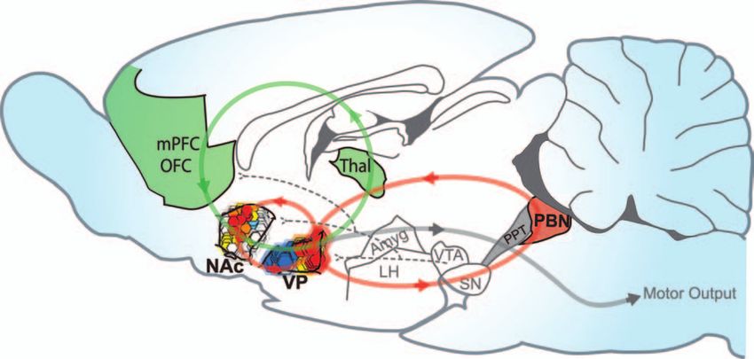
Volume 12, Number 6, 2006 THE NEUROSCIENTIST 505Fig. 5. Brain hedonic hot spots and hedonic circuits. A sagittal brain view of the hedonic hot spots discussed here in the
nucleus accumbens, ventral pallidum, and parabrachial nucleus. Each hedonic hot spot can cause amplification of ‘liking’
reactions to sweetness in response to appropriate drug microinjection within it (e.g., µ-opioid agonist or benzodiazepine).
Anatomical projections are indicated by lines that may create a hedonic circuit by connecting hot spots together (red
circles) or incorporating hot spots into larger mesocorticolimbic loops (green). NAc = nucleus accumbens shell; VP = ven-
tral pallidum; mPFC = medial prefrontal cortex; OFC = orbitofrontal cortex; Thal = thalamus; LH = lateral hypothalamus;
Amyg = amygdala; SN = substantia nigra; VTA = ventral tegmental area; PPT = pedunculopontine tegmentum; PBN =
parabrachial nucleus. Modified from Paxinos and Watson (1998).
them. Thus, the accumbens and ventral pallidum hot spots The notion that the brain stem might code aspects of
may interact with one another, either directly or indirectly, sensory pleasure might come as a surprise to anyone used
and opioid neurotransmission to both hot spots may be to thinking of the brain stem solely in terms of reflexive
required for the overall amplification of taste hedonics by functions. Yet several experiments including recent stud-
either one (Fig. 5). ies in our laboratory have indicated compelling evidence
Supporting the possibility of interaction between hot that brain stem substrates participate in the processing of
spots in the nucleus accumbens and ventral pallidum, taste hedonic signals. The brain stem may even contain a
both structures share reciprocal connections with each hedonic hot spot of its own that uses a different and per-
other (Heimer and Wilson 1975; Phillipson and Griffiths haps surprising neurochemical signal, namely, a benzodi-
1985; Zahm and others 1985; Churchill and Kalivas azepine/GABA signal. This signal functions around the
1994; Usuda and others 1998; Zahm 2000), and each parabrachial nucleus of the pontine hindbrain to enhance
structure can modulate electrophysiological activity in ‘liking’ reactions to sucrose hedonic impact and to simu-
the other (Hakan and others 1992; Hakan and Eyl 1995; late eating behavior (Higgs and Cooper 1996; Peciña and
Napier and Mitrovic 1999). Berridge 1996; Söderpalm and Berridge 2000).
The first evidence that a benzodiazepine/GABA sys-
Hedonic Networks Stretch across tem somewhere in the brain stem might contribute to
the Entire Brain hedonic processing came from a demonstration that a
systemic benzodiazepine drug enhanced positive affec-
The interaction described above highlights the point that tive reactions to sweet tastes even in decerebrate ani-
sensory pleasure does not arise from activity in any one mals, in which the brain had been transected and the
hedonic hot spot, of course, but rather by activation of brain stem surgically separated from connections to the
widespread hedonic brain systems that coordinate multi- forebrain (Berridge 1988).
ple hot spots. Although opioid hot spots in the accum- Even in intact animals, benzodiazepines more effec-
bens shell and ventral pallidum have received the most tively increase positive affective reactions to taste in the
attention in our discussion of hedonic reward so far, brain stem than in the forebrain. Microinjections of low
hedonic hot spots are not limited to the forebrain or to doses of benzodiazepine most effectively increase posi-
opioids. Rather, sensory hedonic systems are distributed tive affective reactions to sucrose taste when injected
in neural circuits that stretch across the brain, possibly into the brain stem ventricle (i.e., fourth ventricle) of nor-
extending caudally to include some brain stem circuits. mal rats than when injected into the forebrain ventricle
506 THE NEUROSCIENTIST Hedonic Hot Spots(i.e., lateral ventricle; Peciña and Berridge 1996). Thus, However, it is not hard to imagine future experiments that
brain stem circuits participate in hedonic enhancements, would help resolve the question, for example, combining
perhaps as the lower rung of a brain hedonic hierarchy. Brain multiple microinjections of different neurotransmitter
stem circuits may even be primary for benzodiazepine- agents simultaneously in separate brain hot spots.
related modulation of affective reactions to tastes.
Recent experiments suggest that the parabrachial Where Other Hedonic Hot Spots May
nucleus in the pons might be a brain stem hot spot for ben- and May Not Be
zodiazepine circuits relevant to taste’s hedonic impact.
Microinjection of the benzodiazepine midazolam directly Thus far, we have discussed hedonic hot spots that have
into the parabrachial nucleus is able to increase the num- been identified in the nucleus accumbens, ventral pal-
ber of hedonic reactions to a sucrose taste, in addition to lidum, and brain stem pons, within a hedonic network
increasing eating behavior, apparently more effectively that stretches across the brain. Are there other hedonic
than into several other brain stem sites (Söderpalm and hot spots in the brain, similarly capable of causing
Berridge 2000). Benzodiazepines have long been sug- increases in the hedonic impact of rewards? There are
gested to augment food hedonics (Cooper and Estall intriguing candidates and also perhaps some surprising
1985; Berridge and Treit 1986). Therefore, it is likely that failures to amplify sensory pleasure. One promising
the parabrachial nucleus plays an important role in brain additional candidate may be regions of the neocortex
stem modulation of taste hedonics and may be embedded that respond specifically to hedonic stimuli, including
within the brain’s distributed circuitry for mediating hedo- especially the orbitofrontal cortex (Kringelbach 2004,
nic pleasure, although the parabrachial hot spot has yet to 2005). In human imaging studies, the orbitofrontal cor-
be mapped by Fos plume techniques (Fig. 5). tex, particularly its caudal region, is preferentially acti-
vated by tastes, flavors, and odors that are rated as
pleasant (O’Doherty and others 2002; Rolls and others
Benzodiazepine/GABA Hedonic Hot Spots
2003; Small and others 2003). The orbitofrontal cortex
Interact with Opioid Hedonic Hot Spots
is also able to track reductions in hedonic impact caused
Consistent with the idea that hedonic networks stretch by eating foods to satiety (O’Doherty and others 2000;
across the entire brain, a recent study suggests that ben- Small and others 2001; Kringelbach and others 2003).
zodiazepine and opioid circuits interact together to Primate and rodent electrophysiology studies generally
amplify hedonic taste reactivity. That is, benzodiazepine- confirm that orbitofrontal cortex neurons fire in response
induced potentiation may in turn involve an opioid link in to palatable sweet tastes and that reward-related firing
the larger neural chain that leads to an increase in ‘liking’ also is diminished after reward satiation (Rolls and oth-
reactions. Richardson and others (2005) showed that ben- ers 1989; Schoenbaum and others 1998; Tremblay and
zodiazepine enhancement of taste hedonic impact may at Schultz 1999; Gutierrez and others 2006; Padoa-
least require permissive activation of endogenous opioid Schioppa and Assad 2006). Some might suggest the ven-
systems somewhere in the brain and is prevented by opi- tromedial prefrontal cortex, insula, and cingulate cortex
oid receptor blockade. They found that prior treatment could be involved in hedonics as well because they play
with an opioid antagonist (naloxone) completely blocked a key role in many positive emotional and affective
the typical 200% elevation of sucrose ‘liking’ reactions processes, including food preferences (Baylis and
that was otherwise caused by diazepam administration Gaffan 1991; Damasio 1994; Francis and others 1999;
(Richardson and others 2005). Bechara and others 2000; De Araujo and others 2003).
How might such interactions be mediated by brain An open question is whether orbitofrontal, ventrome-
hedonic circuits? One possibility is that connections dial prefrontal, or other regions of the cortex actually
between hot spots connect them into a larger distributed cause hedonics and hedonic reactions or instead merely
hedonic network that drugs activate as a whole. Such code and represent them as consequences of hedonic
anatomical connections make direct interaction at least reactions. If the latter, then presumably, causation arises
possible between parabrachial nucleus and forebrain hot from hot spots elsewhere in the brain, such as the ones
spots. Ascending projections connect the parabrachial we described above. Little direct evidence for pleasure
nucleus to the ventral pallidum, and descending projec- causation by the orbitofrontal or ventromedial prefrontal
tions connect both the ventral pallidum and nucleus cortex exists as of yet. Ventral medial prefrontal cortex
accumbens to the parabrachial nucleus (Saper and Loewy lesions may moderately disrupt food selection in mon-
1980; Grove 1988b; Groenewegen and others 1993; Usuda keys, but rats with lesions to the orbitofrontal cortex
and others 1998). The parabrachial nucleus projects also to retain a normal decline in food intake after food has been
a number of structures that in turn target the accumbens, paired with illness, although they are impaired in using
such as the lateral hypothalamus, bed nucleus of the stria conditioned stimulus cues that signal nonreward or
terminalis, and amygdala (Norgren 1976; Lundy and devaluation to guide their choices (Gallagher and others
Norgren 2004). Thus, several circuits allow the potential 1999; Pickens and others 2003; Rolls 2004). Human
for interaction between hedonic hot spots. It is not yet patients with damage to the ventral prefrontal cortex
known whether the opioid/benzodiazepine interaction show fascinating changes in cognition and emotion, but
described above actually involves such separate hot spot it is not clear whether they actually lose any capacities
connections or instead is mediated by multiple neurotrans- for experiencing or reacting to sensory pleasures
mitters interacting in the same site (e.g., brain stem). (Damasio 1994; Bechara and others 2000). Clearly, it
Volume 12, Number 6, 2006 THE NEUROSCIENTIST 507will be of interest to know in the future whether the False Pleasure Transmitter
orbitofrontal or other prefrontal cortical areas contain
hot spots capable of either amplifying or abolishing a Similarly, the transmitter dopamine has been famous as a
basic hedonic reaction to sensory pleasure. If so, the so-called pleasure neurotransmitter for more than 30
hedonic generating network would truly stretch across years, especially within the mesolimbic system that proj-
the entire brain, from brain stem to cortex. ects to the nucleus accumbens (Wise 1985; Hoebel and
others 1999; Shizgal 1999). One reason that claim was
made is that dopamine neurons are turned on by many
False Pleasure Electrode
pleasurable stimuli ranging from foods, sex, and drugs to
Perhaps the most famous and original candidates for social and cognitive rewards (Fiorino and others 1997;
pleasure-generating brain systems come from so-called Schultz 1998; Wise 1998; Ahn and Phillips 1999; Becker
pleasure electrodes, which used brain electrical stimula- and others 2001; Robinson and others 2005; Aragona and
tion to reinforce self-administration behavior such as others 2006). Furthermore, if dopamine was blocked, all
pressing a lever or pushing a button (Olds and Milner rewards appeared to lose certain rewarding properties in
1954). But critical reinspection of the effects of electrode instrumental paradigms (Wise and Bozarth 1985; Hoebel
self-stimulation has indicated that many of the most dra- and others 1999; Shizgal 1999).
matic electrodes may not have been reliable generators of But dopamine is probably not a pleasure neurotrans-
strong pleasure after all. Instead, mesolimbic electrodes mitter. Recent work has shown dopamine to be involved
may have generally produced a false pleasure, that is, by in incentive salience or motivational aspects of reward
generating motivational ‘wanting’ without hedonic ‘lik- (‘wanting’) and to have little if anything to do with gen-
ing’ (Berridge and Valenstein 1991; Berridge 2003). erating hedonic ‘liking’ per se. Even massive destruction
In some early experiments on electrical brain stimula- of ascending dopamine projections does not impair
tion performed in humans (Heath 1972; Sem-Jacobsen affective ‘liking’ reactions elicited by a sweet taste
1976), patients with pleasure electrodes pressed a button (Berridge and others 1989; Berridge and Robinson
that stimulated an electrode in their brain (usually in path- 1998). Nor does dopamine blockade by neuroleptic
ways related to mesolimbic systems) thousands of times drugs reduce ‘liking’ for sweetness (Peciña and others
in a single session of several hours (Heath 1972; 1997).
Valenstein 1974; Sem-Jacobsen 1976). Many textbooks Conversely, activation of dopamine transmission by
cite these cases as examples of intense pleasure elec- genetic manipulation in hyperdopaminergic mice does not
trodes. However, if one reads closely what subjects were enhance hedonic ‘liking’ for sweetness, even though the
reported to have said, it is not at all clear that they experi- same mice are more motivated to obtain sweet rewards
enced intense pleasure per se after stimulation. Pleasure and more resistant to distractions from the goal they
thrills are generally not what was reported, not even in the excessively want (Peciña and others 2003; Cagniard
most extreme brain-stimulation examples. For example, and others 2006). Similarly, amphetamine administration
“B-19,” a young man implanted with stimulation elec- that promotes dopamine release, either directly into the
trodes by Heath and colleagues in the 1960s, voraciously nucleus accumbens or systemically, completely fails to
self-stimulated his electrode and protested when the stim- increase hedonic reactions to taste (Wyvell and Berridge
ulation button was taken away. But there is no clear evi- 2000; Tindell and others 2005). Finally, indirect facilita-
dence that B-19’s electrodes ever caused intense pleasure. tion of dopamine activation by drug-induced neural sensi-
B-19 never was quoted as saying they did. Instead B19’s tization also fails to increase positive ‘liking’ reactions to
electrodes evoked desire to stimulate again and strong sweetness (Wyvell and Berridge 2000; Tindell and others
sexual arousal, although never producing sexual orgasm 2005). Thus, dopamine is neither necessary for normal
or clear evidence of actual pleasure sensation from the hedonic impact of sweet rewards nor sufficient to increase
electrode. The brain stimulation did not serve as a substi- hedonic impact above normal. In short, dopamine appears
tute for sexual acts, but it did instead make him want to do unable to cause changes in basic ‘liking’ reactions to
sexual acts as well as want to press the electrode again. sucrose. It stands in contrast to the hedonic hot spots
What could these electrodes be doing, if not causing described above, which use opioid, cannabinoid, and ben-
pleasure? Among other things, they might be activating zodiazepine signals to powerfully amplify the hedonic
incentive salience attribution to surroundings and per- impact of natural sensory pleasures.
ceived stimuli, especially the act of stimulating the elec-
trode. For example, electrode stimulation of lateral Conclusion
hypothalamus pathways causes rats to want to eat more
without causing them to like food more, similar to the Contemporary neuroscience research techniques have
‘wanting’ without ‘liking’ effects described above made it possible to map hedonic hot spots within the
(Berridge and Valenstein 1991). If human electrodes brain. The ventral pallidum and the nucleus accumbens
caused selective ‘wanting’ in the same way, a person each contain hedonic hot spots for taste rewards, within
might well describe a sudden feeling that life was sud- which activation of µ-opioid receptors causes an increase
denly more attractive, desirable, and compelling to pur- in hedonic valuation of sweet taste stimuli. Accumbens
sue. They might well want to activate their electrode that and ventral pallidum hot spots functionally interact with
produced no pleasure sensation. That would be mere one another in their opioid-mediated amplification of ‘lik-
incentive ‘wanting’ without ‘liking’. ing’ reactions to sweetness, and those limbic hot spots
508 THE NEUROSCIENTIST Hedonic Hot Spotshave connections to other potential hot spots distributed not learning, for a food reward. Neuropsychopharmacology 31:
elsewhere in the brain. Thus, the brain hot spots we have 1362–70.
Caille S, Parsons LH. 2006. Cannabinoid modulation of opiate rein-
described here likely form a larger hot circuit for hedonic forcement through the ventral striatopallidal pathway.
signals that enhance sensory pleasure. Neuropsychopharmacology 31:804–13.
Future work on limbic functional circuitry will be useful Carelli RM, Deadwyler SA. 1997. Cellular mechanisms underlying
to determine what other brain hot spots or transmitters con- reinforcement-related processing in the nucleus accumbens: elec-
trophysiological studies in behaving animals. Pharmacol Biochem
tribute to the hot circuit for hedonic reward. Such knowl- Behav 57:495–504.
edge will help illuminate how mere sensory information Chang JY, Sawyer SF, Lee RS, Woodward DJ. 1994. Electrophysiological
becomes painted with hedonic qualities and liked. In short, and pharmacological evidence for the role of the nucleus accumbens
the future of hedonic research promises to be quite hot. in cocaine self-administration in freely moving rats. J Neurosci 14:
1224–44.
References Churchill L, Kalivas PW. 1994. A topographically organized gamma-
aminobutyric acid projection from the ventral pallidum to the
Ahn S, Phillips AG. 1999. Dopaminergic correlates of sensory-specific nucleus accumbens in the rat. J Comp Neurol 345:579–95.
satiety in the medial prefrontal cortex and nucleus accumbens of Cooper SJ. 1983. Effects of opiate agonists and antagonists on fluid intake
the rat. J Neurosci 19:B1-B6. and saccharine choice in the rat. Neuropharmacology 22:323–8.
Anand BK, Brobeck JR. 1951. Hypothalamic control of food intake in Cooper SJ, Estall LB. 1985. Behavioral pharmacology of food, water
rats and cats. Yale J Biol Med 24:123–40. and salt intake in relation to drug actions at benzodiazepine recep-
Aragona BJ, Liu Y, Yu YJ, Curtis JT, Detwiler JM, Insel TR, and oth- tors. Neurosci Biobehav Rev 9(1):5–19.
ers. 2006. Nucleus accumbens dopamine differentially mediates Cooper SJ, Higgs S. 1994. Neuropharmacology of appetite and taste
the formation and maintenance of monogamous pair bonds. Nat preferences. In: Legg CR, Booth DA, editors. Appetite: neural and
Neurosci 9:133–9. behavioural bases. New York: Oxford University Press. p 212–42.
Bakshi VP, Kelley AE. 1993. Feeding induced by opioid stimulation of Cromwell HC, Berridge KC. 1993. Where does damage lead to
the ventral striatum—role of opiate receptor subtypes. J Pharmacol enhanced food aversion: the ventral pallidum/substantia innomi-
Exp Ther 265:1253–60. nata or lateral hypothalamus? Brain Res 624:1–10.
Baylis LL, Gaffan D. 1991. Amygdalectomy and ventromedial pre- Cromwell HC, Hassani OK, Schultz W. 2005. Relative reward pro-
frontal ablation produce similar deficits in food choice and in sim- cessing in primate striatum. Exp Brain Res 162:520–5.
ple object discrimination learning for an unseen reward. Exp Brain Damasio AR. 1994. Descartes’ error: emotion, reason, and the human
Res 86:617–22. brain. New York: G. P. Putnam.
Bechara A, Damasio H, Damasio AR. 2000. Emotion, decision mak- De Araujo ET, Kringelback ML, Rolls ET, McGlone F. 2003. Human
ing and the orbitofrontal cortex. Cereb Cortex 10:295–307. cortical responses to water in the mouth, and the effects of thirst. J
Becker JB, Rudick CN, Jenkins WJ. 2001. The role of dopamine in the Neurophysiol 90:1865–76.
nucleus accumbens and striatum during sexual behavior in the Doyle TG, Berridge KC, Gosnell BA. 1993. Morphine enhances hedo-
female rat. J Neurosci 21:3236–41. nic taste palatability in rats. Pharmacol Biochem Behav 46:745–9.
Bengtson CP, Osborne PB. 2000. Electrophysiological properties of Fiorino DF, Coury A, Phillips AG. 1997. Dynamic changes in nucleus
cholinergic and noncholinergic neurons in the ventral pallidal region accumbens dopamine efflux during the Coolidge effect in male
of the nucleus basalis in rat brain slices. J Neurophysiol 83:2649–60. rats. J Neurosci 17:4849–55.
Berendse HW, Galis-de Graaf Y, Groenewegen HJ. 1992. Francis S, Rolls ET, Bowtell R, McGlone F, O’Doherty J, Browning A,
Topographical organization and relationship with ventral striatal and others. 1999. The representation of pleasant touch in the brain
compartments of prefrontal corticostriatal projections in the rat. J and its relationship with taste and olfactory areas. Neuroreport
Comp Neurol 316:314–47. 10:453–9.
Berns GS, McClure SM, Pagnoni G, Montague PR. 2001. Fudge JL, Kunishio K, Walsh P, Richard C, Haber SN. 2002.
Predictability modulates human brain response to reward. J Amygdaloid projections to ventromedial striatal subterritories in
Neurosci 21:2793–8. the primate. Neuroscience 110:257–75.
Berridge KC. 1988. Brainstem systems mediate the enhancement of Gallagher M, McMahan RW, Schoenbaum G. 1999. Orbitofrontal cor-
palatability by chlordiazepoxide. Brain Res 447:262–8. tex and representation of incentive value in associative learning. J
Berridge KC. 1996. Food reward: brain substrates of wanting and lik- Neurosci 19:6610–14.
ing. Neurosci Biobehav Rev 20:1–25. Greenwald MK, Johanson CE, Moody DE, Woods JH, Kilbourn MR,
Berridge KC. 2000. Taste reactivity: measuring hedonic impact in Koeppe RA, and others. 2003. Effects of buprenorphine mainte-
infants and animals. Neurosci Biobehav Rev 24:173–98. nance dose on mu-opioid receptor availability, plasma concentra-
Berridge KC. 2003. Pleasures of the brain. Brain Cogn 52:106–28. tions, and antagonist blockade in heroin-dependent volunteers.
Berridge KC. 2004. Pleasure, unconscious affect, and irrational desire. In: Neuropsychopharmacology 28:2000–9.
Manstead ASR, Frijda NH, Fischer AH, editors. Feelings and emo- Grill HJ, Norgren R. 1978. The taste reactivity test. I. Mimetic
tions: the Amsterdam Symposium. Cambridge (UK): Cambridge responses to gustatory stimuli in neurologically normal rats. Brain
University Press. p 43–62. Res 143:263–79.
Berridge KC, Robinson TE. 1998. What is the role of dopamine in Grill HJ, Berridge KC. 1985. Taste reactivity as a measure of the neu-
reward: hedonic impact, reward learning, or incentive salience? ral control of palatability. In: Sprague JM, Epstein AN, editors.
Brain Res Rev 28:309–69. Progress in psychobiology and physiological psychology. Orlando
Berridge KC, Treit D. (1986). Chlordizepoxide directly enhances positive (FL): Academic Press. p 1–61.
ingestive reactions in rats. Pharmacol Biochem Behav 24:217–21. Groenewegen HJ, Berendse HW, Haber SN. 1993. Organization of the
Berridge KC, Valenstein ES. 1991. What psychological process medi- output of the ventral striatopallidal system in the rat: ventral palli-
ates feeding evoked by electrical stimulation of the lateral hypo- dal efferents. Neuroscience 57:113–42.
thalamus? Behav Neurosci 105:3–14. Groenewegen HJ, Vermeulen-Van der Zee E, te Kortschot A, Witter
Berridge KC, Venier IL, Robinson TE. 1989. Taste reactivity analysis MP. 1987. Organization of the projections from the subiculum to
of 6-hydroxydopamine-induced aphagia: implications for arousal the ventral striatum in the rat: a study using anterograde transport
and anhedonia hypotheses of dopamine function. Behav Neurosci of Phaseolus vulgaris leucoagglutinin. Neuroscience 23:103–20.
103:36–45. Grove EA. 1988a. Efferent connections of the substantia innominata in
Cabanac M. 1971. Physiological role of pleasure. Science 173:1103–7. the rat. J Comp Neurol 277:347–64.
Cagniard B, Balsam PD, Brunner D, Zhuang X. 2006. Mice with Grove EA. 1988b. Neural associations of the substantia innominata in
chronically elevated dopamine exhibit enhanced motivation, but the rat: afferent connections. J Comp Neurol 277:315–46.
Volume 12, Number 6, 2006 THE NEUROSCIENTIST 509Gutierrez R, Carmena JM, Nicolelis MA, Simon SA. 2006. Orbitofrontal O’Doherty J, Rolls ET, Francis S, Bowtell R, McGlone F, Kobal G, ensemble activity monitors licking and distinguishes among natural and others. 2000. Sensory-specific satiety-related olfactory activa- rewards. J Neurophysiol 95:119–33. tion of the human orbitofrontal cortex [corrected and republished Hakan RL, Berg GI, Henriksen SJ. 1992. Electrophysiological evi- article originally printed in Neuroreport 2000;11:399–403]. dence for reciprocal connectivity between the nucleus accumbens Neuroreport 11:893–7. septi and ventral pallidal region. Brain Res 581:344–50. Olds J, Milner P. 1954. Positive reinforcement produced by electrical Hakan RL, Eyl C. 1995. Neuropharmacology of the nucleus accum- stimulation of septal area and other regions of rat brain. J Comp bens: iontophoretic applications of morphine and nicotine have Physiol Psychol 47:419–27. contrasting effects on single-unit responses evoked by ventral pal- Olive MF, Anton B, Micevych P, Evans CJ, Maidment NT. 1997. lidal and fimbria stimulation. Synapse 20:175–84. Presynaptic versus postsynaptic localization of mu and delta opioid Heath RG. 1972. Pleasure and brain activity in man: deep and surface receptors in dorsal and ventral striatopallidal pathways. J Neurosci electroencephalograms during orgasm. J Nerv Ment Dis 154:3–18. 17:7471–9. Heimer L, Wilson RD. 1975. The subcortical projections of allocortex: Padoa-Schioppa C, Assad JA. 2006. Neurons in the orbitofrontal cor- similarities in the neural associations of the hippocampus, the peri- tex encode economic value. Nature 441:223–6. form cortex and the neocortex. In: Santini M, editor. Golgi centen- Parker LA, Maier S, Rennie M, Crebolder J. 1992. Morphine- and nial symposium proceedings. New York: Raven Press. p 173–93. naltrexone-induced modification of palatability: analysis by the Higgs S, Cooper SJ. 1996. Hyperphagia induced by direct administra- taste reactivity test. Behav Neurosci 106:999–1010. tion of midazolam into the parabrachial nucleus of the rat. Eur J Paxinos G, Watson C. 1998. The rat brain in stereotaxic coordinates. Pharmacol 313:1–9. San Diego (CA): Academic Press. Hoebel BG, Rada PV, Mark GP, Pothos EN. 1999. Neural systems for Peciña S, Berridge KC. 1995. Central enhancement of taste pleasure reinforcement and inhibition of behavior: relevance to eating, by intraventricular morphine. Neurobiology 3:269–80. addiction, and depression. In: Kahneman D, Diener E, Schwarz N, Peciña S, Berridge KC. 1996. Brainstem mediates diazepam enhance- editors. Well-being: The foundations of hedonic psychology. New ment of palatability and feeding: microinjections into fourth ven- York: Russell Sage Foundation. p 558–72. tricle versus lateral ventricle. Brain Res 727:22–30. Kelley AE, Bakshi VP, Haber SN, Steininger TL, Will MJ, Zhang M. Peciña S, Berridge KC. 2000. Opioid eating site in accumbens shell 2002. Opioid modulation of taste hedonics within the ventral stria- mediates food intake and hedonic liking: map based on microin- tum. Physiol Behav 76:365–77. jection Fos plumes. Brain Res 863:71–86. Kim EM, Quinn JG, Levine AS, O’Hare E. 2004. A bi-directional Peciña S, Berridge KC. 2005. Hedonic hot spot in nucleus accumbens mu-opioid-opioid connection between the nucleus of the accumbens shell: where do mu-opioids cause increased hedonic impact of shell and the central nucleus of the amygdala in the rat. Brain Res sweetness? J Neurosci 25:11777–86. 1029:135–9. Peciña S, Berridge KC, Parker LA. 1997. Pimozide does not shift Kirkham TC, Williams CM. 2001. Endogenous cannabinoids and palatability: separation of anhedonia from sensorimotor suppres- appetite. Nutr Res Rev 14:65–86. sion by taste reactivity. Pharmacol Biochem Behav 58:801–11. Kringelbach ML. 2004. Food for thought: hedonic experience beyond Peciña S, Cagniard B, Berridge KC, Aldridge JW, Zhuang X. 2003. homeostasis in the human brain. Neuroscience 126:807–19. Hyperdopaminergic mutant mice have higher “wanting” but not Kringelbach ML. 2005. The human orbitofrontal cortex: linking “liking” for sweet rewards. J Neurosci 23:9395–402. reward to hedonic experience. Nat Rev Neurosci 6:691–702. Peciña S, Schulkin J, Berridge KC. 2006. Nucleus accumbens Kringelbach ML, O’Doherty J, Rolls ET, Andrews C. 2003. Activation corticotropin-releasing factor increases cue-triggered motivation of the human orbitofrontal cortex to a liquid food stimulus is cor- for sucrose reward: paradoxical positive incentive effects in stress? related with its subjective pleasantness. Cereb Cortex 13:1064–71. BMC Biol 4:8. Lundy RF Jr, Norgren R. 2004. Activity in the hypothalamus, amyg- Peoples LL, West MO. 1996. Phasic firing of single neurons in the rat dala, and cortex generates bilateral and convergent modulation of nucleus accumbens correlated with the timing of intravenous pontine gustatory neurons. J Neurophysiol 91:1143–57. cocaine self-administration. J Neurosci 16:3459–73. Mahler SV, Smith KS, Berridge KC. 2004. What is the “motivational” Phillipson OT, Griffiths AC. 1985. The topographic order of inputs to mechanism for the marijuana munchies? The effects of intra- nucleus accumbens in the rat. Neuroscience 16:275–96. accumbens anandamide on hedonic taste reactions to sucrose Pickel VM, Chan J, Kash TL, Rodriguez JJ, MacKie K. 2004. [Program No. 437.10]. 2004 abstract viewer/itinerary planner. Compartment-specific localization of cannabinoid 1 (CB1) and mu- Washington (DC): Society for Neuroscience. opioid receptors in rat nucleus accumbens. Neuroscience 127:101–12. Maidment NT, Brumbaugh DR, Rudolph VD, Erdelyi E, Evans CJ. Pickens CL, Saddoris MP, Setlow B, Gallagher M, Holland PC, 1989. Microdialysis of extracellular endogenous opioid peptides Schoenbaum G. 2003. Different roles for orbitofrontal cortex and from rat brain in vivo. Neuroscience 33:549–57. basolateral amygdala in a reinforcer devaluation task. J Neurosci Majeed NH, Przewlocka B, Wedzony K, Przewlocki R. 1986. 23:11078–84. Stimulation of food intake following opioid microinjection into the Richardson DK, Reynolds SM, Cooper SJ, Berridge KC. 2005. nucleus accumbens septi in rats. Peptides 7:711–6. Endogenous opioids are necessary for benzodiazepine palatability Miller JM, Vorel SR, Tranguch AJ, Kenny ET, Mazzoni P, van Gorp enhancement: naltrexone blocks diazepam-induced increase of WG, and others. 2006. Anhedonia after a selective bilateral lesion sucrose-liking. Pharmacol Biochem Behav 81:657–63. of the globus pallidus. Am J Psychiatry 163:786–8. Rideout HJ, Parker LA. 1996. Morphine enhancement of sucrose Mogenson GJ, Swanson LW, Wu M. 1983. Neural projections from palatability: analysis by the taste reactivity test. Pharmacol nucleus accumbens to globus pallidus, substantia innominata, and Biochem Behav 53:731–4. lateral preoptic-lateral hypothalamic area: an anatomical and elec- Robinson TE, Berridge KC. 2003. Addiction. Annu Rev Psychol trophysiological investigation in the rat. J Neurosci 3:189–202. 54:25–53. Morley JE, Levine AS. 1983. Involvement of dynorphin and the kappa Robinson S, Sandstrom SM, Denenberg VH, Palmiter RD. 2005. opioid receptor in feeding. Peptides 4:797–800. Distinguishing whether dopamine regulates liking, wanting, and/or Mucha RF, Iversen SD. 1986. Increased food intake after opioid learning about rewards. Behav Neurosci 119:5–15. microinjections into nucleus accumbens and ventral tegmental area Roitman MF, Wheeler RA, Carelli RM. 2005. Nucleus accumbens of rat. Brain Res 397:214–24. neurons are innately tuned for rewarding and aversive taste stimuli, Napier TC, Mitrovic I. 1999. Opioid modulation of ventral pallidal encode their predictors, and are linked to motor output. Neuron inputs. Ann N Y Acad Sci 877:176–201. 45:587–97. Norgren R. 1976. Taste pathways to hypothalamus and amygdala. J Rolls ET. 2004. The functions of the orbitofrontal cortex. Brain Cogn Comp Neurol 166:17–30. 55:11–29. O’Doherty JP, Deichmann R, Critchley HD, Dolan RJ. 2002. Neural Rolls ET, Kringelbach ML, de Araujo IE. 2003. Different representa- responses during anticipation of a primary taste reward. Neuron tions of pleasant and unpleasant odours in the human brain. Eur J 33:815–26. Neurosci 18:695–703. 510 THE NEUROSCIENTIST Hedonic Hot Spots
You can also read