Modeling the Glomerular Filtration Barrier and Intercellular Crosstalk
←
→
Page content transcription
If your browser does not render page correctly, please read the page content below
REVIEW
                                                                                                                                                published: 02 June 2021
                                                                                                                                       doi: 10.3389/fphys.2021.689083

                                                Modeling the Glomerular Filtration
                                                Barrier and Intercellular Crosstalk
                                                Kerstin Ebefors1†, Emelie Lassén 2†, Nanditha Anandakrishnan 2, Evren U. Azeloglu 2 and
                                                Ilse S. Daehn 2*

                                                Department of Physiology, Institute of Neuroscience and Physiology, Sahlgrenska Academy, University of Gothenburg,
                                                1

                                                Gothenburg, Sweden, 2Division of Nephrology, Department of Medicine, Icahn School of Medicine at Mount Sinai,
                                                New York, NY, United States

                                                The glomerulus is a compact cluster of capillaries responsible for blood filtration and
                                                initiating urine production in the renal nephrons. A trilaminar structure in the capillary
                                                wall forms the glomerular filtration barrier (GFB), composed of glycocalyx-enriched
                             Edited by:         and fenestrated endothelial cells adhering to the glomerular basement membrane
                          John D. Imig,         and specialized visceral epithelial cells, podocytes, forming the outermost layer with
          Medical College of Wisconsin,
                          United States
                                                a molecular slit diaphragm between their interdigitating foot processes. The unique
                       Reviewed by:
                                                dynamic and selective nature of blood filtration to produce urine requires the
                  Malgorzata Kasztan,           functionality of each of the GFB components, and hence, mimicking the glomerular
University of Alabama at Birmingham,
                                                filter in vitro has been challenging, though critical for various research applications
                        United States
            Anton Jan Van Zonneveld,            and drug screening. Research efforts in the past few years have transformed our
    Leiden University Medical Center,           understanding of the structure and multifaceted roles of the cells and their intricate
                         Netherlands
                          Carl Öberg,
                                                crosstalk in development and disease pathogenesis. In this review, we present a
              Lund University, Sweden           new wave of technologies that include glomerulus-on-a-chip, three-dimensional
                    *Correspondence:            microfluidic models, and organoids all promising to improve our understanding of
                          Ilse S. Daehn
                                                glomerular biology and to enable the development of GFB-targeted therapies. Here,
                 ilse.daehn@mssm.edu
                                                we also outline the challenges and the opportunities of these emerging biomimetic
     These authors have contributed
     †

   equally to this work and share first         systems that aim to recapitulate the complex glomerular filter, and the evolving
                           authorship           perspectives on the sophisticated repertoire of cellular signaling that comprise the
                                                glomerular milieu.
                     Specialty section:
           This article was submitted to
         Renal and Epithelial Physiology,       Keywords: glomerular filtration barrier, crosstalk, in vitro, podocyte, glomerular endothelial cell, 3D model
                 a section of the journal
                 Frontiers in Physiology            “A model is a lie that helps you see the truth” – Dr. Howard Skipper
             Received: 31 March 2021
              Accepted: 05 May 2021
             Published: 02 June 2021
                                                INTRODUCTION
                            Citation:
                Ebefors K, Lassén E,            The glomerular filtration barrier (GFB) is a highly specialized interface responsible for
 Anandakrishnan N, Azeloglu EU and
                                                blood filtration that is charge and size selective. While its functionality and integrity are
      Daehn IS (2021) Modeling the
    Glomerular Filtration Barrier and
                                                maintained by a constant interaction between glomerular endothelial cells (GECs), the
              Intercellular Crosstalk.          glomerular basement membrane (GBM), and podocytes (Rennke et al., 1975; Rennke and
          Front. Physiol. 12:689083.            Venkatachalam, 1979), they are also influenced by the milieu and dynamics of the renal
   doi: 10.3389/fphys.2021.689083               blood flow. In glomerular diseases, this barrier loses functional integrity, allowing the passage

Frontiers in Physiology | www.frontiersin.org                                         1                                            June 2021 | Volume 12 | Article 689083
Ebefors et al.                                                                                                  Modeling the Glomerular Filtration Barrier

of macromolecules and cells, and results in morphological                 THE FUNCTIONAL BARRIER
changes, increasing the risk of long-term kidney damage
that ultimately leads to kidney failure (USRDS, 2020). This               The glomerulus is the filtering part of the nephron (Figure 1A)
is a growing worldwide health problem that accounts for a                 and consists of three different cell types: podocytes (visceral epithelial
substantial economic burden (Honeycutt et al., 2013). Although            cells), GECs, and mesangial cells. The filtrate from the glomerulus
the etiologies differ among glomerular diseases, damage to                enters the Bowman’s capsule as pre-urine before reabsorption and
the GFB often has the same clinical manifestations, proteinuria           secretion in the tubular system. Glomerular cells are highly specialized
or hematuria, and impaired glomerular filtration rate (GFR).              and interdependent, with fenestrated GECs covering the luminal
    The interconnectivity and structural complexity of the GFB            surface of glomerular capillaries, in direct contact with the blood.
have favored the use of experimental in vivo models, where                Podocytes tightly wrap around the glomerular capillary vessels,
these traits are preserved. Using rodent models is regarded as            with interdigitating foot processes bridged by a slit diaphragm
the gold standard in GFB research. Mice have been used                    (Figure 1B). GECs and podocytes share a common extracellular
extensively to study the GFB, given the advantage that the                matrix (ECM), the glomerular basement membrane (GBM), and
complexity of the GFB microenvironment can be fully                       together, they form the GFB (Figure 1C). Between the capillaries
recapitulated, that there are several available genetically defined       are contractile mesangial cells surrounded by their ECM, providing
strains and the relative ease of single gene targeting (Becker            structural support to the glomerular tuft (Brenner et al., 1978).
and Hewitson, 2013). Also, transgenic lines with fluorescent                  The GFB function relies on its three layers: podocytes, GBM,
reporters in different glomerular cell types provide visual readout       and GECs (Figure 1C). Podocytes are terminally differentiated
and have been useful for determining the origins and fate of              epithelial cells that form the architectural backbone of the GFB
glomerular cells in vivo (Hackl et al., 2013). There are however          anchored to the GBM through transmembrane receptors, such
significant challenges with mimicking human disease in animals,           as integrins (e.g., integrin α3 and laminin β2) and dystroglycan,
as many models do not completely recapitulate human disease               and cover the outer aspect of the glomerular capillary (Pozzi
manifestations and instead allow only for studies of certain              et al., 2008; Meyrier, 2011). They have specialized projections
disease aspects (Becker and Hewitson, 2013). However, the                 that interdigitate to form the slit diaphragm, a key element in
use of animal models is of particular importance for                      the GFB (Perico et al., 2016). The slit diaphragm proteins (e.g.,
pharmacodynamics and pharmacokinetics testing, where the                  nephrin and podocin) anchor to the cytoskeleton at the plasma
effects of pharmaceutical interventions can be examined at                membrane and form bridging structures between the interdigitating
the systemic level to determine drug safety and efficacy before           podocyte projections (foot processes; Kestila et al., 1998; Boute
entering human trials.                                                    et al., 2000). Additional proteins that maintain slit diaphragm
    The use of transgenic zebrafish strains is growing as a               proteins, such as CD2AP, play vital roles in GFB maintenance.
vertebrate model for GFB research (Zhou and Hildebrandt,                  Podocytes are essential in GFB function, underscored by the
2012; Hansen et al., 2020) and has proven to be a useful                  discovery of pathogenic mutations to proteins involved in
tool to investigate glomerular disease development and the                maintaining podocyte structure that are causal to proteinuric forms
effects of drugs on GFB (Schiffer et al., 2015; Müller-Deile              of kidney disease (Vivante and Hildebrandt, 2016; Li et al., 2020a).
et al., 2019). Although studies in zebrafish are more time-                   The GBM is formed by secreted products from both podocytes
and cost-efficient compared with rodent models, there are                 and endothelial cells during glomerulogenesis (St John and
some inherent caveats. It can for instance be difficult to                Abrahamson, 2001). Its role in the barrier function is highlighted
detect proteinuria or the clearance of specific markers of                by genetic studies showing that mutations in key components of
interest in the urine due to the surrounding water volume.                GBM; encoding laminin-α5 and COL4A5, or recessive COL4A3/4,
Also, zebrafish have numerous duplicate genes (Woods et al.,              results in basement membrane nephropathy due to the absence
2000), which complicates the generation of knockout strains,              or inadequate assembly of all collagen chains. These mutations
and they also have the ability to regenerate nephrons de novo             contribute to the development of nephrotic syndrome in pediatric
after injury. Other limitations include the need for                      patients and Alport syndrome, respectively (Tryggvason et al.,
microinjections to the dorsal aorta and cardinal vein for                 1993; Kashtan, 1999; Quinlan and Rheault, 2021).
certain drugs, which limits throughput. Altogether, animal                    Glomerular endothelial cells are highly specialized cells with
work can be expensive, has limited throughput, and poses                  fenestrae and a charged luminal endothelial surface layer, or
challenges for studying intricate crosstalk between the cells             glycocalyx, that is composed of negatively charged networks of
in the glomerulus. Therefore, there is a need for                         proteoglycans, glycoproteins, and glycolipids (Ballermann, 2007;
microphysiological systems that can recapitulate the form                 Fogo and Kon, 2010; Haraldsson and Nystrom, 2012; Khramova
and function of the GFB and offer a controlled environment                et al., 2021) that together with the GBM contribute to the
for studies of isolated pathological events. Current model                maintenance of a charge-selective barrier which is important
systems range from simple to physiologically complex and                  to restrain albumin from the glomerular filtrate (Jeansson et al.,
offer opportunities for examining specific mechanisms                     2009; Singh et al., 2011; Öberg and Rippe, 2013; Boels et al.,
involved in the maintenance as well as damage to the GFB                  2016; Figure 1C). GEC dysfunction can initiate and contribute
(Table 1). Here, we review and discuss some of the current                to GFB breakdown (Haraldsson et al., 2008; Haraldsson and
and future experimental in vitro model systems for studying               Nystrom, 2012; Sun et al., 2013; Daehn, 2018). In addition,
the GFB.                                                                  activated podocytes have been shown to influence endothelial

Frontiers in Physiology | www.frontiersin.org                         2                                          June 2021 | Volume 12 | Article 689083
Ebefors et al. Modeling the Glomerular Filtration Barrier
TABLE 1 | Comparison of in vivo and in vitro models currently used or under development for studies of the glomerular filtration barrier (GFB).
In vitro In vivo
2D monolayer Static Microfluidic Spheroids Animal models
co-culture co-culture organoids
All GFB cell types (Nishinakamura, 2019) No No No No Yes
GBM (Slater et al., 2011; Chew and Lennon, 2018; No Limited Limited Limited Yes
Hale et al., 2018; Petrosyan et al., 2019)
Glycocalyx (Singh et al., 2007; Petrosyan et al., Limited Limited Yes Limited Yes
2019; Koning et al., 2020)
Allows cell differentiation (relevant phenotype; Musah No No Limited Limited Yes
et al., 2017; Bao et al., 2018; Nishinakamura, 2019;
Veissi et al., 2020)
Permselectivity (Li et al., 2016; Petrosyan et al., No Yes Yes Limited Yes
2019; Li et al., 2020b)
Recapitulation of microenvironment (Huh et al., 2013; No Limited Limited Limited Yes
Bhatia and Ingber, 2014; Veissi et al., 2020)
Controlled microenvironment (Anandakrishnan and Yes Yes Yes Yes No
Azeloglu, 2020)
Shear stress (Slater et al., 2012; Musah et al., 2017; No Limited Yes Limited Yes
Yang et al., 2017; Homan et al., 2019)
Bidirectional crosstalk (Li et al., 2016; Casalena et al., No Yes Yes Limited Yes
2020; Veissi et al., 2020)
Material of human origin (Little and Takasato, 2015; Yes Yes Yes Yes No
Musah et al., 2017; Anandakrishnan and Azeloglu,
2020)
High throughput (Boreström et al., 2018; Yes Limited Limited Limited No
Anandakrishnan and Azeloglu, 2020)
Development of personalized/precision medicine Yes Yes Yes Yes No
(Anandakrishnan and Azeloglu, 2020)
Timeline for experiment Short Short Long Long Long
glycocalyx remodeling and loss in experimental FSGS and in vitro Modeling the Glomerular Filtration Barrier
(Ebefors et al., 2019). In diabetic kidney disease, GEC dysfunction The unique environment and complex interactions between the
and glycocalyx damage represent initiating steps in diabetic specialized cells in the GFB make modeling glomerular disease
albuminuria in humans and in experimental models (Zhao et al., particularly challenging. Podocytes are a key target cell for injury
2006; Satchell and Tooke, 2008; Yuen et al., 2012; Dogne et al., in the evolution of segmental sclerosis lesions of proteinuric
2016; Lassen and Daehn, 2020). diseases, and their morphology is critical for glomerular filtration.
Importantly, bidirectional signaling enables cells in the glomeruli However, once isolated, podocytes rapidly dedifferentiate and lose
to function effectively, where podocytes control GEC growth and their specialized morphology, making it difficult to study their
survival via crosstalk of paracrine vascular endothelial growth function in vitro. Immortalized mouse and human podocyte cell
factor alpha (VEGFA and VEGF-R; Sison et al., 2010; Jeansson lines have played a fundamental role in advancing podocyte
et al., 2011). Crosstalk also exists between endothelial and mesangial research, but they lack defined foot processes as well as slit
cells (PDGF-B and PDGFR-β) and between podocytes and mesangial diaphragms. Efforts have been made to improve podocytes in
cells (CCL21 and CCR7; Vaughan and Quaggin, 2008; Schlondorff culture to more closely recapitulate their in vivo phenotypic
and Banas, 2009). Hence, all components contribute to the overall characteristics. By modulating the ECM, which affects most
structure and function of this complex barrier, and model systems aspects of cellular behavior, researchers have established that
that can recapitulate in vivo biology and microenvironment would growing primary rat podocytes in the presence of heparin and
provide a platform for studying cell crosstalk and feedback regulation all-trans retinoic acid on laminin-coated plates resulted in podocytes
and open up the new therapeutic strategies specifically targeting with primary processes that further bifurcated and interdigitated
the GFB. with adjacent cells (Yaoita et al., 2018). Growing podocytes in
Frontiers in Physiology | www.frontiersin.org 3 June 2021 | Volume 12 | Article 689083Ebefors et al. Modeling the Glomerular Filtration Barrier
A B C
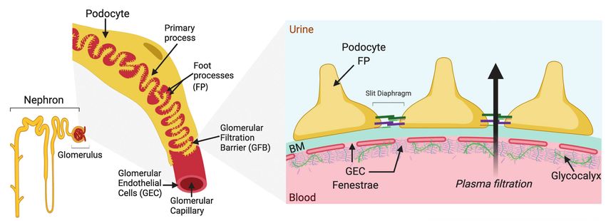
FIGURE 1 | Schematic drawing of a single nephron and glomerulus, a glomerular capillary vessel, and the glomerular filtration barrier (GFB). (A) A single nephron
comprising the blood-filtering glomerulus, enveloped by Bowman’s capsule that connects to the proximal tubule at the start of the urine-modifying tubular system.
(B) The luminal surface of the glomerular capillary vessel is covered by glomerular endothelial cells (GECs), while podocytes wrap around the outside of the vessel
with primary and foot processes (FT), forming an interdigitated pattern. Neighboring FPs are bridged by the slit diaphragm, one of the several essential components
for glomerular permselectivity. The blood is filtered over this capillary barrier, and the pre-urine produced is forwarded from the Bowman’s capsule into the lumen of
the proximal tubule. (C) The schematic cross section of the GFB displays the GEC fenestrae attached to the basement membrane (BM) and covered luminally by
glycoproteins, proteoglycans, and glycosaminoglycans of the glycocalyx, important for maintaining the charge selectivity of the GFB. On the opposite side of the
BM, the podocyte FPs are attached. The FPs interlink by slit diaphragm proteins, such as nephrin and podocin, are important for the restriction of albumin by the
GFB. The arrow shows the direction of plasma filtration over the barrier.
a gelatin microbial transglutaminase platform tuned to the stiffness There are also challenges in obtaining, culturing, and maintaining
of healthy glomeruli promoted the differentiation and maturation GECs in vitro. GECs differ in anatomy to most other endothelial
response of podocytes (Hu et al., 2017). Other approaches involve cells in the body and are defined by their fenestrations, which
culturing podocytes on nanoporous surfaces with grooves. This are important for the function of the filtration barrier (Satchell
method showed that podocytes were better differentiated, had and Braet, 2009; Fogo and Kon, 2010). The fenestrations lack
organized actin cytoskeleton stress fibers, and developed vinculin- diaphragm but are covered with a glycocalyx. Mimicking GEC
positive focal adhesions (Zennaro et al., 2016). Microscale curvature function in vitro has been challenging as they lose fenestrations
surfaces have also been shown to promote podocyte differentiation in culture. This may be due to their dependence on podocyte-
in vitro (Korolj et al., 2018). By growing podocytes on topographic derived growth factors for their viability through intercellular
substrates, the authors showed augmented nephrin expression crosstalk and interactions with the GBM. However, the very first
and structured F-actin arrangement within cells. The curved human glomerular endothelial cell (GEnC) line, developed by
surfaces promoted process formation with interdigitation and Satchell et al. (2006), was shown to have fenestrations in response
improved barrier function compared to podocytes grown on flat to VEGF, and over the years, it has proved to be a useful tool
substrates (Korolj et al., 2018). Bioengineered surfaces that artificially in GFB research, including in studies of glomerular cell interactions
induce branch formation have been developed by growing podocytes (Boor et al., 2010; Byron et al., 2014). The importance of VEGF-C
on a 3D geometry that mechanically enforces the arborization on GEC monolayer permeability has been demonstrated through
of individual podocytes (Ron et al., 2017). The formation of the measurement of trans-endothelial electrical resistance (TEER)
peripheral projections showed increased slit diaphragm proteins as an indicator of the integrity of GEC’s intercellular junctions
(nephrin, podocin, and NEPH1) and synaptopodin, as well as (Ramnath and Satchell, 2020) and the passage of fluorescence-
actinin-4 cross-linked actin stress fibers properly localized within labeled BSA (Foster et al., 2008). The authors found that VEGF-C
these peripheral processes. In addition to observing slit diaphragm- increased TEER and limited albumin passage, in contrast to the
like cell-cell junctions, the authors also demonstrated that on effect of VEGF-A, suggesting that these podocyte-derived growth
these surfaces, podocytes had a significant increase in expression factors regulate the permeability of GECs in the GFB (Foster
of genes related to podocyte function, hence a more mature et al., 2008). Although quantification of the glomerular endothelial
physiological phenotype (Ron et al., 2017). The next steps are glycocalyx in vivo has been achieved by direct labeling or indirect
already underway involving the derivation and generation of measurements (Hjalmarsson et al., 2004; Dane et al., 2015),
human pluripotent stem cells into podocyte-like cells (Yaoita measuring the glycocalyx in cultured GECs has been challenging
et al., 2018; Ge et al., 2020). These will be instrumental for due to the nature of this invisible layer. Recently, atomic force
future studies and high-content screening for podocentric therapies, microscope elastography was used to successfully measure 3D
and for integration into more complex model systems biomechanical properties of the glycocalyx on murine GECs
discussed below. through direct contact by deflection of a cantilever, without
Frontiers in Physiology | www.frontiersin.org 4 June 2021 | Volume 12 | Article 689083Ebefors et al. Modeling the Glomerular Filtration Barrier
exposing cultured cells to fixation or staining procedures that endothelin-1 from GECs exposed to the serum from patients
alter the fragile structure (Ebefors et al., 2019). An additional with preeclampsia resulted in shedding of nephrin from podocytes
requirement of GECs function is fluid flow, which is absent in cell surface via endothelin receptor A after media transfer
monocultures, leading to loss of the influence of shear stress on (Collino et al., 2008). Another study utilized the transfer of
cell shape and signal transduction that is present under physiological purified exosomes from high glucose-treated GECs to podocytes
conditions (Ballermann et al., 1998). One shear stress-inducible and found that TGFβ mRNA, carried by the extracellular
transcription factor is Krüppel-like factor 2 (KLF2; Lee et al., vesicles, contributed to podocyte dedifferentiation epithelial-
2006), an important regulator of hemodynamic signals in endothelial mesenchymal transition (Wu et al., 2017). The authors found
cells that has been shown to be dysregulated in diabetic kidney the same mechanism of exosomes containing TGFβ mRNA
disease. Importantly, the endothelial cell-specific knockout of to contribute to mesangial cell proliferation and matrix production
KLF2 results in worsened endothelial cell and podocyte injury through a similar experimental setup, as well as through tail-
in an experimental model of type 1 diabetes (Zhong et al., 2014). vain injections of the purified exosomes from high glucose-
In addition to the challenges of providing a favorable treated GECs in C57BL/6 mice (Wu et al., 2016). Furthermore,
biophysical environment for glomerular cells, ideal models of the studies of TGFβ-containing exosomes by another group
the GFB should allow for adjustment of the GFR, given that supported the involvement of these extracellular vesicles in
hyperfiltration occurs under physiological conditions, such as glomerular crosstalk following high glucose stimulation (Wang
during pregnancy, and is commonly observed in DKD, polycystic et al., 2018b). Exosomes have emerged as a novel vector for
kidney disease, and sickle-cell anemia (Helal et al., 2012; Cheung cell-cell communication in the kidney, and they are beginning
and Lafayette, 2013). A physiological decline in GFR is conversely to be recognized more and more as a critical player in the
associated with advancing age (Musso and Oreopoulos, 2011). pathogenesis of kidney disease and decline in renal function.
Hence, adjustable GFR is an important consideration for the Co-culture of two or more cell types offers increased complexity
physiological relevance of in vitro GFB models that can over monocultures when studying glomerular crosstalk. Open
be addressed by using microfluidic devices. Innovative tools microfluidics systems allow simultaneous paracrine signaling
are still needed to account for tubuloglomerular feedback (TGF) between two separated cell populations by sharing culture medium
that is regulated via macula densa cells in the distal tubule and hence allow for exchange of soluble factors and transient
and the myogenic response (Vallon, 2003). TGF has mostly signals (Zhang et al., 2020). In transwell systems, two distinct
been studied in vivo due to the challenges of studying the cell types are separated by a porous membrane (Hanspal et al.,
intricate signaling between these cells in vitro. 2017), where a bidirectional exchange of signaling molecules can
Despite some of the challenges mentioned, in vitro models occur with or without direct cell-cell contact (Table 1). Li and
are making substantial progress as an alternative or complement colleagues demonstrated the applicability of their co-culture model
to in vivo experimental models for mechanistic studies of the of the GFB for studies of drug testing and intracellular signaling,
GFB components and intercellular crosstalk. In the following using murine podocytes and GECs on opposite sides of a collagen
sections, we review the recent developments in this evolving field. IV-coated polyethylene terephthalate membrane (Li et al., 2016).
More recently, the same research group successfully exchanged
the murine glomerular cells for human immortalized GECs and
STUDYING GLOMERULAR CELL podocytes, and reported an increase in albumin leak after exposure
CROSSTALK to sera from patients with recurrent FSGS, compared to genetic
or non-recurrent forms (Li et al., 2020b). Casalena et al. have
Two-dimensional (2D) cultures are a simple culture system to demonstrated that both high glucose and serum from diabetic
study glomerular cell-specific effects, as they provide screening mice susceptible to developing diabetic kidney disease disrupt
of large numbers of conditions and treatments that would mitochondrial function and cause oxidative stress in GECs.
otherwise not be possible in vivo (Table 1). To study glomerular Interestingly, the transfer of factors released by the stressed GECs
crosstalk, conditioned medium transfer is necessary when using mediated podocyte cell death in transwell co-cultures, as well as
2D cultures. Despite the inherent limitations of 2D cultures, in media exchange (Casalena et al., 2020). Given that bi-directional
this system allows to chronologically separate cellular signaling communication can still occur while cells are physically separated,
events of pathogenic stimuli that ultimately lead to cell and/ this approach allows for subsequent interrogation of cell-specific
or organ dysfunction. responses. This approach has also been used to define podocyte-
There are different strategies used for the conditioned medium to-GEC-to-podocyte crosstalk in the pathogenesis of FSGS by
transfer, and these have been well described by Hanspal et al. shedding light on the role molecules, such as endothelin-1/
in the context of amyotrophic lateral sclerosis research (Hanspal endothelin receptor type A-mediated glomerular endothelial cell
et al., 2017). The simplest strategy consists of whole medium dysfunction, which was shown to be required for podocyte depletion
transfer from one monoculture to another in separate culture and progression of glomerulosclerosis (Daehn et al., 2014).
vessels. There can also be an intermediate step of extraction Exposure of GECs to laminar shear forces found in vivo
or enrichment of specific media components before medium adds physiological relevance to the transwell co-culture model
transfer to the acceptor cell culture. Insights from this approach of the GFB. Studies by Slater et al. used both conditioned
have provided evidence for the pathologic effects of the milieu medium transfer and co-culture of human GECs and podocytes
in women with preeclampsia, where factors including to investigate how ERK5 activation and KLF2 transcription
Frontiers in Physiology | www.frontiersin.org 5 June 2021 | Volume 12 | Article 689083Ebefors et al. Modeling the Glomerular Filtration Barrier
(associated with endothelial cell shear stress in large vessels) enhanced the permeability to proteins and increased reactive
affected the glomerular microvasculature (Slater et al., 2012). oxygen species production and podocyte detachment (Wang
Their findings demonstrated the existence of intercellular signaling et al., 2017). Musah et al. developed a glomerulus-on-a-chip
from GECs exposed to chronic laminar shear stress that affects with fluidics and strain by using vacuum channels on the side
podocytes. In another study by the same research group, GECs of the channel harboring the GECs and podocytes (Musah
and podocytes were co-cultured on opposite sides of a et al., 2017). The authors developed podocytes derived from
polycaprolactone/electrospun collagen membrane to closer mimic human induced pluripotent stem cells (iPSCs) and used them
the GBM, which was shown to enable cell-cell contact (Slater in combination with human GECs separated by a porous
et al., 2011). Differences in between the conditioned medium polydimethylsiloxane membrane coated with laminin. The
transfer and the co-culture settings suggest that spatial separation mechanical strain was shown to increase the expression of
between crosstalking cell types is an important consideration. nephrin and secretion of VEGF-A by the podocytes. Albuminuria
The models described so far provide robust high-throughput, and podocyte damage were observed with adriamycin treatment,
high-content reductionist assay systems. They have provided underscoring the resemblance to the in vivo setting (Musah
a wealth of information on the fundamental biological and et al., 2017). These models however lack GBM; hence, Petrosyan
disease processes of the GFB. Nevertheless, they provide a et al. developed a glomerulus-on-a-chip without an artificial
limited physiological context of the filtration barrier. Since membrane between GECs and podocytes (Petrosyan et al.,
there is growing awareness of the interconnections between 2019). The authors allowed both cell types to interact and to
cells and the ECM surrounding them, there is substantial effort generate a layer of ECM components. Human GECs and
by the community to develop model systems that can better podocytes were obtained from the same donor; cells were
reflect the complex microenvironment cells encounter in a tissue. separated by collagen I and eventually formed a basement
membrane between the cell layers. GECs were further shown
to develop a glycocalyx layer. The cells could be maintained
3D Culture Models of the GFB in the chip for at least a month, enabling long-term experiments.
Organs-on-a-chip have been developed for complex organs Exposure of chips to puromycin aminonucleoside induced
such as liver (Beckwitt et al., 2018), heart (Agarwal et al., podocyte injury and loss of permselectivity for albumin. Adding
2013), gut (Kim et al., 2012; Kim and Ingber, 2013), lungs serum from patients with membranous nephropathy (MN)
(Huh et al., 2010, 2012), and brain (Moreno et al., 2015). The resulted in albumin leakage, which was prevented by treatment
goal has not been to mimic the whole organs, but rather to with α-MSH. Using podocytes derived from a patient
study complex parts of an organ in a more physiological context. with Alport syndrome rendered improper filtration, supporting
In the renal field, chips for modeling the proximal tubules the chips potential for the use in personalized medicine
(Jang et al., 2013; Hoppensack et al., 2014; Wilmer et al., (Petrosyan et al., 2019).
2016) as well as the filtration barrier are being developed. An Given that the glomerulus in situ has a complex structure
ideal model of the GFB would include cell-to-cell and cell- with intricate microvascular capillary networks in a unique
to-ECM interactions, biomimetic micromechanical properties, geometry that could play a role in the development and function
shear flow, oxygen and nutrient/waste exchange, and a functional of podocytes (Falkenberg et al., 2017), there have been significant
permselective filtration barrier. In the last decade, the efforts to generate 3D models with complex microvascular
development of microfluidic platforms that allow co-culture networks using 3D bioprinting technology. Rayner et al.
of cells under flow (Bhatia and Ingber, 2014) and stretch (Huh demonstrated the use of a multiphoton microscopy-guided 3D
et al., 2013) has emerged (Table 1) and these continue to printing technique to generate perfusable vascular networks
evolve. Here, we describe some examples. with diameters as small as 10 μm (Rayner et al., 2021).
To study the effect of hypertension on the filtration barrier, They further demonstrate bioprinting of a glomerular-like
Zhou et al. developed a glomerulus-on-a-chip using murine microvascular network that supports endothelial lumen
immortalized GECs and podocytes. The cells were separated formation; however, they still require the incorporation of
in the chip by a polycarbonate membrane coated with basement podocytes and mesangial cells to recapitulate the glomerular
membrane extracts, and the authors increased the flow in the physiology and to study cell-cell crosstalk. Other developments
upper channel of the chip harboring the GECs (Zhou et al., include the glomerulus-on-a-plate, recently developed by using
2016). Increasing the mechanical force led to cell damage, loss a microfluidic topographical hollow fiber (Xie et al., 2020).
of junctions, and changes to the cell’s cytoskeleton, leading to This system uses a tubular-like perfusable channel to seed
increased leakage (Zhou et al., 2016). In an in vitro model of GECs in a glomerulus-like knot with microconvex topography,
diabetic kidney disease, Wang et al. developed a glomerulus- filled with hydrogel and covered with murine podocytes. The
on-a-chip using glomeruli isolated from rats. The chip consisted fibers were mounted in specialized 96-well plates with inlet
of five channels, a capillary in the middle and collection channels and outlet wells allowing flow to be applied by either gravity
on the outside, with the channels in between filled with gel. or syringe pump. Perfusing the lumen with albumin showed
Isolated glomeruli were injected in the capillary channel and no leakage of over the barrier, while small molecules could
allowed to attach for the cells to spread and form a barrier readily pass. However, adriamycin treatment was shown to
under flow. GECs and podocytes were identified by CD31 and increase the passage of BSA over the barrier, but only mildly
synaptopodin staining, respectively. High glucose treatment damaged podocytes (Xie et al., 2020).
Frontiers in Physiology | www.frontiersin.org 6 June 2021 | Volume 12 | Article 689083Ebefors et al. Modeling the Glomerular Filtration Barrier
Current GFB 3D culture model technologies have a number opportunity to establish near-physiological models to study human
of drawbacks, such as recirculating instead of a continuous development and diseases. Organoids can be derived from embryonic
flow, long culture times to achieve fully confluent layers, stem cells or iPSCs. The kidney is an anatomically complex organ
lack of a basement membrane, and limited throughput. with numerous different cell types, which makes it difficult to
However, these models still hold great promise for improving get organoids containing all renal structures including a functional
our understanding of glomerular crosstalk and their potential filtration barrier. As of today, organoids are premature, and as
use for personalized and precision medicine. In the future, such, they do not represent ideal modeling systems for studies
chips where cells can form a basement membrane without of the GFB; however, they hold promise to be so in the future.
separating gels or man-made membranes will emerge, and Embryonic kidneys are divided into the metanephric mesenchyme
the inclusion of mesangial cells, pericytes, and parietal epithelial and the ureteric bud. Nephron progenitor cells in the metanephric
cells to the chips would enable all the intricate signaling mesenchyme are the origin of the glomeruli, Bowman’s capsule,
which takes place in the glomerulus. and the renal tubules, and stromal progenitor cells give rise to
interstitial cells. The ureteric bud is the origin of the collecting
Scaffold-Free 3D Cultures ducts. During development, intricate signaling leads to differentiation
Scaffold-free 3D cultures are anchorage-independent models that of cells and the formation of a mature kidney. In order to form
rely on the self-aggregation of cells in specialized culture plates kidney organoids, this signaling needs to be applied to embryonic
with ultra-low attachment coating that promotes spheroid formation. or pluripotent stem cells. With this in mind, the development
Multicellular spheroids have been shown to recapitulate of differentiation protocols for embryonic and iPSCs toward renal
physiological characteristics of tissues and tumors with regard cells (Xia et al., 2013; Taguchi et al., 2014; Takasato et al., 2014)
to cell-cell contact, and allow for natural cell-ECM interactions was rapidly followed by the first reports of kidney organoids
(Sutherland, 1988). Glomeruloid spheres have been developed (Morizane et al., 2015; Takasato et al., 2015). Kidney organoids
using human mesenchymal stem cells, HUVECs, and HEKs (Abe have been characterized via single-cell sequencing and have been
et al., 2019). These spheroids expressed several podocyte markers found to contain developing podocytes, parietal epithelial cells,
and were stable for at least 5 days. Adding serum from patients tubular cells, collecting ducts, and interstitial and stromal cells.
with FSGS resulted in the collapse of the spheres (Abe et al., Missing or underrepresented cells with current methods are GECs,
2019). In 2020, Cho et al. demonstrated a novel pressure-assisted mesangial cells, principal and intercalated cells (Czerniecki et al.,
network for droplet accumulation method for high-throughput 2018; Wu et al., 2018; Combes et al., 2019), and immune cells.
generation of uniform microtissues. As a proof of principle, they Although glomerulus-like structures are formed, they mainly consist
generated glomerulus-like microtissues using immortalized mouse of early podocytes, and these have the potential to be explored
podocytes and mesenchymal stem cells (Cho et al., 2020). More further to study podocytopathies (Sharmin et al., 2016; Kim et al.,
recently, Sobreiro-Almeida et al. observed that the addition of 2017; Hale et al., 2018). Hale et al. describe a protocol for kidney
retinoic acid to an organotypic model of human renal progenitor organoids from iPSCs and compared the expression to human
cells resulted in spheroids with a preferential glomerular immortalized podocyte cell lines. Podocytes derived from organoids
differentiation. Using a hanging drop culture technique to form were shown to have an improved expression profile, as well as
spheroids, they showed that these spheroids remain viable over a GBM (Hale et al., 2018). Genetic modifications targeting podocytes
a period of 28 days and display an elevated expression of PAX2 have also been used in kidney organoids to explore congenital
and NPHS1 in the presence of retinoic acid. Further, co-culture nephrotic syndrome (Kim et al., 2017; Hale et al., 2018; Tanigawa
with microvascular endothelial cells resulted in more compact et al., 2018). In addition, to better study the GFB, improvements
organization of the spheroids (Sobreiro-Almeida et al., 2021). in methods that promote maturation and vascularization of the
These scaffold-free 3D cultures are not barrier models, and organoids have been reported recently, such as culturing kidney
many questions remain: in particular, about the composition organoids on millifluidic chips (Homan et al., 2019), or
of the spheres. And improvement in oxygenation through transplantation of human kidney organoids into the subcapsular
integration of endothelial cells has not been examined in this of mouse kidneys (van den Berg et al., 2018). In the latter, the
setting. Today’s glomeruloid spheres can provide insights for authors demonstrated an improvement in the formation of a GBM
podocyte-ECM interactions and can be adapted to medium- or with the development of a fenestrated endothelium in glomeruli
high-throughput screening assays. There is still the need for (van den Berg et al., 2018). By modulating biophysical cues, such
culture optimization to enhance reproducibility of spheroids as ECM stiffness, Garreta et al. were able to accelerate kidney
in culture and to study GFB components, while maintaining organoid generation from iPSCs (Garreta et al., 2019). They showed
a small enough size for sufficient nutrient exchange. However, that implantation of kidney organoids into chick chorioallantoic
this area of research is moving fast, and we will undoubtedly membrane (CAM) resulted in vascularization of the organoids
see advances in the years to come. within 5 days. They further generated soft hydrogels that display
similar mechanical properties as CAM to study if soft substrates
drive kidney organoid generation compared to stiffer substrates.
Organoids They observed that soft matrix environment resulted in kidney
Attempts to fully culture organs in vitro have led to the development organoids that display similar protein expression as a fetal human
of organoids, self-organized 3D aggregations of cells. Over the kidney. Although the kidney organoids still are embryonic in
last few years, these developments have provided researchers the development and need an in vivo environment for vascularization,
Frontiers in Physiology | www.frontiersin.org 7 June 2021 | Volume 12 | Article 689083Ebefors et al. Modeling the Glomerular Filtration Barrier
further characterization of the role of substrate stiffness can improve which will inevitably streamline time-consuming and costly
kidney organoid differentiation. Another limitation of the current experiments. As we gain our understanding on other aspects that
organoid systems is the heterogeneity and batch-to-batch variation influence GFB function, such as tubuloglomerular crosstalk (Tasnim
during initial formation and maturation. To address this, Dr. and Zink, 2012; Wang et al., 2018a), opportunities to “plug-in”
Little’s group have employed two different approaches for scaling modules will provide insights from the whole nephron’s perspective
up the generation of kidney organoids with less heterogeneity and even distant organ crosstalk. Together with the increasingly
and higher reproducibility. Kumar et al. demonstrated a method quantitative precision medicine approaches that can collate and
to scale up the generation of kidney micro-organoids in suspension combine clinical data with genomic information, these joint efforts
culture (Kumar et al., 2019). Using this method, they were able can help guide the design of novel drug candidates and move
to generate 8,000–10,000 kidney micro-organoids in an even size the field toward the common goal of treating patients with better
range. These organoids are less than 200–300 μm in final size, therapies for diseases of the GFB.
much smaller compared to standard organoids, which allows
efficient nutrient diffusion to the core of the organoids. However,
they showed limited utility with respect to extended long-term
CONCLUSION
cultures due to the absence of vascularization. Lawlor et al. As these experimental model systems continue to evolve and
employed extrusion bioprinting method to plate cell aggregates improve in terms of their physiological context and throughput,
that mature into kidney organoids, which partially eliminates model systems have a huge potential to help unravel the
organoid heterogeneity and enables scaling up of throughput molecular mechanisms of GFB breakdown and the pathogenic
(Lawlor et al., 2021). Using this technique, they were able to crosstalk signaling that may drive disease. These developments
generate 200 organoids in 10 min. In addition to reducing variability, should minimize the use of animal models and accelerate
extrusion bioprinting can also be used to alter the conformation discoveries by enabling the platforms for personalized and
of the organoids, to generate a spheroid or a rectangular cell precision medicine to lower drug-induced adverse events,
aggregate patch based on the extruding tip movement. The authors and identify new targets for treatments of kidney diseases
observe that the rectangular conformation yielded a greater number that affect the filtration barrier.
of nephron units compared to the spheroid conformation (Lawlor
et al., 2021), which with further improvements may be useful
for the development of transplantable kidney tissues. AUTHOR CONTRIBUTIONS
Despite the many challenges that still remain for organoids
to fully resemble mature human kidneys, including less off Conceptualization by KE, EL and ID. KE, EL, NA, EA and
targets cells as described in detail in the review by Geuens ID wrote the manuscript. All authors contributed to the article
et al. (2020), organoid biobanks as repository for drug screening and approved the submitted version.
and development are emerging (Calandrini et al., 2020) and
have the potential for applications in precision medicine.
FUNDING
ID is supported by the National Institutes of Health grant
FUTURE PERSPECTIVE R01DK097253. ID and EA are supported by the Department
The lack of specific treatments for diseases of the GFB is a of Defense CDMRP grants W81XWH-20-1-0836 (ID) and
worldwide health issue. The need for new explorative in vitro W81XWH-20-1-0837 (EA).
models is paramount to elucidate the intricate signaling of cells
in the GFB. Today, there is greater recognition that components ACKNOWLEDGMENTS
of the GFB work as an integrated functional unit. As more and
more new tools become available, such as iPSCs in culture and We acknowledge all front-line workers for all the sacrifices
3D model systems, we shall look to integrate these human-relevant that you and your family are making to help us all get through
in vitro models with data-driven and mechanistic modeling as these tough times dealing with the global pandemic. Words
well as artificial intelligence-driven methods that can assist with are not enough to thank you for your strength, courage, and
in silico drug discovery and modeling (Azeloglu et al., 2014), dedication. Illustrations were created using BioRender.
Anandakrishnan, N., and Azeloglu, E. U. (2020). Kidney tissue engineering
REFERENCES for precision medicine. Nat. Rev. Nephrol. 16, 623–624. doi: 10.1038/
s41581-020-00355-6
Abe, H., Sakurai, A., and Ochi, A. (2019). Induction of steady-state glomeruloid
Azeloglu, E. U., Hardy, S. V., Eungdamrong, N. J., Chen, Y., Jayaraman, G.,
sphere by self-assembly from human embryonic kidney cells. Biochem. Chuang, P. Y., et al. (2014). Interconnected network motifs control podocyte
Biophys. Res. Commun. 508, 654–659. doi: 10.1016/j.bbrc.2018.11.160 morphology and kidney function. Sci. Signal. 7:ra12. doi: 10.1126/scisignal.2004621
Agarwal, A., Goss, J. A., Cho, A., McCain, M. L., and Parker, K. K. (2013). Ballermann, B. J. (2007). Contribution of the endothelium to the glomerular
Microfluidic heart on a chip for higher throughput pharmacological studies. permselectivity barrier in health and disease. Nephron Physiol. 106, 19–25.
Lab Chip 13, 3599–3608. doi: 10.1039/c3lc50350j doi: 10.1159/000101796
Frontiers in Physiology | www.frontiersin.org 8 June 2021 | Volume 12 | Article 689083Ebefors et al. Modeling the Glomerular Filtration Barrier Ballermann, B. J., Dardik, A., Eng, E., and Liu, A. (1998). Shear stress and Daehn, I. S. (2018). Glomerular endothelial cells stress and cross-talk with the endothelium. Kidney Int. Suppl. 67, S100–S108. doi: 10.1046/ podocytes in the development of diabetic kidney disease. Front. Med. 5:76. j.1523-1755.1998.06720.x doi: 10.3389/fmed.2018.00076 Bao, Y.-W., Yuan, Y., Chen, J.-H., and Lin, W.-Q. (2018). Kidney disease models: Dane, M. J., van den Berg, B. M., Lee, D. H., Boels, M. G., Tiemeier, G. L., tools to identify mechanisms and potential therapeutic targets. Zool. Res. Avramut, M. C., et al. (2015). A microscopic view on the renal endothelial 39, 72–86. doi: 10.24272/j.issn.2095-8137.2017.055 glycocalyx. Am. J. Physiol. Renal Physiol. 308, F956–F966. doi: 10.1152/ Becker, G. J., and Hewitson, T. D. (2013). Animal models of chronic kidney ajprenal.00532.2014 disease: useful but not perfect. Nephrol. Dial. Transplant. 28, 2432–2438. Dogne, S., Rath, G., Jouret, F., Caron, N., Dessy, C., and Flamion, B. (2016). doi: 10.1093/ndt/gft071 Hyaluronidase 1 deficiency preserves endothelial function and glycocalyx Beckwitt, C. H., Clark, A. M., Wheeler, S., Taylor, D. L., Stolz, D. B., Griffith, L., integrity in early streptozotocin-induced diabetes. Diabetes 65, 2742–2753. et al. (2018). Liver ‘organ on a chip’. Exp. Cell Res. 363, 15–25. doi: 10.1016/j. doi: 10.2337/db15-1662 yexcr.2017.12.023 Ebefors, K., Wiener, R. J., Yu, L., Azeloglu, E. U., Yi, Z., Jia, F., et al. (2019). Bhatia, S. N., and Ingber, D. E. (2014). Microfluidic organs-on-chips. Nat. Endothelin receptor-A mediates degradation of the glomerular endothelial Biotechnol. 32, 760–772. doi: 10.1038/nbt.2989 surface layer via pathologic crosstalk between activated podocytes and Boels, M. G., Avramut, M. C., Koudijs, A., Dane, M. J., Lee, D. H., van der glomerular endothelial cells. Kidney Int. 96, 957–970. doi: 10.1016/j. Vlag, J., et al. (2016). Atrasentan reduces albuminuria by restoring the kint.2019.05.007 glomerular endothelial glycocalyx barrier in diabetic nephropathy. Diabetes Falkenberg, C. V., Azeloglu, E. U., Stothers, M., Deerinck, T. J., Chen, Y., 65, 2429–2439. doi: 10.2337/db15-1413 He, J. C., et al. (2017). Fragility of foot process morphology in Boor, P., van Roeyen, C. R., Kunter, U., Villa, L., Bucher, E., Hohenstein, B., kidney podocytes arises from chaotic spatial propagation of cytoskeletal et al. (2010). PDGF-C mediates glomerular capillary repair. Am. J. Pathol. instability. PLoS Comput. Biol. 13:e1005433. doi: 10.1371/journal. 177, 58–69. doi: 10.2353/ajpath.2010.091008 pcbi.1005433 Boreström, C., Jonebring, A., Guo, J., Palmgren, H., Cederblad, L., Forslöw, A., Fogo, A. B., and Kon, V. (2010). The glomerulus–a view from the inside–the et al. (2018). A CRISP(e)R view on kidney organoids allows generation of endothelial cell. Int. J. Biochem. Cell Biol. 42, 1388–1397. doi: 10.1016/j. an induced pluripotent stem cell–derived kidney model for drug discovery. biocel.2010.05.015 Kidney Int. 94, 1099–1110. doi: 10.1016/j.kint.2018.05.003 Foster, R. R., Slater, S. C., Seckley, J., Kerjaschki, D., Bates, D. O., Mathieson, P. W., Boute, N., Gribouval, O., Roselli, S., Benessy, F., Lee, H., Fuchshuber, A., et al. et al. (2008). Vascular endothelial growth factor-C, a potential paracrine (2000). NPHS2, encoding the glomerular protein podocin, is mutated in regulator of glomerular permeability, increases glomerular endothelial cell autosomal recessive steroid-resistant nephrotic syndrome. Nat. Genet. 24, monolayer integrity and intracellular calcium. Am. J. Pathol. 173, 938–948. 349–354. doi: 10.1038/74166 doi: 10.2353/ajpath.2008.070416 Brenner, B. M., Hostetter, T. H., and Humes, H. D. (1978). Molecular basis Garreta, E., Prado, P., Tarantino, C., Oria, R., Fanlo, L., Marti, E., et al. (2019). of proteinuria of glomerular origin. N. Engl. J. Med. 298, 826–833. doi: Fine tuning the extracellular environment accelerates the derivation of kidney 10.1056/NEJM197804132981507 organoids from human pluripotent stem cells. Nat. Mater. 18, 397–405. doi: Byron, A., Randles, M. J., Humphries, J. D., Mironov, A., Hamidi, H., Harris, S., 10.1038/s41563-019-0287-6 et al. (2014). Glomerular cell cross-talk influences composition and assembly Ge, X., Zhang, T., Yu, X., Muwonge, A. N., Anandakrishnan, N., Wong, N. J., of extracellular matrix. J. Am. Soc. Nephrol. 25, 953–966. doi: 10.1681/ et al. (2020). LIM-nebulette reinforces podocyte structural integrity by linking ASN.2013070795 actin and vimentin filaments. J. Am. Soc. Nephrol. 31, 2372–2391. doi: Calandrini, C., Schutgens, F., Oka, R., Margaritis, T., Candelli, T., Mathijsen, L., 10.1681/ASN.2019121261 et al. (2020). An organoid biobank for childhood kidney cancers that captures Geuens, T., van Blitterswijk, C. A., and LaPointe, V. L. S. (2020). Overcoming disease and tissue heterogeneity. Nat. Commun. 11:1310. doi: 10.1038/ kidney organoid challenges for regenerative medicine. NPJ Regener. Med. s41467-020-15155-6 5:8. doi: 10.1038/s41536-020-0093-4 Casalena, G. A., Yu, L., Gil, R., Rodriguez, S., Sosa, S., Janssen, W., et al. Hackl, M. J., Burford, J. L., Villanueva, K., Lam, L., Susztak, K., Schermer, B., (2020). The diabetic microenvironment causes mitochondrial oxidative stress et al. (2013). Tracking the fate of glomerular epithelial cells in vivo using in glomerular endothelial cells and pathological crosstalk with podocytes. serial multiphoton imaging in new mouse models with fluorescent lineage Cell Commun. Signal 18:105. doi: 10.1186/s12964-020-00605-x tags. Nat. Med. 19, 1661–1666. doi: 10.1038/nm.3405 Cheung, K. L., and Lafayette, R. A. (2013). Renal physiology of pregnancy. Hale, L. J., Howden, S. E., Phipson, B., Lonsdale, A., Er, P. X., Ghobrial, I., Adv. Chronic Kidney Dis. 20, 209–214. doi: 10.1053/j.ackd.2013.01.012 et al. (2018). 3D organoid-derived human glomeruli for personalised podocyte Chew, C., and Lennon, R. (2018). Basement membrane defects in genetic disease modelling and drug screening. Nat. Commun. 9:5167. doi: 10.1038/ kidney diseases. Front. Pediatr. 6:11. doi: 10.3389/fped.2018.00011 s41467-018-07594-z Cho, C. Y., Chiang, T. H., Hsieh, L. H., Yang, W. Y., Hsu, H. H., Yeh, C. K., Hansen, K. U. I., Siegerist, F., Daniel, S., Schindler, M., Iervolino, A., Blumenthal, A., et al. (2020). Development of a novel hanging drop platform for engineering et al. (2020). Prolonged podocyte depletion in larval zebrafish resembles controllable 3D microenvironments. Front. Cell Dev. Biol. 8:327. doi: 10.3389/ mammalian focal and segmental glomerulosclerosis. FASEB J. 34, 15961–15974. fcell.2020.00327 doi: 10.1096/fj.202000724R Collino, F., Bussolati, B., Gerbaudo, E., Marozio, L., Pelissetto, S., Benedetto, C., Hanspal, M. A., Dobson, C. M., Yerbury, J. J., and Kumita, J. R. (2017). The et al. (2008). Preeclamptic sera induce nephrin shedding from podocytes relevance of contact-independent cell-to-cell transfer of TDP-43 and SOD1 in through endothelin-1 release by endothelial glomerular cells. Am. J. amyotrophic lateral sclerosis. Biochim. Biophys. Acta Mol. Basis Dis. 1863, Physiol. Renal Physiol. 294, F1185–F1194. doi: 10.1152/ajprenal.00442.2007 2762–2771. doi: 10.1016/j.bbadis.2017.07.007 Combes, A. N., Zappia, L., Er, P. X., Oshlack, A., and Little, M. H. (2019). Haraldsson, B., and Nystrom, J. (2012). The glomerular endothelium: new Single-cell analysis reveals congruence between kidney organoids and human insights on function and structure. Curr. Opin. Nephrol. Hypertens. 21, fetal kidney. Genome Med. 11:3. doi: 10.1186/s13073-019-0615-0 258–263. doi: 10.1097/MNH.0b013e3283522e7a Czerniecki, S. M., Cruz, N. M., Harder, J. L., Menon, R., Annis, J., Otto, E. A., Haraldsson, B., Nyström, J., and Deen, W. M. (2008). Properties of the glomerular et al. (2018). High-throughput screening enhances kidney organoid barrier and mechanisms of proteinuria. Physiol. Rev. 88, 451–487. doi: differentiation from human pluripotent stem cells and enables automated 10.1152/physrev.00055.2006 multidimensional phenotyping. Cell Stem Cell 22, 929.e4–940.e4. doi: 10.1016/j. Helal, I., Fick-Brosnahan, G. M., Reed-Gitomer, B., and Schrier, R. W. (2012). stem.2018.04.022 Glomerular hyperfiltration: definitions, mechanisms and clinical implications. Daehn, I., Casalena, G., Zhang, T., Shi, S., Fenninger, F., Barasch, N., et al. Nat. Rev. Nephrol. 8, 293–300. doi: 10.1038/nrneph.2012.19 (2014). Endothelial mitochondrial oxidative stress determines podocyte Hjalmarsson, C., Johansson, B. R., and Haraldsson, B. (2004). Electron microscopic depletion in segmental glomerulosclerosis. J. Clin. Invest. 124, 1608–1621. evaluation of the endothelial surface layer of glomerular capillaries. Microvasc. doi: 10.1172/JCI71195 Res. 67, 9–17. doi: 10.1016/j.mvr.2003.10.001 Frontiers in Physiology | www.frontiersin.org 9 June 2021 | Volume 12 | Article 689083
Ebefors et al.                                                                                                                             Modeling the Glomerular Filtration Barrier

Homan, K. A., Gupta, N., Kroll, K. T., Kolesky, D. B., Skylar-Scott, M., Miyoshi, T.,        Lawlor, K. T., Vanslambrouck, J. M., Higgins, J. W., Chambon, A., Bishard, K.,
   et al. (2019). Flow-enhanced vascularization and maturation of kidney                         Arndt, D., et al. (2021). Cellular extrusion bioprinting improves kidney
   organoids in vitro. Nat. Methods 16, 255–262. doi: 10.1038/s41592-019-0325-y                  organoid reproducibility and conformation. Nat. Mater. 20, 260–271. doi:
Honeycutt, A. A., Segel, J. E., Zhuo, X., Hoerger, T. J., Imai, K., and Williams, D.             10.1038/s41563-020-00853-9
   (2013). Medical costs of CKD in the Medicare population. J. Am. Soc.                      Lee, J. S., Yu, Q., Shin, J. T., Sebzda, E., Bertozzi, C., Chen, M., et al. (2006).
   Nephrol. 24, 1478–1483. doi: 10.1681/ASN.2012040392                                           Klf2 is an essential regulator of vascular hemodynamic forces in vivo. Dev.
Hoppensack, A., Kazanecki, C. C., Colter, D., Gosiewska, A., Schanz, J., Walles, H.,             Cell 11, 845–857. doi: 10.1016/j.devcel.2006.09.006
   et al. (2014). A human in vitro model that mimics the renal proximal                      Li, A. S., Ingham, J. F., and Lennon, R. (2020a). Genetic disorders of the
   tubule. Tissue Eng. Part C Methods 20, 599–609. doi: 10.1089/ten.tec.2013.0446                glomerular filtration barrier. Clin. J. Am. Soc. Nephrol. 15, 1818–1828. doi:
Hu, M., Azeloglu, E. U., Ron, A., Tran-Ba, K. H., Calizo, R. C., Tavassoly, I.,                  10.2215/CJN.11440919
   et al. (2017). A biomimetic gelatin-based platform elicits a pro-differentiation          Li, M., Alfieri, C. M., Morello, W., Cellesi, F., Armelloni, S., Mattinzoli, D.,
   effect on podocytes through mechanotransduction. Sci. Rep. 7:43934. doi:                      et al. (2020b). Assessment of increased glomerular permeability associated
   10.1038/srep43934                                                                             with recurrent focal segmental glomerulosclerosis using an in vitro model
Huh, D., Kim, H. J., Fraser, J. P., Shea, D. E., Khan, M., Bahinski, A., et al.                  of the glomerular filtration barrier. J. Nephrol. 33, 747–755. doi: 10.1007/
   (2013). Microfabrication of human organs-on-chips. Nat. Protoc. 8, 2135–2157.                 s40620-019-00683-2
   doi: 10.1038/nprot.2013.137                                                               Li, M., Corbelli, A., Watanabe, S., Armelloni, S., Ikehata, M., Parazzi, V., et al.
Huh, D., Leslie, D. C., Matthews, B. D., Fraser, J. P., Jurek, S., Hamilton, G. A.,              (2016). Three-dimensional podocyte-endothelial cell co-cultures: assembly,
   et al. (2012). A human disease model of drug toxicity-induced pulmonary                       validation, and application to drug testing and intercellular signaling studies.
   edema in a lung-on-a-chip microdevice. Sci. Transl. Med. 4:159ra147. doi:                     Eur. J. Pharm. Sci. 86, 1–12. doi: 10.1016/j.ejps.2016.02.013
   10.1126/scitranslmed.3004249                                                              Little, M. H., and Takasato, M. (2015). Generating a self-organizing kidney
Huh, D., Matthews, B. D., Mammoto, A., Montoya-Zavala, M., Hsin, H. Y.,                          from pluripotent cells. Curr. Opin. Organ Transplant. 20, 178–186. doi:
   and Ingber, D. E. (2010). Reconstituting organ-level lung functions on a                      10.1097/MOT.0000000000000174
   chip. Science 328, 1662–1668. doi: 10.1126/science.1188302                                Meyrier, A. (2011). Focal and segmental glomerulosclerosis: multiple pathways
Jang, K. J., Mehr, A. P., Hamilton, G. A., McPartlin, L. A., Chung, S., Suh, K. Y.,              are involved. Semin. Nephrol. 31, 326–332. doi: 10.1016/j.semnephrol.
   et al. (2013). Human kidney proximal tubule-on-a-chip for drug transport                      2011.06.003
   and nephrotoxicity assessment. Integr. Biol. 5, 1119–1129. doi: 10.1039/                  Moreno, E. L., Hachi, S., Hemmer, K., Trietsch, S. J., Baumuratov, A. S.,
   c3ib40049b                                                                                    Hankemeier, T., et al. (2015). Differentiation of neuroepithelial stem cells
Jeansson, M., Bjorck, K., Tenstad, O., and Haraldsson, B. (2009). Adriamycin                     into functional dopaminergic neurons in 3D microfluidic cell culture. Lab
   alters glomerular endothelium to induce proteinuria. J. Am. Soc. Nephrol.                     Chip 15, 2419–2428. doi: 10.1039/C5LC00180C
   20, 114–122. doi: 10.1681/ASN.2007111205                                                  Morizane, R., Lam, A. Q., Freedman, B. S., Kishi, S., Valerius, M. T., and
Jeansson, M., Gawlik, A., Anderson, G., Li, C., Kerjaschki, D., Henkelman, M.,                   Bonventre, J. V. (2015). Nephron organoids derived from human pluripotent
   et al. (2011). Angiopoietin-1 is essential in mouse vasculature during                        stem cells model kidney development and injury. Nat. Biotechnol. 33,
   development and in response to injury. J. Clin. Invest. 121, 2278–2289. doi:                  1193–1200. doi: 10.1038/nbt.3392
   10.1172/JCI46322                                                                          Müller-Deile, J., Schenk, H., Schroder, P., Schulze, K., Bolaños-Palmieri, P.,
Kashtan, C. E. (1999). Alport syndrome: an inherited disorder of renal, ocular,                  Siegerist, F., et al. (2019). Circulating factors cause proteinuria in parabiotic
   and cochlear basement membranes. Medicine 78, 338–360. doi:                                   zebrafish. Kidney Int. 96, 342–349. doi: 10.1016/j.kint.2019.02.013
   10.1097/00005792-199909000-00005                                                          Musah, S., Mammoto, A., Ferrante, T. C., Jeanty, S. S. F., Hirano-Kobayashi, M.,
Kestila, M., Lenkkeri, U., Mannikko, M., Lamerdin, J., McCready, P., Putaala, H.,                Mammoto, T., et al. (2017). Mature induced-pluripotent-stem-cell-derived
   et al. (1998). Positionally cloned gene for a novel glomerular protein–                       human podocytes reconstitute kidney glomerular-capillary-wall function on
   nephrin–is mutated in congenital nephrotic syndrome. Mol. Cell 1, 575–582.                    a chip. Nat. Biomed. Eng. 1:0069. doi: 10.1038/s41551-017-0069
   doi: 10.1016/S1097-2765(00)80057-X                                                        Musso, C. G., and Oreopoulos, D. G. (2011). Aging and physiological changes
Khramova, A., Boi, R., Fridén, V., Granqvist, A. B., Nilsson, U., Tenstad, O.,                   of the kidneys including changes in glomerular filtration rate. Nephron
   et al. (2021). Proteoglycans contribute to the functional integrity of the                    Physiol. 119(Suppl. 1), 1–5. doi: 10.1159/000328010
   glomerular endothelial cell surface layer and are regulated in diabetic kidney            Nishinakamura, R. (2019). Human kidney organoids: progress and remaining
   disease. Sci. Rep. 11:8487. doi: 10.1038/s41598-021-87753-3                                   challenges. Nat. Rev. Nephrol. 15, 613–624. doi: 10.1038/s41581-019-0176-x
Kim, H. J., Huh, D., Hamilton, G., and Ingber, D. E. (2012). Human gut-on-                   Öberg, C. M., and Rippe, B. (2013). Quantification of the electrostatic properties
   a-chip inhabited by microbial flora that experiences intestinal peristalsis-like              of the glomerular filtration barrier modeled as a charged fiber matrix
   motions and flow. Lab Chip 12, 2165–2174. doi: 10.1039/c2lc40074j                             separating anionic from neutral Ficoll. Am. J. Physiol. Renal Physiol. 304,
Kim, H. J., and Ingber, D. E. (2013). Gut-on-a-Chip microenvironment induces                     F781–F787. doi: 10.1152/ajprenal.00621.2012
   human intestinal cells to undergo villus differentiation. Integr. Biol. 5,                Perico, L., Conti, S., Benigni, A., and Remuzzi, G. (2016). Podocyte-actin
   1130–1140. doi: 10.1039/c3ib40126j                                                            dynamics in health and disease. Nat. Rev. Nephrol. 12, 692–710. doi: 10.1038/
Kim, Y. K., Refaeli, I., Brooks, C. R., Jing, P., Gulieva, R. E., Hughes, M. R.,                 nrneph.2016.127
   et al. (2017). Gene-edited human kidney organoids reveal mechanisms of                    Petrosyan, A., Cravedi, P., Villani, V., Angeletti, A., Manrique, J., Renieri, A., et al.
   disease in podocyte development. Stem Cells 35, 2366–2378. doi: 10.1002/                      (2019). A glomerulus-on-a-chip to recapitulate the human glomerular filtration
   stem.2707                                                                                     barrier. Nat. Commun. 10:3656. doi: 10.1038/s41467-019-11577-z
Koning, M., van den Berg, C. W., and Rabelink, T. J. (2020). Stem cell-derived               Pozzi, A., Jarad, G., Moeckel, G. W., Coffa, S., Zhang, X., Gewin, L., et al. (2008).
   kidney organoids: engineering the vasculature. Cell. Mol. Life Sci. 77,                       Beta1 integrin expression by podocytes is required to maintain glomerular
   2257–2273. doi: 10.1007/s00018-019-03401-0                                                    structural integrity. Dev. Biol. 316, 288–301. doi: 10.1016/j.ydbio.2008.01.022
Korolj, A., Laschinger, C., James, C., Hu, E., Velikonja, C., Smith, N., et al.              Quinlan, C., and Rheault, M. N. (2021). Genetic basis of type IV collagen
   (2018). Curvature facilitates podocyte culture in a biomimetic platform.                      disorders of the kidney. Clin. J. Am. Soc. Nephrol. 13:CJN.19171220. doi:
   Lab Chip 18, 3112–3128. doi: 10.1039/C8LC00495A                                               10.2215/cjn.19171220
Kumar, S. V., Er, P. X., Lawlor, K. T., Motazedian, A., Scurr, M., Ghobrial, I.,             Ramnath, R. D., and Satchell, S. C. (2020). Glomerular endothelial cells:
   et al. (2019). Kidney micro-organoids in suspension culture as a scalable                     assessment of barrier properties in vitro. Methods Mol. Biol. 2067, 145–151.
   source of human pluripotent stem cell-derived kidney cells. Development                       doi: 10.1007/978-1-4939-9841-8_11
   146:dev172361. doi: 10.1242/dev.172361                                                    Rayner, S. G., Howard, C. C., Mandrycky, C. J., Stamenkovic, S., Himmelfarb, J.,
Lassen, E., and Daehn, I. S. (2020). Molecular mechanisms in early diabetic                      Shih, A. Y., et al. (2021). Multiphoton-guided creation of complex organ-
   kidney disease: glomerular endothelial cell dysfunction. Int. J. Mol. Sci.                    specific microvasculature. Adv. Healthc. Mater. e2100031. doi: 10.1002/
   21:9456. doi: 10.3390/ijms21249456                                                            adhm.202100031 [Epub ahead of print].

Frontiers in Physiology | www.frontiersin.org                                           10                                                  June 2021 | Volume 12 | Article 689083
You can also read