Crosstalk between MicroRNA and Oxidative Stress in Primary Open-Angle Glaucoma
←
→
Page content transcription
If your browser does not render page correctly, please read the page content below
International Journal of
               Molecular Sciences

Review
Crosstalk between MicroRNA and Oxidative Stress in Primary
Open-Angle Glaucoma
Saray Tabak, Sofia Schreiber-Avissar and Elie Beit-Yannai *

                                          Department of Clinical Biochemistry and Pharmacology, Ben-Gurion University of the Negev,
                                          Beer-Sheva 84105, Israel; sarayt@post.bgu.ac.il (S.T.); sofia@bgu.ac.il (S.S.-A.)
                                          * Correspondence: bye@bgu.ac.il; Tel.: +972-8-6477374; Fax: +972-8-6479303

                                          Abstract: Reactive oxygen species (ROS) plays a key role in the pathogenesis of primary open-angle
                                          glaucoma (POAG), a chronic neurodegenerative disease that damages the trabecular meshwork
                                          (TM) cells, inducing apoptosis of the retinal ganglion cells (RGC), deteriorating the optic nerve head,
                                          and leading to blindness. Aqueous humor (AH) outflow resistance and intraocular pressure (IOP)
                                          elevation contribute to disease progression. Nevertheless, despite the existence of pharmacological
                                          and surgical treatments, there is room for the development of additional treatment approaches.
                                          The following review is aimed at investigating the role of different microRNAs (miRNAs) in the
                                          expression of genes and proteins involved in the regulation of inflammatory and degenerative
                                          processes, focusing on the delicate balance of synthesis and deposition of extracellular matrix (ECM)
                                          regulated by chronic oxidative stress in POAG related tissues. The neutralizing activity of a couple of
                                          miRNAs was described, suggesting effective downregulation of pro-inflammatory and pro-fibrotic
                                          signaling pathways, including nuclear factor kappa-light-chain-enhancer of activated B cells (NF-kB),
                                transforming growth factor-beta 2 (TGF-β2), Wnt/β-Catenin, and PI3K/AKT. In addition, with

                                          regards to the elevated IOP in many POAG patients due to increased outflow resistance, Collagen
Citation: Tabak, S.; Schreiber-Avissar,
                                          type I degradation was stimulated by some miRNAs and prevented ECM deposition in TM cells.
S.; Beit-Yannai, E. Crosstalk between
                                          Mitochondrial dysfunction as a consequence of oxidative stress was suppressed following exposure
MicroRNA and Oxidative Stress in
                                          to different miRNAs. In contrast, increased oxidative damage by inhibiting the mTOR signaling
Primary Open-Angle Glaucoma. Int.
                                          pathway was described as part of the action of selected miRNAs. Summarizing, specific miRNAs may
J. Mol. Sci. 2021, 22, 2421. https://
doi.org/10.3390/ijms22052421
                                          be promising therapeutic targets for lowering or preventing oxidative stress injury in POAG patients.

Academic Editor:                          Keywords: primary open angle glaucoma; oxidative stress; trabecular meshwork; intraocular pres-
Antonella Fioravanti                      sure; miRNA; aqueous humor; retinal ganglion cells

Received: 25 January 2021
Accepted: 24 February 2021
Published: 28 February 2021               1. Introduction
                                               Oxidative stress is generated by the imbalance between the production and accumu-
Publisher’s Note: MDPI stays neutral
                                          lation of reactive oxygen species (ROS) and reactive nitrogen species (RNS) in cells and
with regard to jurisdictional claims in
                                          tissues [1], and the efficacy of antioxidant defenses to detoxify them [2]. ROS and RNS are
published maps and institutional affil-
                                          generated as by-products of oxygen and nitric oxide metabolism, and consist of superoxide
iations.
                                          anion, hydrogen peroxide, hydroxyl radicals, and peroxynitrite. Both ROS and RNS have
                                          a dual role as being either useful by participating in cell signaling pathways, or harmful
                                          to the living system by promoting pathological processes such as inflammation, fibrosis,
                                          and apoptosis. Environmental stressors (UV, ionizing radiations, pollutants, smoke, heavy
Copyright: © 2021 by the authors.         metals, and xenobiotics) or endogenous stressors (the mitochondrial electron transport
Licensee MDPI, Basel, Switzerland.
                                          chain and oxidative burst of phagocytes) greatly contribute to the increase in ROS and
This article is an open access article
                                          RNS production. As RNS are produced under ROS exposure, we will use the term ROS
distributed under the terms and
                                          through the review for simplicity. Under physiological homeostasis, ROS participate in a
conditions of the Creative Commons
                                          series of cell signaling, which are essential for the cell’s existence. Increased intracellular
Attribution (CC BY) license (https://
                                          oxidant levels in specific cells or tissues, above homeostasis levels, leads to two main effects:
creativecommons.org/licenses/by/
                                          direct damage to diverse cell components including proteins, nucleotides, and lipids, and
4.0/).

Int. J. Mol. Sci. 2021, 22, 2421. https://doi.org/10.3390/ijms22052421                                        https://www.mdpi.com/journal/ijms
Int. J. Mol. Sci. 2021, 22, 2421                                                                                           2 of 17

                                   secondly, activation of specific signaling pathways leading to morphological damage and
                                   cellular functional weakness. These effects may influence numerous cellular processes
                                   related to the development of age-related diseases [3,4]. Cells display an antioxidant
                                   defensive system based on two arms, the dominant is comprised of enzymatic elements,
                                   for example: superoxide dismutase (SOD), catalase, and glutathione peroxidase [5]. The
                                   second arm involves waste products such as uric acid; active proteins, for example, al-
                                   bumin, cell origin molecules such as glutathione, and vitamins absorbed from the diet
                                   for example ascorbic acid, tocopherol, and others all known as low molecular weight
                                   antioxidants (LMWA).
                                         Oxidative stress is responsible for developing and accelerating ocular diseases, includ-
                                   ing glaucoma disease [6]. Glaucoma is a chronic, degenerative optic neuropathy, damaging
                                   the optic nerve head [7] that is characterized by progressive degeneration of retinal gan-
                                   glion cells (RGC) with a specific pattern of changes in the optic nerve head and retinal
                                   nerve fiber layer [8]. Glaucoma is divided into primary open and closure angle glaucoma
                                   and secondary glaucoma which can result from trauma, medication, and tumor or pseudo-
                                   exfoliation glaucoma [7]. Primary open-angle glaucoma (POAG) is a progressive optic
                                   neuropathy and the prominent cause of irreversible blindness [9]. Increased intraocular
                                   pressure (IOP) is a major risk factor for POAG [10], but additional factors that may affect
                                   the eye were shown to play a significant role, namely, increased glutamate levels [11],
                                   alterations in nitric oxide (NO) metabolism [12], vascular alterations [13,14], and ROS-
                                   associated oxidative damage [15,16]. Mutations of specific genes [17–20] and mechanical
                                   stress [21] due to elevated IOP are also important factors for disease progression. The dam-
                                   age related to IOP is expressed by the occurrence of degenerative phenomena that affect
                                   the sclero-corneal trabecular meshwork (TM) [22], the epithelium responsible for aqueous
                                   humor (AH) drainage from the eye’s anterior chamber [23]. Human TM formed by collagen
                                   lamellae lined by endothelial cells is abundant in the extracellular matrix (ECM), filling the
                                   gaps between the lamellae through which the AH passes. The maximum resistance to the
                                   AH outflow is situated at the periphery of the juxtacanalicular tissue, connected function-
                                   ally and anatomically to the Schlemm’s Canal. The resistance of AH drainage through this
                                   pathway results in IOP increase and TM degeneration [24]. Studies suggest that oxidative
                                   DNA damage accumulates in this degenerating TM, accelerating a neuroinflammation
                                   process which drives the neurodegeneration in POAG pathology [25,26].
                                         During recent years, the increasing investigation of control mechanisms of the gene–
                                   environment interactions led researchers to assume that microRNAs (miRNAs) are molec-
                                   ular mediators that participate in the regulation of oxidative stress and ROS pathways.
                                   miRNAs are formed by short single-stranded nucleotides (18–23 bp length) [27] that bind
                                   specific sequences within their target messenger RNA to regulate the expression of specific
                                   genes at the post-transcriptional level [28]. Growing evidence supports the role of miRNAs
                                   in critical/physiological cellular processes, such as oxidative stress [29], regulated by the
                                   pathophysiology of different disorders, including POAG [27]. Therefore, miRNAs detected
                                   in AH [30,31] represent new candidate biomarkers for the diagnosis, classification, prog-
                                   nosis, and responsiveness to treatment [27,29]. Nevertheless, the detailed mechanisms of
                                   action of miRNAs are not yet fully elucidated. In this review, we will focus on the contri-
                                   bution of oxidative stress to POAG pathological conditions, mediated by different types
                                   of machinery, factors, target genes, and specific miRNAs for seeing potential treatment
                                   strategies targeted at multiple signaling pathways or pathological components.

                                   2. The Role of TM Cells in IOP Regulation and Oxidative Stress Occurrence
                                        The TM is the key component of the AH outflow pathway, contributes to the majority
                                   of outflow resistance, and therefore, regulates IOP. In POAG, the TM undergoes a series of
                                   pathologic changes, causing increased outflow resistance and elevated IOP [32]. Elevated
                                   IOP compresses the structure in and around the optic nerve head, disturbing the axoplasmic
                                   transport within the nerve fibers. This leads to the death of retinal ganglion cells and
                                   their axons, resulting in thinning of the neuro-retinal rim and excavation of optic nerve
Int. J. Mol. Sci. 2021, 22, 2421 3 of 17
head [33]. IOP increase is related to oxidative degenerative processes affecting the TM and
specifically its endothelial cells. Then, ROS reduce local antioxidant activities, inducing
outflow resistance and exacerbating the activities of superoxide dismutase and glutathione
peroxidase in glaucomatous eyes. In this context, in vivo study revealed that lower systemic
antioxidant capacity measured by ferric-reducing activity was involved in the pathogenesis
of POAG via IOP elevation [34]. Furthermore, hydrogen peroxide induces rearrangement
of TM cells and compromises their integrity [35].
3. Oxidative Stress Effects on POAG Related Tissues and Mechanisms
Exposure to sunlight and high oxygen concentration lead to a higher oxidative stress
burden in the eye than other tissues, which can be further complicated by additional
oxidative stressors [36]. With regards to the POAG pathology, we will focus on the main two
tissues influenced by oxidative stress damage, the TM [9] and RGC [37]. The accumulation
of ROS and the immune-stimulatory signaling enhanced by oxidative stress seem to result
from the combination of TM tissue malfunction in the conventional outflow pathway and
the neuroinflammation process [38]. Elevation of oxidative stress-related markers, low
antioxidant resistance, dysfunction/activation of glial cells, activation of the nuclear factor
kappa-light-chain-enhancer of activated B cells (NF-κB) pathway, and the up-regulation of
pro-inflammatory cytokines are all related to the development of POAG [25]. Promoting
the matrix metalloproteinases’ (MMPs) expressions responsible for ECM degradation
by activated NF-κB in the initial stage of glaucoma [39] is meaningful in lowering IOP.
Presently, IOP is the only risk factor affected by medication or glaucoma surgery [8]. Hence,
a new approach to POAG treatment should be considered.
4. Oxidative Stress and Mitochondrial Dysfunction in POAG Pathology
With aging, there is a reduction in the antioxidant network functions, which results in
oxidative damage accumulation in cells and tissues, and a higher susceptibility to morbidity
and mortality [40,41]. Mitochondria contribute to aging through the accumulation of
mitochondrial DNA (mtDNA) mutations, and the production of ROS. Mitochondrial
matrix enzymes, the α-keto acid dehydrogenase complexes [42], the mitochondrial electron
transport chain, and the loss of mitochondrial ability in buffering Ca2+ [43] are all factors
that stimulate ROS production in the mitochondria, resulting in cell death via apoptosis or
necrosis [44]. In POAG, the accumulation of excessive ROS can induce TM damage, which
results in conventional outflow pathway defects [25] and exacerbates the injury to the
optic nerve head and RGC [38]. As high metabolism occurs in RGC, proper mitochondrial
function is essential for these neurons that die in glaucoma [45,46]. Besides, mtDNA
changes and a decrease in the mitochondrial respiratory activity, related to mitochondrial
abnormalities, are more common features than genetic mutations of related POAG genes,
such as MYOC and OPTN [47]. Moreover, in vivo experiments in humans revealed that
both IOP increase and visual field reduction are significantly related to the amount of
oxidative DNA damage affecting TM cells [48].
5. Oxidative Stress-Related TM Damage
The TM is the most sensitive tissue of the anterior segment of the eye and is prone to ox-
idative stress [49]. In glaucoma patients’ TM, significant levels of 8-oxo-20-deoxyguanosine
(8-OH-dG) [26], HSP72 [41], and glutamine synthetase [50] were found, indicating DNA
oxidative stress damage, stress, and excitotoxicity-related protein expression, respectively.
When TM cells are chronically exposed to oxidative stress, significant functional dam-
age to their lysosome system has been reported. Liton et al. described the accumulation of
nondegradable material resulting from diminished autophagy [51], accelerating cell senes-
cence as measured by increased senescence-associated-β-galactosidase and senescence-
associated secretory phenotype (SASP) protein expression/cellular levels [52,53]. These
harmful processes contribute to the functional damage of TM tissue [52], which were shownInt. J. Mol. Sci. 2021, 22, 2421 4 of 17
by Guorong et al. to result from phenotypic changes altering TM tissue microenvironment
and promoting age-associated pathological alterations [53].
For instance, molecular changes in POAG were detected in glaucomatous human
TM cells morphological analysis. These changes included ECM accumulation, cytoskele-
ton disruption, cell death, progressive senescence, NF-κB activation, and the release of
inflammatory markers [39,54]. Interestingly, collagen type I accumulates ROS-scavenging
residues (Tyr/Phe/Met) to prevent mechano-oxidative damage to the tissue, meaning that
mechanical stress on collagen leads to ROS production [55]. Under chronic stress condi-
tions, the endoplasmic reticulum (ER) accumulates reactive oxygen species and promotes
oxidative stress-induced TM damage due to its inability to act in response to unfolded or
misfolded proteins [56,57]. The damaged ER activates inflammatory processes via NF-κB,
mitochondrial changes, and enhanced TM cell apoptosis, which lead to elevated IOP [58],
and activated glial cells, and N-methyl-D-aspartate (NMDA) and AMPA receptors [59].
Wang et al. proposed that the increased expression of the endothelial-leukocyte
adhesion molecule (ELAM-1) in TM cells sustains the IL-1-induced pathogenic role of
oxidative stress in POAG [60]. Elevation in ELAM-1 regulates the NF-kB factor to lower
oxidative stress [48,60].
The highly adaptive complex and efficient antioxidant defense system of TM cells
and AH include two antioxidant classes: enzymatic, such as glutathione peroxidase,
glutathione [61,62], SOD [63], catalase [64], and nonenzymatic, such as ascorbic acid [65].
Nuclear factor erythroid 2-related factor 2 (Nrf2) is a transcription factor activated by
oxidative stress; it binds to antioxidant response elements (ARE) that lead to a cellular
antioxidant response [66]. Low levels of ROS induce antioxidant gene activation, related to
the Nrf2 pathway, while medium levels activate NF-κB signaling and high levels lead to
apoptosis or necrosis [67].
High levels of ROS and particularly hydrogen peroxide levels in TM cells reduce local
antioxidant activities, which, in turn, increase AH outflow resistance thus exacerbating
superoxide dismutase and glutathione peroxidase activities [35]. High levels of hydrogen
peroxide affect the secretion of adhesion proteins to the ECM of TM cells, which result in
cytoskeleton reorganization and cause inadequate adhesion of TM, eventually leading to
cell loss [9].
6. AH Composition Alternations as Response to Oxidative Stress in POAG Patients
AH composition depends on the metabolites produced during its generation and
those acquired during its passage through various anterior segment regions [68]. Since
the proteomic AH profile of POAG patients is completely altered compared to healthy
individuals [59], it is significant to investigate a large number of pro-/anti-oxidation agents
that are found in the AH of POAG patients exposed to oxidative stress. An increase in NO
levels, endothelin 1 (ET-1) [69], hydroxyproline (derived from collagen hydrolysis) [70], and
acetate (regulates outflow dynamics, due to either cell loss or the dysfunction of sub-cellular
structures) increase POAG AH. Both levels of plasminogen activator inhibitor-1 (PAI-1) and
transforming growth factor-beta 2 (TGF-β2) are also elevated in POAG patients [69–71].
Furthermore, the increase in transthyretin (TTR), prostaglandin H2 D-isomerase (PGDS),
and caspase 14 in POAG AH can lead to TM apoptosis [71]. While a significant decrease
in the antioxidant activity in the AH of POAG patients was detected, alternations of
SOD, glutathione peroxidase, catalase, and MDA activities were noticeable [15,37,63]. The
presence of specific proteins in AH, such as junction proteins, chains, and cadherins, which
under physiological conditions contribute to tissue integrity, and determine both RGC and
TM damage degree, was also affected [72].
7. Oxidative Stress and RGC Damage in POAG Patients
Feilchenfeld et al. suggested that low perfusion pressure that compromises ocular
blood flow auto-regulation is affected by vascular insufficiency [73]. Such a condition of
sustained hypoxic insult promotes an increase in glial activity, immune system involve-Int. J. Mol. Sci. 2021, 22, 2421 5 of 17
ment, and IOP [38]. ROS overproduction, lack of ATP supply, mitochondrial function
interruption, high Ca2+ traffic across the neuronal membranes, increased lipid peroxi-
dation and protein carbonyl content are recognized features of neuronal apoptosis that
lead to different neurodegenerations in RGC [41,43,73–76]. Exogenous application of
ROS was found to trigger in vitro RGC apoptosis via a caspase-independent receptor and
mitochondrial pathways [77,78]. The accumulation of advanced-glycation-end-products
(AGEs) that lead to ROS generation was found in the glaucomatous retina and optic nerve
head [79]. The advanced glycation process may be related to the activation of signaling
molecules (mitogen-activated protein kinases, MAPKs, or NF-kB) linked to oxidative stress
in glaucoma. Neurodegenerative injury and glial activation in the course of glaucomatous
degeneration enable tissue healing by evoking an immune response that restores tissue
homeostasis. Nevertheless, oxidative stress and aging-related components may induce a
malfunction in the regulation of innate and adaptive immune response and act as a path for
transforming the beneficial immunity into a neuroinflammatory degenerative process that
results in elevated production of pro-inflammatory molecules TNF-α, NF-κB, nitric oxide
synthase, and cyclooxygenase-2 [80,81]. This, in turn, will contribute to the formation of
ROS and RNS while creating a cycle of responses that aggravate the condition of the cells
and relevant tissues.
8. Functional Roles of Specific miRNAs Found in the Aqueous Humor Related
to POAG
Genetic susceptibility is a crucial factor in POAG, reflected in miRNA expression and
function alternations, thereby leading to POAG occurrence and development. A large
number of miRNAs are involved in the regulation of IOP and play a crucial role in the
increase in IOP found in the majority of POAG patients. Additionally, factors such as
mechanical stress, hypoxia, and inflammation were shown to interfere with optic nerve
head damage through miRNA [27]. Various miRNAs are abundantly expressed in the
human eye and have a clear functional disposition to be used as biomarkers to assist in the
early diagnosis of POAG [82–84]. Several miRNAs were mentioned as POAG treatment
points of interest [85].
miR-29b inhibits collagen I, III, IV synthesis, causing ECM deposition in the TM, and
has an anti-fibrotic effect as was shown in human, rat, and in vitro models [83,84,86–88].
miR-29b is an activator of the Wnt/β-Catenin [89,90] and PI3K/Akt/Sp1 [88,91] signaling
pathways. The Wnt/β-catenin signaling pathway regulates cell–cell adhesion and ECM
expression, while the expression of collagen type I is inhibited by this miRNA through the
PI3K/Akt/Sp1 signaling pathway.
miR-182 is abundantly expressed in the mammalian retina and is necessary for optic
nerve development [92–94]. An increase in miR-182 expression levels was reported in
the AH and TM cells of glaucoma patients [93–95]. miR-182 also has antioxidant and
anti-inflammatory effects and is responsible for the enhancement of SOD activity [95].
miR-182 protects RGC from oxidative stress damage [96] and inhibits the activation of
microglia by targeting Toll-like receptor 4 (TLR4), which activates retinal inflammation as
shown in rat and human cell models [97].
miR-141 prevents the apoptosis of TM cells and RGC. It down-regulates the expression
level of PTEN (phosphatase and tensin homolog) [98] through the PI3K/Akt/mTOR
pathway [99], and thereby promotes cell proliferation and inhibits cell apoptosis. Moreover,
miR-141-3p participates in oxidative stress regulation and inhibits N-methyl-D-aspartate
(NMDA)-induced mouse RGC by inhibiting MAPK signaling [100].
miR-27a exerts a protective role on human TM cells under hydrogen peroxide ad-
ministration. Actually, Salidroside (a strong antioxidant) activates the PI3K/AKT and
Wnt/β-catenin pathways through the enhancement of miR-27a expression in H2 O2 -injured
human TM cells [101].
Another miRNA also involved in glaucoma is miR-17-5p. In human TM cells, this
miRNA has a role in regulating proliferation and apoptosis in response to oxidative stress [102].Int. J. Mol. Sci. 2021, 22, 2421 6 of 17
In POAG patients, neuroinflammation is an important mechanism underlying optic
nerve injury [103]. miR-155 and miR-146a are expressed in activated immune cells and are
essential for B cells immune response, macrophages, and microglia activation [104–106],
which promotes inflammation [107]. In contrast, the main function of miR-146a is to inhibit
inflammation and T cell adhesion [108–110]. Both miRNAs regulate the pro-inflammatory
NF-κB signaling pathway. While miR-155 promotes NF-κB activation [108], miR-146a
inhibits IL-1 receptor-associated kinase 1 and tumor necrosis factor receptor-associated
factor 6 and inhibits inflammation [109].
Neuronal differentiation and anti-inflammatory effects of miR-124 treatment were
shown to modulate the polarization of activated microglia and protect neurons in various
ways [110]. Therefore, reducing the susceptibility of RGC to apoptotic stimuli may have the
potential to strengthen medical effects by neuroprotective agents [111]. Long noncoding
RNA (lncRNA) is a typical non-coding RNA, participating in regulating the transcription
and translation of genes [112]. Emerging evidence has suggested the critical role of lncRNAs
in the occurrence of POAG. These lncRNAs able to favor either the repression or expression
of target mRNAs by sharing miRNA response elements with mRNA and then can alleviate
the inhibition of the miRNA-mediated target gene. Due to the annotated functions of
miRNAs involved in the pathogenesis of POAG, it is essential to discuss the contribution
of cross-regulation between miRNAs and lncRNAs to cellular, physiologic, and pathologic
processes. Yoon J. et al. summarized in their work various examples of direct cross-
regulation among lncRNAs and miRNAs [113]. Concerning POAG, it was documented
that CDR1as (antisense to the cerebellar degeneration-related protein 1 transcript) is a
lncRNA responsible for the repression of miR-7, while Sry (sex-determining region Y)
lncRNA serves as a sponge for miR-138.
9. Mechanisms of miRNAs in POAG
As was previously mentioned, the main reason for IOP elevation in POAG is the
increased resistance to AH drainage through the TM, characterized by abnormal ECM
deposition. Oxidative stress and TGF-β are the main triggers of the expression of ECM
components. Changes in miRNAs are designed to enhance the viability and resist hypoxic
attack accompanied by the oxidative stress response. These beneficial effects are related to
various miRNAs, including miR-29b.
TGF-β2 promotes fibronectin expression [114] through miR-29b inhibition (increases
collagen types I and IV) and the SMAD signaling pathway [115]. ROS in TM cells induces
ECM deposition, which promotes cellular senescence, injury, and apoptosis [116,117]. miR-
29b is significantly down-regulated in TM cells, associated with ECM deposition caused
by oxidative stress. Furthermore, miR-29b and miR-24 were found to be involved in gene
regulation in TM cells [118,119]. Additional anti-oxidation miRNAs, such as miR-182,
miR-187, and miR-126, are involved in optic nerve injury caused by ischemia and hypoxia
and are down-regulated in RGC [96]. Hypoxia down-regulates miR-126, increasing the
expression of MMP-9, which aggravates RGC injury. Downregulation of miR-100 also
has a protective effect against oxidative stress in RGC, protecting them from apoptosis by
activation of the AKT/ERK and tyrosine kinase receptor (TrkB) pathways through phos-
phorylation [119,120]. Researchers have identified several miRNA processes taking place
in neuronal homeostasis under different pathologies. miR-338, miR-7-5p, and miR-138 are
involved in optic nerve damage caused by mechanical stress. Specifically, miR-338 regu-
lates axon respiratory function and neurotransmitter uptake by inhibition of cytochrome C
oxidase IV and ATP synthase (ATP5G1) mRNAs, encoded by mitochondrial genes [121,122].
miR-7-5p is involved in the electrical signal transduction of neurons through axons [123].
miR-138 inhibits nerve fiber demyelination during crush injury [124,125]. Recent findings
demonstrate that miR-200c regulates TM cell contraction, and, as such, it contributes to
IOP lowering in vivo and in vitro [119,126]. The mechanism of miR-200c action is based on
post-transcriptional inhibition of genes associated with contraction regulation of TM cells,
including Zinc finger E-box binding homeobox 1 (ZEB1) and 2 (ZEB2), forming homologyInt. J. Mol. Sci. 2021, 22, 2421 7 of 17
2 domain containing 1 (FHOD1), lysophosphatidic acid receptor 1 (LPAR1/EDG2), en-
dothelin A receptor (ETAR), and Rho-A kinase. Pro-inflammatory cytokines such as TNF-α
and interleukins are significantly up-regulated in the AH and retina in POAG [81,127–129]
and are responsible for microglial activation and lymphocyte infiltration [130–132]. These
findings emphasize the relation between inflammation and optic nerve injury in POAG.
miR-182, miR-27a, miR-155, miR-146a, and miR-125b are all involved in optic nerve injury
caused by inflammation. Activation of retinal local microglia that promote RGC apopto-
sis [130] and activation of the TLR4 pathway [133,134] both contribute to high IOP; hypoxia
and ROS are the main causes of inflammation [103]. Up-regulation of miR-27a found in
the retina of rats with elevated IOP [135], together with miR-182, inhibits inflammation
by targeting TLR4 [97,136]. miR-155, miR-125b, and miR-146a regulate the activation of
microglia in the inflammatory process [137,138]. Only miR-155-5p and miR-125b-5p are
down-regulated in the AH of POAG patients [30,139].
IOP, retinal vascular perfusion pressure, and cerebrospinal fluid pressure are present
around the retina, causing damage to the optic nerve [140]. In POAG, mechanical damage
caused by elevated IOP deteriorates the optic nerve due to axonal transport failure in
RGC [141]. Although not fully clear, it is currently accepted that in the early stage of
hypoxia, there is a transient release of ROS contributing to oxidative stress. It was sug-
gested that hypoxia increases ROS levels by activating the NADPH and xanthine oxidase
pathways [142]. Oxidative stress and miRNAs that promote ROS are up-regulated by
hypoxia [143]. For instance, in the early stage of hypoxia, the Hypoxia-inducible factor
1α (HIF-1α) inhibits apoptosis by up-regulation of miR-21 [144] and miR-210 [145]. Other
results showed that miR-155 [146] and an increase in miR-210 levels [147] weaken the
adaptive response and promote RGC apoptosis.
10. The Role of Oxidative Stress and Related miRNAs in POAG Physiology
and Pathology
It is a well-known fact that increasing specific gene expression may potentially affect
the physiology of AH outflow pathway by contributing to a larger deposition of collagen
and other ECM components in the TM. Nevertheless, the potential involvement of miRNAs
in the alterations in ECM synthesis induced by oxidative stress in TM cells has not been
broadly investigated.
One of the prominent findings is related to miR-29b [116]. It was shown that miR-29b
increased TM cell viability under chronic oxidative stress and physiologic oxygen con-
centrations. At physiological conditions, miR-29b negatively regulated the expression of
collagens (COL1A1, COL1A2, COL4A1, COL5A1, COL5A2, COL3A1), laminin (LAMC1),
and fibrillin (FBN) involved in the synthesis and deposition of ECM in TM cells. Under
chronic oxidative stress conditions, miR-29b down-regulation resulted in increased ex-
pression of these genes. One of the logical and proven explanations for the results is that
miR-29b negatively modulated the expression of collagens and other key components of
the ECM in TM cells and decreased cytotoxicity in the presence of chronic oxidative stress
through NF-kB regulation. It was suggested in the same study that NKRAS2, a negative
modulator of the pro-inflammatory and pro-apoptotic factor NF-kB, was down-regulated
by miR-29b. Additional papers reinforcing these findings are Wenying Ran et al. [148] and
Mingxuan Wang et al. [149]. The decisive conclusion of their work was that up-regulation
of Nrf2 protects TM cells and the RGC cells from the effects of TGF-β2 and fibrosis, caused
by oxidative stress damage, by up-regulating miR-29b. An additional miRNA with a
beneficial physiological effect is miR-141. miR-141 reduces UV light-induced oxidative
stress via the activation of the Keap1- Nrf2 signaling pathway [150]. miR-93, on the other
hand, is elevated in POAG pathology as a response to oxidative stress, inhibits cell viability,
and induces apoptosis of glaucomatous TM via Nrf2 suppression [151].
Examination of Salidroside, a phenolic natural product with pharmacological effects
in human TM cells exposed to oxidative stress, revealed that this type of glucoside of
tyrosol can protect human TM cells against hydrogen peroxide evoked oxidative damageInt. J. Mol. Sci. 2021, 22, 2421 8 of 17
by activation of the PI3K/AKT and Wnt/β-Catenin pathways through enhancement of
miR-27a expression [101].
In another study [152], the pleiotropic effects of Lycium barbarum polysaccharides
(LBPs) on injured human TM cells as a result of exposure to hydrogen peroxide were
investigated. LBPs significantly promoted cell viability by reducing apoptosis, cleaved-
caspase 3/9, and ROS levels in TM cells after hydrogen peroxide administration. Hydrogen
peroxide stimulation down-regulated the protein levels of p-PI3K and p-AKT, while LBPs
countered the down-regulation and resumed the activation of the PI3K/AKT signaling
pathway. The protective effect of LBPs, expressed via PI3K/AKT signaling activation, was
reversed by miR-4295 inhibition. These results indicate that up-regulation of miR-4295 in
human TM cells has a protective effect against oxidative damage.
Oxidative injury of human TM cells was enhanced by miR-7 through mTOR and
MEK/ERK pathways’ down-regulation [153].
So far, the involvement of miRNAs in exposed TM cells to oxidative stress was
presented. We will now focus on RGC cells and their corresponding expression of miRNAs.
miR-182 is a good example of RGC regulation in glaucomatous patients exposed to
oxidative stress. The increase in the antioxidant SOD and decrease of cytochrome C release
from mitochondria were regulated through miR-182 in hydrogen peroxide-treated RGC [96].
These results shed light on the role of miR-182 as an anti-oxidative and anti-apoptotic agent
suppressing the mitochondrial apoptotic pathway.
Overexpression of miR-26a protects RGC cells against cytotoxicity and apoptosis
induced by hydrogen peroxide through down-regulation of PTEN and phosphorylation of
AKT protein downregulation [154].
The last one to be discussed is miR-124. miR-124 prevents oxidative stress and apopto-
sis in human lens epithelial cells by suppressing the activation of the NF-κB pathway [155].
However, other tissues influenced by oxidative stress damage in glaucomatous individuals
were not examined (Table 1).
Table 1. Mechanisms of microRNAs (miRNAs) in primary open-angle glaucoma (POAG).
miRNA Mechanisms Site Effecting Pathway Involved Reference
Electrical Signal
miR-7 Mechanical Stress Optic Nerve, TM Cells Transduction, mTOR, [125]
MEK/ERK
ECM stiffness RGC,
miR-21 Hypoxia TGFβ1, HIF-1α [85,131,156]
Corneal Epithelium
miR-24 ECM deposition TM Cells TGF-β [118]
miR-26a Cytotoxicity, Apoptosis RGC PTEN, AKT [154]
Activation of retina
Optic Nerve, Retina, local microglia, TLR4,
miR-27a Inflammation, Hypoxia [97,100,103,138,142,144,153]
RGC, TM Cells, AH PI3K/AKT,
Wnt/β-Catenin
NF-kB, NKRAS2, Nrf2,
Cell senescence, Injury, TGFβ2, Collagens,
miR-29b Apoptosis, ECM TM Cells, AH LAMC1, FBN, [86–88,98,115–118,148,149]
deposition, Fibrosis Wnt/β-Catenin,
PI3K/Akt/Sp1
miR-93 Apoptosis TM Cells NF-kB, Nrf2 [151]
Apoptosis, Neuronal
miR-100 RGC AKT/ERK, TrkB [119,120]
Growth
Inflammation, Activation of microglia,
miR-124 AH [110,156]
Apoptosis NF-κB
Activation of microglia,
miR-125b Inflammation, Hypoxia Optic Nerve, RGC [103,138,141,142,145,146]
TLR4Int. J. Mol. Sci. 2021, 22, 2421 9 of 17
Table 1. Cont.
miRNA Mechanisms Site Effecting Pathway Involved Reference
miR-126 Ischemia, Hypoxia Optic Nerve, RGC MMP-9 [96]
Nerve Fiber
miR-138 Mechanical Stress Optic Nerve [127,128]
Demyelination
PTEN,
miR-141 Apoptosis TM Cells, RGC, AH PI3K/Akt/mTOR, [98–100]
MAPK
Activation of microglia
Inflammation, Hypoxia, [103,107,109,138,141,142,145,
miR-146a Optic Nerve, RGC, AH and macrophages,
Immune response 146,157,158]
TLR4, NF-κB
Activation of retina
Inflammation, Hypoxia,
local microglia and [103,107,108,133,138,141,142,
miR-155 Apoptoses, Immune Optic Nerve, RGC, AH
macrophages, TLR4, 145,146]
response
NF-κB
Ischemia, Hypoxia,
miR-182 Optic Nerve, RGC, AH Cytochrome C, TLR4 [89–97,144]
Inflammation
Ischemia, Hypoxia,
miR-187 Inflammation, Optic Nerve, RGC P2X7 Receptor [96]
Apoptosis
ZEB1, ZEB2, FHOD1,
miR-200c Mechanical Contraction TM Cells LPAR1/EDG2, ETAR, [119,126]
RHOA Kinase
miR-210 Hypoxia, Apoptosis RGC HIF-1α [132,134]
Cytochrome C Oxidase
miR-338 Mechanical Stress Optic Nerve [123,124]
IV, ATP5G1
miR-4295 Apoptosis TM Cells PI3K/AKT, caspase 3/9 [152]
TM-trabecular meshwork, ECM-extracellular matrix, RGC-retinal ganglion cells, mTOR-mechanistic target of rapamycin, MEK/ERK-
mitogen-activated protein kinase/extracellular signal-regulated kinases, TGF- Transforming growth factor, HIF-Hypoxia induced factor,
PTEN- Phosphatase and tensin homolog, AH-aqueous humor, TLR- toll-like receptors, PI3K- phosphoinositide 3-kinases, NF-kB- nuclear
factor-kappa B, Nrf2-uclear factor erythroid 2–related factor 2, TGFβ- transforming growth factor, TrkB- tropomyosin receptor kinase
B, LAMC- laminin subu-nit gamma, FBN- fibrillin, ZEB- zinc finger e-box binding homeobox, FHOD1- formin homology 2 domain
contain-ing, LPAR-lysophosphatidic acid receptor, RhoA- ras homolog family member A.
11. Concluding Remarks and Future Perspectives
POAG targets a variety of different tissues located in both anterior (TM cells) and
posterior (RGC and optic nerve head) ocular segments. These tissues are highly exposed
to oxidative stress, expressed in neurodegenerative and inflammatory disorders, leading
to cell injury, apoptosis, AH outflow resistance, elevation of IOP, and finally to visual
field loss.
Slowing disease progression and preservation of quality of life are the main goals
for glaucoma treatment, but these do not always succeed in stopping the gradual wors-
ening of visual function, and some patients continue to lose vision despite all currently
available treatments [8].
Another approach to treat POAG patients suggests using hyperbaric oxygen therapy
that exposes the eye to increased oxygen concentration. miRNAs are stable, not degraded
with ease, however, the higher oxygen concentration in the AH and the risk of damage to
TM cells may be greater [159].
miRNAs are stable, not degraded with ease [160], can be stored for a long time,
and most importantly, specifically regulate the expression of target proteins at the post-
transcriptional level. The variety of miRNAs that exist in the AH is beneficial for the
early diagnosis of POAG [139,161,162]. Thus, it should be further investigated whether
regulation of ROS levels, mediated by miRNAs, can protect against POAG progression.
This review demonstrated the favorable role of miRNAs as diagnostic and therapeutic
tools for POAG. We report on five prominent miRNAs that participate in the regulation of
oxidative stress effects in POAG pathology; miR-29b, miR-27a, and miR-124 all protect TM
cells against oxidative damage induced by the expression of NF-kB, by inhibition of the
main signaling pathways: TGF-β2, PI3K/AKT, and Wnt/β-Catenin. miR-29b and miR-27ascriptional level. The variety of miRNAs that exist in the AH is beneficial for the early
diagnosis of POAG [139,161,162]. Thus, it should be further investigated whether regula-
tion of ROS levels, mediated by miRNAs, can protect against POAG progression. This
review demonstrated the favorable role of miRNAs as diagnostic and therapeutic tools
for POAG. We report on five prominent miRNAs that participate in the regulation of ox-
Int. J. Mol. Sci. 2021, 22, 2421 10 of 17
idative stress effects in POAG pathology; miR-29b, miR-27a, and miR-124 all protect TM
cells against oxidative damage induced by the expression of NF-kB, by inhibition of the
main signaling pathways: TGF-β2, PI3K/AKT, and Wnt/β-Catenin. miR-29b and miR-27a
prevent ECM
prevent ECM deposition,
deposition, while
while miR-124
miR-124 mainly
mainly prevents
prevents apoptosis.
apoptosis. miR-182 increases
SOD levels
SOD levels in
in RGC
RGC andand suppresses
suppresses mitochondria
mitochondria dysfunction
dysfunction through negative NF-kB
downregulation. Unlike
downregulation. UnlikemiR-29b,
miR-29b,miR-27a,
miR-27a,miR-124,
miR-124,and
andmiR-182,
miR-182,which
which stand
stand out
out duedueto
to their
their characteristics
characteristics as antioxidants
as antioxidants andand anti-apoptotic
anti-apoptotic agents,
agents, miR-7
miR-7 is aispro-modulator
a pro-modulator of
of oxidative
oxidative injury
injury through
through mTORmTORandand downregulation
downregulation of MEK/ERK
of MEK/ERK pathways
pathways (Figure
(Figure 1).
These data lead to some important conclusions. First, the expression of collagen type I inI
1). These data lead to some important conclusions. First, the expression of collagen type
in the
the ECM ECM of glaucomatous
of glaucomatous patients
patients exposedexposed to oxidative
to oxidative stressbemay
stress may be regulated
regulated by
by specific
specific miRNAs
miRNAs to limit
to limit ECM ECM deposition
deposition to maintain
to maintain normal normal
levels of levels of AH outflow
AH outflow facility.facil-
The
ity. The prominent
prominent risk factorrisk
forfactor
ROS for ROS
levels levels elevation
elevation is NF-kB;is hence,
NF-kB;ahence, a protective
protective mechanism mech- of
anism of
mRNA mRNA should
targeting targeting shouldinhibition
include include inhibition
of TLR4 inofTM
TLR4 andinRGC
TM and RGC tissues.
tissues.
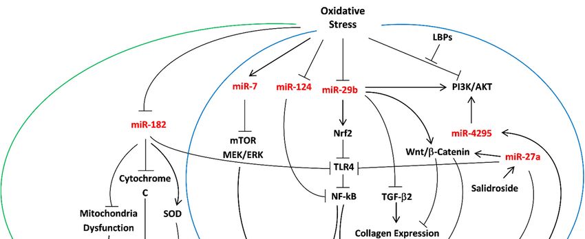
Figure 1. Overview of important miRNAs as mediators of oxidative stress in POAG. Summary of the miRNAs (red)
involved
Figure 1.inOverview
the protective mechanisms
of important against
miRNAs as oxidative
mediatorsstress in POAG.
of oxidative miR-7,
stress miR-24,Summary
in POAG. miR-27a, of
miR-29b, miR-4295
the miRNAs have
(red) in-
an anti-oxidant effect in TM cells (surrounded by the blue arrows), while miR-182 reduces oxidative stress
volved in the protective mechanisms against oxidative stress in POAG. miR-7, miR-24, miR-27a, miR-29b, miR-4295 have damage in RGC
(surrounded by the
an anti-oxidant green
effect arrow).
in TM The following
cells (surrounded byabbreviations referwhile
the blue arrows), to TM (trabecular
miR-182 meshwork),
reduces oxidative RGC
stress(retinal
damageganglion
in RGC
(surrounded by the green arrow). The following abbreviations refer to TM (trabecular meshwork), RGC
cells), LBPs (Lycium barbarum polysaccharides), FBN (fibrillin), ECM (extracellular matrix), SOD (superoxide dismutase), (retinal ganglion
cells), LBPs
TGF-β2 (Lycium barbarum
(transforming polysaccharides),
growth factor-beta FBN(nuclear
2), NF-kB (fibrillin), ECM
factor (extracellular matrix), SOD
kappa-light-chain-enhancer of(superoxide
activated B dismutase),
cells), TLR4
TGF-β2 (transforming growth factor-beta 2), NF-kB (nuclear factor
(toll-like receptor 4), and Nrf2 (nuclear factor erythroid 2-related factor 2).kappa-light-chain-enhancer of activated B cells), TLR4
(toll-like receptor 4), and Nrf2 (nuclear factor erythroid 2-related factor 2).
It is expected that this miR-29b function would enhance apoptosis under chronic
oxidative stress conditions since p53 has pro-apoptotic effects [163,164]. As such, miR-29b
can be an attractive target for interference in POAG. Since the action of TGF-β2 in POAG
is largely mediated through miR-29b [165], further examination of alternation in miR-29b
levels in POAG patients can be used as a tool for disease detection. Examination of relevant
miRNAs in response to the antioxidant, Edaravone, which decreases apoptotic cell death,
oxidative damage to DNA and lipids, and angiogenesis through inhibiting JNK and p38
MAPK pathways in glaucoma, can shed new light regarding facilitating therapy through
MAPK pathway regulation [6]. Furthermore, the possible involvement of oxidative damage
to DNA in POAG pathogenesis may indicate that DNA mutations are involved in a variety
of different human diseases with miRNA treatment as a potential therapeutic strategyInt. J. Mol. Sci. 2021, 22, 2421 11 of 17
that should be investigated. Recent studies emphasized the importance of extracellular
vesicles, and specifically exosomes, as protective signaling mediators in TM cells during
oxidative stress [166]. IL-1β-induced acute neuroinflammation and oxidative stress resulted
in the release of a specific subset of miRNAs via exosomes, potentially regulating the
inflammatory response [167]. The capacity of extracellular vesicles to carry protective
signals following oxidative stress is well documented [168–170]. Exosomes are bi-lipid
layered membranous vesicles with a diameter of approximately 30–100 nm characterized
by specific cell surface markers [171,172]. It has been reported that cell miRNAs reach
the extracellular environment through exosomes and that the exosomal cargo of cellular
proteins, lipids, and miRNAs play an important role as mediators of intercellular crosstalk
between the producing and recipient cells [173]. A genome scan for miRNA-related genetic
variants associated with POAG [118] and a comparison of miRNA expression in AH of
normal and POAG individuals [31] were widely examined. Additionally, appropriate
oxidative stress biomarkers in AH of POAG patients should be further examined, focusing
on the AH-producing cells and the non-pigmented ciliary epithelium (NPCE) located in
the anterior chamber of the eye, exiting the eye through the TM cells in the conventional
pathway [30]. A potential therapeutic target for glaucoma can be achieved by suppression
of miRNAs that are considered as pro-mediators of oxidative stress, such as miR-210, using
long non-coding RNAs transcripts (lncRNAs) [117]. In conclusion, we suggest further
investigating the role of POAG-related miRNAs as antioxidant machinery, examining
their dual role as pro- or anti-inflammatory/apoptotic agents as a response to diverse
concentrations of ROS in TM cells and RGC.
Funding: This research received no external funding.
Conflicts of Interest: The authors declare no conflict of interest.
References
1. Pizzino, G.; Irrera, N.; Cucinotta, M.; Pallio, G.; Mannino, F.; Arcoraci, V.; Squadrito, F.; Altavilla, D.; Bitto, A. Oxidative Stress:
Harms and Benefits for Human Health. Oxid. Med. Cell. Longev. 2017, 2017, 8416763. [CrossRef]
2. Ahmad, A.; Ahsan, H. Biomarkers of inflammation and oxidative stress in ophthalmic disorders. J. Immunoass. Immunochem.
2020, 41, 257–271. [CrossRef]
3. Pham-Huy, L.A.; He, H.; Pham-Huy, C. Free radicals, antioxidants in disease and health. Int. J. Biomed. Sci. 2008, 4, 89–96.
[PubMed]
4. Waris, G.; Ahsan, H. Reactive oxygen species: Role in the development of cancer and various chronic conditions. J. Carcinog.
2006, 5, 14. [CrossRef]
5. Deponte, M. Glutathione catalysis and the reaction mechanisms of glutathione-dependent enzymes. Biochim. Biophys. Acta 2013,
1830, 3217–3266. [CrossRef]
6. Masuda, T.; Shimazawa, M.; Hara, H. Retinal Diseases Associated with Oxidative Stress and the Effects of a Free Radical
Scavenger (Edaravone). Oxid. Med. Cell. Longev. 2017, 2017, 9208489. [CrossRef] [PubMed]
7. Kwon, Y.H.; Fingert, J.H.; Kuehn, M.H.; Alward, W.L. Primary open-angle glaucoma. N. Engl. J. Med. 2009, 360, 1113–1124.
[CrossRef] [PubMed]
8. Weinreb, R.N.; Aung, T.; Medeiros, F.A. The pathophysiology and treatment of glaucoma: A review. JAMA 2014, 311, 1901–1911.
[CrossRef] [PubMed]
9. Izzotti, A.; Bagnis, A.; Sacca, S.C. The role of oxidative stress in glaucoma. Mutat. Res. 2006, 612, 105–114. [CrossRef]
10. Schmidl, D.; Schmetterer, L.; Garhofer, G.; Popa-Cherecheanu, A. Pharmacotherapy of glaucoma. J. Ocul. Pharm. 2015, 31, 63–77.
[CrossRef]
11. Shen, F.; Chen, B.; Danias, J.; Lee, K.C.; Lee, H.; Su, Y.; Podos, S.M.; Mittag, T.W. Glutamate-induced glutamine synthetase
expression in retinal Muller cells after short-term ocular hypertension in the rat. Investig. Ophthalmol. Vis. Sci. 2004, 45, 3107–3112.
[CrossRef]
12. Galassi, F.; Renieri, G.; Sodi, A.; Ucci, F.; Vannozzi, L.; Masini, E. Nitric oxide proxies and ocular perfusion pressure in primary
open angle glaucoma. Br. J. Ophthalmol. 2004, 88, 757–760. [CrossRef] [PubMed]
13. Mozaffarieh, M.; Grieshaber, M.C.; Flammer, J. Oxygen and blood flow: Players in the pathogenesis of glaucoma. Mol. Vis. 2008,
14, 224–233. [PubMed]
14. Nita, M.; Grzybowski, A. The Role of the Reactive Oxygen Species and Oxidative Stress in the Pathomechanism of the Age-Related
Ocular Diseases and Other Pathologies of the Anterior and Posterior Eye Segments in Adults. Oxid. Med. Cell. Longev. 2016,
2016, 3164734. [CrossRef] [PubMed]Int. J. Mol. Sci. 2021, 22, 2421 12 of 17
15. Tang, B.; Li, S.; Cao, W.; Sun, X. The Association of Oxidative Stress Status with Open-Angle Glaucoma and Exfoliation Glaucoma:
A Systematic Review and Meta-Analysis. J. Ophthalmol. 2019, 2019, 1803619. [CrossRef] [PubMed]
16. Moreno, M.C.; Campanelli, J.; Sande, P.; Sanez, D.A.; Keller Sarmiento, M.I.; Rosenstein, R.E. Retinal oxidative stress induced by
high intraocular pressure. Free Radic. Biol. Med. 2004, 37, 803–812. [CrossRef]
17. Menaa, F.; Braghini, C.A.; Vasconcellos, J.P.; Menaa, B.; Costa, V.P.; Figueiredo, E.S.; Melo, M.B. Keeping an eye on myocilin:
A complex molecule associated with primary open-angle glaucoma susceptibility. Molecules 2011, 16, 5402–5421. [CrossRef]
[PubMed]
18. Fingert, J.H. Primary open-angle glaucoma genes. Eye (Lond.) 2011, 25, 587–595. [CrossRef]
19. van Koolwijk, L.M.; Ramdas, W.D.; Ikram, M.K.; Jansonius, N.M.; Pasutto, F.; Hysi, P.G.; Macgregor, S.; Janssen, S.F.; Hewitt, A.W.;
Viswanathan, A.C.; et al. Common genetic determinants of intraocular pressure and primary open-angle glaucoma. PLoS Genet.
2012, 8, e1002611. [CrossRef]
20. Liu, Y.; Allingham, R.R. Major review: Molecular genetics of primary open-angle glaucoma. Exp. Eye Res. 2017, 160, 62–84.
[CrossRef]
21. Ramos, R.F.; Sumida, G.M.; Stamer, W.D. Cyclic mechanical stress and trabecular meshwork cell contractility. Investig. Ophthalmol.
Vis. Sci. 2009, 50, 3826–3832. [CrossRef] [PubMed]
22. Tektas, O.Y.; Lutjen-Drecoll, E. Structural changes of the trabecular meshwork in different kinds of glaucoma. Exp. Eye Res. 2009,
88, 769–775. [CrossRef] [PubMed]
23. Roy Chowdhury, U.; Hann, C.R.; Stamer, W.D.; Fautsch, M.P. Aqueous humor outflow: Dynamics and disease. Investig. Ophthalmol.
Vis. Sci. 2015, 56, 2993–3003. [CrossRef] [PubMed]
24. Abu-Hassan, D.W.; Acott, T.S.; Kelley, M.J. The Trabecular Meshwork: A Basic Review of Form and Function. J. Ocul. Biol. 2014, 2.
[CrossRef]
25. Vernazza, S.; Tirendi, S.; Bassi, A.M.; Traverso, C.E.; Sacca, S.C. Neuroinflammation in Primary Open-Angle Glaucoma.
J. Clin. Med. 2020, 9, 3172. [CrossRef] [PubMed]
26. Izzotti, A.; Sacca, S.C.; Cartiglia, C.; De Flora, S. Oxidative deoxyribonucleic acid damage in the eyes of glaucoma patients.
Am. J. Med. 2003, 114, 638–646. [CrossRef]
27. Wang, Y.; Niu, L.; Zhao, J.; Wang, M.; Li, K.; Zheng, Y. An update: Mechanisms of microRNA in primary open-angle glaucoma.
Brief. Funct. Genom. 2020. [CrossRef] [PubMed]
28. Fioravanti, A.; Pirtoli, L.; Giordano, A.; Dotta, F. Crosstalk between MicroRNA and Oxidative Stress in Physiology and Pathology.
Int. J. Mol. Sci. 2020, 21, 1270. [CrossRef] [PubMed]
29. Engedal, N.; Zerovnik, E.; Rudov, A.; Galli, F.; Olivieri, F.; Procopio, A.D.; Rippo, M.R.; Monsurro, V.; Betti, M.; Albertini, M.C.
From Oxidative Stress Damage to Pathways, Networks, and Autophagy via MicroRNAs. Oxid. Med. Cell. Longev. 2018,
2018, 4968321. [CrossRef] [PubMed]
30. Drewry, M.D.; Challa, P.; Kuchtey, J.G.; Navarro, I.; Helwa, I.; Hu, Y.; Mu, H.; Stamer, W.D.; Kuchtey, R.W.; Liu, Y. Differen-
tially expressed microRNAs in the aqueous humor of patients with exfoliation glaucoma or primary open-angle glaucoma.
Hum. Mol. Genet. 2018, 27, 1263–1275. [CrossRef] [PubMed]
31. Jayaram, H.; Phillips, J.I.; Lozano, D.C.; Choe, T.E.; Cepurna, W.O.; Johnson, E.C.; Morrison, J.C.; Gattey, D.M.; Saugstad, J.A.;
Keller, K.E. Comparison of MicroRNA Expression in Aqueous Humor of Normal and Primary Open-Angle Glaucoma Patients
Using PCR Arrays: A Pilot Study. Investig. Ophthalmol. Vis. Sci. 2017, 58, 28842–28890. [CrossRef] [PubMed]
32. Mao, W.; Millar, J.C.; Wang, W.H.; Silverman, S.M.; Liu, Y.; Wordinger, R.J.; Rubin, J.S.; Pang, I.H.; Clark, A.F. Existence of
the canonical Wnt signaling pathway in the human trabecular meshwork. Investig. Ophthalmol. Vis. Sci. 2012, 53, 7043–7051.
[CrossRef]
33. Yanagi, M.; Kawasaki, R.; Wang, J.J.; Wong, T.Y.; Crowston, J.; Kiuchi, Y. Vascular risk factors in glaucoma: A review.
Clin. Exp. Ophthalmol. 2011, 39, 252–258. [CrossRef] [PubMed]
34. Tanito, M.; Kaidzu, S.; Takai, Y.; Ohira, A. Correlation between systemic oxidative stress and intraocular pressure level. PLoS ONE
2015, 10, e0133582. [CrossRef] [PubMed]
35. Sacca, S.C.; Izzotti, A.; Rossi, P.; Traverso, C. Glaucomatous outflow pathway and oxidative stress. Exp. Eye Res. 2007, 84, 389–399.
[CrossRef] [PubMed]
36. Shaw, P.X.; Stiles, T.; Douglas, C.; Ho, D.; Fan, W.; Du, H.; Xiao, X. Oxidative stress, innate immunity, and age-related macular
degeneration. Aims. Mol. Sci. 2016, 3, 196–221. [CrossRef]
37. Zanon-Moreno, V.; Marco-Ventura, P.; Lleo-Perez, A.; Pons-Vazquez, S.; Garcia-Medina, J.J.; Vinuesa-Silva, I.; Moreno-Nadal, M.A.;
Pinazo-Duran, M.D. Oxidative stress in primary open-angle glaucoma. J. Glaucoma 2008, 17, 263–268. [CrossRef] [PubMed]
38. Tezel, G. Oxidative stress in glaucomatous neurodegeneration: Mechanisms and consequences. Prog. Retin. Eye Res. 2006,
25, 4905–4913. [CrossRef] [PubMed]
39. Sacca, S.C.; Tirendi, S.; Scarfi, S.; Passalacqua, M.; Oddone, F.; Traverso, C.E.; Vernazza, S.; Bassi, A.M. An advanced in vitro
model to assess glaucoma onset. ALTEX 2020, 37, 2652–2674. [CrossRef] [PubMed]
40. Junqueira, V.B.; Barros, S.B.; Chan, S.S.; Rodrigues, L.; Giavarotti, L.; Abud, R.L.; Deucher, G.P. Aging and oxidative stress.
Mol. Asp. Med. 2004, 25, 51–56. [CrossRef]
41. Tezel, G.; Yang, X.; Cai, J. Proteomic identification of oxidatively modified retinal proteins in a chronic pressure-induced rat
model of glaucoma. Investig. Ophthalmol. Vis. Sci. 2005, 46, 3177–3187. [CrossRef] [PubMed]Int. J. Mol. Sci. 2021, 22, 2421 13 of 17
42. Zundorf, G.; Kahlert, S.; Bunik, V.I.; Reiser, G. alpha-Ketoglutarate dehydrogenase contributes to production of reactive oxygen
species in glutamate-stimulated hippocampal neurons in situ. Neuroscience 2009, 158, 610–616. [CrossRef] [PubMed]
43. Crish, S.D.; Calkins, D.J. Neurodegeneration in glaucoma: Progression and calcium-dependent intracellular mechanisms.
Neuroscience 2011, 176, 1–11. [CrossRef] [PubMed]
44. Batandier, C.; Leverve, X.; Fontaine, E. Opening of the mitochondrial permeability transition pore induces reactive oxygen species
production at the level of the respiratory chain complex I. J. Biol. Chem. 2004, 279, 17197–17204. [CrossRef]
45. Ito, Y.A.; Di Polo, A. Mitochondrial dynamics, transport, and quality control: A bottleneck for retinal ganglion cell viability in
optic neuropathies. Mitochondrion 2017, 36, 186–192. [CrossRef] [PubMed]
46. Osborne, N.N.; del Olmo-Aguado, S. Maintenance of retinal ganglion cell mitochondrial functions as a neuroprotective strategy
in glaucoma. Curr. Opin. Pharm. 2013, 13, 16–22. [CrossRef]
47. Abu-Amero, K.K.; Morales, J.; Bosley, T.M. Mitochondrial abnormalities in patients with primary open-angle glaucoma. Inves-
tig. Ophthalmol. Vis. Sci. 2006, 47, 2533–2541. [CrossRef] [PubMed]
48. Sacca, S.C.; Pascotto, A.; Camicione, P.; Capris, P.; Izzotti, A. Oxidative DNA damage in the human trabecular meshwork:
Clinical correlation in patients with primary open-angle glaucoma. Arch. Ophthalmol. 2005, 123, 458–463. [CrossRef] [PubMed]
49. Izzotti, A.; Sacca, S.C.; Longobardi, M.; Cartiglia, C. Sensitivity of ocular anterior chamber tissues to oxidative damage and its
relevance to the pathogenesis of glaucoma. Investig. Ophthalmol. Vis. Sci. 2009, 50, 5251–5258. [CrossRef]
50. Martin, K.R.; Levkovitch-Verbin, H.; Valenta, D.; Baumrind, L.; Pease, M.E.; Quigley, H.A. Retinal glutamate transporter changes
in experimental glaucoma and after optic nerve transection in the rat. Investig. Ophthalmol. Vis. Sci. 2002, 43, 2236–2243. [PubMed]
51. Liton, P.B.; Lin, Y.; Luna, C.; Li, G.; Gonzalez, P.; Epstein, D.L. Cultured porcine trabecular meshwork cells display altered
lysosomal function when subjected to chronic oxidative stress. Investig. Ophthalmol. Vis. Sci. 2008, 49, 3961–3969. [CrossRef]
[PubMed]
52. Fossel, M. Cell senescence in human aging and disease. Ann. N. Y. Acad. Sci. 2002, 959, 14–23. [CrossRef] [PubMed]
53. Li, G.; Luna, C.; Qiu, J.; Epstein, D.L.; Gonzalez, P. Modulation of inflammatory markers by miR-146a during replicative
senescence in trabecular meshwork cells. Investig. Ophthalmol. Vis. Sci. 2010, 51, 2976–2985. [CrossRef]
54. Vernazza, S.; Tirendi, S.; Scarfi, S.; Passalacqua, M.; Oddone, F.; Traverso, C.E.; Rizzato, I.; Bassi, A.M.; Sacca, S.C. 2D- and
3D-cultures of human trabecular meshwork cells: A preliminary assessment of an in vitro model for glaucoma study. PLoS ONE
2019, 14, e0221942. [CrossRef] [PubMed]
55. Zapp, C.; Obarska-Kosinska, A.; Rennekamp, B.; Kurth, M.; Hudson, D.M.; Mercadante, D.; Barayeu, U.; Dick, T.P.;
Denysenkov, V.; Prisner, T.; et al. Mechanoradicals in tensed tendon collagen as a source of oxidative stress. Nat. Commun 2020,
11, 2315. [CrossRef] [PubMed]
56. Anholt, R.R.; Carbone, M.A. A molecular mechanism for glaucoma: Endoplasmic reticulum stress and the unfolded protein
response. Trends Mol. Med. 2013, 19, 586–593. [CrossRef] [PubMed]
57. Cullinan, S.B.; Diehl, J.A. Coordination of ER and oxidative stress signaling: The PERK/Nrf2 signaling pathway. Int. J. Biochem.
Cell Biol. 2006, 38, 317–332. [CrossRef] [PubMed]
58. Rao, R.V.; Ellerby, H.M.; Bredesen, D.E. Coupling endoplasmic reticulum stress to the cell death program. Cell Death Differ. 2004,
11, 372–380. [CrossRef] [PubMed]
59. Izzotti, A.; Longobardi, M.; Cartiglia, C.; Sacca, S.C. Proteome alterations in primary open angle glaucoma aqueous humor.
J. Proteome Res. 2010, 9, 4831–4838. [CrossRef]
60. Wang, N.; Chintala, S.K.; Fini, M.E.; Schuman, J.S. Activation of a tissue-specific stress response in the aqueous outflow pathway
of the eye defines the glaucoma disease phenotype. Nat. Med. 2001, 7, 304–309. [CrossRef] [PubMed]
61. Gherghel, D.; Griffiths, H.R.; Hilton, E.J.; Cunliffe, I.A.; Hosking, S.L. Systemic reduction in glutathione levels occurs in patients
with primary open-angle glaucoma. Investig. Ophthalmol. Vis. Sci. 2005, 46, 877–883. [CrossRef] [PubMed]
62. Gherghel, D.; Mroczkowska, S.; Qin, L. Reduction in blood glutathione levels occurs similarly in patients with primary-open
angle or normal tension glaucoma. Investig. Ophthalmol. Vis. Sci. 2013, 54, 3333–3339. [CrossRef]
63. Ferreira, S.M.; Lerner, S.F.; Brunzini, R.; Evelson, P.A.; Llesuy, S.F. Oxidative stress markers in aqueous humor of glaucoma
patients. Am. J. Ophthalmol. 2004, 137, 62–69. [CrossRef]
64. Ghanem, A.A.; Arafa, L.F.; El-Baz, A. Oxidative stress markers in patients with primary open-angle glaucoma. Curr. Eye Res.
2010, 35, 295–301. [CrossRef] [PubMed]
65. Shahani, S.B.; Shaikh, P.; Memon, S.G.; Memon, S. Possible prevention of reactive oxygen species induced human trabecular
meshwork cell damage by resveratrol and ascorbic acid. Prof. Med. J. 2019, 26, 1036–1041. [CrossRef]
66. Motohashi, H.; Yamamoto, M. Nrf2-Keap1 defines a physiologically important stress response mechanism. Trends Mol. Med. 2004,
10, 549–557. [CrossRef]
67. Gloire, G.; Legrand-Poels, S.; Piette, J. NF-kappaB activation by reactive oxygen species: Fifteen years later. Biochem. Pharm. 2006,
72, 1493–1505. [CrossRef]
68. Myer, C.; Perez, J.; Abdelrahman, L.; Mendez, R.; Khattri, R.B.; Junk, A.K.; Bhattacharya, S.K. Differentiation of soluble aqueous
humor metabolites in primary open angle glaucoma and controls. Exp. Eye Res. 2020, 194, 108024. [CrossRef]
69. Zhao, Y.; Zhu, H.; Yang, Y.; Ye, Y.; Yao, Y.; Huang, X.; Zhang, Y.; Shu, X.; Chen, X.; Yang, Y.; et al. AQP1 suppression by ATF4
triggers trabecular meshwork tissue remodelling in ET-1-induced POAG. J. Cell Mol. Med. 2020, 24, 3469–3480. [CrossRef]You can also read