Review Article Quercetin and Cancer Chemoprevention
←
→
Page content transcription
If your browser does not render page correctly, please read the page content below
Hindawi Publishing Corporation
Evidence-Based Complementary and Alternative Medicine
Volume 2011, Article ID 591356, 15 pages
doi:10.1093/ecam/neq053

Review Article
Quercetin and Cancer Chemoprevention

          Lara Gibellini,1 Marcello Pinti,1 Milena Nasi,1 Jonas P. Montagna,1 Sara De Biasi,1
          Erika Roat,1 Linda Bertoncelli,1 Edwin L. Cooper,2 and Andrea Cossarizza1
          1 Department   of Biomedical Sciences, University of Modena and Reggio Emilia School of Medicine, 41125 Modena, Italy
          2 David   Geffen School of Medicine, UCLA Medical Center (CHS), Los Angeles, CA, USA

          Correspondence should be addressed to Andrea Cossarizza, andrea.cossarizza@unimore.it

          Received 24 November 2009; Accepted 9 April 2010
          Copyright © 2011 Lara Gibellini et al. This is an open access article distributed under the Creative Commons Attribution License,
          which permits unrestricted use, distribution, and reproduction in any medium, provided the original work is properly cited.

          Several molecules present in the diet, including flavonoids, can inhibit the growth of cancer cells with an ability to act as
          “chemopreventers”. Their cancer-preventive effects have been attributed to various mechanisms, including the induction of cell-
          cycle arrest and/or apoptosis as well as the antioxidant functions. The antioxidant activity of chemopreventers has recently
          received a great interest, essentially because oxidative stress participates in the initiation and progression of different pathological
          conditions, including cancer. Since antioxidants are capable of preventing oxidative damage, the wide use of natural food-derived
          antioxidants is receiving greater attention as potential anti-carcinogens. Among flavonoids, quercetin (Qu) is considered an
          excellent free-radical scavenging antioxidant, even if such an activity strongly depends on the intracellular availability of reduced
          glutathione. Apart from antioxidant activity, Qu also exerts a direct, pro-apoptotic effect in tumor cells, and can indeed block the
          growth of several human cancer cell lines at different phases of the cell cycle. Both these effects have been documented in a wide
          variety of cellular models as well as in animal models. The high toxicity exerted by Qu on cancer cells perfectly matches with the
          almost total absence of any damages for normal, non-transformed cells. In this review we discuss the molecular mechanisms that
          are based on the biological effects of Qu, and their relevance for human health.

1. Introduction                                                           substantially alter the natural history of carcinogenesis, and
                                                                          that an inverse correlation between a high consumption
1.1. Chemoprevention and Diet. The field of cancer chemo-                 of fruits and vegetables and the incidence of some cancers
prevention, defined as the long-term intervention with                    does exist [3, 4]. Indeed, a high consumption of fruits
natural or synthetic molecules to prevent, inhibit or reverse             and vegetables was associated with a reduced risk of
carcinogenesis, is gaining increasing importance, especially              intestinal cancer, especially of colon cancer, in former
at a time when the use of complementary and alternative                   smokers or in no-smokers, whereas no preventive effect
medicine (CAM) and natural health products is consistently                was found in current smokers [5]. Consumption of
increasing [1]. At present, the use of CAM in oncology                    fruits, particularly citrus fruit, as well as vegetables, likely
represents a challenging area of interest since remarkable                correlates with decreased esophageal cancer risk [6, 7].
scientific evidences suggest that natural dietary factors can             Green leafy vegetables, rather than fruit, might also have
inhibit the process of carcinogenesis and can effectively                  a protective effect against lung cancer [8, 9]. High intake
influence the risk of cancer in humans. The idea of cancer                of cruciferous vegetables may be associated with reduced
chemoprevention arises from both statistical and epidemi-                 risk of aggressive prostate cancer [10]. With regard to
ological data showing that a modification of the lifestyle is             renal cancer, although the results of case-control studies
associated with lower incidence of certain types of cancers               are not fully consistent and often contradictory, analyses
(i.e., colorectal, stomach, lung and esophageal cancers) and              performed on very large number of patients have found
that, in particular, such modifications include a vegetable-              an inverse association for the intake of total fruit [11],
and fruit-rich diet [2].                                                  total vegetables [12], and some subgroups of vegetables
     Accumulating evidences from observational and                        such as cruciferous vegetables, dark green vegetables and
prospective studies indicate that dietary components may                  yellow-orange vegetables [13]. Conversely, positive effects of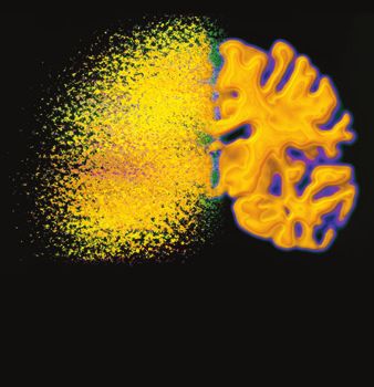
2                                                                  Evidence-Based Complementary and Alternative Medicine

fruit and vegetable consumption, combined or separately,          antioxidant enzymes, MnSOD and GSH peroxidase-1 (Gpx-
were not found as far as the risk of developing bladder or        1), determines an increased incidence of neoplasms in mice
ovary cancer was concerned [14].                                  [38].

1.2. Natural Chemopreventers. The majority of the case-           2.2. Reactive Oxygen Species Induced Carcinogenesis in Human
control studies focused on the use of fruits and vegeta-          Cells. The involvement of reactive oxygen species (ROS) in
bles because these items, which include soybean, ginger,          tumor progression has also been demonstrated in human
onion, cabbage, cauliflower, turmeric, are the basis of           cells. NADPH oxidase 1 (Nox1), an enzyme that produces
most diets throughout the world, and represent important          superoxide (which is in turn dismuted to hydrogen perox-
sources of potentially non-toxic molecules (dietary phyto-        ide), is overexpressed in colon and prostate cancer cell lines
chemicals). These molecules can exert a cancer-preventive         [39, 40], while its downregulation reverses tumor growth
effect and therefore are termed as “chemopreventers” [15].         [41]. Decreased levels of MnSOD and rapid cell doubling
Among them, the most studied are curcumin, quercetin              time have been reported in human pancreatic cancer cell
(Qu), resveratrol, luteolin, genistein, (−)-epigallocatechin-     lines at various levels of differentiation [42].
3-gallate (EGCG), lycopene and, in general, flavonoids and            These studies are consistent with the observation that
polyphenols [16–19]. A majority of studies have analyzed          a significant shift of cellular oxidative balance could lead
the biological properties (e.g., antioxidant, antimicrobial,      to tumor promotion or progression, as ROS are involved
antiproliferative, pro- or anti-apoptotic) of the aforemen-       in damaging of DNA as well as in mitogenic signaling
tioned molecules that are present, along with other com-          [43–45]. Endogenous ROS are generally supposed to cause
pounds, in aqueous extracts from plants [20–25].                  DNA damage [46], through the production of oxidized
    A relevant interest is present on the mechanism(s)            bases and DNA strand breaks, and to be a relevant factor
of action of chemopreventers, especially concerning the           contributing to chromosome instability and accumulation
identification of molecular and cellular targets of CAM com-      of mutations and deletions, finally leading to cancer [47].
pounds, and the molecular basis of their cancer-preventive        About 1% of oxygen consumption results in the production
action. At the biochemical level, chemopreventers usually         of ROS [46], thus implying that, in every cell, ROS can
act as modulators of signal transduction pathways that are        damage ∼20 000 DNA bases per day [48]. Cells have evolved
involved in almost all biological processes: cell prolifera-      several antioxidant defenses, including repair and detoxifying
tion, apoptosis, cell migration, cell differentiation, oxidative   enzymes, and small scavenger molecules, such as GSH.
balance and inflammation [26–29]. Chemopreventers often           Nevertheless, the presence of these intracellular protective
have preferentially an antioxidant activity; however, they are    systems is not sufficient to ensure an adequate and complete
also able to exert anti-proliferation and anti-inflammation       removal of oxidative damages.
actions. Indeed, they can directly modulate several proteins          Apart from the direct action on DNA, it has to be
that are involved in cell cycle and cellular homeostasis          noted that ROS act as secondary messengers in several
and whose deregulation can play a role in carcinogenesis,         pathways, which can potentially promote carcinogenic pro-
such as p53, p73, p21, Bax, Bcl-2, COX-2, NF-kB, catalase,        cesses, including resistance to apoptosis, increase in cell
glutathione (GSH)-peroxidase [4, 30, 31]. The antioxidant         proliferation and production of metastasis [32]. Indeed, ROS
activity of chemopreventers is nowadays gaining more              have been involved in the transcriptional activation of several
importance because of the observations, both in vitro and in      proto-oncogenes, such as c-FOS, c-JUN and c-MYC. In
vivo, that the deregulation of free-radical homeostasis can be    human hepatoma cells, ROS modulate the expression of c-
involved in carcinogenesis [32].                                  FOS and c-JUN through PKB pathway [49]. Furthermore,
                                                                  p66Shc, which is involved in the regulation of ROS signaling,
2. Carcinogenesis and Reactive Oxygen Species                     is responsible for androgenic proliferation signals through
                                                                  ROS production in prostate cancer cells that are positive
2.1. Reactive Oxygen Species Induced Carcinogenesis in Ani-       to androgen receptor [50]. Finally, in anaplastic large cell
mal Models. The evidence of a strong association between          lymphomas, the use of nordihydroguaiaretic acid, which is
the production of free radicals and carcinogenesis mainly         an inhibitor of lipoxygenase, results in the inhibition of
derives from in vitro studies showing that some pro-oxidant       several pathways which are involved in antiapoptotic and
chemicals promote tumors in several animal models, whereas        pro-mitogenic functions [51].
primary endogenous antioxidant enzymes could interfere
with tumor promotion. For instance, 1,2-dimethylhydrazine
induces colon carcinogenesis in rats [33, 34], whereas            3. Flavonoids and Their Possible Role in
benzoylperoxide promotes papillomas and carcinomas after             Cancer Chemoprevention
7,12-dimethylbenz[a]anthracene initiation in mice [35]. In
rat liver epithelial cells treated with N-methyl-N  -nitro-      3.1. Flavonoids in the Diet. Among chemopreventers, one
N-nitrosoguanidine, hydrogen peroxide exerts a tumor              of the most studied group of antioxidant compounds are
promoting activity [36]. Conversely, the overexpression of        flavonoids. Flavonoids are a large heterogeneous group of
manganese superoxide dismutase (MnSOD) reduces tumor              benzo-γ-pyrone derivatives that share a common carbon
incidence in a multistage skin carcinogenesis mouse model         skeleton of dyphenylpropanes [52] and can be divided into
[37]. A combined deficiency in two mitochondrial-localized        six different classes, namely flavonols, flavones, flavanones,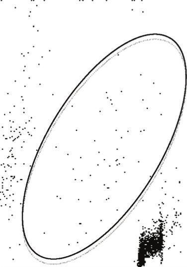
Evidence-Based Complementary and Alternative Medicine                                                                         3

flavanols, isoflavones and anthocyanidins, according to their    are important in cancer progression. Chromatin is remod-
molecular structure [53].                                        eled by chemical modifications of DNA and histones, such as
    Flavonoids are largely present in fruits, vegetables, aro-   DNA methylation and multiple histone modifications, such
matic plants, medical herbs, tea and red wine [54]. It is        as methylation, phosphorylation, acetylation, sumoylation
extremely difficult to estimate the daily human intake of          and ubiquitination; for example, resveratrol activates sirtuin
flavonoids, especially because of the lack of standardized       (SIRT)-1, a member of histone deacetylase (HDAC) family,
analytical methods [55]. However, the average daily intake       which plays key roles in cell survival and apoptosis [77]. The
of the most abundant flavonoids, catechins, is ∼100 mg           network of SIRT1-modulated signals is wide and complex,
[56]. Similar to daily intake, it is also quite complex to       and involves SIRT1 direct interactions with several proteins
assess and quantify the bioavailability of flavonoids [57].      involved in cell survival (p53, bax, E2F1, FOXO3, Dif1), DNA
Nevertheless, metabolized forms of flavonoids present in         repair (WRN, Ku70, RAD51) and cell cycle/apoptosis (β-
blood significantly differ from the native compounds, and         catenin, survivin, NFκB) [78]. The activation of SIRT1 by
plasma concentration of total metabolites can have a range       resveratrol induces the formation of SIRT1-p300 complexes,
0–4 μmol L−1 with an intake of 50 mg of aglycone, which is       causing the inactivation of p300 acetyltransferase and a
the non-sugar compound left after partial metabolization of      reduction in the acetylation of both β-catenin and NFκB-
the original flavonoid [58].                                     p65. The main consequence of this phenomenon is the
                                                                 downregulation of the multidrug resistance (MDR)-1 and
3.2. Cancer Chemoprevention by Flavonoids: Molecular             Bcl-xL genes with the subsequent stimulation of cell death,
Mechanisms. Results from cell culture and animal models          as well as the reduction of chemoresistance in breast tumor
reveal that flavonoids exert positive preventive effects in       cells [79].
carcinogenesis and neurodegenerative disorders essentially           Several catechol-containing dietary polyphenols are
because of their antioxidant activity, their capacity to         capable of modulating DNA methylation. Among them,
affect the expression of several detoxifying enzymes [59],        EGCG is a potent and efficacious in vitro inhibitor of DNA
and their ability to modulate protein signaling cascades         methyltransferase (DNMT)-1, whereas Qu can demethylate
[60]. Flavonoids can interfere with specific stages of the       the p16INK4a gene promoter, whose hypermethylation is
carcinogenic process, and can inhibit cell proliferation and     present in human colon cancer cells [80]. Qu also activates
induce apoptosis in several types of cancer cells.               histone deacetylase enzymatic activity, thus reducing the
    EGCG is one of the most intensively studied flavonoids       acetylation of histone H3 in human prostate cancer cells. The
as it is the major polyphenolic component of green tea.          deacetylation of H3 could be responsible for the inhibition of
EGCG inhibits cell proliferation and induces apoptosis in        survivin expression, and for the subsequent sensitization to
several human tumor cell lines, including CaSki and HeLa         TRAIL-induced apoptosis [81].
cervical cells [61], Hep-2 cells [62], laryngeal squamous
carcinoma cells [63], SW780 and TCCSUP bladder urothelial        3.3. Cancer Chemoprevention by Flavonoids in Human.
cells [64], melanoma cells [65], adrenal NCI-H295 cancer         Regarding flavonoids as chemopreventers in humans, con-
cells [66] and A549 lung cancer cells [67]. The mechanisms       trasting results have been reported, and indeed some studies
by which apoptosis is triggered differ depending on the cell      showed an inverse correlation between the intake of total
line and include via death receptor, or via mitochondrial and    dietary flavonoids and the risk of cancer [82–85], whereas
endoplasmic reticulum-dependent pathways.                        others did not evidence any association [86]. Furthermore,
    The cancer-preventive properties of flavonoids can be        the importance of risk factors such as smoke has to be
attributed to their capacity of quenching ROS, reactive nitro-   taken into account, since in different groups of patients,
gen species (RNS) and other radicals. Tea catechins, espe-       a limited evidence for a preventive effect of flavonoids on
cially EGCG, react with superoxide radical, hydroxyl radical,    the development of pancreatic cancer has been reported.
peroxyl radical and peroxynitrite [68]. Resveratrol, present     For example, no association between flavonoid intake and
in red wine, grapes and peanuts, is a scavenger of superoxide    pancreatic cancer risk was found in male current smokers
and peroxynitrite radicals [69], and genistein, mainly derived   [87]. The Multiethnic Cohort Study, on the contrary,
from soy, can scavenge exogenous or endogenous hydrogen          reported that the intake of total flavonols was associated with
peroxide in cell models [70]. Moreover, flavonoids exert their   a reduced pancreatic cancer risk among current smokers,
protective antioxidant effect not only by quenching ROS,          but not in never or former smokers [88]. Lack of pre-
but also by modulating the activity of several detoxifying       ventive effect was described in the case of ovarian can-
enzymes, including lipoxygenase, cycloxygenase, inducible        cer [89]. Conversely, several other epidemiological studies
nitric oxide synthase, monoxygenase, xanthine oxidase and        confirmed the protective role of a high flavonoid intake
NADH oxidase [71–75]. Among enzymes that are inhibited           against colorectal [90, 91] and lung cancers [92]. Interest-
by flavonoids, thioredoxin reductases have to be quoted,         ingly, although natural chemopreventers have undergone
as they are involved in cellular redox control, and are          extensive mechanistic investigation at the molecular and
overexpressed in different aggressive tumors [76].                cellular level, their preclinical efficacy needs to be further
    Growing evidences suggest that flavonoids (in particular,    explored, and clinical trials have only recently started to
resveratrol and quercetin) may contribute to chromatin           investigate the potential preventive role of these compounds
remodeling and thus interfere with epigenetic alterations that   [93].
4                                                                 Evidence-Based Complementary and Alternative Medicine

4. Qu and Its Molecular Role in                                  of GSQ is reversible and glutathionyl-Qu adducts can be
   Cancer Chemoprevention                                        continuously dissociated into QQ and GSH [107]. As a result,
                                                                 in the presence of high GSH concentrations, oxidized Qu
4.1. The Importance of the Diet. Qu (3,3 ,4 ,5,7-penta-
                                                                 reacts with GSH to form GSQ again, and the reversibility
hydroxyflavone) is an important dietary flavonoid, present
                                                                 of the reaction ensures the protection against QQ toxicity.
in different vegetables, fruits, seeds, nuts, tea and red wine
                                                                 In the presence of low GSH content, oxidized Qu reacts
[94–96]. The average daily intake of Qu can reach 30 mg in
                                                                 with protein thiols, exerting a toxic effect within cells
most Western countries [97], and its bioavailability depends
                                                                 [107, 108]. Similarly, long exposure to Qu along with high
on the metabolic form present in the food. Indeed, Qu
                                                                 Qu concentration, causes a reduction in GSH content,
obtained from plant source is in the form of Qu-glucose
                                                                 suggesting the inability of Qu to cope with ROS for that
conjugates (Qu glucosides), which are absorbed in the
                                                                 period. As a consequence, the pro-oxidant effect of Qu
apical membrane of the enterocytes. Once absorbed, Qu
                                                                 could prevail over the antioxidant effect and result in cell
glucosides are hydrolyzed to generate Qu aglycone which is
                                                                 death by damaging cellular compartments [109, 110]. As
further metabolized to the methylated, sulfonylated and glu-
                                                                 shown in Figure 1, when high levels of GSH are present, Qu-
curonidated forms by the enterocytic transferases [98]. Qu
                                                                 derived semiquinone and quinoidal products are constantly
metabolites are then transported first to the intestinal lumen
                                                                 reduced, thus limiting Qu cytotoxicity and enabling Qu
[98], and then to the liver, where other conjugation reactions
                                                                 to act as antioxidant rather than as pro-oxidant [111].
take place to form Qu-3-glucuronide and Qu-3 -sulfate,
                                                                 The antioxidant capability of Qu strongly depends on the
which are the major Qu-derived circulating compounds in
                                                                 intracellular availability of GSH, since, in Qu-treated cells,
human plasma [99, 100]. According to recent studies on
                                                                 alterations typical of apoptosis appear when intracellular
Qu bioavailability [58], when Qu is absorbed in the form
                                                                 GSH is completely depleted. Indeed, in different cellular
of Qu glucosides, the peak plasma concentration ranges
                                                                 models low concentrations of Qu induce cell proliferation
from 3.5 to 5.0 μmol L−1 . In the unconjugated form, Qu
                                                                 and increase the antioxidant capacity of the cells, whereas
absorption is less efficient, and peak plasma concentration
                                                                 higher concentrations of Qu decrease antioxidant capacity
is
Evidence-Based Complementary and Alternative Medicine 5
Quercetin (Qu)
MMP collapse
Oxidative Qu
stress
Qu-quinone
Caspases Apoptosis
(QQ) p53
Low GSH High GSH Qu
p21
Qu
Cyclin E
Glutathionyl CDK2
quercetin (GSQ) G1/S checkpoint
p27 Cyclin A
G1 S CDK2
DNA and protein
damages Cyclin D
p21
CDK4/6
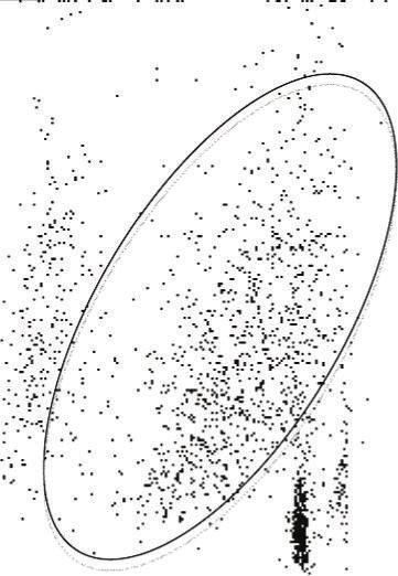
Figure 1: Antioxidant and pro-oxidant effects of Qu in the presence
of low and high levels of reduced GSH. The antioxidant and M G2
pro-oxidant effects of Qu strongly depend upon the availability p21
G2/M checkpoint Qu
of intracellular reduced GSH. During an oxidative stress, in
Cyclin B
the presence of peroxidases, Qu reacts with H2 O2 to form a CDK1
semiquinone radical that is rapidly oxidized to QQ. QQ has a Qu
pro-oxidant effect; its high reactivity towards protein thiols and
DNA leads to cell damage and cytotoxicity. QQ is also highly Qu
reactive towards thiols, and preferentially reacts with GSH to form
relatively stable protein-oxidized Qu adducts such as 6-GSQ and Figure 2: Effects of Qu on cell cycle. Qu is able to regulate cell cycle
8-GSQ. The reversibility of this reaction allows the continuous by directly binding several molecular targets and, depending on the
dissociation of GSQ into GSH and QQ. In the presence of high GSH cell type and tumor origin, it blocks the cell cycle at G2/M or at
concentrations, QQ reacts with GSH to form GSQ again, and QQ the G1/S transition. At the G1/S transition, Qu blocks cell-cycle
cannot exert its cytotoxic effects, whereas when low levels of GSH progression through the up-regulation of p21 and p27 and p53.
are present, QQ reacts with protein thiols, thus leading to cellular p21 exerts an inhibitory activity on several CDKs. In particular, p21
damage. inhibits CDK2-cyclin E, with the consequent inhibition of CDK2-
dependent phosphorylation of pRb and the sequestration of E2F1,
thus inhibiting gene transcription induced by E2F1 and progression
into and through S phase. p21 also inhibits CDK2-cyclin A and
Qu to directly poison TopoII through the stabilization of CDK1-cyclin B, which are essential for progression through S phase
double strand breaks in the TopoII-DNA cleavage complexes and G2, respectively. p27 exerts several effects on cell cycle, but only
under certain conditions it can inhibit the complexes CDK4-cyclin
could account for genetic rearrangements leading primary
D and CDK6-cyclin D. The tumor suppressor p53, once activated,
hematopoietic progenitor cells to develop mixed-lineage can induce several different cellular responses, including growth
leukemia [120]. arrest and apoptosis. Growth arrest is essentially elicited through
the up-regulation of the genes that encode for inhibitors of cell-cycle
4.4. Direct Pro-Apoptotic Effects of Qu. Collectively, the pro- progression, including p21 and p27. In different cellular models,
Qu stabilizes p53 both at mRNA and protein levels. Apart from
apoptotic effects of Qu may result from multiple pathways.
blocking cell growth through the direct action on key modulators of
First, in MDA-MB-231 cells, Qu treatment increases cytoso- cell cycle, Qu is able to induce apoptosis via mitochondrial pathway:
lic Ca2+ levels and reduces the mitochondrial membrane indeed, Qu can disrupt MMP, which in turn provokes the release
potential (MMP), thus promoting activation of caspase-3, of cytochrome c in the cytoplasm, a phenomenon that activates
-8 and -9 [26]. The capability of Qu to induce apoptosis multiple caspases, such as caspase-3 and -7.
via mitochondrial pathway has been confirmed in U937 cell
line [109, 121]. In these cells, Qu disrupts MMP [109, 121],
which in turn provokes the release of cytochrome c in
the cytoplasm [122], and subsequently activates multiple Qu exposure results in proteasomal degradation of survivin
caspases, such as caspase-3 and -7 [123]. Second, Qu [127], according to another proposed model, Qu treatment
inhibits cell growth and apoptosis by down-regulating the raises cyclin B1 and p53 proteins that, in turn, increase
transcriptional activity of β-catenin/Tcf signaling, with the survivin and p21 protein expression, thereby inhibiting
consequent down-regulation of cyclin D1 and survivin [124, apoptosis [126]. Third, Qu likely triggers apoptosis through
125]. The Qu-induced regulation of apoptosis through the the generation of ROS and the subsequent activation of
modulation of survivin has been demonstrated to have a AMPKα1 and ASK1 which is, in turn, accompanied by p38
controversial fashion in glioma cells as well as in lung activation and recruitment of caspases [63]. Fourth, the
carcinoma cell lines [126, 127]. Thus, while in glioma cells antiproliferative and pro-apoptotic effects could be related
6                                                                  Evidence-Based Complementary and Alternative Medicine

to the capability of Qu to directly bind tubulin, provoking       these genes functional [139]. It is interesting to note that in a
the depolymerization of cellular microtubules [128]. Fifth,       model of mild or severe H2 O2 -induced stress, cells knocked
Qu is a potent enhancer of TNF-related apoptosis-inducing         out by p53 exhibit much higher ROS levels than controls
ligand (TRAIL)-induced apoptosis, through the induction           [140, 141]. Furthermore, p53 deficiency sensitizes cells to
of the expression of death receptor (DR)-5, a phenomenon          H2 O2 damage, reducing viability and triggering apoptosis,
that specifically occurs in prostate cancer cells [129]. The      and causes an excessive oxidation of DNA after challenge
up-regulation of DR5, together with the down-regulation           with H2 O2 [140, 141].
of c-FLIP (which is an inhibitor of caspase-8), are two
mechanisms involved in Qu-induced recovery of TRAIL               4.6. Normal Cells Still Continue to Behave Normally. Despite
sensitivity, at least in hepatocellular carcinoma cells [130].    the fact that the effects of Qu have been analyzed in a
Of note, the enhancement of TRAIL-induced apoptosis by            variety of cancer cells, little is known about its effects
Qu also occurs through the inhibition of the expression of        on normal, non-transformed human cells. In general, the
survivin in the ERK-MSK1 signal pathway [81].                     most striking difference between normal and tumor cells
    Thus, the capability of Qu to induce apoptosis in             is that tumor cell lines are prone to cell-cycle arrest and
cancer cells (via both the intrinsic and extrinsic pathways)      apoptosis at Qu concentrations that have no or little effect
undoubtedly renders this molecule an interesting tool in the      on non-transformed cells [142]. For example, in human lung
oncology field.                                                   embryonic fibroblasts and human umbilical vein endothelial
                                                                  cells, Qu exerts cytotoxicity by increasing intracellular ROS
4.5. Qu Influences p53 Activity. Several studies have investi-    levels only when present at very high concentrations, that
gated the role of p53 in the antiproliferative and proapoptotic   is, from 100 to 500 μM [142]. Recently, we have shown
action of Qu on tumor cell lines. In HepG2 cells, Qu              that treating human peripheral blood lymphocytes (PBL)
causes cell-cycle arrest and apoptosis by inducing p53            with Qu causes a loss of mitochondrial membrane potential
phosphorylation and by stabilizing p53 both at the mRNA           only in a small amount of cells, and that this effect occurs
and protein level [131]. In HCT116 colon carcinoma cells,         only at high concentrations, that is, >100 μM [101]. In
p53 contributes to Qu-mediated higher expression of NAG-          activated or proliferating PBL, cell death seemed to be
1, which in turn triggers apoptosis [132]. It is interesting to   independent from the entrance into the cell cycle of resting
note that the presence of p53 limits the effect of Qu, since       lymphocytes (a phenomenon that typically occurs after
when p53 is inhibited, cells become more sensitive to Qu-         antigenic stimulation). In this model, Qu did not even
related cytotoxicity and Qu-related apoptosis. p53 elevates       increase PBL susceptibility to CD95-induced apoptosis [101,
the p21 level, which may attenuate the proapoptotic effects        113]. It is important to note that the doses that were not
of Qu in p53-wild-type tumor cells [126]. The H1299 lung          effective for PBL (i.e.,
Evidence-Based Complementary and Alternative Medicine                                                                                               7

                                                                                 p53

                                                              Unstressed or                    Stressed
                                                            low-stressed cells                   cells

                                              Upregulation of                                     Upregulation of
                                             antioxidant genes                                    pro-oxidant and
                                                                                                pro-apoptotic genes
                                                       e
                                                     on
                                                  thi ase
                                                 a
                                               ut ct
                                             Gl redu

                                                                   SG
                                                                GS
                                                       SH
                                                    2G                           H2 O
                                                                         e
                                                                      ion e
                                                                  tath idas
                                                                 u
                                                               Gl erox
                                                                 p
                                          Superoxide                                          Mn-superoxide
                                                                                               dismutase
                                          dismutase                  Catalase                •O2 −            H2 O2
                                                                                                                      Catalase
                                                                                                                                 H2 O
                                      −
                                •O2                         H2 O2                H2 O

Figure 3: Effects of p53 in the intracellular oxidant system. According to a new model of p53-dependent regulation of ROS, p53 acts on the
intracellular antioxidant system in both unstressed and low-stressed cells through the up-regulation of a series of genes (indicated in light
gray boxes), including Gpx1, Mn-SOD2 and catalase. GPX catalyzes the oxidation of GSH into GSSG; Mn-SOD2 is localized in mitochondria
and catalyzes the dismutation of superoxide (O2 − ) into oxygen and hydrogen peroxide (H2 O2 ), whereas catalase catalyzes the decomposition
of H2 O2 to water and oxygen. When high levels of stress are present, p53 induces the transcription of several pro-oxidant as well as pro-
apoptotic genes, such as bax (in dark gray circle), with the consequent release of cytochrome c from mitochondria, activation of caspases
and induction of apoptosis.

presence of Qu, a significant decrease in neuronal apoptosis                           in rat colon [148]. Qu was tested as a possible treatment
has been observed, which is caused by the inhibition of                                for primary and invasive mammary carcinoma induced by
inflammatory mediators, including interleukin (IL)-6, IL-8                             dimethyl-benz-(a)-anthracene [149]. A direct injection of
and monocyte chemoattractant protein 1, and the increase                               Qu into the tumor mass once a week for 4 weeks significantly
in the expression of superoxide dismutase and thioredoxin                              reduced the volume of the neoplastic lesions.
mediators [146].                                                                           In a recent study, treatment with both N-nitroso-
     In summary, what emerges from the few studies on                                  diethylamine as cancer-inducer and Qu as preventer pro-
healthy cells is that the concentrations of Qu that can be                             tected rats against hepatocarcinoma; this was accompa-
obtained with a diet rich in these flavonoids are capable of                           nied by the maintenance of a correct intracellular oxi-
exerting significant effects on tumor cells, while not affecting                         dant/antioxidant status. In the presence of Qu, lipid perox-
cell cycle or cell activation of normal, non-transformed cells.                        idation was inhibited, and GSH and GSH peroxidase exerted
                                                                                       protective effects against oxidative damage [150]. When
                                                                                       administered by intraperitoneal injection to mice previously
5. In Vivo Studies and Their Problems                                                  engrafted with lung tumor cells, Qu had a growth-inhibitory
5.1. Carcinogenesis in Different Animal Models. Although                                activity [143]. Other than a significant dose-dependent delay
different aspects of the molecular mechanisms involved in                               in tumor growth, together with apigenin Qu displays a
the preventive effects of Qu on cancer have been covered,                               potent anti-invasive activity on the highly metastatic B16-
its efficacy in vivo as chemopreventer or chemoterapeutical                              BL6 melanoma cells in vitro [143]. Qu, in combination with
has to be further elucidated. For this purpose, in recent years                        resveratrol and catechins, was able to reduce distal metastatic
the effects of Qu have been studied in several animal models                            invasions, especially to liver and bone, in nude mice through
of carcinogenesis. Administration of Qu before the initiation                          the up-regulation of the forkhead box O1 (FOXO1) and
stage of carcinogenesis reduced benzo(a)pyrene-induced                                 NFKBIA (IkappaBalpha) genes, which activate apoptosis and
lung tumor burden in mice which showed an increase in                                  inhibit NFκB activity [151].
the activity of antioxidant enzymes, including superoxide
dismutase, catalase, GSH peroxidase, GSH-S-transferase,                                5.2. The Problem of Solubility in Water and Possible Solutions.
GSH reductase and a decrease in the levels of lipid peroxides                          The limited solubility of Qu in water presents a major
[147]. Similarly, administration of Qu supplements prior to                            problem for its administration as a chemopreventer. Accord-
exposure of azoxymethane as carcinogen drastically reduced                             ingly, many studies analyzed possible complexes able to
the incidence of aberrant crypt foci and preneoplastic lesions                         transport Qu to various tissues [152, 153]. Promising studies
8                                                                                                       Evidence-Based Complementary and Alternative Medicine

                                                                                                                                       Physical parameters
                                                                1023                                 1023

                                        Side scatter
                                                                       0                                0
                                                                           0                  1023          0                  1023
                                                                               Forward scatter                  Forward scatter
                                                                                    (a)                              (b)
                                                                1023                                 1023

                                        Side scatter

                                                                                                                                       Apoptosis
                                                                       0 0                             0 0
                                                                        10 101 102 103 104              10 101 102 103 104
                                                                           PI fluorescence                 PI fluorescence
                                                                                 (c)                             (d)
                                                                     400                             400
                                              Relative cell number

                                                                     320                             320

                                                                                                                                      H2 O2 content
                                                                     240                             240
                                                                     160                             160
                                                                      80                              80
                                                                       0                               0
                                                                        0.1 1 10 100 1000               0.1 1 10 100 1000
                                                                        H2 DCF-DA fluorescence          H2 DCF-DA fluorescence
                                                                                (e)                             (f)
                                                                     400                             400
                                              Relative cell number

                                                                     320                             320
                                                                                                                                  •O2 − content
                                                                     240                             240
                                                                     160                             160
                                                                      80                              80
                                                                       0                               0
                                                                        0.1     1 10 100 1000           0.1      1 10 100 1000
                                                                               HE fluorescence                  HE fluorescence
                                                                                     (g)                              (h)
                                                                     400                             400
                                              Relative cell number

                                                                     320                             320
                                                                                                                                       GSH content

                                                                     240                             240
                                                                     160                             160
                                                                      80                              80
                                                                       0                               0
                                                                        0.1      1 10 100 1000          0.1       1 10 100 1000
                                                                               MBB fluorescence                 MBB fluorescence
                                                                                     (i)                              (j)

Figure 4: Changes in cell viability, apoptosis and content of H2 O2 , O2 − and GSH in U937 tumor cell line treated for 24 h with
Qu 100 μM. U937 cells treated with 100 μM Qu were separately stained with four fluorescent dyes: propidium iodide (PI), 2 ,7 -
dichlorodihydrofluorescein diacetate (H2 -DCFDA), hydroethidine (HE) and monobromobimane (MBB) for the quantification of nuclear
DNA, intracellular H2 O2 , intracellular O2 − and intracellular GSH content, respectively. Cells were then analyzed by flow cytometry. In (a) and
(b) the physical parameters (identified by forward and side scatter for cell dimension and granularity, resp.) of the cells under investigation
are represented. Treating cells with 100 μM Qu causes an increase in the number of cells with reduced forward scatter and increased side
scatter, typical of apoptosis (indicated in b by an arrow). (c) and (d) Changes in DNA content (PI fluorescence) and side scatter in control
and Qu-treated cells. In the ellipse, cells with hypodyploid DNA content and increased side scatter (i.e., those apoptotic) are present, and
increase after treatment with Qu. (e) and (f) Show intracellular H2 O2 content in both control and treated cells. Qu causes a shift to the left
of the fluorescence peak (see, in (e), the histogram shift in relation to the fix position of the red bars) indicating a small reduction in H2 O2
content, likely because of the concomitant increase in O2 − . Qu also causes a small change in the percentage of cells that do not bind the dye,
that is, those undergoing apoptosis (arrow), which are those evidenced in (d). (g) and (h) Represent intracellular O2 − content in the absence
or in the presence of 100 μM Qu. Treating cells with Qu significantly increases the amount of cells with high HE fluorescence (arrow), which
represents an increase of intracellular O2 − content. (i) and (j) Show GSH content in the absence or after treatment with Qu 100 μM. In the
presence of Qu, MBB fluorescence decreases in a consistent number of cells (arrow), indicating that Qu is able to deplete intracellular GSH.
Evidence-Based Complementary and Alternative Medicine                                                                           9

have been obtained with polyethylene glycol (PEG) and sul-         oral administration of Qu at doses up to 1000 mg day−1 ,
fobutyl ether-7beta-cyclodextrin (SBE7betaCD) [154, 155].          corresponding to a high daily supplementation, for up to 12
The association of Qu to PEG (Q-PEGL) has been tested              weeks. Only one old study reported the presence of urinary
in different mouse models through intravenous injections.           bladder and intestinal tumors in rats fed with a dietary
The biodistribution and the antitumor activity of Qu have          supplement of Qu of the order of 50 mg kg−1 body weight per
been evaluated in mice bearing lung cancer and in mice             day [162]. However, these results were not confirmed by any
bearing colon adenocarcinoma and hepatoma. Interestingly,          other long-term study performed with several-fold higher
Q-PEGL has a better solubility in water, and prolongs the          doses of Qu [163, 164], and in fact the occurrence of the
circulation times of Qu in blood, enhancing its antitumor          aforementioned cancers was ascribed to the potential cross-
activity [155].                                                    contamination of the bracken fern diet with the carcinogen
    The use of Qu bound to SBE7betaCD carrier has been             ptaquiloside [165].
studied in the BDF1 mouse model of melanoma, after oral                Parallel to safety studies, exhaustive studies on efficacy of
administration of such compounds. Qu-SBE7betaCD com-               Qu in human beings are also needed. The sole clinical study
plex significantly improved anti-cancer activity of Qu, with       performed with Qu has determined the serum concentration
decreased density of the microvessel within the melanoma           of a water-soluble pro-drug form of Qu, named QC12, after
[154].                                                             oral or intravenous administration to patients with different
    Finally, another strategy to bypass the poor water-            types of tumor, but did not evaluate any clinical effect [166].
solubility of Qu was the use of a vesiculated form of Qu in        It would be of extreme interest to expand this observation,
galactosylated liposomes, able to bind galactosyl receptors        and plan studies on different types of patients, ranging from
on the surface of hepatic cells. In rats, Qu-galactosylated        those with treatable forms of cancer, to those who have failed
liposomes have been tested against diethyl nitrosamine             any form of chemo- or radiotherapy.
induced hepatocarcinoma: a decrease in the number of both
                                                                       At present, single natural chemicals are investigated in
hyperplastic nodules and preneoplastic lesions in the rat liver
                                                                   clinical trials to evaluate their potential chemopreventive
have been reported [156].
                                                                   activity on different types of cancer. For instance, lycopene
                                                                   and genistein are investigated for the prevention of prostate
6. Relevance for Human Health                                      cancer, resveratrol and curcumin particularly for colon
                                                                   cancer, while green tea preferentially for solid tumors, lung
The capacity of Qu to act as a chemotherapeutical compound         and esophageal cancers [93]. The inclusion of Qu in this
is poorly studied, even if the combination of curcumin and         group will likely expand the possibility to fight against this
Qu appears to reduce the number and size of ileal and rectal       or other types of human diseases. Finally, it is to note
adenomas in patients with familial adenomatous polyposis,          that the use of Qu is likely to be much higher than what
without the onset of an appreciable toxicity [157]. It is to       we imagine, as it is freely sold almost everywhere, and
note that all the epidemiological studies that often report        easily available as dietary supplement (e.g., in the General
contrasting data, do not have the possibility to evaluate the      Nutrition Corporation—GNC stores).
activity of Qu as such, but have to cope with the dietary
intake of this flavonoid.
    Qu can be considered a very interesting candidate for          7. Conclusions
clinical applications in the prevention (or even in the treat-
ment) of some forms of cancer, as several data consistently        The studies of Qu on cellular models offer an almost
support its safety for human health and the lack of adverse        exhaustive explanation of the mechanisms that link Qu to the
effects, at least at the levels of the estimated dietary intake     oxidative cell balance and to the control of cell-cycle phases.
[158]. With particular regard to dietary supplementation,          Promising results have been obtained in the evaluation of
apparently controversial effects have been observed in vitro        the biological effects of Qu on both cancer and normal
and in vivo. Indeed, although in vitro studies demonstrated        cells: the high toxicity of Qu for cancer cells, along with
the existence of a Qu-related mutagenicity, long-term in vivo      the characteristic to exert antiproliferative and proapoptotic
toxicity studies failed to show any Qu-related promotion of        effects on normal cells only at high concentrations are crucial
carcinogenicity after oral administration.                         aspects in the field of anticancer research, whose important
    Two mechanisms have been proposed to explain this dual         goal is the identification of drugs that selectively kill tumor
aspect. First, Qu scarce absorption, together with almost          cells without damaging normal cells.
complete metabolism in the intestinal tract, support the               Results from cellular models invite major attention to
importance of Qu degradation for the elimination of its            study Qu in more complex and sophisticated animal models,
toxicity [159]. As a result of the first-pass effect, oral admin-   such as those represented by animals with genetic defects in
istration of Qu causes its almost complete metabolization,         one or more genes that control oncogenesis, or with primary
and metabolites still retain antioxidant properties [160].         or secondary immune deficiencies. Furthermore, controlled
Conversely, severe toxic effects can be exerted when Qu             clinical trials are needed to assess both the chemopreventive
is administered intraperitoneally [161]. Second, multiple          and chemoterapeutic effects of this molecule in a pure form.
detoxifying mechanisms exist, in vivo, to limit the pro-           For this reason, investigations focused on pharmacokinetic
oxidant effects of Qu. As far as human clinical trials are          and bioavailability in different regions of the organism are
concerned, no significant adverse effects were reported after       urgently needed.
10                                                                        Evidence-Based Complementary and Alternative Medicine

Funding                                                                   [14] F. L. Buchner, H. B. Bueno-de-Mesquita, M. M. Ros, E.
                                                                               Kampman, L. Egevad, and K. Overvad, “Consumption of
MIUR-PRIN 2008 (Project: “Regulation of Lon protease                           vegetables and fruit and the risk of bladder cancer in
expression in response to oxidative damage to mitochondria”                    the European Prospective Investigation into Cancer and
to A.C., partial).                                                             Nutrition,” International Journal of Cancer, vol. 125, pp.
                                                                               2643–2651, 2009.
                                                                          [15] Y.-J. Surh, “Cancer chemoprevention with dietary phyto-
Acknowledgments                                                                chemicals,” Nature Reviews Cancer, vol. 3, no. 10, pp. 768–
                                                                               780, 2003.
The authors gratefully acknowledge Partec GmbH (Mu”
                                                                          [16] W. L. Ki and J. L. Hyong, “The roles of polyphenols in cancer
nster, Germany) for continuous help and technical support.                     chemoprevention,” BioFactors, vol. 26, no. 2, pp. 105–121,
                                                                               2006.
References                                                                [17] M. H. Ravindranath, V. Ramasamy, S. Moon, C. Ruiz, and S.
                                                                               Muthugounder, “Differential growth suppression of human
  [1] D. A. Kennedy, J. Hart, and D. Seely, “Cost effectiveness of              melanoma cells by tea (Camellia sinensis) Epicatechins
      natural health products: a systematic review of randomized               (ECG, EGC and EGCG),” Evidence-Based Complementary
      clinical trials,” Evidence-Based Complementary and Alterna-              and Alternative Medicine, vol. 6, pp. 523–530, 2009.
      tive Medicine, vol. 6, no. 3, pp. 297–304, 2009.                    [18] M. H. Ravindranath, T. S. Saravanan, C. C. Monteclaro
  [2] G. Danaei, S. Vander Hoorn, A. D. Lopez, C. J. L. Murray,                et al., “Epicatechins purified from green tea (Camellia
      and M. Ezzati, “Causes of cancer in the world: comparative               sinensis) differentially suppress growth of gender-dependent
      risk assessment of nine behavioural and environmental risk               human cancer cell lines,” Evidence-Based Complementary and
      factors,” Lancet, vol. 366, no. 9499, pp. 1784–1793, 2005.               Alternative Medicine, vol. 3, no. 2, pp. 237–247, 2006.
  [3] P. Knekt, J. Kumpulainen, R. Järvinen et al., “Flavonoid           [19] S. Salvioli, E. Sikora, E. L. Cooper, and C. Franceschi,
      intake and risk of chronic diseases,” American Journal of                “Curcumin in cell death processes: a challenge for CAM of
      Clinical Nutrition, vol. 76, no. 3, pp. 560–568, 2002.                   age-related pathologies,” Evidence-Based Complementary and
  [4] M. M. Manson, “Cancer prevention—the potential for                       Alternative Medicine, vol. 4, no. 2, pp. 181–190, 2007.
      diet to modulate molecular signalling,” Trends in Molecular         [20] M. C. Bufalo, J. M. Candeias, and J. M. Sforcin, “In
      Medicine, vol. 9, no. 1, pp. 11–18, 2003.                                vitro cytotoxic effect of Brazilian Green Propolis on human
  [5] F. J. B. Van Duijnhoven, H. B. Bueno-De-Mesquita, P.                     laryngeal epidermoid carcinoma (HEp-2) cells,” Evidence-
      Ferrari et al., “Fruit, vegetables, and colorectal cancer risk:          Based Complementary and Alternative Medicine, vol. 6, pp.
      the European Prospective Investigation into Cancer and                   483–487, 2009.
      Nutrition,” American Journal of Clinical Nutrition, vol. 89, no.    [21] K. Chatelain, S. Phippen, J. McCabe, C. A. Teeters, S.
      5, pp. 1441–1452, 2009.                                                  O’Malley, and K. Kingsley, “Cranberry and grape seed
  [6] L. A. Anderson, R. G. Watson, S. J. Murphy, B. T. Johnston,              extracts inhibit the proliferative phenotype of oral squamous
      H. Comber, and J. Mc Guigan, “Risk factors for Barrett’s                 cell carcinomas,” Evidence-Based Complementary and Alter-
      oesophagus and oesophageal adenocarcinoma: results from                  native Medicine, 2008.
      the FINBAR study,” World Journal of Gastroenterology, vol.          [22] S. Mandal, B. Hazra, R. Sarkar, S. Biswas, and N. Mandal,
      13, pp. 1585–1594, 2007.                                                 “Assessment of the antioxidant and reactive oxygen species
  [7] P. Terry, J. Lagergren, H. Hansen, A. Wolk, and O. Nyrén,               scavenging activity of methanolic extract of Caesalpinia
      “Fruit and vegetable consumption in the prevention of                    crista leaf,” Evidence-Based Complementary and Alternative
      oesophageal and cardia cancers,” European Journal of Cancer              Medicine, 2009.
      Prevention, vol. 10, no. 4, pp. 365–369, 2001.                      [23] N. Matsunaga, Y. Chikaraishi, M. Shimazawa, S. Yokota, and
  [8] A. J. Alberg, M. V. Brock, and J. M. Samet, “Epidemiology                H. Hara, “Vaccinium myrtillus (Bilberry) extracts reduce
      of lung cancer: looking to the future,” Journal of Clinical              angiogenesis in vitro and in vivo,” Evidence-Based Comple-
      Oncology, vol. 23, no. 14, pp. 3175–3185, 2005.                          mentary and Alternative Medicine, 2007.
  [9] D. Feskanich, R. G. Ziegler, D. S. Michaud et al., “Prospective     [24] T. T. Ou, C. J. Wang, G. U. Hung, C. H. Wu, and H. J. Lee,
      study of fruit and vegetable consumption and risk of lung                “Aqueous extract of Shi-Liu-Wei-Liu-Qi-Yin induces G2/M
      cancer among men and women,” Journal of the National                     phase arrest and apoptosis in human bladder carcinoma
      Cancer Institute, vol. 92, no. 22, pp. 1812–1823, 2000.                  cells via fas and mitochondrial pathway,” Evidence-Based
 [10] V. A. Kirsh, U. Peters, S. T. Mayne et al., “Prospective study of        Complementary and Alternative Medicine, 2009.
      fruit and vegetable intake and risk of prostate cancer,” Journal    [25] E. Szliszka, Z. P. Czuba, J. Bronikowska, A. Mertas, A.
      of the National Cancer Institute, vol. 99, no. 15, pp. 1200–             Paradysz, and W. Krol, “Ethanolic extract of Propolis
      1209, 2007.                                                              augments TRAIL-induced apoptotic death in prostate can-
 [11] A. Wolk, G. Gridley, S. Niwa et al., “International renal                cer cells,” Evidence-Based Complementary and Alternative
      cell cancer study. VII. Role of diet,” International Journal of          Medicine, 2009.
      Cancer, vol. 65, no. 1, pp. 67–73, 1996.                            [26] S.-Y. Chien, Y.-C. Wu, J.-G. Chung et al., “Quercetin-
 [12] F. Bravi, C. Bosetti, L. Scotti et al., “Food groups and renal           induced apoptosis acts through mitochondrial- and caspase-
      cell carcinoma: a case-control study from Italy,” International          3-dependent pathways in human breast cancer MDA-MB-
      Journal of Cancer, vol. 120, no. 3, pp. 681–685, 2007.                   231 cells,” Human and Experimental Toxicology, vol. 28, no.
 [13] J.-M. Yuan, M. Gago-Dominguez, E. Castelao, J. H. Hankin,                8, pp. 493–503, 2009.
      R. K. Ross, and M. C. Yu, “Cruciferous vegetables in relation       [27] R. Naithani, L. C. Huma, R. M. Moriarty, D. L. McCormick,
      to renal cell carcinoma,” International Journal of Cancer, vol.          and R. G. Mehta, “Comprehensive review of cancer chemo-
      77, no. 2, pp. 211–216, 1998.                                            preventive agents evaluated in experimental carcinogenesis
Evidence-Based Complementary and Alternative Medicine 11
models and clinical trials,” Current Medicinal Chemistry, vol. [42] J. J. Cullen, C. Weydert, M. M. Hinkhouse et al., “The role of
15, no. 11, pp. 1044–1071, 2008. manganese superoxide dismutase in the growth of pancreatic
[28] M. Notarbartolo, P. Poma, D. Perri, L. Dusonchet, M. adenocarcinoma,” Cancer Research, vol. 63, no. 6, pp. 1297–
Cervello, and N. D’Alessandro, “Antitumor effects of cur- 1303, 2003.
cumin, alone or in combination with cisplatin or dox- [43] S. P. Hehner, R. Breitkreutz, G. Shubinsky et al., “Enhance-
orubicin, on human hepatic cancer cells. Analysis of their ment of T cell receptor signaling by a mild oxidative shift in
possible relationship to changes in NF-kB activation levels the intracellular thiol pool,” Journal of Immunology, vol. 165,
and in IAP gene expression,” Cancer Letters, vol. 224, pp. 53– no. 8, pp. 4319–4328, 2000.
65, 2005. [44] G. Poli, G. Leonarduzzi, F. Biasi, and E. Chiarpotto, “Oxida-
[29] M. R. Vijayababu, A. Arunkumar, P. Kanagaraj, P. Venkatara- tive stress and cell signalling,” Current Medicinal Chemistry,
man, G. Krishnamoorthy, and J. Arunakaran, “Quercetin vol. 11, no. 9, pp. 1163–1182, 2004.
downregulates matrix metalloproteinases 2 and 9 proteins [45] M. Valko, M. Izakovic, M. Mazur, C. J. Rhodes, and J.
expression in prostate cancer cells (PC-3),” Molecular and Telser, “Role of oxygen radicals in DNA damage and cancer
Cellular Biochemistry, vol. 287, no. 1-2, pp. 109–116, 2006. incidence,” Molecular and Cellular Biochemistry, vol. 266, no.
[30] A. Gescher, U. Pastorino, S. M. Plummer, and M. M. 1-2, pp. 37–56, 2004.
Manson, “Suppression of tumour development by substances [46] A. L. Jackson and L. A. Loeb, “The contribution of endoge-
derived from the diet—mechanisms and clinical implica- nous sources of DNA damage to the multiple mutations in
tions,” British Journal of Clinical Pharmacology, vol. 45, no. cancer,” Mutation Research, vol. 477, no. 1-2, pp. 7–21, 2001.
1, pp. 1–12, 1998. [47] A. Klungland, I. Rosewell, S. Hollenbach et al., “Accu-
[31] J. A. Milner, S. S. McDonald, D. E. Anderson, and P. mulation of premutagenic DNA lesions in mice defective
Greenwald, “Molecular targets for nutrients involved with in removal of oxidative base damage,” Proceedings of the
cancer prevention,” Nutrition and Cancer, vol. 41, no. 1-2, pp. National Academy of Sciences of the United States of America,
1–16, 2001. vol. 96, no. 23, pp. 13300–13305, 1999.
[32] S. P. Hussain, L. J. Hofseth, and C. C. Harris, “Radical causes [48] K. B. Beckman and B. N. Ames, “Oxidative decay of DNA,”
of cancer,” Nature Reviews Cancer, vol. 3, no. 4, pp. 276–285, Journal of Biological Chemistry, vol. 272, no. 32, pp. 19633–
2003. 19636, 1997.
[33] T. Devasena, V. P. Menon, and K. N. Rajasekharan, “Pre-
[49] S. L. Liu, X. Lin, D. Y. Shi, J. Cheng, C. Q. Wu, and
vention of 1,2-dimethylhydrazine-induced circulatory oxida-
Y. D. Zhang, “Reactive oxygen species stimulated human
tive stress by bis-1,7-(2-hydroxyphenyl)-hepta-1,6-diene-
hepatoma cell proliferation via cross-talk between PI3-
3,5-dione during colon carcinogenesis,” Pharmacological
K/PKB and JNK signaling pathways,” Archives of Biochemistry
Reports, vol. 58, no. 2, pp. 229–235, 2006.
and Biophysics, vol. 406, pp. 173–182, 2002.
[34] D. C. Liebler and J. A. Burr, “Effects of UV light and tumor
[50] S. Veeramani, T.-C. Yuan, F.-F. Lin, and M.-F. Lin, “Mito-
promoters on endogenous vitamin E status in mouse skin,”
chondrial redox signaling by p66Shc is involved in regulating
Carcinogenesis, vol. 21, no. 2, pp. 221–225, 2000.
androgenic growth stimulation of human prostate cancer
[35] T. J. Slaga, A. J. P. Klein-Szanto, L. L. Triplett, L. P. Yotti,
cells,” Oncogene, vol. 27, no. 37, pp. 5057–5068, 2008.
and J. E. Trosko, “Skin tumor-promoting activity of benzoyl
peroxide, a widely used free radical-generating compound,” [51] K. Thornber, A. Colomba, L. Ceccato, G. Delsol, B. Payrastre,
Science, vol. 213, no. 4511, pp. 1023–1025, 1981. and F. Gaits-Iacovoni, “Reactive oxygen species and lipoxy-
genases regulate the oncogenicity of NPM-ALK-positive
[36] R.-P. Huang, A. Peng, M. Z. Hossain, Y. Fan, A. Jagdale, and
anaplastic large cell lymphomas,” Oncogene, vol. 28, no. 29,
A. L. Boynton, “Tumor promotion by hydrogen peroxide in
pp. 2690–2696, 2009.
rat liver epithelial cells,” Carcinogenesis, vol. 20, no. 3, pp.
485–492, 1999. [52] S. Ramos, “Effects of dietary flavonoids on apoptotic
[37] Y. Zhao, L. Chaiswing, T. D. Oberley et al., “A mechanism- pathways related to cancer chemoprevention,” Journal of
based antioxidant approach for the reduction of skin carcino- Nutritional Biochemistry, vol. 18, no. 7, pp. 427–442, 2007.
genesis,” Cancer Research, vol. 65, no. 4, pp. 1401–1405, 2005. [53] L. Bravo, “Polyphenols: chemistry, dietary sources, metab-
[38] Y. Zhang, Y. Ikeno, W. Qi, A. Chaudhuri, Y. Li, and A. olism, and nutritional significance,” Nutrition Reviews, vol.
Bokov, “Mice deficient in both Mn superoxide dismutase and 56, no. 11, pp. 317–333, 1998.
glutathione peroxidase-1 have increased oxidative damage [54] C. Rice-Evans, “Flavonoid antioxidants,” Current Medicinal
and a greater incidence of pathology but no reduction in Chemistry, vol. 8, pp. 797–807, 2001.
longevity,” The Journals of Gerontology. Series A, vol. 64, pp. [55] A. Scalbert and G. Williamson, “Dietary intake and bioavail-
1212–1220, 2009. ability of polyphenols,” Journal of Nutrition, vol. 130, pp.
[39] M. Fukuyama, K. Rokutan, T. Sano, H. Miyake, M. Shimada, 2073S–2085S, 2000.
and S. Tashiro, “Overexpression of a novel superoxide- [56] F. Perez-Vizcaino, J. Duarte, R. Jimenez, C. Santos-Buelga,
producing enzyme, NADPH oxidase 1, in adenoma and well and A. Osuna, “Antihypertensive effects of the flavonoid
differentiated adenocarcinoma of the human colon,” Cancer quercetin,” Pharmacological Reports, vol. 61, no. 1, pp. 67–75,
Letters, vol. 221, no. 1, pp. 97–104, 2005. 2009.
[40] S. D. Lim, C. Sun, J. D. Lambeth et al., “Increased Nox1 and [57] G. L. Russo, “Ins and outs of dietary phytochemicals in
hydrogen peroxide in prostate cancer,” Prostate, vol. 62, no. 2, cancer chemoprevention,” Biochemical Pharmacology, vol.
pp. 200–207, 2005. 74, no. 4, pp. 533–544, 2007.
[41] R. S. Arnold, J. He, A. Remo et al., “Nox1 expression [58] C. Manach, G. Williamson, C. Morand, A. Scalbert, and C.
determines cellular reactive oxygen and modulates c-fos- Remesy, “Bioavailability and bioefficacy of polyphenols in
induced growth factor, interleukin-8, and Cav-1,” American humans. I. Review of 97 bioavailability studies,” American
Journal of Pathology, vol. 171, no. 6, pp. 2021–2032, 2007. Journal of Clinical Nutrition, vol. 81, pp. 230S–242S, 2005.12 Evidence-Based Complementary and Alternative Medicine
[59] C. Chen and T. K. Ah-Ng, “Dietary cancer-chemopreventive [73] T. Schewe, H. Kühn, and H. Sies, “Flavonoids of cocoa inhibit
compounds: from signaling and gene expression to pharma- recombinant human 5-lipoxygenase,” Journal of Nutrition,
cological effects,” Trends in Pharmacological Sciences, vol. 26, vol. 132, no. 7, pp. 1825–1829, 2002.
no. 6, pp. 318–326, 2005. [74] S.-Y. Sheu, C.-H. Lai, and H.-C. Chiang, “Inhibition of
[60] R. J. Williams, J. P. E. Spencer, and C. Rice-Evans, xanthine oxidase by purpurogallin and silymarin group,”
“Flavonoids: antioxidants or signalling molecules?” Free Anticancer Research, vol. 18, no. 1, pp. 263–267, 1998.
Radical Biology and Medicine, vol. 36, no. 7, pp. 838–849, [75] M. H. Siess, J. P. Mas, M. C. Canivenc-Lavier, and M.
2004. Suschetet, “Time course of induction of rat hepatic drug-
[61] Y. Qiao, J. Cao, L. Xie, and X. Shi, “Cell growth inhibition metabolizing enzyme activities following dietary administra-
and gene expression regulation by (-)-epigallocatechin-3- tion of flavonoids,” Journal of Toxicology and Environmental
gallate in human cervical cancer cells,” Archives of Pharmacal Health, vol. 49, pp. 481–496, 1996.
Research, vol. 32, no. 9, pp. 1309–1315, 2009. [76] J. Lu, L. V. Papp, J. Fang, S. Rodriguez-Nieto, B. Zhivotovsky,
[62] X. Wang, M. W. Hao, K. Dong, F. Lin, J. H. Ren, and and A. Holmgren, “Inhibition of mammalian thioredoxin
H. Z. Zhang, “Apoptosis induction effects of EGCG in reductase by some flavonoids: implications for myricetin and
laryngeal squamous cell carcinoma cells through telomerase quercetin anticancer activity,” Cancer Research, vol. 66, no. 8,
repression,” Archives of Pharmacal Research, vol. 32, pp. pp. 4410–4418, 2006.
1263–1269, 2009. [77] M. T. Borra, B. C. Smith, and J. M. Denu, “Mechanism of
[63] Y. K. Lee, J. T. Hwang, D. Y. Kwon, Y. J. Surh, and O. J. Park, human SIRT1 activation by resveratrol,” Journal of Biological
“Induction of apoptosis by quercetin is mediated through Chemistry, vol. 280, no. 17, pp. 17187–17195, 2005.
AMPKalpha1/ASK1/p38 pathway,” Cancer Letters, vol. 292, [78] M. C. Haigis and D. A. Sinclair, “Mammalian sirtuins:
pp. 228–236, 2010. biological insights and disease relevance,” Annual Review of
[64] B. J. Philips, C. H. Coyle, S. N. Morrisroe, M. B. Chancellor, Pathology, vol. 5, pp. 253–295.
and N. Yoshimura, “Induction of apoptosis in human [79] L. Y. Bourguignon, W. Xia, and G. Wong, “Hyaluronan-
bladder cancer cells by green tea catechins,” Biomedical mediated CD44 interaction with p300 and SIRT1 regulates
Research, vol. 30, no. 4, pp. 207–215, 2009. beta-catenin signaling and NFkappaB-specific transcription
[65] M. Nihal, H. Ahsan, I. A. Siddiqui, H. Mukhtar, N. Ahmad, activity leading to MDR1 and Bcl-xL gene expression and
and G. S. Wood, “(-)-Epigallocatechin-3-gallate (EGCG) chemoresistance in breast tumor cells,” The Journal of
sensitizes melanoma cells to interferon induced growth Biological Chemistry, vol. 284, pp. 2657–2671, 2009.
inhibition in a mouse model of human melanoma,” Cell
[80] S. Tan, C. Wang, C. Lu et al., “Quercetin is able to
Cycle, vol. 8, no. 13, pp. 2057–2063, 2009.
demethylate the p16INK4a gene promoter,” Chemotherapy,
[66] P. P. Wu, S. C. Kuo, W. W. Huang, J. S. Yang, K. C. Lai, and vol. 55, no. 1, pp. 6–10, 2008.
H. J. Chen, “(-)-Epigallocatechin gallate induced apoptosis
[81] Y.-H. Kim, D.-H. Lee, J.-H. Jeong, Z. S. Guo, and Y. J.
in human adrenal cancer NCI-H295 cells through caspase-
Lee, “Quercetin augments TRAIL-induced apoptotic death:
dependent and caspase-independent pathway,” Anticancer
involvement of the ERK signal transduction pathway,” Bio-
Research, vol. 29, pp. 1435–1442, 2009.
chemical Pharmacology, vol. 75, no. 10, pp. 1946–1958, 2008.
[67] R. Yamauchi, K. Sasaki, and K. Yoshida, “Identification
of epigallocatechin-3-gallate in green tea polyphenols as a [82] C. Bosetti, M. Rossi, J. K. McLaughlin et al., “Flavonoids
potent inducer of p53-dependent apoptosis in the human and the risk of renal cell carcinoma,” Cancer Epidemiology
lung cancer cell line A549,” Toxicology in Vitro, vol. 23, no. Biomarkers and Prevention, vol. 16, no. 1, pp. 98–101, 2007.
5, pp. 834–839, 2009. [83] C. Bosetti, L. Spertini, M. Parpinel et al., “Flavonoids and
[68] S. Valcic, J. A. Burr, B. N. Timmermann, and D. C. breast cancer risk in Italy,” Cancer Epidemiology Biomarkers
Liebler, “Antioxidant chemistry of green tea catechins. New and Prevention, vol. 14, no. 4, pp. 805–808, 2005.
oxidation products of (-)-epigallocatechin gallate and (-)- [84] T. Hirvonen, P. Pietinen, M. Virtanen et al., “Intake of
epigallocatechin from their reactions with peroxyl radicals,” flavonols and flavones and risk of coronary heart disease in
Chemical Research in Toxicology, vol. 13, no. 9, pp. 801–810, male smokers,” Epidemiology, vol. 12, no. 1, pp. 62–67, 2001.
2000. [85] E. De Stefani, P. Boffetta, H. Deneo-Pellegrini et al., “Dietary
[69] N. J. Miller and C. A. Rice-Evans, “Antioxidant activity of antioxidants and lung cancer risk: a case-control study in
resveratrol in red wine,” Clinical Chemistry, vol. 41, no. 12, Uruguay,” Nutrition and Cancer, vol. 34, no. 1, pp. 100–110,
p. 1789, 1995. 1999.
[70] N.-E. Es-Safi, S. Ghidouche, and P. H. Ducrot, “Flavonoids: [86] M. G. Hertog, P. C. Hollman, M. B. Katan, and D. Kromhout,
hemisynthesis, reactivity, characterization and free radical “Intake of potentially anticarcinogenic flavonoids and their
scavenging activity,” Molecules, vol. 12, no. 9, pp. 2228–2258, determinants in adults in The Netherlands,” Nutrition and
2007. Cancer, vol. 20, pp. 21–29, 1993.
[71] M. J. Laughton, P. J. Evans, M. A. Moroney, J. R. Hoult, and [87] G. Bobe, S. J. Weinstein, D. Albanes et al., “Flavonoid intake
B. Halliwell, “Inhibition of mammalian 5-lipoxygenase and and risk of pancreatic cancer in male smokers (Finland),”
cyclo-oxygenase by flavonoids and phenolic dietary additives. Cancer Epidemiology Biomarkers and Prevention, vol. 17, no.
Relationship to antioxidant activity and to iron ion-reducing 3, pp. 553–562, 2008.
ability,” Biochemical Pharmacology, vol. 42, pp. 1673–1681, [88] U. Nöthlings, S. P. Murphy, L. R. Wilkens, B. E. Henderson,
1991. and L. N. Kolonel, “Flavonols and pancreatic cancer risk: the
[72] G. M. Raso, R. Meli, G. Di Carlo, M. Pacilio, and R. Di multiethnic cohort study,” American Journal of Epidemiology,
Carlo, “Inhibition of inducible nitric oxide synthase and vol. 166, no. 8, pp. 924–931, 2007.
cyclooxygenase-2 expression by flavonoids in macrophage [89] M. A. Gates, S. S. Tworoger, J. L. Hecht, I. De Vivo, B.
J774A.1,” Life Sciences, vol. 68, no. 8, pp. 921–931, 2001. Rosner, and S. E. Hankinson, “A prospective study of dietaryYou can also read