Tertiary Lymphoid Structures in Cancer: The Double-Edged Sword Role in Antitumor Immunity and Potential Therapeutic Induction Strategies - Frontiers
←
→
Page content transcription
If your browser does not render page correctly, please read the page content below
REVIEW
                                                                                                                                              published: 29 July 2021
                                                                                                                                    doi: 10.3389/fimmu.2021.689270

                                           Tertiary Lymphoid Structures in
                                           Cancer: The Double-Edged Sword
                                           Role in Antitumor Immunity
                                           and Potential Therapeutic
                                           Induction Strategies
                                           Wendi Kang 1, Zhichao Feng 1,2, Jianwei Luo 1, Zhenhu He 1, Jun Liu 1, Jianzhen Wu 1
                                           and Pengfei Rong 1,2*
                                           1 Department of Radiology, The Third Xiangya Hospital of Central South University, Changsha, China, 2 Molecular Imaging
                         Edited by:        Research Center, Central South University, Changsha, China
          Catherine Sautes-Fridman,
   U1138 Centre de Recherche des
  Cordeliers (CRC) (INSERM), France        The complex tumor microenvironment (TME) plays a vital role in cancer development and
                        Reviewed by:       dramatically determines the efficacy of immunotherapy. Tertiary lymphoid structures
               Marie-Cecile Michallet,
            University of Lyon, France
                                           (TLSs) within the TME are well recognized and consist of T cell-rich areas containing
                          Yona Keisari,    dendritic cells (DCs) and B cell-rich areas containing germinal centers (GCs).
             Tel Aviv University, Israel   Accumulating research has indicated that there is a close association between tumor-
                 *Correspondence:          associated TLSs and favorable clinical outcomes in most types of cancers, though a
                        Pengfei Rong
             rongpengfei66@163.com         minority of studies have reported an association between TLSs and a poor prognosis.
                                           Overall, the double-edged sword role of TLSs in the TME and potential mechanisms need
                   Specialty section:      to be further investigated, which will provide novel therapeutic perspectives for antitumor
 This article was submitted to Cancer
       Immunity and Immunotherapy,
                                           immunoregulation. In this review, we focus on discussing the main functions of TLSs in the
                a section of the journal   TME and recent advances in the therapeutic manipulation of TLSs through multiple
               Frontiers in Immunology
                                           strategies to enhance local antitumor immunity.
           Received: 31 March 2021
            Accepted: 05 July 2021         Keywords: tertiary lymphoid structures, tumor immunity, lymphoid neogenesis, bioengineering,
            Published: 29 July 2021        immunotherapy, LIGHT

                           Citation:
 Kang W, Feng Z, Luo J, He Z, Liu J,
    Wu J and Rong P (2021) Tertiary
 Lymphoid Structures in Cancer: The
                                           INTRODUCTION
        Double-Edged Sword Role in
                                           Tumors originate and develop in a complicated and dynamic microenvironment, and there are
   Antitumor Immunity and Potential
    Therapeutic Induction Strategies.
                                           endothelial cells, immune cells, and stromal cells existing around or within the tumor
         Front. Immunol. 12:689270.        microenvironment (TME) and interacting with tumor cells (1, 2). Effective antitumor immunity is
   doi: 10.3389/fimmu.2021.689270          recognized to require the existence and activation of a variety of immune cells, including B cells,

Frontiers in Immunology | www.frontiersin.org                                     1                                          July 2021 | Volume 12 | Article 689270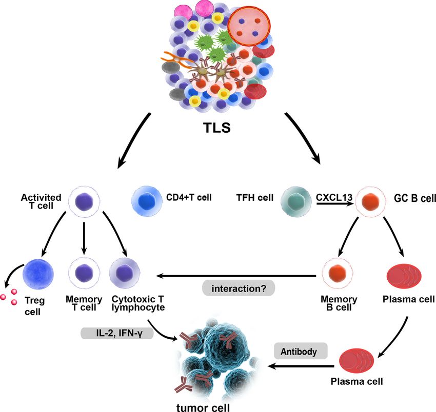
Kang et al.                                                                                                                        Tertiary Lymphoid Structure Induction

CD8+ T cells, and CD4+ T cells, etc. This concept is confirmed by                            review, we briefly summarize the development of TLSs and
the presence of intratumoral tertiary lymphoid structures (TLSs),                           focus on discussing the function of TLSs and multiple
which are well-organized tumor-infiltrating lymphocyte (TIL)                                 approaches that had been developed to induce TLS formation.
clusters and may generate an advanced immune response (3). As
is known to us, immunotherapy can utilize positive feedback to
activate the immune system and boost the infiltration of
endogenous T cells into tumors and subsequent destruction of                                DEVELOPMENT AND FORMATION
tumor cells (4–6). However, only 5%-30% of patients with                                    OF TLSS
malignancies exhibit activated intratumoral T cell immunity after
                                                                                            SLO development is a highly organized process that is initiated and
anti-programmed cell death protein-1 (PD-1)/programmed death-
                                                                                            continued during embryogenesis, which has similarities to TLS
ligand 1 (PD-L1) immunotherapy (7, 8). This failure is mainly due
                                                                                            formation and provides a classical model for understanding TLS
to the extensive immunosuppressive mechanisms in the TME that
                                                                                            development (33). However, there are some differences between the
lead to the decreased number and dysfunction of infiltrating T cells
                                                                                            canonical SLOs and TLSs. A chronic inflammatory state is sufficient
(9–11).
                                                                                            to induce TLS formation even in the absence of lymphoid tissue
    TLSs are ectopic lymphoid organs that can develop at sites of
                                                                                            inducer (LTi) cells (34), indicating that chronic inflammation may
chronic inflammation, such as those associated with infection
                                                                                            be an important factor that favors lymphoid neogenesis and
and autoimmunity, but also form within the TME (12, 13). TLSs
                                                                                            promotes TLS formation (35). Some studies have revealed that
share similar structural and functional characteristics with
                                                                                            DCs (36), T helper 17 (Th17) cells (37, 38), B cells (39), M1
secondary lymphoid organs (SLOs) (14). However, TLSs lack a
                                                                                            macrophages (40), and T follicular helper (TFH) cells (41) can
capsule and can form in various nonlymphoid tissues, such as
                                                                                            initiate TLS neogenesis in various pathological conditions. In
stroma and epithelium (15). The prognostic impact of TLSs has
                                                                                            addition, group 3 innate lymphoid cells (ILC3s) are associated
been widely explored and most reports have indicated that TLSs
                                                                                            with ectopic lymphoid aggregates (42). Tumor-infiltrating ILC3s
are associated with positive immunoreactivity and favorable
                                                                                            may interact with fibroblasts and lung tumor cells to facilitate
clinical outcomes in most types of cancers (12, 16–20). For
                                                                                            cytokine release, contributing to protective TLS formation (43).
example, TLSs are shown to be associated with relapse-free
                                                                                            Lymphotoxin (LT) signaling plays a vital role in TLS formation (44,
survival in patients with oral carcinoma or early-stage
                                                                                            45). The LTa1b2-LTb receptor (LTbR) interaction initiates
hepatocellular carcinoma (21, 22). Moreover, germinal centers
                                                                                            signaling that leads to the production of various chemokines and
(GCs) in TLSs may determine the prognostic value of TLSs (23,
                                                                                            adhesion molecules, such as CCL19, CCL21, CXCL13, and CXCL12
24). Even though, TLSs show an association with poor prognosis
                                                                                            (46). CCR7-expressing T cells are recruited by their homologous
in a minority of studies (25–27). It is urgent to comprehensively
                                                                                            ligands CCL21 and CCL19, and this recruited population forms a T
illustrate the function of TLSs in the TME.
                                                                                            cell zone. In addition, B cells can express CXCR5 and CXCR4 on
    SLOs, such as lymph nodes (LNs), provide three-dimensional
                                                                                            their surface to transmigrate to the follicle through the activity of the
structures for immune cells to optimize cell-cell interactions and
                                                                                            CXCL12-CXCR4 and CXCL13-CXCR5 axis (47, 48). These
produce an effective immune response (28, 29). Effector T (Teff)
                                                                                            findings show that the CCL19/CCL21-CCR7 and CXCL13-
cells are activated after being instructed by DCs, and migrate
                                                                                            CXCR5 axes are vital for regulating TLS development (49).
from external draining LNs into the tumor to exert their function
                                                                                            Although the knowledge of TLS formation mechanisms is widely
(30, 31). Increasing studies have shown that the antitumor
                                                                                            researched, the possible mechanisms and factors need to be further
immune response originates not only in LNs but also directly
                                                                                            explored in future studies. The main molecular and cellular
in TLSs (32). In general, the cells and molecules that regulate the
                                                                                            mechanisms of TLS formation are shown in Figure 1.
signaling underlying TLS formation and promote immune
responses within TLS remain to be further studied. In this

                                                                                            THE FUNCTION OF TLSS IN THE TME
Abbreviations: TLSs, tertiary lymphoid structures; TILs, tumor-infiltrating
lymphocytes; TME, tumor microenvironment; PD-1, programmed cell death                       Favorable Impact of TLSs on
protein-1; PD-L1, programmed death-ligand 1; SLOs, secondary lymphoid
organs; GCs, germinal centers; HEVs, high endothelial venules; LN, lymph                    Antitumor Properties
node; DCs, dendritic cells; Teff, effector T cell; FDCs, follicular DCs; PCs,               Mature TLSs are similar to SLOs, which contain T cell-rich areas
plasma cells; PNAd, peripheral node addressing; TFH cells, T follicular helper              with CD3+ T cells and dendritic cell (DC)-lysosomal associated
cells; Treg cells, regulatory T cells; LTi, lymphoid tissue inducer; LT, lymphotoxin;       membrane protein+ (DC-LAMP) mature DCs and follicular
LTbR, lymphotoxin-b receptor; IL, interleukin; ICB, immune checkpoint
blockade; APCs, antigen-presenting cells; TGF-b, transforming growth factor-b;
                                                                                            CD20+ B cell-rich zones (12). The B cell follicles in TLSs
TNFR, tumor necrosis factor receptor; TNF-a, tumor necrosis factor-a; ACT,                  comprise follicular dendritic cells (FDCs), B cells, plasmablasts,
adoptive cell transfer; VEGF, vascular endothelial growth factor; VTP, vascular             and TFH cells required for GC formation and B cell
targeting peptide; TLRs, Toll-like receptors; IFN-g, interferon-gamma; ILC3s,               differentiation (50). Macrophages, neutrophils, and regulatory
group 3 innate lymphoid cells; CTL, cytotoxic T lymphocyte; PLG, poly (lactide-             T (Treg) cells have been discovered in the TLSs of lung cancer,
coglycolide); TRAIL, TNF-related apoptosis-inducing ligand; MDSCs, myeloid-
derived suppressor cells; GM-CSF, granulocyte-macrophage colony-stimulating
                                                                                            pancreatic cancer, and ovarian cancer (51–54). TLSs are divided
factor; ICAM-1, intercellular cell adhesion molecule 1; VCAM1, vascular cell                into classical and nonclassical structures. Classical structures are
adhesion molecule 1; MADCAM1, mucosal addressable cell adhesion molecule 1.                 mature and contain T cells, DCs, B cells, and FDC compartments

Frontiers in Immunology | www.frontiersin.org                                           2                                       July 2021 | Volume 12 | Article 689270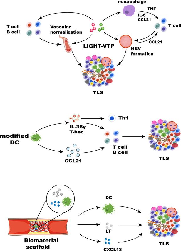
Kang et al. Tertiary Lymphoid Structure Induction
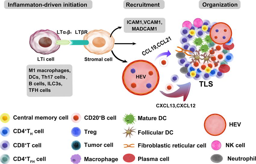
FIGURE 1 | Main molecular and cellular mechanisms of TLS formation in tumors. The development of TLSs is similar to that of SLOs. A chronic inflammatory state
is sufficient to induce TLS formation in the absence of lymphoid tissue inducer (LTi) cells. Many immune cells can be used as LTi cells, such as B cells, DCs, M1
macrophages, Th17 cells, ILC3s, and TFH cells. Immune and stromal cell cross-talk mediates TLS formation mainly through the binding of lymphotoxin (LT) ab and
LTbR, which can further release many chemokines (CXCL13, CXCL12, CCL21, and CCL19) and adhesion molecules (VCAM1, ICAM1, and MADCAM1). These
chemokines recruit lymphocytes from HEVs and form T and B cell zones. ILC3s, group 3 innate lymphoid cells; VCAM1, vascular cell adhesion molecule 1; ICAM1,
intercellular adhesion molecule 1; MADCAM1, mucosal addressable cell adhesion molecule 1.
and comprise more active components than nonclassical promote antitumor immunity, which indicates that TLSs may
structures, mainly B cells. Nonclassical TLSs usually contain B facilitate coordinated antitumor responses involving the
cells that are less activated than those in classical structures (14). combined actions of cytolytic T cells and PCs (58). B cells in
TLSs are distributed intratumorally and peritumorally and are TLSs are organized and highly differentiated and can produce
more abundant in the invasive margin than in the tumor core tumor-specific antibodies in adenocarcinomas and ovarian
(12). Intratumoral TLSs may have greater prognostic cancer (59). In omental metastases from ovarian cancer,
significance, but this has not been widely established. In some memory B lymphocytes essentially located within TLSs had
studies, intratumoral TLSs are a favorable prognosticator in higher clonality and somatic hypermutation rates, and they
pancreatic cancer and hepatocellular carcinoma (20, 21). One produced chemokines attracting DCs, T cells, and natural killer
study proposed a hypothesis to explain the better prognosis of (NK) cells. The density of B cells also correlated with that of
intratumoral TLSs. Tumors with, rather than without, mature DCs in the stroma of tumors (53). Recent studies have
intratumoral TLSs are less invasive, especially regarding blood shown the important role of TLSs and B cells in immunotherapy.
vessel invasion, and have a role related to the immune response. The frequencies of memory B cells, PCs, and GC-like B cells in
These tumors retain a relatively complete vascular network to the tumors of responders treated with immune checkpoint
transport immune cells and other molecules into the tumor and blockade (ICB) therapy are significantly higher than those in
initiate a more effective antitumor immune response (17). nonresponders. Increased B cell proliferation indicating GC
Increasing evidence shows that TLSs play an important role in activity and formation within TLSs has been observed (60).
controlling tumor invasion. Mature TLSs exhibit evidence for the High expression of B-lineage markers is associated with
formation of GCs (24) and GC B cells in TLSs are characterized improved prognosis and TLS formation in sarcoma (61).
by FDCs and Ki67+ proliferating B cells (51). Oligoclonal B cell B cells within TLSs can predict favorable prognosis in
responses have been identified in melanoma, which suggests an melanoma patients receiving ICB therapy. In addition, B cell-
active humoral antitumor response within TLSs that is driven by rich tumors are associated with elevated levels of initial and
B cells (55). High PC counts are associated with higher numbers memory T cells. T cells in tumors without TLSs possess a
of TLSs and B cells in breast cancer and neck carcinomas (56, dysfunctional molecular phenotype, which indicates that TLSs
57). PCs surrounded by TLSs are associated with the highest have a key role in the melanoma TME by conferring distinct T
levels of TILs and cytotoxicity-related gene products in ovarian cell phenotypes (62). In summary, these studies demonstrate a
cancer. This study showed that CD8+ TILs can predict prognosis major role of TLS-associated B cells in TLS function. B cells
only in combination with PCs, CD20+ TILs, and CD4+ TILs, probably act together with key immune constituents of TLSs by
suggesting that these four lymphocyte subsets work in concert to altering T cell activation and function. Memory B cells may act as
Frontiers in Immunology | www.frontiersin.org 3 July 2021 | Volume 12 | Article 689270Kang et al. Tertiary Lymphoid Structure Induction
antigen-presenting cells (APCs) to drive the expansion of both prognosis, indicating that antigen presentation allows local T
memory and naive T cell responses. B cells can also activate and cells to initiate responses to tumor-associated antigens in TLSs
recruit other immune effector cells by secreting an array of (67, 68). Whether TLS-associated DCs present tumor antigens
cytokines (60). The potential functions of TLSs in the TME are directly to CD8+ T cells or whether CD4+ Th cells participate in
shown in Figure 2. the production of CD8+ cytotoxic T cell responses in TLSs
The existence of TLSs at metastatic tumor sites is the key remains to be further studied. TFH cells produce CXCL13,
factor in the level of TILs, which directly determines the potentially resulting in the formation of TLSs to trigger the GC
antitumor effect (19). Moreover, the presence of TLSs led to B cell response (69). FDCs can also produce chemokines and
increased infiltration and activation of T cells and other immune cytokines involved in B cell proliferation and migration in LN,
cells and was associated with a good prognosis in liver cancer and such as interleukin (IL) -6 and CXCL13 (70). B cells produce
pancreatic carcinoma (21, 63, 64). There is evidence that TLSs LTa1b2, which has a crucial function in the differentiation of
can activate effector T cells in tumors (65). In MC38 tumors, FDCs within TLSs (71).
T cells from TLSs exhibited a largely enhanced baseline level of HEVs in TLSs are associated with T and B cell infiltration and
IFN-g (interferon-gamma) release. This finding revealed indicate favorable outcomes in oral carcinoma and breast cancer
successful antitumor T cell priming activity within induced (72, 73). The emergence of HEVs also contributes to the
TLSs, and TLSs may act as immune factories where T cells formation of TLSs (74). There is ample evidence that the
activate effector cells to mediate synergistic antitumor effects function of HEVs in TLSs is similar to that in LNs, providing
(66). Studies of lung and ovarian cancers showed that TLS- a channel for immune cells to accumulate in the tumor. HEVs in
associated DCs establish unique immune states characterized by TLSs express molecules similar to those expressed in LNs, such as
a strong T helper (Th) 1 orientation and facilitate a good CCL21 and peripheral node addressing (PNAd), and cells
FIGURE 2 | Potential functions of tertiary lymphoid structures in the TME. As in canonical SLOs, TLSs may constitute a critical site where specific T and B cells can
undergo terminal differentiation into effector cells. GC B cells can differentiate into memory B cells and plasma cells in TLSs, and fully differentiated B cells and exert
their antitumor effects. T cells can differentiate and expand, and they are activated as effector cells that exert cytotoxic effects. B cells and T cells in TLSs may interact
with each other and play a synergistic role, which needs to be confirmed by more studies. Treg cells within TLSs could exert a negative influence on the capacity of
TLSs to generate effector and memory lymphocytes.
Frontiers in Immunology | www.frontiersin.org 4 July 2021 | Volume 12 | Article 689270Kang et al. Tertiary Lymphoid Structure Induction
expressing CCR7 and L-selectin ligands for receptors on HEVs costimulatory levels, the immunosuppression of Treg cells to
are found in TLSs (75). PNAd expression indicates that HEVs DCs is relieved after Treg cell depletion, and the TLS
are essential for recruiting lymphocytes to lymphoid organs (76), microenvironment may become more immunostimulatory to
an event that orchestrates the extravasation of Lselectin+ and promote antitumor responses by T cells (54). The recruitment of
CCR7+ immune cells into TLSs (77, 78). LTab plays a key role in Treg cells and myeloid-derived suppressor cells (MDSCs) to
PNAd expression in HEV (79). Single-cell analysis revealed the lymphoid aggregates in mouse B16 melanomas expressing
heterogeneity of HEVs in LN. LTbR signaling and inflammation CCL21 was found to correlate with the promotion of tumor
also have crucial effects on HEV transcriptomes (80). However, growth (89). Therefore, TLS-associated Treg cells and MDSC
the study showed that HEV neogenesis is dependent on tumor presence may exert a negative influence on the capacity of TLSs
necrosis factor receptor (TNFR) rather than LTbR signaling in to generate effector and memory lymphocytes.
Treg-depleted tumors, suggesting another mechanism for HEV
formation. The expression of PNAd is not dependent on the
LTbR signal but is stimulated by activation of TNFR mediated by
LTa3 derived from CD8+ T cells (81). HEV formation is
DEVELOPMENT OF MULTIPLE
associated with increased T and B lymphocyte infiltration and APPROACHES TO INDUCE THE
activation in murine pancreatic cancer and breast cancer (82, 83). In TLS FORMATION
mouse models of melanoma and lung cancer, the LN-like
vasculature in tumors, characterized by the expression of PNAd A variety of LN modifications to improve the efficacy of tumor
and chemokine CCL21, induced by effector lymphocytes allows immunotherapy have been widely discussed and researched.
naive T cells to enter tumors and enhance antitumor immunity. Targeting LN can affect the efficacy of cancer vaccines, ICB
Vasculogenesis is regulated by a mechanism involving CD8+ T cells therapy, and adoptive cell transfer (ACT) at the cellular level.
that secrete IFN-g and LTa3 (84). In summary, T cells may Macroscopic biomaterials mimicking LN characteristics can be
contribute to the formation of the peripheral vasculature and HEV. used as immune niches for cell reprogramming and in vivo
transmission and can be used for preclinical testing of drugs and
The Adverse Impact of TLSs on Tumors vaccines in vitro at the tissue level (90). TLSs may be the first line
Nevertheless, a few studies have indicated that TLSs have a of T cell differentiation and expansion and are the key to
negative impact on prognosis in colorectal cancer and breast inducing intratumoral immune sensitization in situ. Therefore,
cancer (25–27). The studies showed that TLSs that develop in the similar principles can be used for developing strategies to induce
inflamed liver during hepatitis can function as a niche for tumor TLS formation, and a new antitumor immune strategy can be
progenitor cells in hepatocellular carcinoma and are associated constructed. Although biomaterials for transporting or recruiting
with an increased risk of late recurrence and decreased survival. APCs can mimic the cellular characteristics of SLOs, other
It can be postulated that TLSs, which persist in the liver and are strategies aim to induce TLS formation specifically, as observed
associated with a viral infection, play a different role than TLSs in situ. These strategies aim to mimic the chemokine and
induced by tumors (25). Lymphoid aggregates are associated inflammatory signals of the main molecular and cellular
with more advanced diseases and indicate an adverse prognosis mechanisms of TLS formation. In the next section, we discuss
in colorectal cancers. These structures form in association with strategies that induce TLS formation through the delivery of
more advanced tumors, suggesting that they are a reaction to chemokine-expressing cells or chemokines, implantation of
progressive tumor invasion, and their prognostic significance biomaterial scaffolds containing these inflammatory factors and
varies with disease progression and according to the inherent agents, and multiple therapeutic approaches.
immunogenicity of the tumor (26). TLSs are associated with
adverse prognosis in renal carcinoma with lung metastasis. TLSs Chemokines and Cytokines
are rarely found in lung metastasis of renal carcinoma, and A chemokine delivery strategy for TLSs provides a convenient
studies speculated that the presence of T cells may not be way to generate ectopic lymphoid tissue in tumors. Recent
educated in peritumoral TLSs but may reflect a chronic electronic screening techniques involving the identification of
inflammatory response, which is known to be harmful to the TLS-related chemokine genes that induce lymphocyte
host. At the same time, the high expression of vascular chemotaxis have offered a framework for a more effective
endothelial growth factor (VEGF) and IL-6 genes in renal design of TLSs (91, 92). A 12-chemokine gene signature also
carcinoma may also inhibit the differentiation of DCs, resulting provided a promising starting point for the potential
in an impaired T cell response and poor prognosis (85). TLS Treg construction of designed TLSs (93–96). In early studies,
cells are detected in breast and colorectal cancers (86, 87). The chemokines produced by lymphoid structures were expressed
decrease in the number of TLS Treg cells is associated with tumor in various ways, which led to the formation of lymphoid tissue
regression in metastatic prostate cancer (88). Treg cells are structures. For example, transgenic mice expressed B lymphocyte
present in TLSs in tumor-bearing lungs and exhibit activated chemokines in pancreatic islets, and the expression of B
phenotypes. Costimulatory ligand expression by DCs and T cell lymphocyte chemokines resulted in the formation of LN-like
proliferation rates increased in TLSs after Treg cell depletion, structures that included HEVs, interstitial cells, and B and T cell
enhancing the antitumor immune response. The reason may be zones and illustrated that the maintenance of B lymphocyte
that Treg cells in TLSs regulate DC function by reducing chemokine-induced lymphoid structures depends on LTbR
Frontiers in Immunology | www.frontiersin.org 5 July 2021 | Volume 12 | Article 689270Kang et al. Tertiary Lymphoid Structure Induction
signaling (97). CCL21 exhibits a stronger capacity than CCL19 to pathway of the specialized network, and remodeling reactive
induce more organized infiltrates in the islets of transgenic mice LNs (117). The LTbR signaling pathway plays a critical role in
(98). Intratumoral injection of CCL21 facilitated lymphocyte HEV differentiation and function in LN (44). Because of the
infiltration into pancreatic tumors (99), and targeting similarity between SLOs and TLSs, it is speculated that LTbR
lymphotoxin-a can induce lymphocyte infiltration and signaling is also involved in HEV differentiation and function in
lymphoid-like tissue formation in B16 melanoma (100). LN- TLSs. Further studies are required to understand the precise
like lymphocyte infiltration was also found in transgenic mice mechanisms by which HEV formation in TLSs is induced and
expressing CCL21 driven by the thyroglobulin promoter in the the effects of HEVs on different types of cancer. This knowledge
thyroid gland and transgenic pancreas (101, 102). Type I may guide the therapeutic objectives of cancer interventions.
interferon can also drive B cell recruitment by CXCR5– Other means can also be used to deliver LIGHT to tumor sites,
CXCL13 signaling and initiate ectopic GC formation within TLSs and the oncolytic activity of attenuated Salmonella Typhimurium
in pulmonary virus infection (103). In the salivary glands of adult was enhanced by the stable insertion of the gene encoding
mice, IL-7 regulates lymphatic vessel expansion and promotes the LIGHT. Attenuated S. Typhimurium expressing LIGHT
neogenesis of TLSs in the first phase, and LTbR signaling regulates inhibited the growth of primary tumors and the spread of lung
TLS neogenesis in the second phase (104). Th17 cytokines can metastasis (118). The findings suggest that avirulent bacteria can
regulate TLS development and function. For instance, IL-22 be used as targeted carriers for the local production of therapeutic
modulates CXCL12, CXCL13, and IL-23 production, contributing proteins in tumors. In recent years, the potential use of exosomes
to the formation of TLSs (105–107). In conclusion, these studies in the treatment and control of many diseases has expanded
show that many chemokines and cytokines involved in lymphoid because of their inherent characteristics in regulating complex
structure formation can be used as novel and feasible inducers in intracellular pathways. The characteristics of exosomes can also
combination with other stimulants and multiple methods to induce be exploited to induce TLSs. Exosomes are extracellular vesicles
the formation of TLSs. derived from endosomes and have a diameter of approximately
LIGHT, the 14th member of the tumor necrosis factor 40-160 nm. They can carry a variety of substances, such as
superfamily (TNFSF14), is a protein primarily expressed on proteins and DNA, to allow these substances to be absorbed by
activated T cells and immature DCs (108). LIGHT can function other cells (119, 120). Therefore, we hypothesize that exosomes
as both a soluble and cell surface-bound type II membrane can be used as carriers to load many chemokines and cytokines to
protein and interact with its two primary functional receptors: induce the formation of TLSs.
Herpes Virus Entry Mediator and LTbR (109). LIGHT can Toll-like receptors (TLRs) have also been researched concerning
interact with Herpes Virus Entry Mediator and deliver co- TLS formation. Myofibroblasts were stimulated with TLR agonists
stimulatory signals to T cells (44). LTbR is found on the surface and cytokines in giant cell arteritis, which upregulated B cell-
of epithelial, stromal, and myeloid cells (110). LIGHT-LTbR activating factor and CXCL13 and resulted in the formation of
signaling plays an important role in immune responses, TLSs (121). Inhalation of TLR9 agonists can generate profound
functioning to repair tumor vasculature and to support effector remodeling of tumor-bearing lungs and lead to TLS formation in
cells cell trafficking to and infiltration into tumors (111). Recently, adjacent tumors (122). In addition, both the anti-HBV response to
LTbR signal transduction induced by LIGHT has become a focus the TLR7 agonist GS-9620 and TLR4 agonists in mouse models of
of the investigation. When combined with an anti-VEGF myasthenia gravis can induce TLS generation (123, 124).
antibody, LIGHT can activate LTbR signaling and mediate Transforming growth factor-b (TGF-b) plays a noncanonical role
chemokine production to recruit T cells (112). In pancreatic in coordinating immune responses against ovarian cancer. CD8+ T
cancer, targeting LIGHT for homing to tumor vessels via a cells in the presence of TGF-b upregulate the secretion of CD103
vascular targeting peptide (VTP), LIGHT-VTP showed a dual and CXCL13, and CD8+ TILs play a role in mediating B cell
ability to induce TLS formation and regulate the angiogenic recruitment and TLS formation (125).
vasculature (83). LIGHT targeting to tumor vessels induces
vessel normalization, and HEVs and TLS formation may occur Cells
through a self-amplifying loop in pancreatic cancer. The An alternative approach to produce TLSs is to deliver cells that
mechanism may involve the LIGHT-triggered expression of express chemokines or to engineer chemokines that are associated
inflammatory cytokines in macrophages, such as IL-1b, IL-6, with lymphomagenesis. DC-based therapeutic strategies can be
CXCL13, TNF, and CCL21, These chemokines further recruit T used therapeutically to promote the extranodal priming of
cells. Macrophages and T cells have been deemed essential for antitumor immunity (126). DCs expressing T cell chemokines
HEV and TLS formation (83, 113) (Figure 3A). LIGHT-VTP in were injected into melanoma tumors, which yielded rapid T cell
combination with ICB therapy can produce intratumoral memory infiltration and initiation of intratumor responses (127).
T cells and Teff cells and improve prognosis (114). In addition, Additionally, intraperitoneal injection of murine DCs promoted
LIGHT-VTP combined with anti-VEGF and ICB therapy can the acute infiltration of immature T cells and NK cells into the TME,
increase the frequency of HEVs and normalize tumor vessels and an effect related to upregulated expression of NK and T cell
the accumulation of T cells in glioblastoma and lung metastases recruitment chemokines by murine DCs (128). DCs engineered
(115, 116). The LT-LIGHT axis provides key differentiation to overexpress T-bet suppressed the growth of sarcomas in vivo after
signals guiding the differentiation of the reticular network and intratumoral injection and prolonged the overall survival of mice
vascular system, maintaining the mesenchymal differentiation (126, 128). DCs promote LT signaling through LTbR for HEV
Frontiers in Immunology | www.frontiersin.org 6 July 2021 | Volume 12 | Article 689270Kang et al. Tertiary Lymphoid Structure Induction
A
B
C
FIGURE 3 | Strategies for therapeutic induction of TLS formation. TLS inducers, such as chemokines, cytokines, DCs, and therapeutic approaches, can induce TLS
formation in different ways. (A) Cytokines and chemokines involved in lymphogenesis, including LIGHT, CCL21, CXCL13, LT, and IFN-g, can lead to the formation of
TLSs. LIGHT-VTP targeting tumor vessels can induce vessel normalization, and HEV and TLS formation may occur through a self-amplifying loop. The mechanism
may be related to the LIGHT-triggered expression of the inflammatory cytokines IL-6, TNF, and CCL21 in macrophages. These chemokines can recruit T cells.
Macrophages and T cells play an important role in the formation of HEVs and TLSs (83). HEV formation and vascular normalization can also recruit more immune
cells. (B) Some immune cells, such as modified DCs and stromal cells, leading to the formation of TLSs. DCs engineered to secrete IL-36g also initiate therapeutic
TLS formation, which can upregulate the expression of T-bet. T-bet and IL-36g cooperate to reinforce their expression and recruit immune cells, leading to TLS
formation. DCs modified with the CCL21 gene can significantly increase T cell infiltration. Activated DCs can also upregulate many factors associated with TLS
formation. (C) Biomaterials can provide 3D scaffolds in situ and deliver cells and chemokines. Collagen scaffolds containing LT, CCL21, CXCL13, and activated DCs
were transplanted into mice. The recruited lymphocytes can form artificial lymph node structures.
differentiation and function in LN (129). DCs, which coordinate DCs (131). IL-36g is involved in polarizing type-1 immune
adaptive immune responses, have historically been a promising responses. It is a downstream target of the type-1 transactivator
target. DCs are a source of LT, and homeostatic chemokines T-bet and can induce T-bet expression in target cells (132). Research
(CXCL12, CXCL13, CCL19, and CCL21) are known to contribute has shown that IL-36g predominantly expressed by M1
to TLS formation in the lungs of influenza virus-infected mice. macrophages and vasculature cells, including smooth muscle cells
Similar to the depletion of DCs, blockade of LTbR signaling after and HEVs, mediates polarization toward a type 1 immune response.
virus clearance leads to the disintegration of TLSs and GC reactions. This pattern of IL-36g expression increased CD4+ central memory
It is suggested that the DC-mediated LTBR pathway contributes to T cell infiltrate and the density of B cells and led to TLS formation in
the formation of TLSs (36). Other methods have focused on human colorectal cancer (133). The injection of tumors with DCs
modifying and editing DCs to express the transcription factor T- engineered to secrete a bioactive form of mIL-36g also initiated
bet and secrete IL-36g to play a vital role. IL-36 cytokines are an IL-1 therapeutic TLS formation and slowed tumor progression in a
subfamily consisting of three agonists that signal through the mouse model of colorectal carcinoma. Furthermore, DC.IL-36g
common heterodimeric receptor IL-36R (130), which is expressed cells show strongly upregulated expression of T-bet, suggesting that
on endothelial cells and many immune cells, including T cells and T-bet and IL-36g cooperate to reinforce each other’s expression in
Frontiers in Immunology | www.frontiersin.org 7 July 2021 | Volume 12 | Article 689270Kang et al. Tertiary Lymphoid Structure Induction
DCs, rendering them competent to promote TLS formation in the support. Scaffolds are usually three-dimensional microporous
therapeutic TME (134) (Figure 3B). In lung cancer, autologous DCs structures designed to achieve cell encapsulation in vitro or cell
transduced with an adenoviral vector modified with the CCL21 gene penetration in vivo while providing mechanical support, cell
significantly reduced the tumor load and T cell infiltration (135), adhesion capability, and a continuous supply of biological cues to
accompanied by enhanced expression of IFN-g, IL-12, and CXCL10, promote cell migration and interaction (144). Biomaterial
as well as molecules related to reduced immunosuppression in the scaffolds can boost the efficacy of immunotherapies, such as
TME (136). Mice vaccinated with DCs charged with apoptotic/ cancer vaccines and ACT (145–147). For instance, biomaterials
necrotic B16 cells are protected against B16 challenge, and TLSs loaded with signaling molecules and engineered T cells have been
form at the vaccination site (137). In conclusion, DCs play an evaluated in vitro. These biomaterials were surgically implanted
important role in inducing TLS formation. These results provide a near the tumor or under a resected tumor bed, where they
framework for the usage of DCs. Promoting the expression of maintained continuous proliferation and release of specific T
multiple chemokines by targeting DCs is a valuable strategy to cells (148).
induce TLS formation. In early cases, collagen scaffolds containing both thymus-
LTi-like cells from newborn mouse LNs were injected derived stromal cells expressing LTa and activated DCs were
intradermally into adult mice and formed TLSs in the skin, and transplanted into the renal subcapsular space of mice. The
the results indicated that hyperactivated lymphocytes can fulfill the recruited lymphocytes formed artificial lymph nodes (ALN)
role of LTi cells during inflammatory responses (138). structures, which contained FDC, T cell, and B cell regions and
Subcutaneous injection of the LN-derived stromal cell line HEV-like structures. ALN induced a potent immune response in
resulted in the formation of TLSs that promote infiltration of vivo and the accumulation of memory and effector T and B cells.
immune cell subsets and inhibit tumor growth by improving the The engineered structures elicited a humoral response after
antitumor activity of TILs (66). Lymphoid tissue-like organoids vaccination and could be transplanted into immunodeficient
were constructed by transplantation of stromal cells embedded in mice to secrete antibodies after secondary immunization (139,
biocompatible scaffolds into the renal subcapsular space in mice. 149, 150) (Figure 3C). Based on this strategy, cell-free
The structure is similar to that of SLOs and contains clusters of B biomaterials have been explored. Hydrogels can provide a
and T cells and HEVs, DCs, and FDC networks (139). Other cells, controlled cellular microenvironment for immune cells so that
such as immune fibroblasts, bone marrow mesenchymal stem cells they can be recruited, expanded, and activated in vitro and in vivo
(MSCs), adipocytes, and macrophages, also play their roles. (151). Hydrogels can be used to deliver antigens, chemokines, and
Research on autoimmune conditions demonstrated that external other factors to DCs and induce T and B cell responses, and they can
triggers at mucosal sites can induce gradual differentiation of effectively encapsulate immunomodulators and immune cells. DCs
stromal cell populations into immune fibroblast networks, which can be activated in vitro in hydrogels before implantation and can be
supports the establishment of TLSs at an early stage. This process is recruited and activated in gels by immobilized stimulators, as in a
mediated mainly by paracrine and autocrine signals regulated by IL- bioreactor (152, 153). In another study, collagen sponge scaffolds
13. Once lymphocytes are recruited, the initial fibroblast network is embedded with sustained-release gel beads containing LTa1b2, and
expanded by local production of IL-22 and lymphotoxin. This many chemokines were transplanted into the subcapsular space of
finding demonstrates the role of immune fibroblasts in maintaining mice to establish ALN-like TLSs, recruiting memory T cells and B
TLS and supporting their formation and identifies new therapeutic cells and induced a strong antigen-specific immune response (154).
targets (140). Human MSCs stimulated with TNF-a and IL-1b A synthetic immune priming center consisting of an in situ cross-
significantly increased the expression of CCL19, VCAM1, ICAM1. linking hydrogel delivering chemokines and particles loaded with
Stimulated MSCs can induce CD4+ T cell proliferation. MSCs could DNA and siRNA can attract numerous DCs and can both generate a
play a key role as LTo cells in promoting the early inflammatory and strong transition to a T helper 1 response and increase the cytotoxic
initiating the formation of kidney-specific TLSs (141). Mucosa- T lymphocyte (CTL) response. The multimode injectable system
resident CXCL13+CX3CR1hi macrophages are responsible for can simultaneously deliver chemokines, DNA, and siRNA antigens
recruiting B cells and CD4+ T cells to sites of Salmonella invasion to DCs. This system constitutes a novel strategy to regulate
and subsequently activating them, resulting in TLS formation and a immunotherapy in situ and could provide an effective vaccine
local pathogen-specific IgA response (142). Recently, the strategy to prevent cancer (155).
combination of TNF-a and lipopolysaccharide was shown to Other biomaterials include polylactide-coglycolide (PLG),
directly induce adipocytes to produce TLS-related chemokines, nano-sapper, and nanoparticles. A macroporous PLG matrix was
thereby coordinating the formation of functional TLSs in the used to deliver granulocyte-macrophage colony-stimulating factor
mesentery affected by Crohn’s disease (143). In summary, these (GM-CSF), tumor antigens, and danger signals in vivo. GM-CSF
studies have further proved the various initiating factors and recruited DCs and significantly enhanced their homing to LNs, and
mechanisms of the formation of TLSs and provided more danger signals and cancer antigens further activated the recruited
references and insights for inducing the TLS formation. DCs. These materials elicited protective antitumor immunity and
showed prospects as cancer vaccines (156). A study demonstrated
Biomaterials improved immune function by targeting DCs with adjuvant vector
Biomaterials can support the formation of TLSs by locally and cells engineered from MKT cell ligands loaded with tumor antigen
controllably releasing chemokines and providing cellular mRNAs. This method also enhanced the local immune response via
Frontiers in Immunology | www.frontiersin.org 8 July 2021 | Volume 12 | Article 689270Kang et al. Tertiary Lymphoid Structure Induction
TLS formation (157). Importantly, these polymers may be designed and size reduction. Although their size tended to normalize after 2
to program the transport of various types of cells in vivo. Nano- weeks, the apoptotic rate remained high, suggesting active and
sapper was co-loaded with an antifibrotic protein and a plasmid continuous proliferation in residual irradiated cells and providing
expressing LIGHT. By normalizing the tumor vasculature, reducing them with a window to optimize their unique function (165). Low-
collagen deposition, and stimulating the expression of lymphocyte- dose stimulator of interferon genes (STING) agonist treatment can
recruiting chemokines, Nano-sapper induces TLS formation to upregulate the expression of various cytokines and increase the
promote CTL infiltration and remodel the TME (158). infiltration of T cells and DCs to establish a proinflammatory
Recognition of ectopic HEVs in human pancreatic ductal TME, which can also lead to normalization of the tumor
adenocarcinoma by engineered MECA79-coated nanoparticles vasculature, ultimately inducing the formation of TLSs and
can increase the transport of Taxol to the tumor and distinctly controlling tumor growth. Stimulating DC maturation and local
reduce tumor growth (159). Nanomaterials are promising for production of vascular normalization-promoting and TLS-
inducing TLS formation. Local delivery of engineered promoting factors, such as CCL19, CCL21, LTa, LTb, and LIGHT
biomaterials can play a role by establishing synthetic immune (166). A study showed that Treg elimination can activate CD8+ T cells
niches to enhance antitumor immunity. Immunotherapy based and promote the development of HEVs in tumors. The study
on biomaterials will facilitate the development of the next proposed a model in which a positive feedback loop of T cell
generation of tumor therapies. activation by Treg cell depletion can promote HEV development, T
cell infiltration, and tumor destruction (81). A prostate cancer study
Other Therapeutic Approaches showed that Treg cells and cyclooxygenase 2 are attractive therapeutic
Multiple cancer therapeutic strategies, such as cancer vaccines, targets that can be used to strengthen TLS-driven tumor immunity.
ICB therapy, antiangiogenic therapy, radiotherapy, and In particular, the existence of HEVs and lymphatic vessels suggests
chemotherapy, contribute to TLS formation. After therapeutic that TLSs can also be used as a platform for cell-based or
vaccination against human papillomavirus serotype 16 with E6/ cyclooxygenase 2 blockade therapy to control tumor growth (88).
E7 antigens, significant immune changes in the TME were PD-L1+, PD-L2+, LAG3+, and TIM3+ cells were detected in
observed in subjects with CIN2/3, and TLSs formed in the some breast cancer-related TLSs, and PD-1 was used as a marker
immune-infiltrated cervical tissues. At the molecular level, of T cell activity in both the T and B cell areas in TLSs. The
these histological changes in the matrix were characterized by expression levels of immune checkpoint molecules were
increased gene expression and associated with immune associated with the level of TILs and TLS formation (167). In a
activation (CXCR3) and effector function (T-bet and IFN-b) group of patients with renal carcinoma, the low expression of
(160). A prominent study of patients with resected pancreatic immune checkpoints and the localization of mature DCs in TLSs
cancer showed that 33 of the 39 patients treated with the GM- are associated with a better prognosis (168) (NCT03387761).
CSF vaccine exhibited TLS formation after 2 weeks. Further Recently, some prominent studies have shown that B cell and
analysis showed that these structures could regulate adaptive TLS formation promote the immunotherapy response in patients
immunity. Inhibition of the Treg signaling pathway and with melanoma and sarcoma after ICB therapy (60–62). In a
enhancement of the Th17 signaling pathway in TLS aggregates study of locoregionally advanced urothelial carcinoma, the
were associated with increased survival and intratumoral Teff: Treg formation of TLSs was observed in responding patients after
ratios and upregulation of the mechanism of immunosuppression treatment with combined CTLA-4 and PD-1 blockade therapy,
(161). The findings help to guide the production of the next which could be an effective preoperative treatment strategy (169).
generation of effective cancer vaccines and facilitate better Another study compared the metabolic, transcriptional, and
responses to ICB therapy. TLSs containing lymphocytes and APCs functional characteristics of intratumoral CD8+ T cell subtypes
appeared in all 11 patients who received cisplatin neoadjuvant with high, moderate, and no PD-1 expression from patients with
chemotherapy in a study on hepatoblastoma, indicating that non-small cell lung carcinoma. PD-1+ high T lymphocytes
cisplatin can induce TLS infiltration and synergistically induce the produce CXCL13, which mediates the recruitment of immune
death of immunogenic cells and trigger an antitumor immune cells to TLSs and has the potential to be predicted after treatment
response. This may involve so-called immunogenic cell death, a with PD-1 blockade therapy (170). Combination therapy with
controlled cell death process that produces damage-associated anti-VEGFR2 and anti-PD-L1 antibodies can induce HEV
molecular patterns that can be used as adjuvants to initiate an formation in pancreatic and breast cancer. LTbR signaling
immune response through the recruitment and activation of DCs plays an important role in the generation and activation of
(162). Administration of preoperative chemoradiotherapy tumor HEVs. HEV formation can increase the activity of
(neoadjuvant chemotherapy, NAC) was associated with increased CTLs, which makes tumors sensitive to ICB therapy (82). An
TLS formation and may affect the immunological composition of the anti-mouse LTbR agonistic antibody increased TIL infiltration in
TME and confer a favorable prognosis in patients with pancreatic a mouse model of colon cancer. Agonistic monoclonal antibodies
ductal adenocarcinoma (163). However, corticosteroid therapy targeting LTbR are a novel method for treating colorectal cancer
during chemotherapy impaired GC formation and reduced TLS and potentially other types of cancer (171). Considering that the
prognostic value in patients with lung cancer (164). After formation of TLSs is strongly related to the LTBR signaling
radiotherapy, apoptosis in tumors with TLSs increased pathway, targeting LTbR can also be used as an approach to
significantly. The TLSs also showed an acute increase in apoptosis induce TLS formation and enhance antitumor immunity.
Frontiers in Immunology | www.frontiersin.org 9 July 2021 | Volume 12 | Article 689270Kang et al. Tertiary Lymphoid Structure Induction
TLSs formation induced by multiple therapeutic strategies therapy deserves more research in the design of new
may involve a complex network of mechanisms, such as various immunotherapies, and a more in-depth understanding of the
types of cells, chemokines, and molecular mechanisms. We mechanisms in terms of the types of cytokines and chemokines
speculate that the main reason for TLS formation may be that leading to the formation of HEVs in different types of cancer is
the immune suppression in the TME is relieved after multiple needed. In the future, we need to focus on the combination of
therapeutic strategies, and the function of many immune cells methods inducing HEV and TLSs formation with new
can be restored. These cells interact with each other to activate therapeutic strategies that can alleviate immunosuppression,
LTbR signaling and other pathways and induce the production such as chemotherapies, radiotherapies, and ICB therapies.
of various chemokines and cytokines, which can ultimately lead These strategies may promote the formation of TLSs as well to
to the formation of TLSs. TLSs formation further enhances synergistically enhance adaptive immunity and provide insight
antitumor immunity, which may explain why the existence of into ultimately effective immune-mediated tumor control.
TLSs is related to a more favorable prognosis after therapy.
AUTHOR CONTRIBUTIONS
CONCLUSION AND FUTURE PERSPECTIVES Conceptualization: WK and ZH. Writing original draft: WK.
In summary, current research has revealed the significance of Supervision: ZF and PR. All authors contributed to the article
TLSs in tumor immunotherapy. TLSs may constitute a privileged and approved the submitted version.
niche for educating T cells and B cells, which can activate and
enhance immune responses. Although the major cellular and
molecular mechanisms of TLSs have been elucidated, how to FUNDING
utilize them as an important part of the immune-related cancer
control strategy is still being developed. Targeting the molecular This work was supported by funding from the National Natural
pathways of TLSs development to induce formation is a Science Foundation of China [grant numbers: 82071986,
promising immunotherapeutic strategy, which may directly 81771827] and the Province Natural Science Foundation of
enhance the antitumor response in situ. HEV induction Hunan [grant numbers: 2020JJ4855, 2020JJ4841].
REFERENCES 12. Sautès-Fridman C, Petitprez F, Calderaro J, Fridman WH. Tertiary
Lymphoid Structures in the Era of Cancer Immunotherapy. Nat Rev
1. Sautès-Fridman C, Cherfils-Vicini J, Damotte D, Fisson S, Fridman WH, Cancer (2019) 19(6):307–25. doi: 10.1038/s41568-019-0144-6
Cremer I, et al. Tumor Microenvironment is Multifaceted. Cancer 13. Di Caro G, Castino GF, Bergomas F, Cortese N, Chiriva-Internati M, Grizzi
Metastasis Rev (2011) 30(1):13–25. doi: 10.1007/s10555-011-9279-y F, et al. Tertiary Lymphoid Tissue in the Tumor Microenvironment: From
2. Maman S. Witz IP. A History of Exploring Cancer in Context. Nat Rev its Occurrence to Immunotherapeutic Implications. Int Rev Immunol (2015)
Cancer (2018) 18(6):359–76. doi: 10.1038/s41568-018-0006-7 34(2):123–33. doi: 10.3109/08830185.2015.1018416
3. Paijens ST, Vledder A, de Bruyn M, Nijman HW. Tumor-Infiltrating 14. Rodriguez AB, Engelhard VH. Insights Into Tumor-Associated Tertiary
Lymphocytes in the Immunotherapy Era. Cell Mol Immunol (2020) 18 Lymphoid Structures: Novel Targets for Antitumor Immunity and Cancer
(4):842–59. doi: 10.1038/s41423-020-00565-9 Immunotherapy. Cancer Immunol Res (2020) 8(11):1338–45. doi: 10.1158/
4. Schumacher TN, Schreiber RD. Neoantigens in Cancer Immunotherapy. Sci 2326-6066.CIR-20-0432
(New York NY) (2015) 348(6230):69–74. doi: 10.1126/science.aaa4971 15. Pimenta EM, Barnes BJ. Role of Tertiary Lymphoid Structures (TLS) in
5. Topalian SL, Weiner GJ, Pardoll DM. Cancer Immunotherapy Comes of Anti-Tumor Immunity: Potential Tumor-Induced Cytokines/Chemokines
Age. J Clin Oncol (2011) 29(36):4828–36. doi: 10.1200/JCO.2011.38.0899 That Regulate TLS Formation in Epithelial-Derived Cancers. Cancers (2014)
6. Wei SC, Levine JH, Cogdill AP, Zhao Y, Anang N-AAS, Andrews MC, et al. 6(2):969–97. doi: 10.3390/cancers6020969
Distinct Cellular Mechanisms Underlie Anti-CTLA-4 and Anti-PD-1 16. Munoz-Erazo L, Rhodes JL, Marion VC, Kemp RA. Tertiary Lymphoid
Checkpoint Blockade. Cell (2017) 170(6):1120–33.e17. doi: 10.1016/ Structures in Cancer - Considerations for Patient Prognosis. Cell Mol
j.cell.2017.07.024 Immunol (2020) 17(6):570–5. doi: 10.1038/s41423-020-0457-0
7. Christofi T, Baritaki S, Falzone L, Libra M, Zaravinos A. Current 17. Hiraoka N, Ino Y, Yamazaki-Itoh R. Tertiary Lymphoid Organs in Cancer
Perspectives in Cancer Immunotherapy. Cancers (2019) 11(10):1472. Tissues. Front Immunol (2016) 7:244. doi: 10.3389/fimmu.2016.00244
doi: 10.3390/cancers11101472 18. Li K, Guo Q, Zhang X, Dong X, Liu W, Zhang A, et al. Oral Cancer-
8. Feng Z, Rong P, Wang W. Meta-Analysis of the Efficacy and Safety of PD-1/ Associated Tertiary Lymphoid Structures: Gene Expression Profile and
PD-L1 Inhibitors Administered Alone or in Combination With Anti-VEGF Prognostic Value. Clin Exp Immunol (2020) 199(2):172–81. doi: 10.1111/
Agents in Advanced Hepatocellular Carcinoma. Gut (2020) 69(10):1904–6. cei.13389
doi: 10.1136/gutjnl-2019-320116 19. Lee M, Heo S-H, Song IH, Rajayi H, Park HS, Park IA, et al. Presence of
9. Devalaraja S, To TKJ, Folkert IW, Natesan R, Alam MZ, Li M, et al. Tumor- Tertiary Lymphoid Structures Determines the Level of Tumor-Infiltrating
Derived Retinoic Acid Regulates Intratumoral Monocyte Differentiation to Lymphocytes in Primary Breast Cancer and Metastasis. Mod Pathol (2019)
Promote Immune Suppression. Cell (2020) 180(6):1098–114.e16. 32(1):70–80. doi: 10.1038/s41379-018-0113-8
doi: 10.1016/j.cell.2020.02.042 20. Hiraoka N, Ino Y, Yamazaki-Itoh R, Kanai Y, Kosuge T, Shimada K.
10. Thommen DS. Schumacher TN. T Cell Dysfunction in Cancer. Cancer Cell Intratumoral Tertiary Lymphoid Organ Is a Favourable Prognosticator in
(2018) 33(4):547–62. doi: 10.1016/j.ccell.2018.03.012 Patients With Pancreatic Cancer. Br J Cancer (2015) 112(11):1782–90.
11. Togashi Y, Shitara K, Nishikawa H. Regulatory T Cells in Cancer doi: 10.1038/bjc.2015.145
Immunosuppression - Implications for Anticancer Therapy. Nat Rev Clin 21. Li H, Wang J, Liu H, Lan T, Xu L, Wang G, et al. Existence of Intratumoral
Oncol (2019) 16(6):356–71. doi: 10.1038/s41571-019-0175-7 Tertiary Lymphoid Structures Is Associated With Immune Cells Infiltration
Frontiers in Immunology | www.frontiersin.org 10 July 2021 | Volume 12 | Article 689270Kang et al. Tertiary Lymphoid Structure Induction
and Predicts Better Prognosis in Early-Stage Hepatocellular Carcinoma. Cells During Atherosclerosis-Related Lymphoid Neogenesis. Cardiovasc Res
Aging (Albany NY) (2020) 12(4):3451–72. doi: 10.18632/aging.102821 (2014) 101(3):434–43. doi: 10.1093/cvr/cvt263
22. Li Q, Liu X, Wang D, Wang Y, Lu H, Wen S, et al. Prognostic Value of 41. Jones GW, Jones SA. Ectopic Lymphoid Follicles: Inducible Centres for
Tertiary Lymphoid Structure and Tumour Infiltrating Lymphocytes in Oral Generating Antigen-Specific Immune Responses Within Tissues.
Squamous Cell Carcinoma. Int J Oral Sci (2020) 12(1):24. doi: 10.1038/ Immunology (2016) 147(2):141–51. doi: 10.1111/imm.12554
s41368-020-00092-3 42. Shikhagaie MM, Björklund ÅK, Mjösberg J, Erjefält JS, Cornelissen AS, Ros
23. Posch F, Silina K, Leibl S, Mündlein A, Moch H, Siebenhüner A, et al. XR, et al. Neuropilin-1 Is Expressed on Lymphoid Tissue Residing LTi-Like
Maturation of Tertiary Lymphoid Structures and Recurrence of Stage II and Group 3 Innate Lymphoid Cells and Associated With Ectopic Lymphoid
III Colorectal Cancer. Oncoimmunology (2018) 7(2):e1378844. doi: 10.1080/ Aggregates. Cell Rep (2017) 18(7):1761–73. doi: 10.1016/j.celrep.2017.01.063
2162402X.2017.1378844 43. Carrega P, Loiacono F, Di Carlo E, Scaramuccia A, Mora M, Conte R, et al. NCR
24. Siliņ a K, Soltermann A, Attar FM, Casanova R, Uckeley ZM, Thut H, et al. (+)ILC3 Concentrate in Human Lung Cancer and Associate With Intratumoral
Germinal Centers Determine the Prognostic Relevance of Tertiary Lymphoid Structures. Nat Commun (2015) 6:8280. doi: 10.1038/ncomms9280
Lymphoid Structures and Are Impaired by Corticosteroids in Lung 44. Tang H, Zhu M, Qiao J, Fu Y-X. Lymphotoxin Signalling in Tertiary
Squamous Cell Carcinoma. Cancer Res (2018) 78(5):1308–20. Lymphoid Structures and Immunotherapy. Cell Mol Immunol (2017) 14
doi: 10.1158/0008-5472.CAN-17-1987 (10):809–18. doi: 10.1038/cmi.2017.13
25. Finkin S, Yuan D, Stein I, Taniguchi K, Weber A, Unger K, et al. Ectopic 45. Wolf MJ, Seleznik GM, Zeller N, Heikenwalder M. The Unexpected Role of
Lymphoid Structures Function as Microniches for Tumor Progenitor Cells Lymphotoxin Beta Receptor Signaling in Carcinogenesis: From Lymphoid
in Hepatocellular Carcinoma. Nat Immunol (2015) 16(12):1235–44. Tissue Formation to Liver and Prostate Cancer Development. Oncogene
doi: 10.1038/ni.3290 (2010) 29(36):5006–18. doi: 10.1038/onc.2010.260
26. Bento DC, Jones E, Junaid S, Tull J, Williams GT, Godkin A, et al. High 46. Colbeck EJ, Ager A, Gallimore A, Jones GW. Tertiary Lymphoid Structures
Endothelial Venules are Rare in Colorectal Cancers But Accumulate in in Cancer: Drivers of Antitumor Immunity, Immunosuppression, or
Extra-Tumoral Areas With Disease Progression. Oncoimmunology (2015) 4 Bystander Sentinels in Disease? Front Immunol (2017) 8:1830.
(3):e974374. doi: 10.4161/2162402X.2014.974374 doi: 10.3389/fimmu.2017.01830
27. Figenschau SL, Fismen S, Fenton KA, Fenton C, Mortensen ES. Tertiary 47. Nerviani A, Pitzalis C. Role of Chemokines in Ectopic Lymphoid Structures
Lymphoid Structures Are Associated With Higher Tumor Grade in Primary Formation in Autoimmunity and Cancer. J Leukoc Biol (2018) 104(2):333–
Operable Breast Cancer Patients. BMC Cancer (2015) 15:101. doi: 10.1186/ 41. doi: 10.1002/JLB.3MR0218-062R
s12885-015-1116-1 48. Tokunaga R, Naseem M, Lo JH, Battaglin F, Soni S, Puccini A, et al. B Cell
28. Willard-Mack CL. Normal Structure, Function, and Histology of Lymph Nodes. and B Cell-Related Pathways for Novel Cancer Treatments. Cancer Treat
Toxicol Pathol (2006) 34(5):409–24. doi: 10.1080/01926230600867727 Rev (2019) 73:10–9. doi: 10.1016/j.ctrv.2018.12.001
29. Rosenberg SA. Cancer Immunotherapy Comes of Age. Nat Clin Pract Oncol 49. Schulz O, Hammerschmidt SI, Moschovakis GL, Förster R. Chemokines and
(2005) 2(3):115. doi: 10.1038/ncponc0101 Chemokine Receptors in Lymphoid Tissue Dynamics. Annu Rev Immunol
30. Menares E, Gá lvez-Cancino F, Cá ceres-Morgado P, Ghorani E, Ló pez E, (2016) 34:203–42. doi: 10.1146/annurev-immunol-041015-055649
Dı́az X, et al. Tissue-Resident Memory CD8 T Cells Amplify Anti-Tumor 50. Teillaud J-L, Dieu-Nosjean M-C. Tertiary Lymphoid Structures: An Anti-
Immunity by Triggering Antigen Spreading Through Dendritic Cells. Nat Tumor School for Adaptive Immune Cells and an Antibody Factory to Fight
Commun (2019) 10(1):4401. doi: 10.1038/s41467-019-12319-x Cancer? Front Immunol (2017) 8:830. doi: 10.3389/fimmu.2017.00830
31. Garris CS, Arlauckas SP, Kohler RH, Trefny MP, Garren S, Piot C, et al. 51. Germain C, Gnjatic S, Tamzalit F, Knockaert S, Remark R, Goc J, et al.
Successful Anti-PD-1 Cancer Immunotherapy Requires T Cell-Dendritic Presence of B Cells in Tertiary Lymphoid Structures is Associated With a
Cell Crosstalk Involving the Cytokines IFN-g and IL-12. Immunity (2018) 49 Protective Immunity in Patients With Lung Cancer. Am J Respir Crit Care
(6):1148–61.e7. doi: 10.1016/j.immuni.2018.09.024 Med (2014) 189(7):832–44. doi: 10.1164/rccm.201309-1611OC
32. Drayton DL, Liao S, Mounzer RH, Ruddle NH. Lymphoid Organ 52. Stromnes IM, Hulbert A, Pierce RH, Greenberg PD. Hingorani SR. T-Cell
Development: From Ontogeny to Neogenesis. Nat Immunol (2006) 7 Localization, Activation, and Clonal Expansion in Human Pancreatic Ductal
(4):344–53. doi: 10.1038/ni1330 Adenocarcinoma. Cancer Immunol Res (2017) 5(11):978–91. doi: 10.1158/
33. Jones GW, Hill DG, Jones SA. Understanding Immune Cells in Tertiary 2326-6066.CIR-16-0322
Lymphoid Organ Development: It Is All Starting to Come Together. Front 53. Montfort A, Pearce O, Maniati E, Vincent BG, Bixby L, Böhm S, et al. A
Immunol (2016) 7:401. doi: 10.3389/fimmu.2016.00401 Strong B-Cell Response Is Part of the Immune Landscape in Human High-
34. Furtado GC, Pacer ME, Bongers G, Bé né zech C, He Z, Chen L, et al. Tnfa- Grade Serous Ovarian Metastases. Clin Cancer Res (2017) 23(1):250–62.
Dependent Development of Lymphoid Tissue in the Absence of Rorgt⁺ doi: 10.1158/1078-0432.CCR-16-0081
Lymphoid Tissue Inducer Cells. Mucosal Immunol (2014) 7(3):602–14. 54. Joshi NS, Akama-Garren EH, Lu Y, Lee D-Y, Chang GP, Li A, et al.
doi: 10.1038/mi.2013.79 Regulatory T Cells in Tumor-Associated Tertiary Lymphoid Structures
35. Luo S, Zhu R, Yu T, Fan H, Hu Y, Mohanta SK, et al. Chronic Inflammation: Suppress Anti-Tumor T Cell Responses. Immunity (2015) 43(3):579–90.
A Common Promoter in Tertiary Lymphoid Organ Neogenesis. Front doi: 10.1016/j.immuni.2015.08.006
Immunol (2019) 10:2938. doi: 10.3389/fimmu.2019.02938 55. Selitsky SR, Mose LE, Smith CC, Chai S, Hoadley KA, Dittmer DP, et al.
36. GeurtsvanKessel CH, Willart MA, Bergen IM, van Rijt LS, Muskens F, Prognostic Value of B Cells in Cutaneous Melanoma. Genome Med (2019)
Elewaut D, et al. Dendritic Cells Are Crucial for Maintenance of Tertiary 11(1):36. doi: 10.1186/s13073-019-0647-5
Lymphoid Structures in the Lung of Influenza Virus-Infected Mice. J Exp 56. Seow DYB, Yeong JPS, Lim JX, Chia N, Lim JCT, Ong CCH, et al. Tertiary
Med (2009) 206(11):2339–49. doi: 10.1084/jem.20090410 Lymphoid Structures and Associated Plasma Cells Play an Important Role in
37. Peters A, Pitcher LA, Sullivan JM, Mitsdoerffer M, Acton SE, Franz B, et al. Th17 the Biology of Triple-Negative Breast Cancers. Breast Cancer Res Treat
Cells Induce Ectopic Lymphoid Follicles in Central Nervous System Tissue (2020) 180(2):369–77. doi: 10.1007/s10549-020-05548-y
Inflammation. Immunity (2011) 35(6):986–96. doi: 10.1016/j.immuni.2011.10.015 57. Lechner A, Schlößer HA, Thelen M, Wennhold K, Rothschild SI, Gilles R,
38. Grogan JL, Ouyang W. A Role for Th17 Cells in the Regulation of Tertiary et al. Tumor-Associated B Cells and Humoral Immune Response in Head
Lymphoid Follicles. Eur J Immunol (2012) 42(9):2255–62. doi: 10.1002/ and Neck Squamous Cell Carcinoma. Oncoimmunology (2019) 8
eji.201242656 (3):1535293. doi: 10.1080/2162402X.2018.1535293
39. Lochner M, Ohnmacht C, Presley L, Bruhns P, Si-Tahar M, Sawa S, et al. 58. Kroeger DR, Milne K, Nelson BH. Tumor-Infiltrating Plasma Cells Are
Microbiota-Induced Tertiary Lymphoid Tissues Aggravate Inflammatory Associated With Tertiary Lymphoid Structures, Cytolytic T-Cell Responses,
Disease in the Absence of RORgamma T and LTi Cells. J Exp Med (2011) 208 and Superior Prognosis in Ovarian Cancer. Clin Cancer Res (2016) 22
(1):125–34. doi: 10.1084/jem.20100052 (12):3005–15. doi: 10.1158/1078-0432.CCR-15-2762
40. Guedj K, Khallou-Laschet J, Clement M, Morvan M, Gaston A-T, Fornasa G, 59. Schlößer HA, Thelen M, Lechner A, Wennhold K, Garcia-Marquez MA,
et al. M1 Macrophages Act as Ltbr-Independent Lymphoid Tissue Inducer Rothschild SI, et al. B Cells in Esophago-Gastric Adenocarcinoma Are
Frontiers in Immunology | www.frontiersin.org 11 July 2021 | Volume 12 | Article 689270You can also read


