MOLECULAR PATHOGENESIS OF MERKEL CELL CARCINOMA - DECAPRIO ...
←
→
Page content transcription
If your browser does not render page correctly, please read the page content below
PM16CH03_DeCaprio ARjats.cls November 16, 2020 16:40
Annual Review of Pathology: Mechanisms of Disease
Molecular Pathogenesis of
Merkel Cell Carcinoma
James A. DeCaprio1,2,3
Annu. Rev. Pathol. Mech. Dis. 2021.16. Downloaded from www.annualreviews.org
1
Department of Medical Oncology, Dana-Farber Cancer Institute, Boston, Massachusetts
Access provided by Harvard University on 11/30/20. For personal use only.
02215, USA; email: james_decaprio@dfci.harvard.edu
2
Department of Medicine, Brigham and Women’s Hospital, Boston, Massachusetts 02115, USA
3
Department of Medicine, Harvard Medical School, Boston, Massachusetts 02115, USA
Annu. Rev. Pathol. Mech. Dis. 2021. 16:3.1–3.23 Keywords
The Annual Review of Pathology: Mechanisms of Disease
Merkel cell polyomavirus, neuroendocrine carcinoma, MYCL, ATOH1,
is online at pathol.annualreviews.org
LSD1, INSM1
https://doi.org/10.1146/annurev-pathmechdis-
012419-032817 Abstract
Copyright © 2021 by Annual Reviews.
Merkel cell carcinoma (MCC) is an aggressive neuroendocrine carcinoma of
All rights reserved
the skin with two distinct etiologies. Clonal integration of Merkel cell poly-
omavirus DNA into the tumor genome with persistent expression of viral
T antigens causes at least 60% of all MCC. UV damage leading to highly
mutated genomes causes a nonviral form of MCC. Despite these distinct
etiologies, both forms of MCC are similar in presentation, prognosis, and
response to therapy. At least three oncogenic transcriptional programs fea-
ture prominently in both forms of MCC driven by the virus or by mutation.
Both forms of MCC have a high proliferative growth rate with increased
levels of cell cycle–dependent genes due to inactivation of the tumor sup-
pressors RB and p53, a strong MYC signature due to MYCL activation by
the virus or gene amplification, and an attenuated neuroendocrine differen-
tiation program driven by the ATOH1 transcription factor.
·.•�-
3.1
, Review in Advance first posted on
November 23, 2020. (Changes may
still occur before final publication.)
PM16CH03_DeCaprio ARjats.cls November 16, 2020 16:40
MERKEL CELLS
Research into the development, physiology, and molecular features of normal Merkel cells has
MCC: Merkel cell helped guide our understanding of Merkel cell carcinoma (MCC) tumor biology. Merkel cells
carcinoma were first described in 1875 by the German anatomist Friedrich Sigmund Merkel as Tastzellen or
Atonal bHLH touch cells (1). He found these cells to be in close association with nerve endings and proposed that
transcription factor 1 they function as mechanoreceptors. Merkel cells are typically located in the basal layer of the skin
(ATOH1): epithelium and surround hair follicles. Clusters of Merkel cells also form touch domes, specialized
a transcription factor structures present in areas of highly sensitive skin. Merkel cells make synaptic connections to
required for Merkel
slowly adapting type I afferent sensory nerves (1).
cell development
Lineage tracing in mice identified a series of transcription factors essential for the development
of normal Merkel cells. The ATOH1 (atonal bHLH transcription factor 1) gene is required for
the development of Merkel cells (2). Conditional knockout of ATOH1 using a Cre deletor strain
driven by the epidermal cytokeratin 14 (KRT14, CK14) promoter eliminated the development
Annu. Rev. Pathol. Mech. Dis. 2021.16. Downloaded from www.annualreviews.org
of Merkel cells. In contrast, Cre driven by the neural crest–specific WNT1 promoter had no
Access provided by Harvard University on 11/30/20. For personal use only.
impact on the number or location of Merkel cells (3). This important experiment indicated that
normal Merkel cells are likely derived from an epidermal skin precursor and not from the neural
crest. SOX2 and ISL1 (Islet1) form a heterodimeric transcription factor that cooperates to sustain
ATOH1 expression (4, 5).
Merkel cells express cytokeratins CK8, CK18, CK19, and CK20, characteristic of epidermal
cells. They also express several markers common to neuroendocrine cells, including neuron-
specific enolase, synaptophysin, chromogranin A, CD56, and INSM1 (Insulinoma1) (1, 6). In ad-
dition to these epithelial and neuroendocrine markers, Merkel cells express PIEZO2, which func-
tions as a mechanically activated ion channel that can detect and convert mechanical stimuli into
electrical signals (7). PIEZO2 is required for the Merkel cell touch sensation (8). Recent structural
analysis of the homologous PIEZO1 revealed a homotrimer that assembles into a three-bladed
propeller-shaped pattern that can flex in response to mechanical force (9). Given the overall ho-
mology to PIEZO1, it is likely that PIEZO2 forms a similar three-propeller structure that serves
to transmit gentle touch pressure in the Merkel cell to the neural synapse (10).
Several signaling pathways have been described to influence Merkel cell development. Notch
signaling antagonizes ATOH1 signaling by activating the HES family of the helix-loop-helix fam-
ily of DNA binding proteins that competes with ATOH1 for specific binding to DNA. Knockout
of HES1 or the essential Notch coactivator RBPJ results in increased numbers of Merkel cells
(11). Polycomb repressive complex 1 (PRC1) and PRC2 repress the development of Merkel cells
in neonatal skin, and loss of PRC1 catalytic activity also results in an increased number of Merkel
cells (12, 13). Hedgehog and bone morphogenetic protein signaling contribute to Merkel cell
development, particularly during formation of the touch dome and hair follicle (14–16).
MCC
MCC was first described in 1972 by Cyril Toker (17) as a trabecular carcinoma of the skin with
carcinoid features. Later, he reported the presence of neurosecretory granules—membrane-bound
granules containing dense cores—within the tumor cells. This feature is indistinguishable from
tumor cells of neural crest origin and is also present in normal Merkel cells (18). The tumor name
was changed to MCC to reflect the similarity in appearance of tumor cells to Merkel cells (19, 20).
MCC is an aggressive neuroendocrine carcinoma of the skin that frequently metastasizes to
draining lymph nodes and distant organs, including liver, bone, pancreas, lung, and brain (21).
MCC typically presents as a rapidly growing, erythematous lesion in the dermal layer of the skin.
·.•�-
The most common presentation of MCC is in older, fair-skinned adults with a lifelong history of
3.2 DeCaprio
, Review in Advance first posted on
November 23, 2020. (Changes may
still occur before final publication.)
PM16CH03_DeCaprio        ARjats.cls   November 16, 2020      16:40

                                                                                          intense UV exposure from the sun. MCC occurs less frequently in non-sun-exposed skin as well as
                                                                                          in children, young adults, and dark-skinned persons. Residence in a latitude closer to the equator
                                                                                          is associated with increased incidence of MCC in North American men, but not women, possibly
                                                                                          due to occupational sunlight exposure patterns (22). Risk for developing MCC is also increased in
                                                                                          patients with severely immunocompromising conditions, including HIV/AIDS, or from medical
                                                                                          treatment of autoimmune diseases, solid organ transplantation, and other types of cancers (23).
                                                                                          The AEIOU mnemonic accounts for 90% of all MCC presentation: asymptomatic/lack of ten-
                                                                                          derness, expanding rapidly, immune suppression, older than 50 years, and UV-exposed/fair skin
                                                                                          (24).
                                                                                               The most recent MCC staging system from the American Joint Committee on Cancer esti-
                                                                                          mates a 5-year overall survival rate of 51% for local disease, 35% for nodal involvement, and 14%
                                                                                          for metastatic disease (25, 26). Surgery and radiation therapy can be curative for local and nodal
                                                                                          MCC, but systemic therapy is usually required for extensive, metastatic, and recurrent disease.
Annu. Rev. Pathol. Mech. Dis. 2021.16. Downloaded from www.annualreviews.org

                                                                                          Cytotoxic chemotherapy, based on cisplatin and etoposide regimens, has a high response rate but
                                                                                          is limited by a short duration with a mean progression-free survival of just 94 days (27). A revolu-
   Access provided by Harvard University on 11/30/20. For personal use only.

                                                                                          tion in MCC care began recently when it was determined that checkpoint blockade therapy with
                                                                                          antibodies to PD-1 or PD-L1 could induce frequent and durable responses (28–31). Predictions
                                                                                          for overall survival may improve as experience with checkpoint blockade therapy increases.

                                                                                          HISTOLOGY OF MCC
                                                                                          MCC can vary from a pure neuroendocrine histology to a variant form with mixed histologic
                                                                                          features. High-grade neuroendocrine MCC cells have a high nuclear to cytoplasmic ratio with
                                                                                          scant cytoplasm, giving them the appearance of small blue cell tumors when stained by hema-
                                                                                          toxylin and eosin. The tumor nuclei have an open, pepper-and-salt-appearing chromatin pattern
                                                                                          with frequent mitotic figures indicative of a high proliferative rate (Figure 1). Immunohistochem-
                                                                                          istry (IHC) staining of MCC for neuroendocrine markers is typically positive for chromogranin,
                                                                                          synaptophysin, CD56, and neurofilament. MCC also stains specifically for CK20, which typically
                                                                                          shows a paranuclear dot-like pattern. In contrast, CK20 staining in normal Merkel cells is more
                                                                                          uniformly distributed throughout the cytoplasm. CK20 staining can distinguish MCC from other
                                                                                          more common neuroendocrine tumors such as small cell lung carcinoma (SCLC) (32). SCLC
                                                                                          stains positive for TTF-1 (thyroid-specific transcription factor 1, encoded by the NKX2-1 gene),
                                                                                          while MCC is negative for this stain. INSM1 has recently emerged as a useful IHC marker for
                                                                                          MCC and Merkel cells as well as for other neuroendocrine carcinomas (6).
                                                                                              There are several reports of combined MCC tumors with a mixed histology showing a neu-
                                                                                          roendocrine component and other tumor types such as squamous cell carcinoma. There have been
                                                                                          suggestions that these tumors represent collision tumors from two separate origins. However, sev-
                                                                                          eral reports demonstrate that certain mutations and histologic markers are shared between the two
                                                                                          components, which suggests that the MCC tumor developed from a preexisting neoplasm (33, 34).

                                                                                          POLYOMAVIRUS-POSITIVE MCC COMPARED TO NONVIRAL MCC
                                                                                          A pathogenic cause for MCC was first suspected when it was recognized that the incidence of
                                                                                          MCC was increased more than tenfold in people living with HIV-1/AIDS compared to the gen-
                                                                                          eral population (35). The risk for developing MCC is also increased in patients with medically
                                                                                          induced immunosuppression for autoimmune conditions such as rheumatoid arthritis and solid
                                                                                          organ transplantation (36). Recognizing the increased risk for developing MCC by immunosup-

                                                                                      ·.•�-
                                                                                          pression, Huichen Feng and Masahiro Shuda in the laboratory of Yuan Chang and Patrick Moore

                                                                                                                                         www.annualreviews.org   •   Pathogenesis of Merkel Cell Carcinoma   3.3

                                                                                      ,          Review in Advance first posted on
                                                                                                 November 23, 2020. (Changes may
                                                                                                 still occur before final publication.)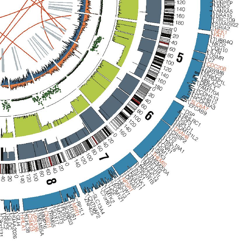
PM16CH03_DeCaprio ARjats.cls November 16, 2020 16:40
a 10 mm b 250 µm
Annu. Rev. Pathol. Mech. Dis. 2021.16. Downloaded from www.annualreviews.org
Access provided by Harvard University on 11/30/20. For personal use only.
c 1 mm d 500 µm
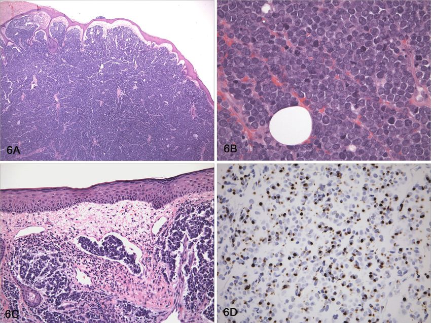
Figure 1
Merkel cell carcinoma. (a) A predominantly dermal nodule is shown consisting of small round blue cells. (b) The cells have a high
nuclear to cytoplasmic ratio and pale chromatin with nuclear molding. (c) Lymphovascular invasion (arrow) is a common feature.
(d) Perinuclear dot-like reactivity is shown for CK20 (hematoxylin and eosin stain). Figure adapted from Reference 128.
began a search for a pathogenic cause for MCC (37). They performed whole transcriptome se-
MCPyV: Merkel cell
polyomavirus quencing and searched for pathogens by first subtracting all human genes from their analysis. In
the remaining unmapped sequences, novel transcripts distantly related to polyomaviruses were
Merkel cell
detected. Complete sequencing of the viral genome in MCC tumors led to the determination
carcinoma, nonviral
(MCCN): often that it corresponded to a new human polyomavirus called Merkel cell polyomavirus (MCPyV or
contains a high tumor MCV) (37). MCPyV viral DNA was shown to be integrated into the tumor genome by Southern
mutational burden blotting in 8 of 10 tested MCC tumors. Evidence that MCPyV DNA was likely causative or at
with a strong UV least an early event in MCC tumorigenesis was implied by an identical restriction fragment length
mutational signature
polymorphism pattern observed in a primary skin tumor and a metastatic lymph node from the
Tumor mutational same patient.
burden (TMB): Additional sequencing studies of MCC tumors revealed significant differences in MCC tumors
somatic mutations per
containing MCPyV and those without a virus. Nonviral MCC (MCCN) tumors have a predom-
megabase; high TMB
gives rise to inant UV mutational signature with a very high tumor mutational burden (TMB), often greater
neoantigens than 20 somatic mutations per megabase (38–40). In contrast, polyomavirus-associated MCC
(MCCP) tumors have a low TMB of 6 or less, without a UV mutational signature (41). Whole
MCCP: Merkel cell
carcinoma, genome sequencing revealed evidence for a highly damaged genome in MCCN with many somatic
polyomavirus- single nucleotide variants, copy number alterations, and translocations. In contrast, MCCP tumors
·.•�-
associated typically have near-normal diploid genomes with few somatic mutations (Figure 2) (42, 43).
3.4 DeCaprio
, Review in Advance first posted on
November 23, 2020. (Changes may
still occur before final publication.)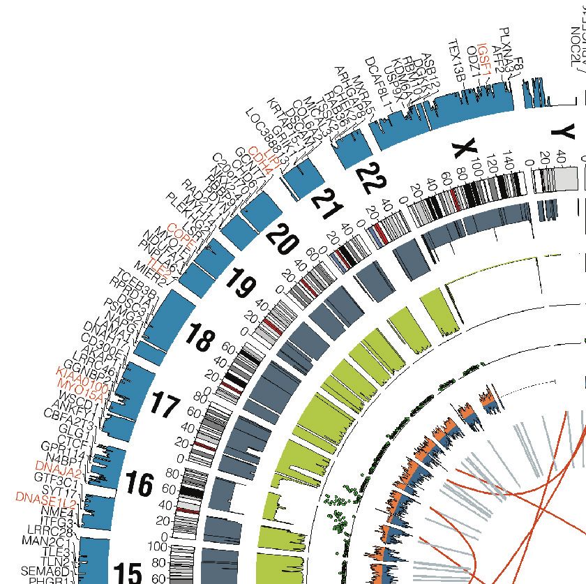
PM16CH03_DeCaprio ARjats.cls November 16, 2020 16:40
a
Annu. Rev. Pathol. Mech. Dis. 2021.16. Downloaded from www.annualreviews.org
Access provided by Harvard University on 11/30/20. For personal use only.
Chromosome
Lesser number
Density of
allele Karyotype somatic variants
Density of fraction
heterozygous (LAF)
SNPs
0
Density of 20
homozygous 40 UTP3
SNPs 60
80
100
4
120
140
160 SPOCK3
b 180 STOX2
Somatic Loss of Called Gene symbols
junctions heterozygosity level for impacted
(LOH) genes
Chromosome
position (Mb)
(Caption appears on following page)
·.•�-
www.annualreviews.org • Pathogenesis of Merkel Cell Carcinoma 3.5
, Review in Advance first posted on
November 23, 2020. (Changes may
still occur before final publication.)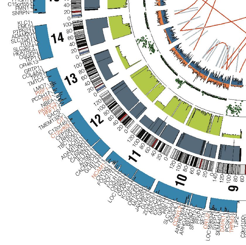
PM16CH03_DeCaprio             ARjats.cls        November 16, 2020   16:40

                                                                                     Figure 2 (Figure appears on preceding page)
                                                                                     Circos plots and functional annotation of genomic alterations in Merkel cell carcinoma tumors. The MCPyV-negative tumor (a) and
                                                                                     MCPyV-positive tumor (b) show large differences in the number and type of mutations. Abbreviations: Mb, megabase; MCPyV, Merkel
                                                                                     cell polyomavirus; SNP, single-nucleotide polymorphism. Figure adapted from Reference 42 (CC BY).

                                                                                                                          Commonly mutated genes in MCCN include loss-of-function mutations in the tumor sup-
                                                                                     KMT2C: lysine                    pressor genes RB1 and TP53 (Figure 3). Both TP53 and RB1 are usually wild-type in MCCP,
                                                                                     methyl transferase 2C,           but inactivating mutations have been reported (44, 45). Loss-of-function mutations in NOTCH1,
                                                                                     MLL3                             KMT2C, and KMT2D are also frequently observed in MCCN (41, 43). PI3K signaling is likely
                                                                                     KMT2D: lysine                    activated in both MCCP and MCCN with activating mutations present in PIK3CA or loss of the
                                                                                     methyl transferase 2D,           negative regulators PTEN, TSC1, and TSC2 (39, 46).
                                                                                     MLL4                                 In addition to loss of tumor suppressors, amplification of the MYC paralog MYCL (MYCL1
                                                                                     MYCL: L-MYC;                     or L-MYC) is frequently observed in MCCN (43, 47). MYCL was first described in a subset of
                                                                                     paralog of MYC and               SCLC (48). Amplification of MYC, MYCN, and MYCL occurs frequently in SCLC in a mutu-
Annu. Rev. Pathol. Mech. Dis. 2021.16. Downloaded from www.annualreviews.org

                                                                                     MYCN;                            ally exclusive pattern, which indicates that these paralogs likely provide overlapping oncogenic
   Access provided by Harvard University on 11/30/20. For personal use only.

                                                                                     heterodimerizes with
                                                                                                                      functions (49). MYCL is not required for normal mouse development, although it is expressed in
                                                                                     MAX
                                                                                                                      developing kidney, lung, and brain (50). More recently, MYCL has been shown to be required for
                                                                                                                      the development of the Batf3-dependent subset of classical dendritic (cDC1) cells (51, 52).

                                                                                      TMB status       100
                                                                                      (mutations        50
                                                                                                         0
                                                                                      per Mb)
                                                                                                                                                      High (≥20 mutations/Mb)   Intermediate (6−19 mutations/Mb)    Low (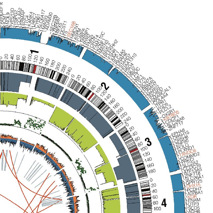
PM16CH03_DeCaprio ARjats.cls November 16, 2020 16:40
MERKEL CELL POLYOMAVIRUS
MCPyV was the fifth human polyomavirus to be identified. There are now 13 known human poly-
omaviruses. MCPyV DNA can be readily detected on the skin of healthy individuals, although the LT: large T antigen
specific skin cells that support viral replication in vivo are not known. Two additional human poly-
ST: small T antigen
omaviruses, HPyV6 and HPyV7, were identified from the skin or hair follicles of healthy adults
using a technique called rolling circle amplification, which takes advantage of the small circular na-
ture of the polyomavirus double-stranded DNA (dsDNA) genome (53). In immunocompromised
conditions, HPyV6 and HPyV7 can replicate and cause a hyperkeratotic skin condition charac-
terized by pruritic and brown plaques with epidermal hyperplasia and virus-laden keratinocytes
(54, 55). The condition is called HPyV6- and HPyV7-associated pruritic and dyskeratotic
dermatoses.
Rolling circle amplification identified a fourth human polyomavirus associated with the skin
and was detected in a patient with a rare skin disease named trichodysplasia spinulosa (56). In
Annu. Rev. Pathol. Mech. Dis. 2021.16. Downloaded from www.annualreviews.org
this condition, trichodysplasia spinulosa–associated polyomavirus (TSPyV) replicates in the inner
Access provided by Harvard University on 11/30/20. For personal use only.
root sheath of the hair follicle, destroying the normal hair structure and leading to alopecia and
folliculitis. Given that these four human polyomaviruses can be isolated from the skin and TSPyV
can replicate in hair follicles, while HPyV6 and HPyV7 replicate in keratinocytes, it is likely that
MCPyV can also replicate in one or both of these cell types. Of note, an image was reported
of scalp folliculitis that presented in a double-lung transplant recipient that was immunostained
with antibodies to MCPyV large T antigen (LT), suggesting that MCPyV could replicate in hair
follicles and cause destruction in a manner similar to TSPyV (57). It should be noted that MCC
typically presents on hairy skin, which is consistent with the idea that MCPyV may replicate in
hair follicles. Although there have been reports of MCC presenting in mucosal tissues, most cases
of mucosal MCC appear to be nonviral. However, at least some MCC tumors of the nasopharynx
may contain MCPyV (58, 59).
The MCPyV dsDNA circular genome is approximately 5.4 kb and can be divided into three
regions: The early viral gene region (EVGR) encodes genes that are expressed prior to the onset of
viral DNA replication; the late viral gene region (LVGR) encodes genes expressed after viral DNA
replication commences; and the regulatory region, called the noncoding control region (NCCR),
contains the viral origin of replication and the promoters and enhancers that drive expression of
the early and late viral genes.
The EVGR encodes LT, a spliced form of LT called 57kT, and small T antigen (ST). In addi-
tion, ALTO is encoded in an alternative open reading frame from LT (60). The LVGR encodes the
viral coat proteins VP1 and VP2 as well as a microRNA that regulates T antigen levels. The poly-
omavirus virion is composed of 72 pentamers of VP1, with each pentamer lined by one molecule
of VP2 on the inner surface (61). When expressed in bacteria or yeast, VP1 will spontaneously
form pentamers and assemble into virus-like particles that can serve as a useful capture antigen to
detect antibodies in serum indicative of prior infection (62, 63). Based on the VP1 serology assay,
infection with MCPyV occurs as early as several months of age and increases in frequency until
adulthood, when 70–90% of all adults show evidence for persistent infection (64–66).
While antibodies to MCPyV VP1 are widespread in the general population, antibodies to LT
and ST are present in less than 1% of healthy individuals. In contrast, antibodies to MCPyV ST
and LT can be detected in at least half of patients with MCCP (67). When present, antibody titers
to the T antigens can decrease upon successful treatment of MCCP and can be used as a biomarker
to follow disease status (67). Of note, MCC patients often have higher titers of antibodies to
VP1 than do normal healthy individuals, although the significance of this observation is unclear
(68).
·.•�-
www.annualreviews.org • Pathogenesis of Merkel Cell Carcinoma 3.7
, Review in Advance first posted on
November 23, 2020. (Changes may
still occur before final publication.)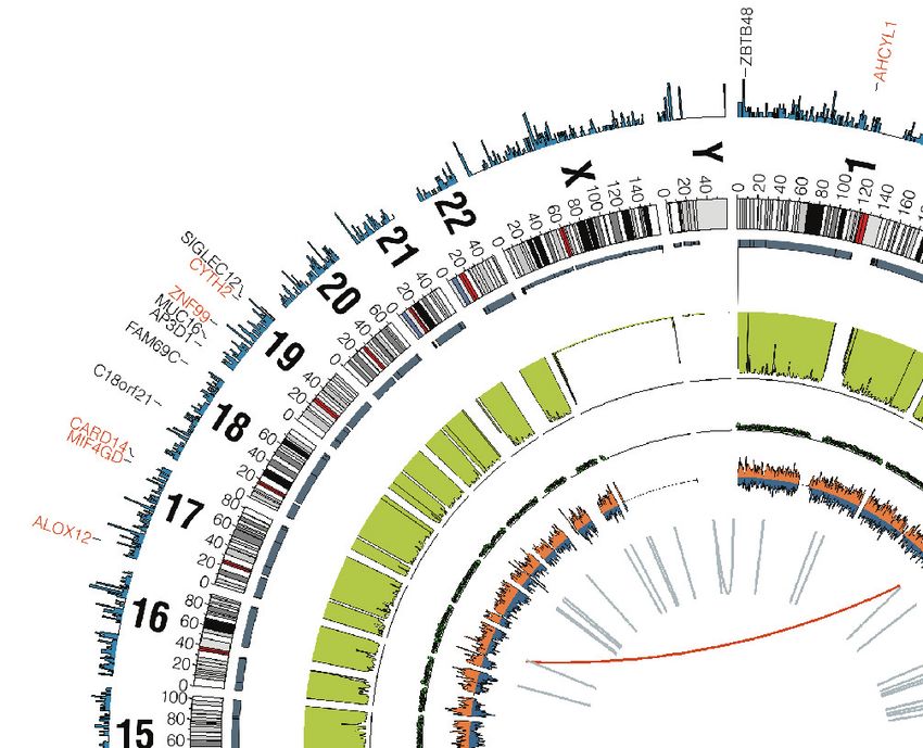
PM16CH03_DeCaprio ARjats.cls November 16, 2020 16:40
a b
LT LT VP1
NCCR
ST VP2
0-439
0-876
0-708
0-186
0-1967
0-981
0-1492
0-462
Annu. Rev. Pathol. Mech. Dis. 2021.16. Downloaded from www.annualreviews.org
Access provided by Harvard University on 11/30/20. For personal use only.
c d
8 A20S
8 A20S
6
Count
7
4
6
S
1Y
00
2
N1
H4
5
F
L
54
93
Count
0
S2
S2
4 DnaJ
L3 K P3 300 94V
P3 5N30403S L
3
9G
P A2
F
44
D1 142
V
R
65
Q A
S
H1 161 H
L4
2
P1 68 L
P 42
N
D7 E
V
C
R2235 Y
L6 R
7 Y
S
L6 24P
D203F
A480F
1Y
E4 16R
3 C
74Y
S
26
52
I
T6 0I
T3 6S
95
82
S 11
00
29
S2 7T
41
I
S21S
P263
28
22
0
8
H4
Q7
1
S3
T5
S4
T5
P4
1
0
DnaJ OBD Core helicase/ATPase
LXCXE NLS
Figure 4
MCPyV integration. (a) MCPyV coverage and mutations from virus-positive MCC cases. Read coverage for MCPyV is shown in gray,
and each plot represents a single patient. Scales for the coverage plots are set from 0 to the maximum read coverage per patient. The
illustration above indicates the relative position within the MCPyV genome. Limits of the minimal conserved region in MCCP tumors
are indicated by dotted vertical lines. (b) Representative assembly graph of a partially duplicated MCPyV genome integrated into the
tumor genome. The path for linearization of the assembly graph is shown by the dark gray line with arrowheads. (c,d) Residue changes
in MCPyV LT and ST, respectively. Lollipop plots of LT and ST show missense mutations relative to the MCPyV reference; the height
reflects the number of observations in the cohort, and the residue change is labeled above the position. The LT and ST domains are
highlighted by colored boxes. Abbreviations: LT, large T antigen; MCC, Merkel cell carcinoma; MCCP, polyomavirus-associated
Merkel cell carcinoma; MCPyV, Merkel cell polyomavirus; NCCR, noncoding control region; NLS, nuclear localization sequence;
OBD, origin-binding domain; ST, small T antigen. Figure adapted from Reference 43 (CC BY).
VIRAL INTEGRATION
The MCPyV viral genome becomes highly mutated when it becomes clonally integrated in
MCCP (37). In MCCP tumors, the NCCR and at least part of the EVGR region are retained,
including an intact ST and a truncated form of LT (Figure 4a) (43). It can be assumed that the
NCCR is conserved to promote expression of LT and ST, although additional functions of the
NCCR may also contribute to oncogenesis. The N termini of LT and ST are shared and encode
for a DnaJ or J domain. The J domain is usually wild-type or contains very few mutations in MCCP.
The unique region of ST is also near wild-type in most MCCP tumors. In contrast, the second
·.•�-
exon of LT contains many mutations, including point substitutions, deletions, and frameshifts that
3.8 DeCaprio
, Review in Advance first posted on
November 23, 2020. (Changes may
still occur before final publication.)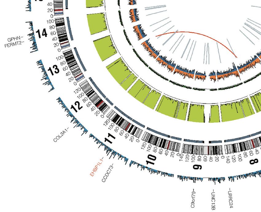
PM16CH03_DeCaprio        ARjats.cls    November 16, 2020      16:40

                                                                                          truncate LT. It should be noted that viral integration is not a normal phase of the MCPyV life cy-
                                                                                          cle. The integrated viral DNA can no longer produce any viable virus. The integration of viral
                                                                                          DNA can be viewed as a random genetic accident. However, when the combination of an intact
                                                                                          NCCR, ST, and a truncated form of LT integration occurs in the appropriate cell type that permits
                                                                                          expression of the T antigens, an MCC tumor emerges.
                                                                                              Integrated MCPyV DNA in the tumor genome can exist as multiple copies with the same or
                                                                                          near-identical mutations in each copy of the viral genome. This observation implies that the viral
                                                                                          DNA was already mutated prior to its insertion into the tumor DNA. In addition to amplification
                                                                                          of the viral genome, some MCCP tumors show that the surrounding cellular DNA was coampli-
                                                                                          fied with the virus (42, 43, 69). These observations have led to a model where the mutated viral
                                                                                          DNA integrates into the tumor genome, generating a circular DNA form that is subsequently am-
                                                                                          plified by rolling circle amplification before resolving the amplified insertion back into the host
                                                                                          genome (Figure 4b). Additional studies are needed to provide a more accurate molecular descrip-
Annu. Rev. Pathol. Mech. Dis. 2021.16. Downloaded from www.annualreviews.org

                                                                                          tion of the amplification and insertion process. Clarification of this viral mechanism could also
                                                                                          provide insight into the process of gene amplification observed for cellular oncogenes.
   Access provided by Harvard University on 11/30/20. For personal use only.

                                                                                          MCPyV LARGE T ANTIGEN
                                                                                          The wild-type full-length LT encodes a protein of 817 residues. An alternatively spliced form of
                                                                                          LT, 57kT, has an in-frame deletion of the central region that deletes most of the DNA origin bind-
                                                                                          ing and helicase domains but retains the C-terminal 100 residues of full-length LT (70, 71). The
                                                                                          function of 57kT is not known, although the C-terminal 100 residues have growth-suppressing
                                                                                          activities (71, 72).
                                                                                              MCPyV LT binds specifically to the retinoblastoma tumor suppressor protein RB to inac-
                                                                                          tivate RB and thereby activate E2F cell cycle–regulated genes (45, 73, 74). The LXCXE motif
                                                                                          is responsible for binding to RB and is an essential component of the transforming activity of
                                                                                          MCPyV LT. An interesting report demonstrated that an MCCP tumor and a derivative cell line,
                                                                                          LoKe, contained a deletion in the RB1 gene and that continued LT expression was not required for
                                                                                          proliferation of this cell line (45). This result indicates that LT binding and inactivation of RB are
                                                                                          required to maintain proliferation of this MCCP tumor. Other LT activities may also contribute
                                                                                          to MCCP oncogenesis but are not essential for the maintenance of the tumor cells.
                                                                                              Unlike LT from other polyomaviruses such as SV40, MCPyV LT does not bind or inacti-
                                                                                          vate p53 (71, 72). Instead, MCPyV LT, through its association with RB, activates p53 (75). LT
                                                                                          may also activate p53 indirectly through its association with USP7 (76). USP7 (also known as
                                                                                          HAUSP) normally functions as a deubiquitinating enzyme. An important substrate of USP7 is
                                                                                          MDM2, and deubiquitination of MDM2 by USP7 leads to increased levels of MDM2, which can
                                                                                          in turn bind to p53 and decrease p53 levels. Truncated LT in MCCP may inhibit USP7 activ-
                                                                                          ity or affect its ability to deubiquitinate MDM2. It has been reported that USP7 increases the
                                                                                          binding of full-length LT to the viral origin of replication and reduces viral DNA replication
                                                                                          (76). In contrast, BRD4 binding to full-length LT has been reported to promote viral replication
                                                                                          (77).
                                                                                              LT can bind specifically to VPS39 (also known as Vam6) (78, 79). LT binding to VPS39 se-
                                                                                          questers it from involvement in lysosomal trafficking, although the significance of this activity
                                                                                          in MCC or in MCPyV replication is not known (79). An LT point substitution, W209A, that
                                                                                          disrupts binding to VPS39 has wild-type transforming potential (74). Phosphorylation of LT at
                                                                                          residues S220 and S239 has been reported (80). Point substitution of these serine residues leads
                                                                                          to increased levels of LT and may represent binding sites to FBW7, although this observation has

                                                                                      ·.•�-
                                                                                          not been confirmed (81).

                                                                                                                                          www.annualreviews.org   •   Pathogenesis of Merkel Cell Carcinoma   3.9

                                                                                      ,          Review in Advance first posted on
                                                                                                 November 23, 2020. (Changes may
                                                                                                 still occur before final publication.)
PM16CH03_DeCaprio         ARjats.cls   November 16, 2020      16:40

                                                                                          a                                                                 b

                                                                                          200 µm                                                           200 µm
Annu. Rev. Pathol. Mech. Dis. 2021.16. Downloaded from www.annualreviews.org

                                                                                     Figure 5
   Access provided by Harvard University on 11/30/20. For personal use only.

                                                                                     Large T antigen (LT) appears in the nucleus and cytoplasm. Immunohistochemistry staining for Merkel cell polyomavirus LT with
                                                                                     CM2B4 antibody is shown with (a) predominantly nuclear and (b) both nuclear and cytoplasmic staining. Figure adapted from
                                                                                     Reference 75.

                                                                                                                   All MCC tumors reported to date express a truncated form of LT due to mutations to the
                                                                                                               integrated viral genome (70, 82). In MCCP tumors, the first exon of LT, encoding a DnaJ or J
                                                                                                               domain, is usually intact with very few point substitutions (Figure 4) (43). In contrast, almost
                                                                                                               immediately after the start of the second exon of LT, MCCP tumors contain point substitutions
                                                                                                               and deletions. The mutations spare the LXCXE (RB-binding) motif, but many other residues sur-
                                                                                                               rounding the LXCXE motif have been reported to be mutated. The LXCXE motif is surrounded
                                                                                                               by the MCPyV-unique regions MUR-1 and MUR-2, but they are unlikely to contribute to onco-
                                                                                                               genesis because deletions or point substitutions have been identified in several different MCC
                                                                                                               tumors and cell lines (43, 74). The nuclear localization sequence follows the MUR-2 domain, is
                                                                                                               retained in only about half of MCCP tumors, and is not required for cellular transformation (74).
                                                                                                               The truncated forms of LT that lose the nuclear localization sequence can be detected in the cy-
                                                                                                               toplasm and nucleus and are apparently able to enter the nucleus because of their smaller size
                                                                                                               (Figure 5). The LT origin binding and helicase domains are necessary for normal polyomavirus
                                                                                                               replication but are typically absent in MCCP. Disruption of the origin binding and helicase activi-
                                                                                                               ties eliminates the possibility that LT could bind to the integrated viral origin of replication within
                                                                                                               the NCCR and initiate the process of replicating viral DNA replication that leads to a DNA dam-
                                                                                                               age response (70, 83, 84). These LT truncating mutations will also reduce the number of potential
                                                                                                               viral antigens that could trigger an antitumor immune response. In addition, growth-suppressing
                                                                                                               activities in the C terminus of LT are lost with these deletions (71, 72).
                                                                                                                   ALTO is typically truncated by mutation or not expressed in MCC tumors. The C terminus
                                                                                                               of ALTO has a hydrophobic region required for binding to cellular membranes (60). Loss of a
                                                                                                               similar C-terminal hydrophobic region in mouse polyomavirus middle T antigen (MT) disables
                                                                                                               the transforming activity of MT (85). Given the similarity of ALTO to MT, it is unlikely that the
                                                                                                               truncated ALTO contributes to the transformed phenotype in MCCP.

                                                                                                               MCPyV SMALL T ANTIGEN
                                                                                                               The MCPyV ST is an essential contributor of MCPyV transforming activity (86, 87). While LT
                                                                                                               is heavily mutated in MCCP tumors, ST is typically wild-type and intact. Wild-type MCPyV ST

                                                                                      ·.•�-
                                                                                                               contains 186 residues and shares the N-terminal 79 residues with LT. The remaining C-terminal

                                                                                                        3.10   DeCaprio

                                                                                      ,            Review in Advance first posted on
                                                                                                   November 23, 2020. (Changes may
                                                                                                   still occur before final publication.)
PM16CH03_DeCaprio ARjats.cls November 16, 2020 16:40
a
PP2A
ST
CK1α
EP400
L
MYC
MDM4 MDM2 p53
b
ncBAF ATOH1
Neuroendocrine
Annu. Rev. Pathol. Mech. Dis. 2021.16. Downloaded from www.annualreviews.org
differentiation
Access provided by Harvard University on 11/30/20. For personal use only.
LSD1 INSM1
RCOR2
Figure 6
The ST-MYCL-EP400 complex activates downstream target genes. (a) The ST-MYCL-EP400 complex
activates MDM2, MDM4, and CK1α, which assemble into a ubiquitin ligase that inhibits p53. The
ST-MYCL-EP400 complex has additional target genes not shown (75, 92). (b) Additional ST-MYCL-EP400
complex downstream target genes include components of the LSD1-RCOR2-INSM1 complex that form a
transcriptional repressor complex that opposes the ATOH1 transcription factor and the ncBAF complex (75,
92, 109). Abbreviations: ncBAF, noncanonical BAF complex; ST, small T antigen. Panel a adapted from
References 75 and 92; panel b adapted from Reference 109.
region of ST is unique. Although a few point substitutions have been reported, at least one point
substitution, A20S, may be related to differences in MCPyV strains (43).
The ST in all mammalian polyomaviruses binds to the protein phosphatase 2A (PP2A) com-
plex. Mammalian PP2A consists of at least three subunits. The PP2A scaffold A subunit forms EP400 complex:
transcription activator
a horseshoe-like structure containing multiple HEAT domains that recruit the regulatory B and
complex composed of
catalytic C subunits (88, 89). Polyomavirus ST typically displaces the PP2A B subunit and forms EP400 (p400),
a trimeric complex with the A and C subunits (90). The PP2A scaffold subunit Aα (PPP2R1A) TRRAP, KAT5
form is more abundant than the Aβ form (PPP2R1B). There are two forms of the PP2A catalytic (TIP60), ACTL6A
C subunit (PPP2CA and PPP2CB). MCPyV ST can bind to both forms of the A and C subunits (BAF53A),
RUVBL1/RUVBL2
(91, 92). ST binding to PP2A contributes to the transforming activities of SV40 and mouse poly-
(TIP49/TIP48),
omavirus ST (93–95). However, it is not clear if MCPyV ST binding to PP2A contributes to its MEAF6, MRGBP,
transforming potential (91). YEATS4 (GAS41),
MCPyV ST forms a complex with the MYC paralog MYCL (L-MYC) and its het- MORF4L1/MORF4L2
erodimeric partner MAX (92). In addition, ST and MYCL/MAX recruit the EP400 (p400) (MRG15/MRGX),
DMAP1, BRD8,
chromatin remodeling complex (Figure 6a). The EP400 complex is made up of at least 15
VPS72 (YL1),
unique proteins, including EP400 (p400), TRRAP, KAT5 (TIP60), ACTL6A (BAF53A), RU- EPC1/EPC2,
VBL1/RUVBL2 (TIP49/TIP48), MEAF6, MRGBP, YEATS4 (GAS41), MORF4L1/MORF4L2 MBTD1, and ING3
(MRG15/MRGX), DMAP1, BRD8, VPS72 (YL1), EPC1/EPC2, MBTD1, and ING3. The
·.•�-
EP400 complex has also been referred to as the p400, Tip60, BAF53, or TRRAP complex in
www.annualreviews.org • Pathogenesis of Merkel Cell Carcinoma 3.11
, Review in Advance first posted on
November 23, 2020. (Changes may
still occur before final publication.)PM16CH03_DeCaprio ARjats.cls November 16, 2020 16:40
mammalian cells and the NuA4 complex in yeast (96–99). The EP400 complex has both histone
acetylation and chromatin remodeling activities that participate in transcription and DNA
damage responses.
While MYC, MYCN, and MYCL have each been reported to bind to the EP400 complex, or
at least various components of the EP400 complex, MCPyV ST is notable for its apparent ability
to increase the stability of the interaction of the complete 15-protein EP400 complex with MYCL
(92). Although MYC, MYCN, and MYCL have been reported to bind several additional cellular
factors, including Host cell factor 1 (HCF-1 or HCFC1) and WDR5, it is not known if MCPyV
ST can also bind these other factors together with MYCL and MAX (99).
MCPyV ST-MYCL-EP400 COMPLEX ACTIVATES KEY DOWNSTREAM
TARGET GENES
Annu. Rev. Pathol. Mech. Dis. 2021.16. Downloaded from www.annualreviews.org
The ST-MYCL-EP400 complex binds together to the transcriptional start sites of several hun-
dred genes and functions to activate their expression. These ST-MYCL-EP400 complex–activated
Access provided by Harvard University on 11/30/20. For personal use only.
downstream target genes contribute to MCPyV oncogenesis. Chromatin immunoprecipitation of
ST, MAX, and EP400 followed by next-generation sequencing (ChIP-Seq) from MCCP cell lines
identified similar and overlapping specific DNA binding sites that were predominantly located
near the transcription start sites of several hundred genes (92). Notably, there was specific en-
richment of binding of all three factors to the E-Box (CACGTG) or canonical MYC binding
sites in gene promoters. Depletion of EP400, MYCL, or ST by RNA interference (RNAi) led
to significantly decreased levels of genes whose promoters were bound by the ST-MYCL-EP400
complex. Identification of the ST-MYCL-EP400 target genes revealed a large number of known
MYC target genes involved in ribosomal biogenesis, splicing, glycolysis, and other basic metabolic
functions. These observations are consistent with a role for MYCL and the MAX heterodimer
functioning similar to the MYC paralog (98, 99).
An additional set of ST-MYCL-EP400 complex target genes involves regulation of p53 ac-
tivity. As indicated above, most MCCP tumors contain the wild-type TP53 gene. Furthermore,
expression of MCPyV LT can activate p53 at least in part through inactivation of the RB tumor
suppressor protein (75). An important question is whether MCPyV can also reduce p53 activity
in MCCP tumors containing wild-type TP53. This was addressed by recognizing that the ST-
MYCL-EP400 complex increased levels of MDM2, an E3 ubiquitin ligase, which specifically binds
p53 and promotes its ubiquitination and subsequent degradation by the proteasome (Figure 6a).
Perhaps similar to MYCL in MCCP, MDM2 had been previously recognized as a MYCN target
gene functioning to inhibit p53 activity in neuroblastoma (100–102). In addition to MDM2, the
ST-MYCL-EP400 complex increases levels of MDM4 and CK1α (CSNK1A1), which cooperate
to activate the E3 ubiquitin ligase activity of MDM2 (75). It is likely that additional ST-MYCL-
EP400 complex target genes cooperate with MDM2 to promote the degradation of p53 and its
loss of function. Importantly, the ability of the ST-MYCL-EP400 complex to transactivate lev-
els of MDM2 implies that inhibitors of MDM2 could prove to be effective at activating p53 in
MCCP containing wild-type p53.
Expression of ST can increase levels of several proteins specifically involved in cell motility.
Overexpression of MCPyV ST in HEK293 cells led to increased levels of proteins involved
in microtubule destabilization, including CDC42, CFL1, CTTN, and RHOA, which lead to a
motile and migratory phenotype. Of note, there is considerable overlap between the ST-MYCL-
EP400 complex target genes and the proteomic analysis of ST expression in HEK293 cells, which
include CDK2, CORO1C, CTNNA1, KPNA3, MAPT, MTPAP, RHOA, and PFN1 (92, 103,
·.•�-
3.12 DeCaprio
, Review in Advance first posted on
November 23, 2020. (Changes may
still occur before final publication.)PM16CH03_DeCaprio ARjats.cls November 16, 2020 16:40
104). It is possible that these ST-MYCL-EP400 target genes contribute to the highly metastatic
potential of MCC.
LSD1: KDM1A;
lysine-specific
LSD1, RCOR2, AND INSM1 OPPOSE ATOH1
demethylase that
While the ST-MYCL-EP400 complex functions as a transcriptional activator, it was recognized forms a transcription
that when ST, MYCL, and EP400 were depleted by RNAi in MCCP cell lines, gene expression repressor complex
with RCOR2 and
levels for a substantial number of genes increased. These genes were not directly repressed by
INSM1
the ST-MYCL-EP400 complex. Instead, it was demonstrated that the ST-MYCL-EP400 com-
plex could transactivate several components of the lysine-specific demethylase 1 (LSD1) repressor Noncanonical BAF
chromatin remodeler
complex (Figure 6b). LSD1 functions to remove activating H3K4me2 and H3K4me1 marks and
(ncBAF): complex
thereby reduce transcriptional activity. LSD1 forms a complex with several proteins, including that contains BRD9
the CoREST factor RCOR2, HDAC1/2, and INSM1. INSM1 is a member of the SNAG do- and GLTSCR1
Annu. Rev. Pathol. Mech. Dis. 2021.16. Downloaded from www.annualreviews.org
main protein family that includes Snail (SNAI1), Slug (SNAI2), Scratch (SCRT1, SCRT2), GFI1, functions to open
GFI1B, OVOL1, and OVOL2 (105). Each of the SNAG domain–containing proteins contains a chromatin and
Access provided by Harvard University on 11/30/20. For personal use only.
cooperates with
highly conserved SNAG motif at the N terminus that can become methylated and bind directly
ATOH1 in MCCP
to LSD1 (106, 107).
Certain inhibitors target the LSD1 demethylating activity and lead to persistence of the methy-
lated histone mark in treated cells. In addition, several groups have reported that some LSD1
inhibitors disrupt the interaction between LSD1 and SNAG domain–containing proteins (107,
108). This can lead to decreased DNA binding of the LSD1 complex, with loss of its repressive
activity and increased levels of target genes. In MCC, LSD1 inhibitors disrupt binding of LSD1
to INSM1, which destabilizes the complex and leads to decreased DNA binding by LSD1 and
RCOR2 (109). Decreased binding results in loss of repression by the LSD1 complex with corre-
sponding increased levels of target genes.
Identification of LSD1-RCOR2 DNA binding sites by ChIP-Seq revealed an enrichment
for ATOH1 binding sites in MCC cell lines. Further testing demonstrated that ATOH1 com-
peted with the LSD1-RCOR2 complex for binding to promoters of ATOH1-dependent genes
(Figure 6b). This result implies that the LSD1-RCOR2-INSM1 complex functions at least in
part to repress ATOH1 transcriptional activity.
A genome-wide CRISPR-Cas9 screen was performed to identify genes that, when lost, enable
MCC cell lines to survive in the presence of LSD1 inhibitors. As expected, loss of KMT2D or
KMT2C, lysine methyl transferases for H3K4me2 and H3K4me1, allowed cells to tolerate LSD1
inhibitors (109). An unexpected result was that loss of several components of the noncanonical
BAF (ncBAF) complex could also cause resistance to LSD1 inhibitors. The ncBAF complex func-
tions to open chromatin and allow gene expression. In this context, the ncBAF complex may coop-
erate with ATOH1 to promote neuroendocrine differentiation gene expression (Figure 6b) (109).
These recent results imply that the LSD1-RCOR2-INSM1 complex functions in part to oppose
ATOH1- and ncBAF-dependent gene expression. In this manner, it appears that a major function
of the LSD1 complex in MCC is to repress ATOH1-driven expression of neural specific genes.
Whether ATOH1 functions to directly recruit the ncBAF complex is not known.
Two recent reports support a role for MCPyV T antigens in modulating ATOH1 activity.
RNAi-mediated depletion of the T antigens in MCCP cell lines induced a neuron-like differ-
entiation pattern with increased levels of neural-related genes and neurite outgrowths capable of
supporting sodium-dependent action potentials (5). T antigen knockdown reduced levels of SOX2
and ATOH1. Conversely, T antigen overexpression in MCCN cell lines or fibroblasts led to in-
creased levels of SOX2 and ATOH1. Furthermore, overexpression of ATOH1 in MCCN cell lines
·.•�-
www.annualreviews.org • Pathogenesis of Merkel Cell Carcinoma 3.13
, Review in Advance first posted on
November 23, 2020. (Changes may
still occur before final publication.)PM16CH03_DeCaprio ARjats.cls November 16, 2020 16:40
Cell cycle MYCL ATOH1 attenuated
LT inactivates RB
MCCP ST-MYCL-EP400 Reduced by LSD1
ST inactivates p53
LSD1 RCOR2 ATOH1 Common
PP2A
S
ST
EP400
L
MYC
G1 G2
M
–4,000 0 4,000 –4,000 0 4,000 –4,000 0 4,000 –4,000 0 4,000
MCCN RB1 and TP53 mutated MYCL amplified Chromatin factors mutated
Annu. Rev. Pathol. Mech. Dis. 2021.16. Downloaded from www.annualreviews.org
Figure 7
Access provided by Harvard University on 11/30/20. For personal use only.
Three transcriptional pathways deregulated in MCCP and MCCN by distinct mechanisms. MCPyV LT binds and inactivates RB,
while the ST-MYCL-EP400 complex functions to inactivate p53. In MCCN, RB1 and TP53 are mutated. The ST-MYCL-EP400
complex induces an MYCL transcriptional program, while MYCL is frequently amplified in MCCN. The activity of the ATOH1
transcription factor is partially repressed by the LSD1-RCOR2-INSM1 complex, while chromatin factors such as KMT2D and
KMT2C are frequently mutated, perhaps partially disabling ATOH1 signaling. Abbreviations: LSD1, lysine-specific demethylase 1; LT,
large T antigen; MCCN, nonviral Merkel cell carcinoma; MCCP, polyomavirus-associated Merkel cell carcinoma; MCPyV, Merkel cell
polyomavirus; ST, small T antigen. Figure adapted from Reference 109.
changed their growth pattern from adherent cells to suspension cells that reflect a neuroendocrine
growth pattern similar to classical MCCP cell lines (110).
MOLECULAR FEATURES COMMON TO MCCP AND MCCN
Analysis of how MCPyV perturbs cellular function and contributes to oncogenesis in MCCP
reveals that at least some of the same signaling pathways are perturbed by mutations in MCCN.
These pathways can be grouped into three distinct gene expression patterns that affect the cell
cycle, MYC activity, and ATOH1 signaling (Figure 7).
The first set of genes perturbed in MCC comprises the cell cycle regulatory genes. A key fea-
ture of the cell cycle genes is regulation by the tumor suppressor proteins RB and p53. RB and
p53 act as checkpoints when they respond to external and internal cellular stresses and reduce
cell cycle gene expression. RB can inhibit cell cycle progression by binding to and repressing the
E2F family of transcription factors. E2F transcription factors bind and activate the promoters
of genes required for entry into S phase. The principal mechanism for p53 control of the cell
cycle is through direct activation of p21 (CDKN1A), which functions to inhibit the activity of
cyclin-dependent kinases CDK1 and CDK2 during the G1/S and G2/M phases of the cell cy-
cle. In the absence of both RB and p53, cells can enter into S phase unfettered by any extrinsic
checkpoint that controls Cyclin D–CDK4 or Cyclin E–CDK2 activity. Furthermore, in the ab-
sence of p53 and p21, there is reduced inhibition of Cyclin E–CDK2 and Cyclin A–CDK1/2
activity. MCCN tumors have a high frequency of loss-of-function mutations in the RB1 and TP53
genes (38, 40). Conversely, in MCCP tumors, MCPyV LT binds and inactivates the RB protein
while the ST-MYCL-EP400 complex transactivates MDM2 to promote the degradation of p53.
Whether the absence of RB and p53, with the resulting deregulation of cell cycle–dependent gene
expression, represents a significant vulnerability for targeted therapy in both forms of MCC is not
·.•�-
known.
3.14 DeCaprio
, Review in Advance first posted on
November 23, 2020. (Changes may
still occur before final publication.)PM16CH03_DeCaprio ARjats.cls November 16, 2020 16:40
The second set of genes deregulated in MCC comprises the MYC-dependent genes. As de-
scribed above, MCPyV ST recruits MYCL to the EP400 complex to activate MYC-dependent
gene expression. The ST-MYCL-EP400 complex promotes the expression of a variety of genes in-
volved in MYC signaling pathways. MYC signaling is also activated in MCCN by amplification of
the MYCL gene (43, 47). Although it is not clear whether amplification of MYCL functions equiv-
alently to the ST-MYCL-EP400 complex, it is notable that amplification of MYC or MYCN is
not typically observed in MCC (41). Given this observation, it is likely that MYCL provides at
least some unique oncogenic activity in MCC that cannot be readily substituted with MYC or
MYCN amplification.
The third set of genes deregulated in MCC is controlled by the ATOH1 transcription factor.
While ATOH1 is expressed in both MCCP and MCCN, it is clear that its transcriptional activity
is at least partially attenuated, since MCC cells do not become fully differentiated into Merkel
or neural cells. Indeed, decreased levels of MCPyV T antigens or overexpression of ATOH1 led
Annu. Rev. Pathol. Mech. Dis. 2021.16. Downloaded from www.annualreviews.org
to a terminally differentiated neural phenotype (5, 110). As shown recently, ATOH1 transcrip-
tional activity is opposed by the LSD1-RCOR2-INSM1 complex in MCCP tumors (109). While
Access provided by Harvard University on 11/30/20. For personal use only.
ATOH1 is expressed in MCCN, the levels may be lower than those observed in MCCP (109,
111). Furthermore, loss of KMT2D and KMT2C, lysine methyl transferases whose activities op-
pose LSD1, may result in reduced ATOH1 activity. Of note, inactivating mutations in KMT2C
and KMT2D are among the most frequently mutated genes in MCCN tumors (41).
It is likely that there are additional signaling pathways that are perturbed in MCC. PI3K sig-
naling is often activated in both MCCP and MCCN. Heterozygous loss of chromosome 10 occurs
in more than 30% of MCCN and MCCP tumors and leads to reduced levels of PTEN, thereby
increasing AKT activity. Activation of PIK3CA by point mutations is observed in MCCP and
MCCN. Other signaling pathways that may be perturbed in both forms of MCC include Notch,
Hedgehog, and bone morphogenetic protein signaling pathways (38, 109, 112).
PRC2 activity is required for the proper development of Merkel cells and may play an im-
portant role in MCC. Low levels of EZH2 expression in MCC tumors, as determined by IHC,
correlate with an improved prognosis compared to tumors that have moderate or strong EZH2
expression (113). Recently, it was shown that PRC2 coordinated transcriptional silencing of the
major histocompatibility complex class I (MHC-I) antigen processing pathway in an MCC cell
line (114). MCC tumors and cell lines typically have low levels of MHC-I expression, including
HLA-A, HLA-B, and HLA-C, which could contribute to immune evasion (115, 116). At least
some MCC cells can increase their levels of MHC-I in response to interferons as well as HDAC1
and EZH2 inhibitors (114, 115, 117).
CELL OF ORIGIN
The cell of origin for MCC remains a critical question in the field. A better understanding of what
is the original cell type that leads to the development of MCC could lead to improved models that
are more reflective of the disease and support preclinical therapeutic trials. Identification of the
cell of origin could provide insight into risk factors and preventive strategies needed to minimize
the risk of developing MCC.
The high degree of UV-associated DNA mutations in MCCN points to the likelihood that its
cell of origin is a sun-exposed skin cell. The high TMB burden due to UV mutations observed
in MCCN is also found in melanoma, invasive cutaneous squamous cell carcinoma, and basal
cell carcinomas. It is highly likely that MCCN tumors derive from an epithelial cell in the ker-
atinocyte lineage. One opposing argument against a keratinocyte origin for MCCN is the typical
·.•�-
presentation of MCC tumors in the dermal layer. However, there are several reports of the in
www.annualreviews.org • Pathogenesis of Merkel Cell Carcinoma 3.15
, Review in Advance first posted on
November 23, 2020. (Changes may
still occur before final publication.)PM16CH03_DeCaprio ARjats.cls November 16, 2020 16:40
situ appearance of MCC associated with a cutaneous squamous cell carcinoma consistent with the
possibility that MCCN can originate in the epidermal layer (34, 118, 119).
Several mouse models support a keratinocyte lineage for MCCP (120, 121). Notably, coex-
pression of MCPyV ST and ATOH1 using keratinocyte specific promoters (KRT5) led to the de-
velopment of neuroendocrine Merkel-like tumor cells (122). Further advancement of this mouse
model could be used to explore additional phenotypes, including metastasis, invasion, and immune
evasion, as well as provide an opportunity to test checkpoint blockade therapy in combination with
additional agents.
Since MCCP tumors do not have a UV mutational signature, it is unlikely that they will orig-
inate from a sun-exposed keratinocyte. It is possible that keratinocytes deep within a hair follicle
may avoid extensive UV-induced damage to the genome and permit transformation by MCPyV.
Perhaps another requirement for the MCCP cell of origin is that it should be capable of expressing
MYCL and ATOH1, since MYCL is required for MCPyV ST oncogenic activity and ATOH1 is
Annu. Rev. Pathol. Mech. Dis. 2021.16. Downloaded from www.annualreviews.org
required for the neuroendocrine phenotype. It could be imagined that the highly specialized cell
types associated with hair follicles, including progenitor cells, would have the necessary malleabil-
Access provided by Harvard University on 11/30/20. For personal use only.
ity to induce expression of MYCL and ATOH1 in the presence of MCPyV.
Trichoblastomas are benign tumors that arise from hair follicle cells in the skin and are typically
removed to rule out basal cell carcinoma. Trichoblastomas contain somatic mutations and often
harbor a large number of normal-appearing Merkel cells (123). An interesting report noted the
presence of an MCCP tumor within a trichoblastoma. Identical somatic mutations were observed
in the trichoblastoma component as well as in the MCCP tumor, which suggests that the MCCP
arose within a preexisting trichoblastoma (124).
Since all examples of MCCP contain mutations that truncate LT and inactivate its ability to
support viral DNA replication, it is likely that the cell of origin for MCCP can support viral
replication. If the MCCP cell of origin was unable to support viral replication, then there would be
less selective pressure to eliminate the viral replication potential by eliminating the origin binding
and helicase domains of LT. It is plausible that expression of wild-type MCPyV LT may promote
replication and amplification of the mutated viral genome and associated cellular genome as the
initial oncogenic event in MCCP. Viral replication may be an early event in the pathogenesis of
MCCP that contributes to amplification of the viral genome (43, 69).
It is plausible that the cell of origin for MCCP derives from a nonepithelial cell type. Candidate
cell types proposed include B lymphocytes due to PAX5 expression detected in some MCC tu-
mors (125, 126). Alternatively, dermal fibroblasts have been suggested as a potential cell of origin
due to the facility of MCPyV replication in this cell type (127). Both B lymphocytes and dermal
fibroblasts could partially explain the typical presentation of MCC tumors in the dermal layer of
the skin.
SUMMARY POINTS
1. Our understanding of Merkel cell carcinoma (MCC) has been informed by studies of
the development and physiology of Merkel cells. The ATOH1 transcription factor is
essential for the development of Merkel cells.
2. There are two forms of MCC with similar presentation and prognosis but completely
different genetic causes. One form is caused by integration of Merkel cell polyomavirus
(MCPyV) with persistent expression of the viral T antigens. The nonviral form of MCC
is caused by extensive UV-induced mutations.
·.•�-
3.16 DeCaprio
, Review in Advance first posted on
November 23, 2020. (Changes may
still occur before final publication.)PM16CH03_DeCaprio ARjats.cls November 16, 2020 16:40
3. The viral oncogenes are the major contributors to the pathogenesis of polyomavirus-
associated MCC (MCCP). MCPyV LT inactivates RB, while MCPyV ST binds to
MYCL and the EP400 transcription complex to activate downstream MYC target
genes.
4. There are many downstream targets of the ST-MYCL-EP400 complex. These target
genes include factors that inactivate p53 and others that contribute to cellular motility
and invasion properties.
5. A key downstream target of the ST-MYCL-EP400 complex is the LSD1-RCOR2-
INSM1 complex. The LSD1 complex functions as a lysine-specific demethylase that
serves to repress genes. A relevant class of genes repressed by the LSD1 complex in
MCC cells is driven by the ATOH1 transcription factor.
6. Mutations in oncogenes and tumor suppressor genes observed in nonviral MCC
Annu. Rev. Pathol. Mech. Dis. 2021.16. Downloaded from www.annualreviews.org
(MCCN) disrupt similar pathways that are targeted by the MCPyV T antigens in MCCP.
Access provided by Harvard University on 11/30/20. For personal use only.
These include the RB, p53, MYCL, and ATOH1 pathways.
FUTURE ISSUES
1. Downstream targets of the ST-MYCL-EP400 complex may represent opportunities for
targeted therapy in MCCP tumors. Inhibitors of MDM2 and LSD1 may prove to be
useful in the treatment of MCC. Additional downstream targets may also prove to be
required for the survival of MCCP tumors.
2. The cell of origin for MCCP and MCCN remains incompletely described. It is assumed
that MCCN requires a sun-exposed cell type to account for the UV mutational signa-
ture, while MCCP requires a non-sun-exposed cell type to account for the lack of a UV
mutational signature. Additional requirements for the MCCP cell of origin include the
requirement for expression of ATOH1 and MYCL.
3. The high response rate of MCCP and MCCN tumors to checkpoint blockade therapy
with antibodies to PD-1 or PD-L1 remains unexplained. The high tumor mutational
burden (TMB) in MCCN likely contributes to an increased number of neoantigens that
represent targets for immune cell detection. The viral antigens in MCCP may similarly
provoke a strong immune response to checkpoint blockade therapy. A better understand-
ing of the specific mutations in MCCN and the activities of the MCPyV T antigens in
MCCP may provide further insight into why these tumors respond so well and how to
improve the response rate and durability of response.
4. Lessons learned by comparing the oncogenic activities in MCCN and MCCP can inform
insights into other high-grade neuroendocrine carcinomas.
DISCLOSURE STATEMENT
J.A.D. received research funding from Constellation Pharmaceuticals. J.A.D. has served as a con-
sultant to Merck & Co. and EMD Serono.
·.•�-
www.annualreviews.org • Pathogenesis of Merkel Cell Carcinoma 3.17
, Review in Advance first posted on
November 23, 2020. (Changes may
still occur before final publication.)You can also read