The adrenergic-induced ERK3 pathway drives lipolysis and suppresses energy dissipation - Genes Dev
←
→
Page content transcription
If your browser does not render page correctly, please read the page content below
Downloaded from genesdev.cshlp.org on September 26, 2020 - Published by Cold Spring Harbor Laboratory Press

The adrenergic-induced ERK3 pathway
drives lipolysis and suppresses energy
dissipation
Rabih El-Merahbi,1,4 Jonathan Trujillo Viera,1,4 Angel Loza Valdes,1,2 Katarzyna Kolczynska,1,2
Saskia Reuter,1 Mona C. Löffler,1 Manuela Erk,1 Carsten P. Ade,3 Till Karwen,1 Alexander E. Mayer,1
Martin Eilers,3 and Grzegorz Sumara1,2
1
 Rudolf Virchow Center for Experimental Biomedicine, University of Würzburg, 97080 Würzburg, Germany; 2Nencki Institute of
Experimental Biology, PAS, 02-093 Warsaw, Poland; 3Theodor Boveri Institute, Biocenter, University of Würzburg, Am Hubland,
97074 Würzburg, Germany

Obesity-induced diabetes affects >400 million people worldwide. Uncontrolled lipolysis (free fatty acid release from
adipocytes) can contribute to diabetes and obesity. To identify future therapeutic avenues targeting this pathway, we
performed a high-throughput screen and identified the extracellular-regulated kinase 3 (ERK3) as a hit. We dem-
onstrated that β-adrenergic stimulation stabilizes ERK3, leading to the formation of a complex with the cofactor
MAP kinase-activated protein kinase 5 (MK5), thereby driving lipolysis. Mechanistically, we identified a down-
stream target of the ERK3/MK5 pathway, the transcription factor FOXO1, which promotes the expression of the
major lipolytic enzyme ATGL. Finally, we provide evidence that targeted deletion of ERK3 in mouse adipocytes
inhibits lipolysis, but elevates energy dissipation, promoting lean phenotype and ameliorating diabetes. Thus,
ERK3/MK5 represents a previously unrecognized signaling axis in adipose tissue and an attractive target for future
therapies aiming to combat obesity-induced diabetes.
[Keywords: lipolysis; ATGL/ERK3; obesity; MK5; FOXO1; PKA; adrenalin; UCP1]
Supplemental material is available for this article.
Received October 8, 2019; revised version accepted February 11, 2020.

Adaptation to changes in nutrient availability is pivotal                   Schweiger et al. 2017), indicating that induction of
for the survival of living organisms. Adipose tissue is a                   lipolysis can result in different physiological outcomes
central storage organ in the body. Upon nutrient inges-                     depending on the nutritional status of the organism. How-
tion, insulin promotes triglycerides (TG) synthesis and                     ever, the signaling pathways that link the nutritional and
storage in the adipocytes. During food deprivation or in-                   hormonal status with temporarily controlled lipolytic
creased energy demand, lipolysis of triglycerides (TG)                      response remain obscure.
stored in lipid droplets of adipocytes provides peripheral                     So far, several hormones are known to regulate the rate
tissues with essential nutrients: free fatty acids (FFAs)                   of lipolysis in response to different physiological challeng-
and glycerol. Consequently, this leads to the reduction                     es. Catecholamines, acting through the β3-adrenergic re-
of adipose tissue mass. However, uncontrolled induction                     ceptor, represent major endocrine factors inducing TG
of lipolysis independently of the organism’s nutritional                    degradation (Zechner et al. 2012). Recently, we proposed
demands is also associated with the development of mul-                     that gut-derived serotonin promotes lipolysis by acting
tiple metabolic diseases including type 2 diabetes and can-                 through the HTR2B receptor (Sumara et al. 2012; El-Mer-
cer-associated cachexia (Haemmerle et al. 2006; Das et al.                  ahbi et al. 2015). These and other hormones, as well as
2011; Arner and Langin 2014; Schreiber et al. 2015). Para-                  several inflammatory mediators, activate assembly of
doxically, inhibition of specific components of lipolytic                   the lipolytic machinery in the lipid droplets (Zechner
machinery can ameliorate diet and genetically induced                       et al. 2012). In contrast, insulin-induced signaling attenu-
obesity (Sekiya et al. 2004; Schreiber et al. 2015;                         ates expression and activity of lipolytic enzymes and in-
                                                                            hibits lipolysis rate (Zechner et al. 2017).
4
 These authors contributed equally to this work.
Corresponding author: grzegorz.sumara@uni-wuerzburg.de
Article published online ahead of print. Article and publication date are
online at http://www.genesdev.org/cgi/doi/10.1101/gad.333617.119. Free-     © 2020 El-Merahbi et al. This article, published in Genes & Develop-
ly available online through the Genes & Development Open Access             ment, is available under a Creative Commons License (Attribution 4.0 In-
option.                                                                     ternational), as described at http://creativecommons.org/licenses/by/4.0/.

                  GENES & DEVELOPMENT 34:1–16 Published by Cold Spring Harbor Laboratory Press; ISSN 0890-9369/20; www.genesdev.org                 1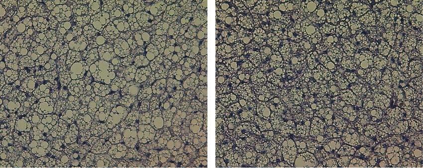
Downloaded from genesdev.cshlp.org on September 26, 2020 - Published by Cold Spring Harbor Laboratory Press

El-Merahbi et al.

   In the lipid droplets, three enzymes were described to             Results
mediate lipolysis: adipose triglyceride lipase (ATGL, also
known as patatin-like phospholipase domain-containing                 siRNA-based screen in adipocytes reveals ERK3
protein 2), hormone-sensitive lipase (HSL), and monoglyc-             as a central regulator of lipolysis
eride lipase (MGL) (Zechner et al. 2012). As revealed by
mouse genetic experiments, deletion of ATGL results in                We designed a screening strategy to assess the impact of
almost complete inhibition of FFAs and glycerol release,              kinase-mediated signaling on the rate of lipolysis evoked
while silencing of HSL or MGL in adipose tissue have a                by the β-adrenergic agonist, isoproterenol (Iso.), and the
moderate effect on lipolysis rate in mice (Osuga et al.               HTR2B agonist, BW-723C86, in differentiated adipocyte-
2000; Haemmerle et al. 2002, 2006; Taschler et al. 2011).             like cells 3T3L1 (Supplemental Fig. S1a). Cotreatment of
Additionally, the rate of lipolysis is regulated by a number          adipocytes with Iso. and BW-723C86 resulted in maximal
of lipid droplet-associated proteins including perilipins,            stimulation of glycerol and FFAs release (Supplemental
CGI-58, and G0S2, which are required for efficient stimu-             Fig. S1c,d). We verified our screening strategy using
lation of lipolysis (Zechner et al. 2017).                            siRNA-mediated silencing of ATGL. Indeed, depletion of
   However, signaling events inducing lipolysis in adipo-             ATGL resulted in a strong reduction of FFAs and glycerol
cytes in response to the catecholamines and serotonin                 release from adipocytes (Supplemental Fig. S1b–d). The
stimulation remain poorly characterized. Activation of                primary screen revealed that silencing of 48 kinases
protein kinase A (PKA) is one known signaling event re-               resulted in decreased lipolysis (FFAs output), whereas
quired for induction of lipolytic enzymes upon activation             depletion of 69 kinases enhanced it in 3T3L1-derived adi-
of the β3-adrenergic receptor. PKA directly phosphory-                pocytes (Supplemental Table 1). In a secondary screen (us-
lates HSL to induce its activity (Zechner et al. 2017). It            ing a different set of siRNA pools) we confirmed that
also phosphorylates perilipin and CGI-58, which allows                silencing of 28 kinases reduced glycerol and FFA release,
CGI-58 to get into a complex with ATGL and to activate                while silencing of 23 enhanced it (Fig. 1A,B). Of note,
this lipase (Zechner et al. 2017). However, how PKA can               Prkar1a, Prkar2b, Ndrg1, Raf1, Lats2, Peg3, and Trib3,
mediate the signaling maintaining the transcription of                which appeared in our screen, were previously implicated
the lipolytic machinery in response to hormonal stimula-              in the regulation of lipolysis and different aspects of adipo-
tion is not understood.                                               cytes function (Curley et al. 2005; Qi et al. 2006; Kalderon
   Therefore, we designed an unbiased screen to identify              et al. 2012; An et al. 2013; Cai et al. 2017; Torres-Quesada
the factors that could control lipolysis. We reasoned that            et al. 2017). These confirm the accuracy of our screening
such a response is likely to occur rapidly through post-              strategy.
translational mechanisms and chose to screen through                     Interestingly, silencing of Extracellular regulated kinase
genes encoding for all known protein kinases. Unexpect-               3 (Erk3) inhibited lipolysis to the largest extent in both
edly, we identified a high number of kinases potentially              screens (Fig. 1A,B; Supplemental Table 1). Consistently,
implicated in the regulation of lipolysis. Among them, si-            silencing of Erk3 in adipocytes derived from primary stro-
lencing of Erk3 resulted in the highest suppression of li-            mal vascular cells or 3T3L1 cells (by specific siRNAs and
polysis rate. ERK3 (also known as MAPK6) is an atypical               shRNA) resulted in almost complete suppression of glyc-
member of the MAPK family. ERK3 is a constitutively ac-               erol and FFAs release evoked by β-agonists and HTR2B ag-
tive kinase; therefore, its abundance determines the rate             onists (Fig. 1C,D; Supplemental Fig. S2C–G) comparable
of substrates phosphorylation (Coulombe et al. 2003,                  with the silencing of Atgl (Fig. 1C,D; Supplemental Fig.
2004). In quiescent cells, ERK3 is subjected to rapid pro-            S2C,D).
teasome-mediated degradation (Coulombe et al. 2003,
2004). Interestingly, we demonstrated that β-adrenergic-
induced PKA signaling stabilizes ERK3 by promoting                    β-Adrenergic activation of PKA leads to stabilization
the formation of the complex between ERK3 and MAP ki-                 of ERK3
nase-activated protein kinase 5 (MK5), which protects
both kinases from degradation. Moreover, we demonstrat-               In quiescent cells, ERK3 is subjected to rapid proteasome-
ed that ERK3/MK5 pathway activates the translocation of               mediated degradation (Coulombe et al. 2003, 2004).
Forkhead box protein O1 (FOXO) to the nucleus, which                  Consistent with this, incubation of adipocytes with pro-
promotes ATGL expression. Consistently, the deletion                  teasome inhibitors (Mg132 or lactacystin) stabilized
of Erk3 in adipose tissue or inhibition of MK5 in mice re-            ERK3 (Fig. 2A,B). In addition, incubation of adipocytes
sults in a decrease of Atgl expression and lipolysis. Surpris-        with β-adrenergic agonists (Iso. and CL316) also increased
ingly, mice deficient for Erk3 specifically in adipocytes             ERK3 levels (Fig. 2A,B), while mRNA levels of Erk3 were
are resistant to diet-induced obesity and diabetes but dis-           unaffected (Fig. 2C). The abundance of ectopically ex-
play elevated energy expenditure, suggesting that the bal-            pressed Myc-tagged ERK3 was also stabilized by the β-ag-
ance between the nutritional demands and lipolysis rate is            onist and the proteasome inhibitor (Fig. 2D). Finally,
perturbed in the absence of ERK3. We propose that the                 blocking translation in adipocytes with cycloheximide de-
ERK3/MK5 pathway represents a missing link down-                      creased ERK3 levels over time, which was inhibited by
stream from PKA required for the fine-tuning of the lipo-             Iso. (Fig. 2E). This demonstrates that β-agonists stabilize
lytic transcriptional signaling and an attractive target for          ERK3 at the protein level, likely via inhibiting its protea-
future antiobesity and antidiabetic therapies.                        somal degradation.

2       GENES & DEVELOPMENT
Downloaded from genesdev.cshlp.org on September 26, 2020 - Published by Cold Spring Harbor Laboratory Press

                                                                                             Adrenergic control of adipocyte’s function

A                                                                                      Figure 1. ERK3 is required for induction of
                                                                                       lipolysis. (A,B) Relative rate of glycerol re-
                                                                                       lease (A) and FFA release (B) (Z-score; n = 4)
                                                                                       from 3T3L1 cells transfected with the indi-
                                                                                       cated siRNA pools. (siNTC) Nontargeting
                                                                                       control. (C,D) FFA (C) and glycerol (D) re-
                                                                                       lease from stromal–vascular cell (SVC)-de-
                                                                                       rived adipocytes transfected with specific
                                                                                       siRNAs and stimulated with the β-agonist
                                                                                       isoproterenol (Iso.) and/or the HTR2b ago-
                                                                                       nist BW-723C86 as indicated (n = 3). Data
                                                                                       are presented as average ± SEM, (∗∗∗ ) P ≤
                                                                                       0.001.

B

C                                          D

  We next sought to identify the mechanism by which β-             PKA-mediated phosphorylation of MK5 promotes ERK3
adrenergic agonists stimulate ERK3 levels and promote li-          stabilization and lipolysis
polysis in adipocytes. β-Agonists promote cAMP levels in
cells, thereby activating protein kinase A (PKA) (Arner            Formation of a complex between ERK3 and MK5 protects
and Langin 2014). Indeed, the elevation of cAMP levels             both kinases from degradation and leads to the activation
independently of β-adrenergic receptors (by forskolin              of MK5 (Schumacher et al. 2004; Seternes et al. 2004). It
or IBMX) was sufficient to stabilize ERK3 (Fig. 2B). More-         has been also shown that PKA phosphorylates MK5 at
over, inhibition of PKA with either H89 or PKI 14-22               Ser115 and might regulate the subcellular localization of
prevented β-agonist-induced stabilization of ERK3 in adi-          MK5 (Kostenko et al. 2011). Interestingly, our primary
pocytes (Fig. 2F). Additionally, stimulation with β-ago-           screen revealed that silencing of Mk5 reduced the rate of
nists, compounds promoting cAMP levels, or treatment               lipolysis (Supplemental Table 1). Therefore, we hypothe-
with proteasomal inhibitors also stabilized ERK3 in                sized that a PKA/ERK3/MK5 signaling axis may regulate
Hek293 and undifferentiated 3T3L1 cells (Fig. 2G; Supple-          lipolysis induced by β-adrenergic stimulation. Consistent
mental Fig. S3a). Thus, β-adrenergic stimulation promotes          with this hypothesis, we found that β-adrenergic stimula-
PKA-dependent ERK3 stabilization.                                  tion also stabilized Mk5 (Fig. 3A), which was partially

                                                                                                   GENES & DEVELOPMENT               3
Downloaded from genesdev.cshlp.org on September 26, 2020 - Published by Cold Spring Harbor Laboratory Press

El-Merahbi et al.

                                                                             C
            A                     B                                                                     D

            E                                                F                            G

Figure 2. β-Adrenergic signaling promotes ERK3 protein levels in a PKA-dependent manner. (A,B,F ) Western blot for ERK3 on differen-
tiated 3T3L1 cells stimulated as indicated for 2 h. (Iso.) Isoproterenol; (proteasome inhibitor) Mg132 and lactacystin; (cAMP elevators
Forsk.) Forskolin and IBMX; (PKA inhibitors) H89 and PKI 14-22. (C) Relative expression of Erk3 in 3T3L1 cells stimulated as shown
for 2 h. (D) Western blot analysis of indicated proteins on 3T3L1-derived adipocytes expressing Myc-tagged ERK3 and stimulated as shown
for 2 h. (E) ERK3 levels on 3T3L1 cell treated with cycloheximide for indicated time points and stimulated with Iso. for 2 h prior to cy-
cloheximide treatment (G) Western blot analysis of indicated proteins on Hek293T cells stimulated as indicated for 2 h. n = 3 for each ex-
periment. For graphs, data are presented as average ± SEM.

blocked by PKA inhibition (Fig. 3B), as shown previously                3H). Of note, the abundance and expression of different
for ERK3 (Fig. 2F). Silencing of Erk3 in adipocytes de-                 MK5 mutants were equal (Fig. 3G; Supplemental Fig.
creased the abundance of MK5 (Fig. 3C), while mRNA                      S3g). Ectopic expression of MK5 mutants also did not af-
levels were unaffected (Supplemental Fig. S3b). Similarly,              fect ERK3 at the transcriptional level (Supplemental Fig.
silencing of MK5 resulted in decreased abundance of                     S3h). Importantly, in cells stimulated with β-adrenergic
ERK3 but did not affect its expression (Fig. 3D; Sup-                   agonist, overexpression of the wild-type form of MK5
plemental Fig. S3c). Consistently, silencing of Mk5                     was sufficient to stabilize ERK3 to a higher extent than
significantly reduced FFAs and glycerol output from adi-                in the control cells (Fig. 3I). Finally, β-adrenergic stimula-
pocytes upon β-agonist stimulation (Fig. 3E; Supplemental               tion of adipocytes promoted the formation of the complex
Fig. S3d). Thus, β-adrenergic-stimulated MK5 also pro-                  between MK5 and ERK3, while inhibition of PKA activity
motes lipolysis in adipocytes.                                          blocked this interaction (Fig. 3J). Taken together, these re-
   PKA-dependent phosphorylation of MK5 promotes its                    sults indicate that PKA-dependent phosphorylation of
translocation from the nucleus to the cytoplasm (Kos-                   MK5 on Ser115 is required to stimulate MK5 nuclear ex-
tenko et al. 2011). This raises the possibility that, upon ad-          port and a complex formation with ERK3, which results
ipocyte stimulation, PKA-dependent phosphorylation of                   in stabilization of both kinases and induction of lipolysis.
MK5 might allow for the formation of a complex between
MK5 and ERK3 as well as stabilization of both kinases,
                                                                        The ERK3/MK5 pathway promotes nuclear translocation
and thereby induction of lipolysis. Consistent with this,
                                                                        of FOXO1 to drive Atgl expression
stimulation of adipocytes with β-agonist resulted in redis-
tribution of MK5 from the nucleus to the cytoplasm,                     To identify the mechanism by which ERK3/MK5 pro-
which was abolished by PKA inhibition (Fig. 3F). Impor-                 motes lipolysis, we subjected control and ERK3-deficient
tantly, leptomycin B, which blocks nuclear export, pre-                 adipocytes to RNA sequencing. Global transcriptomic
vented β-agonist-induced ERK3 stabilization (Fig. 3F;                   analysis revealed that ERK3 is required for expression of
Supplemental Fig. S3e), indicating that translocation of                genes inducing lipolysis (Fig. 4A; Supplemental Fig. S4a).
MK5 to the cytoplasm is required for ERK3 stabilization.                Consistently, qPCR confirmed the reduced expression of
To test this directly, we generated phospho-deficient and               the major lipolytic enzyme Atgl (Zechner et al. 2012) in
phospho-mimetic mutants of MK5 on Ser115 (MK5-                          adipocytes depleted of Erk3 or Mk5 (Supplemental Fig.
S115A and MK5-S115E, respectively). MK5-S115A local-                    S4b). Similarly, ATGL protein was reduced in adipocytes
ized primarily to the nucleus, while MK5-S115E displayed                depleted of Erk3 (Fig. 4B).
cytoplasmic localization (Supplemental Fig. S3f). Impor-                   Forkhead box protein O family members are transcrip-
tantly, MK5-S115E mutation promoted the formation of                    tion factors that function broadly in regulating energy ho-
a complex with endogenous ERK3 (Fig. 3G). Consistently,                 meostasis, including insulin and glucose metabolism
expression of MK5-S115E, but not wild-type MK5 or                       (Eijkelenboom and Burgering 2013). FOXO proteins can
MK5-S115A, was sufficient to stabilize ERK3 (Fig. 3G,I)                 be phosphorylated by MK5, and FOXO1 was proposed to
and evoke lipolysis even in the absence of β-agonist (Fig.              promote ATGL expression (Chakrabarti and Kandror

4       GENES & DEVELOPMENT
Downloaded from genesdev.cshlp.org on September 26, 2020 - Published by Cold Spring Harbor Laboratory Press
Adrenergic control of adipocyte’s function
A B C D
E F G
H I J
Figure 3. Stabilization of ERK3 by β-adrenergic stimulation requires PKA-dependent phosphorylation of MK5. (A) Western blot analysis
for MK5 and actin after blockage of translation by cycloheximide for indicated time points, cells were pretreated with isoproterenol or
vehicle for 2 h before the addition of cycloheximide. (B) Western blot analysis for MK5 on differentiated 3T3L1 cells stimulated as indi-
cated (cycloheximide, isoproterenol, Mg132, and PKA inhibitors H89 and PKI 14-22) for 2 h. (C,D) Western blot analysis on ERK3 and MK5
protein levels in 3T3L1 adipocytes transfected with Erk3- or Mk5-specific siRNA respectively and compared with nontargeting control
transfected cells. Cells were treated with vehicle or isoproterenol for 2 h before collection. (E) FFAs release from Mk5-depleted (siMk5)
and nontargeting control (siNTC) 3T3L1 cells in response to stimulation with isoproterenol for 2 h. (F) MK5 levels in cytoplasmic and
nuclear fractions of 3T3L1 cells treated as indicated for 2 h. (Lept) Leptomycin B. (G, left panel, input) ERK3 and MK5 levels in adipocytes
transduced with MK5 mutants as shown. (Right panel) Immunoprecipitation (IP) using Flag antibody of indicated Flag-tagged mutants of
MK5, followed by Western blot using specified antibodies against coprecipitated proteins. (H) FFA release from adipocytes expressing in-
dicated MK5 mutants and transfected with specific siRNAs and treated as shown (for 2 h). (I ) Western blot for indicated proteins on ex-
tracts isolated from cells expressing indicate mutants of MK5 stimulated as shown for 2 h. (J) IP using Flag antibody on extracts prepared
from cells stimulated as indicated for 2 h, followed by Western blot for ERK3 and Flag. n = 3 for each experiment. For graphs, data are pre-
sented as average ± SEM, (∗∗ ) P ≤ 0.01; (∗∗∗ ) P ≤ 0.001.
2009b; Kress et al. 2011; Chow et al. 2013). Consistently, promoter (Fig. 4F,G; Supplemental Fig. S4e). Moreover,
we showed that FOXO1 binds to the Atgl promoter in ad- expression of the phospho-mimetic FOXO1 mutant
ipocytes (Fig. 4C), and depletion of FOXO1 reduced Atgl (S215D), but not wild-type or phospho-deficient (S215A)
expression as well as lipolysis (Supplemental Fig. S4b,c). FOXO1, restored binding to the Atgl promoter in adipo-
MK5 phosphorylates members of the FOXO family on cytes depleted of ERK3 (Fig. 4H). To further assess wheth-
Ser215, which is conserved among all the FOXO members er FOXO1 mediates β-adrenergic and ERK3/MK5-induced
(including FOXO1) (Kress et al. 2011; Chow et al. 2013). expression of Atgl, we have used adipocytes express-
We have generated phospho-deficient and phospho- ing MK5-S115E (which present increased abundance of
mimetic mutants of FOXO1 (FOXO1 S215A and FOXO1 ERK3) and wild-type form of MK5, depleted from
S215D, respectively). Importantly, FOXO1 S215A dis- FOXO1. As expected stimulation of cells with β-agonist
played primarily cytoplasmic localization while FOXO1 or expression of MK5-S115E mutant resulted in induction
S215D localized mainly in the nucleus (Supplemental of Atgl expression, which was ameliorated by silencing of
Fig. S4d). Silencing of either Erk3 or Mk5 in adipocytes re- FOXO1 (Fig. 4I). Next, we check whether the reintroduc-
duced the nuclear localization of FOXO1 (Fig. 4D,E). Addi- tion of ATGL to the adipocytes is sufficient to restore the
tionally, in ERK3-deficient white and brown adipocytes, lipolytic rate in cells depleted from ERK3. In fact, overex-
we observed reduced binding of FOXO1 to the Atgl pression of ATGL in 3T3L1 cells depleted from ERK3
GENES & DEVELOPMENT 5Downloaded from genesdev.cshlp.org on September 26, 2020 - Published by Cold Spring Harbor Laboratory Press
El-Merahbi et al.
A B C
D E
F G
H I J
Figure 4. ERK3/MK5 promotes FOXO1-mediated transcription of Atgl. (A) RNA sequencing-based heat map of the expression patterns of
indicated genes. (B) Western blot for ATGL on 3T3L1 adipocytes transfected with control or Erk3 siRNA and treated with Iso. for 30 or 60
min. (C ) Chromatin immunoprecipitation of Atgl promoter using FOXO1 and actin antibody from 3T3L1 (D,E) Representative images of
FOXO1 staining on 3T3L1 cells transfected with indicated siRNAs, stimulated as shown for 2 h and corresponding quantifications of spe-
cific localizations. (F–H) Chromatin immunoprecipitation of Atgl promoter using FOXO1 antibody from 3T3L1 cells transfected with
Erk3 or NTC siRNA (F ) and SVC-derived brown adipocytes (G) stimulated for 2 h as shown, or expressing specific FOXO1 mutants
(H). (I) Relative expression of Atgl in 3T3L1 cells expressing the indicated mutants of MK5, transfected with NTC or Foxo1-
specific siRNA and stimulated with Iso. for 2 h. (J) FFAs release from 3T3L1 cells ectopically expressing Atgl (Atgl tg), transfected
with NTC or Erk3-specific siRNA and stimulated with Iso. for 2 h. N = 3–4 for each experiment. For graphs, data are presented as aver-
age ± SEM. (∗ ) P ≤ 0.05; (∗∗ ) P ≤ 0.01; (∗∗∗ ) P≤0.001.
6 GENES & DEVELOPMENTDownloaded from genesdev.cshlp.org on September 26, 2020 - Published by Cold Spring Harbor Laboratory Press
Adrenergic control of adipocyte’s function
restored the lipolysis rate induced by β-agonist to the level for sustaining lipolysis after the initial phase of the TG
observed in wild-type cells (Fig. 4J). Altogether, these data release.
suggest that ERK3/MK5 drives lipolysis by promoting
FOXO1 nuclear translocation and FOXO1-dependent
Murine adipose tissue requires ERK3 to perform lipolysis
transcription of Atgl.
β-Adrenergic stimulation rapidly activates lipolysis. Next, we decided to investigate whether our finding of
However, transcriptional events that are apparently used ERK3/MK5 pathway is also relevant in vivo. ERK3 is a
by EKR3/MK5 pathway require longer periods in order constitutively active kinase (Coulombe et al. 2003,
to be translated into biological effects. Therefore, we 2004) whose abundance is regulated during tissues and tu-
have assessed the detailed timing of β-adrenergic activa- mor development. Consistently, ERK3 has been implicat-
tion of ERK3. Already, 15 min after stimulation by β-ago- ed in lung development and in carcinogenesis (Klinger
nist, ERK3 has been stabilized in adipocytes and the peak et al. 2009; Long et al. 2012). However, its role in adipose
of its abundance has been observed from 2 to 8 h later tissue has not been described. Food deprivation induces
(Supplemental Fig. S4g). Interestingly, β-adrenergic stimu- stress hormones (catecholamines) and, consequently, li-
lation, induced Atgl expression already after 30 min and polytic response in adipose tissue depots, with the stron-
abundance of the protein was substantially increased 2 h gest response in epi-gonadal white adipose tissue
later (Supplemental Fig. S4g,h). Of note, β-adrenergic in- (EpiWAT). Consistently, we found elevated ERK3 levels
duced expression and abundance of ATGL, but not the ex- in EpiWAT of mice subjected to fasting (Fig. 5A). Similar-
pression of Mk5, was ameliorated by the silencing of Erk3 ly, β-adrenergic stimulation of mice led to an increase of
(Supplemental Fig. S4g–i). Although induction of lipolysis ERK3 levels in EpiWAT (Fig. 5B), suggesting that ERK3
was rapid upon β-agonist, FFAs release kept increasing at controls lipolysis rate also under physiological conditions.
later time points in control cells but not in the ERK3-de- To test whether ERK3 controls lipolytic response in ad-
ficient adipocytes (Supplemental Fig. S4j). Therefore, a β- ipose tissue, we generated mice deficient for Erk3 specifi-
adrenergic-induced ERK3/MK5 pathway is required rather cally in adipocytes (Erk3Adipo.Δ/Δ) (Supplemental Fig. S5a,
A B
C D
E
F G
Figure 5. ERK3 is required for induction of lipolysis in adipose tissue of mice. (A) ERK3 protein levels in EpiWAT of mice fasted for indi-
cated time points (n = 3). (B) ERK3 levels in mice injected with β-agonist CL316243 or vehicle control (n = 3). (C,D) FFAs and glycerol release
in circulation respectively after injection of β-agonist (CL316243) for indicated times in mice with adipocyte-specific deletion of Erk3
(Erk3Adipo.Δ/Δ) or control animals (Erk3f/f) (n = 6). (E) Relative Atgl expression in EpiWAT from Erk3Adipo.Δ/Δ and control mice. (F,G)
ATGL and MK5 levels, respectively, in EpiWAT isolated from Erk3f/f and Erk3Adipo.Δ/Δ. Data are presented as average ± SEM. (∗ ) P ≤
0.05; (∗∗ ) P ≤ 0.01; (∗∗∗ ) P ≤ 0.001.
GENES & DEVELOPMENT 7Downloaded from genesdev.cshlp.org on September 26, 2020 - Published by Cold Spring Harbor Laboratory Press
El-Merahbi et al.
b). Consistently, with the results obtained in vitro mura et al. 2015). In the HFD-fed Erk3Adipo.Δ/Δ mice, we
β-adrenergic stimulation failed to induce FFAs and glycer- found increased expression of Ucp1 and associated genes
ol levels in the blood of Erk3Adipo.Δ/Δ mice, in contrast to in SubWAT and brown adipose tissue (BAT) (Fig. 6G; Sup-
control animals (Fig. 5C,D). Thus, ERK3 is required for plemental Fig. S6n). Consistently, UCP1 protein levels
stimulation-induced lipolysis in adipocytes in animals. were elevated in SubWAT (Fig. 6H). Moreover, silencing
Consistent with the mechanism of ERK3 action predicted of Mk5, Erk3 or FoxO1 was sufficient to promote expres-
based on in vitro data, deletion of ERK3 in adipocytes re- sion of Ucp1 and associated thermogenic genes in adipo-
sulted in decreased Atgl transcription and protein levels cytes derived from SVC isolated from subWAT (Fig. 6I;
and reduction in MK5 abundance (Fig. 5E–G). Thus, Supplemental Fig. S6o) and BAT (Fig 6J; Supplemental
ERK3 is required for lipolysis in adipocytes in vitro and Fig. S6p). Moreover, we showed that in response to
in vivo. β-adrenergic stimulation, FOXO1 binds to the promoter
of Ucp1 of white (Fig. 6K) and brown (Fig. 6L) primary ad-
ipocytes in ERK3-dependent manner. All of these suggest
Deletion of ERK3 in adipocytes promotes energy
that ERK3/MK5/FOXO1 axis exerts negative feedback
expenditure
on Ucp1 expression and presumably energy dissipation
Typically, reduced lipolysis leads to accumulation of fat by adipocytes. Indeed, isolated primary brown adipocytes
tissue and the development of obesity and diabetes. Para- from Erk3Adipo.Δ/Δ mice had an increased oxygen
doxically, it has been shown that reducing ATGL action consumption rate and decoupling activity (Fig. 6M,N).
can also protect against diabetes (Haemmerle et al. Negative energy balance generated by elevated Ucp1 ex-
2006; Arner and Langin 2014; Schoiswohl et al. 2015; pression in adipose tissue of mice could be ameliorated
Schreiber et al. 2015). In addition to its role regulating by housing animals at thermoneutral conditions (Löffler
Atgl expression, FOXO1 promotes differentiation of adi- et al. 2018). In fact, housing HFD fed Erk3Adipo.Δ/Δ mice
pocytes and suppresses expression of uncoupling protein and corresponding control animals at thermoneutral con-
1 (UCP1), which drives energy expenditure and therefore ditions resulted in similar body weight gain (Supplemen-
counteracts lipid accumulation (Nakae et al. 2003, 2008, tal Fig. S6q). This suggests that Erk3Adipo.Δ/Δ mice are
2012; Liu et al. 2016; Kita et al. 2019; Peng et al. 2019). protected from HFD-induced obesity due to the increased
Because of the direct action of ERK3/MK5 complex on thermogenesis.
the FOXO1-mediated transcriptional signaling, we decid- Thus, depletion of ERK3 inhibits lipolysis by reducing
ed to test the impact of ERK3 on the development of obe- FOXO1-mediated ATGL expression but at the same
sity and diabetes in vivo. Erk3Adipo.Δ/Δ mice gained time promoting Ucp1 expression and energy dissipation
significantly less weight and presented reduced adiposity by brown as well as white adipose tissue, thereby protect-
than control animals when fed a high-fat diet (HFD) (Fig. ing against obesity and diabetes. By promoting FOXO1 nu-
6A; Supplemental Fig. S6a). Moreover, histological analy- clear localization, ERK3 may fine-tune the transcriptional
sis revealed reduced adipocytes’ size and multilocular response leading to stimulation of Atgl and reduction of
cells within subcutaneous adipose tissue (SubWAT) (Fig. Ucp1 expression. Thus, at least in the context of HFD,
6B,C) as well as smaller lipid droplets in brown adipose tis- FOXO1-dependent suppression of Ucp1 expression and
sue (Fig. 6D). Erk3Adipo.Δ/Δ mice also displayed improved energy expenditure appears to exert a dominant role
glucose tolerance and insulin sensitivity (Fig. 6E; Supple- over ATGL-dependent lipolysis. Thus, Erk3Adipo.Δ/Δ mice
mental Fig. S6c) when fed HFD. Interestingly, Erk3Adipo.Δ/Δ provide us with the unique experimental model to test
mice fed HFD presented strongly improved fasting glyce- the physiological relevance of both transcriptional
mia and normalization of glucose during glucose toler- responses.
ance test to the initial values strongly ameliorated the
differences between genotypes (Supplemental Fig. S6b).
Inactivation of ERK3 in obese mice prevents further body
In the case of insulin challenge, a relative drop in glucose
weight gain and improves insulin sensitivity
levels compared with values at fasting was similar in
Erk3Adipo.Δ/Δ and Erk3f/f mice (Supplemental Fig. S6d). To corroborate this hypothesis, we tested whether dele-
On normal diet (ND), Erk3Adipo.Δ/Δ mice presented similar tion of ERK3 in mice with established obesity can inhibit
body weight gain as the corresponding control animals, further body weight gain and improve glycaemia. We
while glucose clearance and insulin sensitivity were im- have crossed ERK3f/f with tamoxifen-inducible Adiponec-
proved only due to the reduced fasting glucose levels (Sup- tin promoter-driven CRE line (Erk3Ind.Adipo.Δ/Δ) and in-
plemental Fig. S6e–i). These phenotypes prompted us to duced deletion of ERK3 with tamoxifen administration
test the effect of ERK3 on energy expenditure. Indeed, after 9 wk of HFD feeding. After induction of ERK3 dele-
Erk3Adipo.Δ/Δ mice fed HFD revealed increased energy ex- tion in Erk3Ind.Adipo.Δ/Δ (Supplemental Fig. S7a), these
penditure when compared with control animals (Fig. 6F), mice did not further gain weight like corresponding con-
while energy intake and voluntary movements were un- trol animals (Fig. 7A). Consistent with previous results,
changed independently of the diet (Supplemental Fig. Erk3Ind.Adipo.Δ/Δ mice displayed elevated energy expendi-
S6j–m). Elevated expression of Ucp1 in different adipose ture (Fig. 7B), but deletion of ERK3 did not influence food
tissue depots drives energy dissipation of adipocytes, intake and voluntary movements (Supplemental Fig.
which can increase the energy expenditure of the entire S7b,c). As in the case of constitutive deletion of ERK3 in
organism and prevent the development of obesity (Kaji- the adipocytes, induction of ERK3 removal in mice with
8 GENES & DEVELOPMENTDownloaded from genesdev.cshlp.org on September 26, 2020 - Published by Cold Spring Harbor Laboratory Press
Adrenergic control of adipocyte’s function
A B C
D E F
G H I
J K L M N
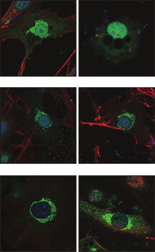
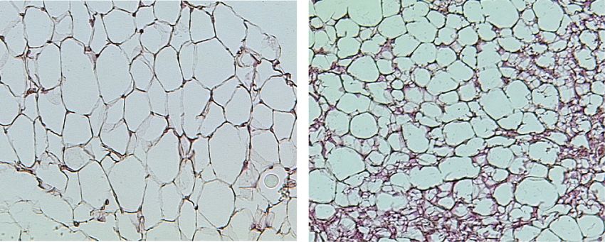
Figure 6. Deletion of Erk3 in adipocytes promotes energy dissipation and protects against obesity. (A,E) Bodyweight evolution and glu-
cose tolerance test (GTT), respectively, on Erk3Adipo.Δ/Δ and Erk3f/f mice fed a high-fat diet (HFD) (n = 11). (B,C) Representative H&E stain-
ing of subcutaneous adipose tissue and quantification of the average adipocyte size of indicated mice fed HFD (n = 8). (D) Representative
H&E staining of brown adipose tissue (BAT) on the indicated mice fed HFD. (F) Energy expenditure of Erk3Adipo.Δ/Δ and Erk3f/f mice fed
HFD (n = 8). (G) Relative expression of indicated genes in SubWAT from the indicated mice (n = 6). (H) Western blot for UCP1 in SubWAT
from Erk3Adipo.Δ/Δ and Erk3f/f mice fed HFD (n = 6). (I,J) Relative expression of Ucp1 in subWAT-derived (I) and BAT-derived (J) adipocytes
depleted of specific proteins by siRNA (n = 4). (K,L) Chromatin immunoprecipitation of Ucp1 promoter using FOXO1 antibody from and
SVC-derived brown adipocytes (K ) and white adipocytes (L) stimulated for 2 h as shown (n = 4). (M,N) Oxygen consumption rate (OCR) in
response to the indicated substances (M) as well as OCR annotated to the indicated cellular processes (N) in Erk3-deficient SVCs differ-
entiated into brown adipocytes. Data are presented as average ± SEM. (∗ ) P ≤ 0.05; (∗∗ ) P ≤ 0.01; (∗∗∗ ) P ≤ 0.001.
established obesity resulted in elevated expression of been already developed and approved for phase II clinical
Ucp1 and other thermogenic genes in subWAT and BAT trial (Westhovens et al. 2013). One-week administration
(Fig. 7C; Supplemental Fig. S7d,e). Remarkably, induced of GLPG0259 in mice significantly reduced β-agonist-in-
deletion of ERK3 in obese mice improved glucose duced FFAs and glycerol levels as well as Atgl expression
tolerance and insulin sensitivity (Fig. 7D,E). Therefore, in- compared with control animals (Fig. 7F,G). Finally, inhibi-
hibition of ERK3/MK5 pathway might ameliorate obesity- tion of MK5 resulted in the induction of thermogenic
induced diabetes. In fact, MK5 inhibitor (GLPG0259) has genes expression in subWAT and BAT (Supplemental
GENES & DEVELOPMENT 9Downloaded from genesdev.cshlp.org on September 26, 2020 - Published by Cold Spring Harbor Laboratory Press
El-Merahbi et al.
A B C
D E
F G H
Figure 7. Inhibiting ERK3/MK5 pathway in obese mice promotes energy dissipation and prevents further body weight gain.
(A–E) Bodyweight evolution (A), energy expenditure (B), expression of thermogenic genes (C), glucose tolerance test (D), and insulin tol-
erance test (E) of mice with inducible deletion of Erk3 (Erk3Ind.Adipo.Δ/Δ) and control animals fed HFD upon administration of tamoxifen at
indicated time point. (F ) Glycerol release in circulation after injection of β-agonist (CL316243) for indicated times in BL6/J mice that have
been orally administered with GLPG0259 inhibitor (10 mg/kg) or vehicle control for one consecutive week (n = 5). (G) Relative Atgl ex-
pression in SubWAT from GLPG0259 administered mice and their respective vehicle control mice (n = 5). (H) Proposed model of action
of the β-adrenergic-induced ERK3/MK5/FOXO1 pathway. Data are presented as average ± SEM. (∗∗ ) P ≤ 0.005; (∗∗∗ ) P ≤ 0.01.
Fig. S7f,g). Altogether, these indicate that inhibition of wohl et al. 2015; Schreiber et al. 2015). Ablation of the ma-
MK5 with GLPG0259 might be a valid strategy to target jor enzyme involved in the regulation of lipolysis, ATGL,
ERK3/MK5/FOXO1 pathway in adipocytes. in adipose tissue and in whole-body results in the increase
Taken together, we identified a signaling axis that can of TG accumulation in adipocytes and improved insulin
link and fine-tune the transcriptional response leading sensitivity (Trites and Clugston 2019). Paradoxically,
to lipolysis and to energy dissipation in adipocytes. We pharmacological inhibition of major lipase ATGL by
speculate that inhibition of ERK3/MK5 signaling, which atglistatin has been shown to protect mice from the devel-
could be achieved by specific inhibitors, might represent opment of obesity and diabetes (Schweiger et al. 2017).
an attractive strategy to ameliorate diabetes and obesity However, atglistatin is not effective in humans (Schweiger
(Fig. 7H). et al. 2017). Therefore, there is an urgent need for identifi-
cation of targets, inhibition of which would decrease the li-
polysis rate and could potentially protect from the
Discussion development of obesity-evoked diabetes. Kinases can be
in general easily targeted by pharmacological agents.
Aberrant activation of lipolytic machinery in adipocytes Here, we developed a screening strategy to identify kinases
contributes to the development of obesity-evoked diabetes implicated in the regulation of lipolysis rate. Our primary
(Haemmerle et al. 2006; Arner and Langin 2014; Schois- and secondary screen revealed 50 kinases implicated in the
10 GENES & DEVELOPMENTDownloaded from genesdev.cshlp.org on September 26, 2020 - Published by Cold Spring Harbor Laboratory Press
Adrenergic control of adipocyte’s function
regulation of lipolysis induced by adrenergic and seroto- pression of major lipase, ATGL, in a FOXO1-dependent
nergic stimulation. Among them, several kinases were manner. These data explain how β-adrenergic signaling
previously implicated in the regulation of lipolysis or adi- is linked to the transcription of lipolytic enzymes in par-
pocyte function. For example, our screen revealed that si- ticular FOXO1-dependent transcription. At this point,
lencing of two regulatory subunits of multisubunit we cannot exclude the possibility that ERK3/MK5 path-
complex of PKA (Prkar1a and Prkar2b, which inhibits way may, in addition, promote lipolysis in a transcription-
PKA activation) (Torres-Quesada et al. 2017) enhances al-independent manner. Also, we cannot exclude that
the lipolysis rate. On the other hand, silencing of Ndrg1, ERK3 partially drives lipolysis and thermogenesis in an
Raf1, Lats2, Peg3, and Trib3, which were previously MK5 independent manner. In fact, the silencing of Mk5
shown to promote lipolysis or to regulate other aspects of was less effective in suppressing lipolysis than the silenc-
adipocytes function (Curley et al. 2005; Qi et al. 2006; Kal- ing of Erk3. However, these discrepancies between pheno-
deron et al. 2012; An et al. 2013; Cai et al. 2017), resulted in types might also reflect differences in siRNA efficiency.
inhibition of lipolysis. These results confirm the appropri- We showed that the ERK3/MK5 pathway drives
ate design and relevance of our screen. Among the known lipolysis by promoting Atgl expression. β-Adrenergic
factors not identified in the screen were the classical mem- stimulation induces lipolytic machinery within minutes,
bers of MAPK family, extracellular-regulated kinase (ERK) while transcriptional response requires at least 1 h in or-
1/2 and c-Jun N-terminal kinase (JNK) 1/2/3 reported pre- der to translate into a biological effect. In fact, the silenc-
viously to promote triglycerides degradation in adipocytes ing of ERK3 resulted in a significant decrease in lipolysis
(Greenberg et al. 2001; Gehart et al. 2010; Hong et al. 2018). rate only 2 h after β-adrenergic stimulation, indicating
This might be explained by the fact that in our screen we that the ERK3/MK5 pathway is responsible for sustaining
silenced each of the isoforms of a given family of kinases lipolysis rather than initializing it. However, we cannot
separately. Since members of the MAPK family presents exclude that the deletion of Erk3 leads to the reduction
high homology, high redundancy in function can occur be- of Atgl expression prior to the β-adrenergic stimulation.
tween different isoforms. Alternatively, in our screen, we FOXO1 regulates a broad spectrum of transcriptional
stimulated lipolysis with a mixture of β-adrenergic and targets implicated in different aspects of adipocytes func-
HTR2B agonists, which might not necessarily lead to the tion. We and others showed that FOXO1 promotes the ex-
activation of classical members of the MAPK family. Ex- pression of Atgl (Chakrabarti and Kandror 2009b;
citingly, the screen revealed that atypical member of Chakrabarti et al. 2011; Jung et al. 2019). Interestingly, a
MAPK family, ERK3, is a critical regulator of lipolysis. number of reports showed that FOXO1 suppresses the ex-
We showed that deletion of ERK3 in adipose tissue of pression of genes regulating energy dissipation, including
mice suppressed the production of FFAs and glycerol in re- UCP1, in white and brown adipocytes (Nakae et al. 2008,
sponse to the β-adrenergic stimulation, ultimately con- 2012; Liu et al. 2016; Kita et al. 2019; Peng et al. 2019).
firming results from the screen. However, a recent study indicated that in brown adipose
ERK3 is a constitutively active kinase (Coulombe et al. tissue of mice fed ND, FOXO1 does not affect UCP1 and
2003, 2004) whose protein levels are tightly regulated. We in vitro under specific culture conditions deletion of
show that in adipocytes ERK3 activity correlates with its FOXO1 might even decrease Ucp1 expression in brown
abundance. Moreover, we propose a mechanism whereby adipocytes (Jung et al. 2019). Another study also indicates
the formation of a complex between ERK3 and MK5 ki- that in the mice with decreased insulin signaling, FOXO1
nase depends on β-adrenergic-induced PKA activation might drive Ucp1 expression (Ortega-Molina et al. 2012).
and promotes the stability of both kinases. In line with We showed that expression of Ucp1 was markedly in-
our model, β-adrenergic stimulation promotes interaction duced in white and, even more relevant to the total energy
between MK5 and ERK3. Similarly, the phosphomimetic balance of animals also, in brown adipocytes deficient for
mutant of MK5 (MK5-S115E) presented a higher affinity FOXO1, MK5, or ERK3. Consistent with these observa-
to endogenous ERK3 than the wild-type form of MK5 in tions, we showed that deletion of ERK3 in adipocytes of
the unstimulated cells. However, the expression of phos- mice inhibits the development of obesity or further
phomimetic mutant of MK5 also induced stability of weight gain in obese mice, at least partially due to the in-
ERK3, therefore, theoretically enhanced interaction be- creased Ucp1 expression and increased energy dissipation.
tween MK5 and ERK3 might be a result of increased avail- Several reports indicate that the induction of lipolysis is
ability of these proteins. These results provide the first required for UCP1-dependent energy dissipation by adipo-
link between adrenergic signaling and atypical MAPKs cytes (Ahmadian et al. 2011; Li et al. 2014; Schreiber et al.
and define ERK3/MK5 as crucial components of adrener- 2017). Deletion of ATGL in the whole body or all of the de-
gic-induced signaling. Moreover, our data confirm pub- pots of adipose tissue results in decreased energy dissipa-
lished evidence that ERK3 is subjected to ubiquitin– tion in mice upon cold exposure especially during the
proteasome-mediated degradation (Coulombe et al. time of food deprivation (Ahmadian et al. 2011; Schreiber
2003, 2004). In future, it will be important to understand et al. 2017). However, in mice deficient for ATGL in the
the molecular mechanisms by which the formation of a whole body (except for the cardiac muscle) and mice treat-
complex between ERK3 and MK5 protects both kinases ed with atglistatin, which were fed HFD and maintained
from proteasome-mediated proteolysis. at room temperature, energy expenditure was not de-
Our results demonstrate that β-adrenergic-induced creased (Schreiber et al. 2015, 2017). Moreover, mice treat-
ERK3/MK5 pathway promotes lipolysis by driving the ex- ed with Atglistatin display higher UCP1 levels in adipose
GENES & DEVELOPMENT 11Downloaded from genesdev.cshlp.org on September 26, 2020 - Published by Cold Spring Harbor Laboratory Press
El-Merahbi et al.
tissue (Schweiger et al. 2017). Similarly, deletion of mice as described previously in Cai et al. (2017). Concisely,
CGI-58 in brown adipose tissue, which partially blocks li- mice subcutaneous white adipose tissue (SubWAT) and brown ad-
polysis, does not block energy dissipation by adipocytes in ipose tissue (BAT) were sliced and digested in PBS containing
mice fed ad libitum (Shin et al. 2017, 2018). Mice deficient 2 mg/mL collagenase D (Sigma-Aldrich), 5 mM CaCl2, and 1%
BSA for 40 min at 37°C. Adipose tissue are then passed through
for ERK3 show about 50% reduced Atgl expression, and at
a 40-µm mesh, washed in PBS by centrifugation, and plated on
the same time, increased Ucp1 expression in white as well Matrigel-coated (Corning) plates in DMEM/F-12 containing
as in brown adipose tissue when fed HFD. Moreover, these 10% FBS, 1% NEAA, and 40 µg/mL gentamycin. Two days after
mice show increased energy expenditure when fed HFD confluence, preadipocyte differentiation was induced by 0.2 µM
and maintained under room temperature. These results indomethacin, 0.5 mM IBMX, 1 µM dexamethasone, and 1.5
are in line with the fact that FOXO1 promotes Atgl expres- µg/mL insulin for the first 48 h. Afterward, cells were maintained
sion but suppresses Ucp1 transcription (Nakae et al. 2008, in complete medium with 1.5 µg/mL insulin for up to six addi-
2012; Chakrabarti and Kandror 2009b; Chakrabarti et al. tional days. In preadipocytes derived from BAT, additional T3
2011; Liu et al. 2016; Kita et al. 2019; Peng et al. 2019). (2 nM; Sigma-Aldrich) was added to the medium throughout dif-
Therefore, it appears that at least upon HFD feeding, sup- ferentiation. 3T3-L1 cells were differentiated into adipocytes ac-
cording to the standard procedure described in Cai et al. (2017).
pression of Ucp1 expression and energy expenditure
Prior to treatment, differentiated adipocytes were washed twice
would play a dominant role over ATGL-dependent lipoly- with phenol-free DMEM and then serum-starved for 2 hr in phe-
sis. We postulate that increased expression of Ucp1 in nol red-free DMEM supplemented with 2% BSA; afterward and as
mice deficient for ERK3 in adipocytes fed ad libitum indicated in experiments, treatments were added in the following
with HFD at least partially contributes to the enhanced concentrations: 1.10 µM isoproterenol (Tocris Biotech), 10 µM
energy expenditure observed in these animals, which in Mg-132 (Tocris Biotech), 20 μM Forskolin (Sigma-Aldrich),0.5
turn protects from HFD-induced obesity. However, at mM IBMX (Sigma-Aldrich), 10 μM CL316,243 (Tocris Biotech),
this point we cannot exclude additional mechanisms pro- 20 µM lactacystin (Sigma-Aldrich), 100 μg/mL cycloheximide
moting energy expenditure in the absence of ERK3. (Tocris Biotech), 20 μM H89 (Sigma-Aldrich), 20 μM PKI14-22
Based on our results, pharmacological targeting of the (Sigma-Aldrich), 50 nM leptomycin B1 (Sigma-Aldrich), and
10 μM BW-723C86 (Tocris Biotech).
ERK3/MK5/FOXO1 pathway might be a valid strategy
to treat obesity and associated diabetes. Moreover, the ex-
istence of MK5 inhibitor, GLPG0259, which was already Transient transfection with siRNA
shown not to have toxic side effects in humans
(Westhovens et al. 2013), raises a possibility for effective Differentiated adipocytes were transfected with siRNA always in
suspension as follows: Mature adipocytes were detached with
antiobesity therapy. In fact, we have shown that adminis-
Accutase solution for 10 min, spun down at 300g for 5 min, and
tration of GLPG0259 in mice reduces Atgl expression, counted. Indicated siRNAs and Dharmafect-Duo transfection re-
suppresses lipolysis in mice, and increases the expression agent (Dharmacon) were diluted in Opti-MEM I reduced serum
of thermogenic genes, indicating that this inhibitor effec- medium separately before being mixed by pipetting. The
tively targets ERK3/MK5/FOXO1 pathway. However, siRNA-Duofect mix was added to Matrigel-coated culture plates
since the deletion of Atgl had a deleterious effect on heart or 96-well Seahorse plates and left to incubate for 30 min at room
function and currently available inhibitors against ATGL temperature. The cells were resuspended in culture medium and
are not effective in humans (Trites and Clugston 2019), added on top of the preincubated siRNA-Duofect mix. The final
targeting directly ATGL for treatment of obesity and dia- concentrations of Duofect, siRNA, and cell number were adjust-
betes is difficult. Targeting ERK3/MK5 not only partially ed per surface area in a ratio of 2.1 uL/cm2 duofect, 1.25 nM/cm2
siRNA, and 16.4 × 10⁴/cm2 cells, respectively. The cells were
inhibits lipolysis but also promotes energy dissipation;
used for experiments 48 h after transfection. All siRNA sequenc-
thus, inhibitors targeting this pathway might be of use es were purchased as smart pools from Dharmacon.
for the treatment of obesity and associated diseases. How-
ever, further studies are required to define whether inhib-
itors directed against the ERK3/MK5 pathway might Generation of stable cell lines
overcome the potential side effects of ATGL inhibitors.
The Dharmacon GIPZ lentiviral shRNA system was used to in-
Taken together, we identified the β-adrenergic-induced duce long-term gene silencing of the Erk3 gene, shRNA Erk3
ERK3/MK5/FOXO1 pathway as a central regulator of li- oligo was synthesized by Eurofins (shErk3: 5′ -TGCTGTTGAC
polysis and energy expenditure and is a potential target AGTGAGCGAACACCTGTAACTACAAAACAATAGTGAAG
for future antiobesity therapy (Fig. 7G). CCACAGATGTATTGTTTTGTAGTTACAGGTGTGTGCCT
ACTGCCTCGGA-3′ ) and cloned into a pGIPZ shRNA mir (a
generous gift from Eilers laboratory, Biocenter, Würzburg) be-
tween XhoI and EcoRI cloning sites. The pBABE- Puro retroviral
Materials and methods
vector system was used to overexpress ERK3 protein. Concisely,
Myc tagged Erk3 coding sequence was cloned from human cDNA
Preadipocyte culture and differentiation
vector and inserted between EcoR1 and Sal1 in the pBABE vector.
3T3-L1 preadipocytes, Platinum-E, and HEK293T cells were cul- HA-Foxo1 and 3xFlag-Mk5, Myc-mPnpla2 overexpressing vec-
tured and maintained in Dulbecco’s modified Eagle’s medium tors were all synthesized by Vectorbuilder by the insertion of
(DMEM), 10% fetal bovine serum (FBS), 1% nonessential amino their respective coding sequences in PLV-Puro lentivirus plas-
acids (NEAA), and 40 µg/mL gentamycin. Stromal vascular cells mid. Wild-type sequences were point mutated as mentioned
(SVCs) were isolated from subcutaneous white adipose tissue above using Q5 site-directed mutagenesis kit (NEB). Lentiviral
(sWAT) or from brown adipose tissue (BAT) of 5- to 9-wk-old particles were packaged in HEK293T cells with psPAX and
12 GENES & DEVELOPMENTDownloaded from genesdev.cshlp.org on September 26, 2020 - Published by Cold Spring Harbor Laboratory Press
Adrenergic control of adipocyte’s function
pMD2.G and transduced in 3T3L1 cells. Retroviral particles were flox mice (Erk3f/f ) were used for the generation of targeted dele-
produced using PlatinumE cells. 3T3L1 cells were spinfected tion in tissues. For targeted deletion of Erk3 in adipocytes,
with viral supernatant with the addition of polybrene then select- Erk3f/f mice were cross-bred with adiponectin promoter-driven
ed by puromycin 48 h later. Cre mice adiponectin-Cre. As for the inducible model, Erk3f/f
mice were cross-bred with adiponectin promoter-driven Cre-
ERT2 mice 100 mg/kg tamoxifen (Sigma-Aldrich) was then ad-
Lipolysis assay
ministered orally when indicated in experiments above for five
Determination of free glycerol and free fatty acids was performed consecutive days to initiate deletion Erk3.
as described before (Löffler et al. 2018). In brief, adipocytes were
washed twice with phenol-free DMEM then serum-starved for
Animal experiments
2 h in phenol red-free DMEM supplemented with 2% fatty
acid-free BSA, stimulated afterward with 1 µM isoproterenol for For induction of lipolysis, we injected Erk3Adipo.Δ/Δ and their cor-
2 h. Conditioned medium was then collected and analyzed. responding control Erk3f/f mice, BL6/j mice either treated with
FFAs in the medium were measured using NEFA reagents vehicle or MK5 inhibitor (GLPG0259) as indicated above, with
(Wako) and glycerol was quantified by free glycerol reagent β-adrenergic agonist CL316,243 (CL) intraperitoneally (i.p.) at a
(Sigma-Aldrich) according to the manufacturers’ instructions. concentration of 2 mg/kg in 0.9% NaCl. Blood was sampled
Values were normalized to the cells’ total DNA content by from the tail vein for metabolic studies as indicated on the corre-
Hoechst 33342 staining. sponding figures.
Bodyweight gain of mice was monitored every other week. In-
direct calorimetry measurements were done at the end using
Oxygen consumption
the Phenomaster system (TSE) as described before (Viera et al.
Mitochondrial respiration of brown adipocytes derived from the 2016; Mayer et al. 2019). Briefly, mice were housed individually
stromal-vascular fraction was determined by measuring oxygen with ad libitum access to indicated food and water. After a 48-h
consumption rate (OCR) using the Seahorse XF Cell Mito stress acclimation period, parameters were sampled for 72 h in the fed
test (Agilent Technologies 103015-100) in a Seahorse XFe96 ana- stage. Locomotor activity was determined by consecutive photo-
lyzer according to the manufacturer’s protocol. Briefly, cells were beam breaks occurring in adjacent beams. All measurements
incubated for 1 h with 180 µL Seahorse assay medium containing were obtained every 10 min during a full light/dark cycle.
1 mM sodium pyruvate, 2 mM glutamine, and 5 mM glucose. For blood glucose tolerance tests, mice were fasted overnight,
Subsequently, the cells were stimulated with 2 µM oligomycin, and glucose (2 g/kg) was administered intraperitoneally. For insu-
1 µM FCCP, and 0.75 µM rotenone/antimycin A (Sigma-Aldrich). lin sensitivity tests, mice were fasted for 4 h before receiving an
Cells are then fixed and the total DNA content was assessed by intraperitoneal injection of 0.5 U/kg human insulin. A blood
crystal violet staining. Absorbance was read at 595 in a Spark drop was collected from the tail tip directly onto the test strip
10M microplate reader (Tecan) and the measurements were for the blood glucose measurement (Accu-Chek, Roche). Glucose
used to normalize the seahorse values. concentrations were sampled in both conditions before and at 15,
30, 60, 90, and 120 min after injection.
Generation of mouse models
Histological analysis and cell size analysis
All experiments with mouse models were approved by the local
institutional animal care (Regierung von Unterfranken, Germa- For histological analysis, adipose tissues were fixed in 4% parafor-
ny) and conducted according to the guidelines and state regula- maldehyde and embedded in paraffin. The sections (5 µm) were
tions. Experiments were performed under animal protocol stained with hematoxylin–eosin (H&E) according to the standard
numbers AK 55.2-2531.01-124/13 and 55.2-2532-2-741. Mice procedure. Digital images were captured using a Leica light mi-
were maintained in a specific pathogen-free facility with the am- croscope at 20× magnification. Measurement of adipocytes size
bient temperature set at either 23°C or 30°C (for thermoneutral- and distribution was performed in a blinded fashion. Approxi-
ity experiment), following a 12-h light–dark cycle and given ad mately 400 adipocytes per sample were measured using ImageJ
libitum access to water and standard chow diet (sniff Spezial- software with the additional Adiposoft plugin.
diäten), which was exchanged under indicated experimental con-
ditions to high-fat diet (HFD). All mice were closely monitored by
Western blotting and immunoprecipitation and subcellular
the authors, facility technicians, and by an independent veteri-
fractionations
nary scientist responsible for animal welfare. Bl6/j mice were ad-
ministered orally by gavage with either vehicle or 10 mg/kg MK5 Total proteins were extracted from cells and tissues by a standard
inhibitor (GLPG0259; Medkoo 15986) for 1 consecutive week. RIPA buffer. Subcellular fractionations were performed using the
Erk3 floxed mice (Erk3fl/fl) were genetically engineered on the NE-PER kit (Thermo Scientific) according to the manufacturer’s
C57BL/6 background. Briefly, the targeting vector containing instructions. For immunoprecipitation (IP), indicated cells were
part of the Erk3 gene was constructed as follow: The selection lysed by Pierce IP lysis buffer (Thermo Scientific) and then IP us-
neomycin (Neo)-containing cassette flanked by two FLP sites ing Flag tag magnetic beads (Millipore M8823) according to the
was inserted between Exons (E) 3 and 4 together with LoxP site. manufacturer’s instructions. The lysis and wash buffers were all
Second LoxP site was inserted between E2 and E3. At the 3′ -end supplemented with protease and phosphatase inhibitors mix.
of the construct negative selection cassette containing diphtheria Protein concentration was quantified using a BCA kit (Thermo
toxin was inserted (Supplemental Fig. S3c). The resulting con- Scientific). Reduced protein extracts were separated on 10%
struct was linearized and electroporated into the BL6-derived ES SDS-PAGE gels by electrophoresis and transferred PVDF mem-
cells. Upon selection of positive clones and expansion of ES cells branes with wet transfer cells. Membranes were blocked in 5%
in which homologs recombination took place, the ES cells were (w/v) skim milk in TBS-T before overnight probing with the indi-
injected into the blastocyst and implanted into a pseudo-pregnant cated primary antibodies at 4°C, followed by TBST washes and in-
female. The resulting chimeric mice were crossed with the FLPe cubation with the corresponding secondary antibody. The signals
knock-in mice to remove the Neo cassette. The resulting Erk3 were detected on autoradiography film with enhanced
GENES & DEVELOPMENT 13You can also read