The Interplay between Peripherin 2 Complex Formation and Degenerative Retinal Diseases - MDPI
←
→
Page content transcription
If your browser does not render page correctly, please read the page content below
Review

The Interplay between Peripherin 2 Complex
Formation and Degenerative Retinal Diseases
Lars Tebbe, Mashal Kakakhel, Mustafa S. Makia, Muayyad R. Al-Ubaidi * and Muna I. Naash *
 Department of Biomedical Engineering, University of Houston, Houston, 77204 TX, USA;
 ltebbe@Central.UH.EDU (L.T.); mkakakhe@central.uh.edu (M.K.); msmakia@Central.UH.EDU (M.S.M.)
 * Correspondence: malubaid@central.uh.edu (M.R.A.-U.); mnaash@central.uh.edu (M.I.N); Tel.: 713-743-1651
    (M.I.N.)

 Received: 04 February 2020; Accepted: 20 March 2020; Published: 24 March 2020

 Abstract: Peripherin 2 (Prph2) is a photoreceptor-specific tetraspanin protein present in the outer
 segment (OS) rims of rod and cone photoreceptors. It shares many common features with other
 tetraspanins, including a large intradiscal loop which contains several cysteines. This loop enables
 Prph2 to associate with itself to form homo-oligomers or with its homologue, rod outer segment
 membrane protein 1 (Rom1) to form hetero-tetramers and hetero-octamers. Mutations in PRPH2
 cause a multitude of retinal diseases including autosomal dominant retinitis pigmentosa (RP) or
 cone dominant macular dystrophies. The importance of Prph2 for photoreceptor development,
 maintenance and function is underscored by the fact that its absence results in a failure to initialize
 OS formation in rods and formation of severely disorganized OS membranous structures in cones.
 Although the exact role of Rom1 has not been well studied, it has been concluded that it is not
 necessary for disc morphogenesis but is required for fine tuning OS disc size and structure.
 Pathogenic mutations in PRPH2 often result in complex and multifactorial phenotypes, involving
 not just photoreceptors, as has historically been reasoned, but also secondary effects on the retinal
 pigment epithelium (RPE) and retinal/choroidal vasculature. The ability of Prph2 to form complexes
 was identified as a key requirement for the development and maintenance of OS structure and
 function. Studies using mouse models of pathogenic Prph2 mutations established a connection
 between changes in complex formation and disease phenotypes. Although progress has been made
 in the development of therapeutic approaches for retinal diseases in general, the highly complex
 interplay of functions mediated by Prph2 and the precise regulation of these complexes made it
 difficult, thus far, to develop a suitable Prph2-specific therapy. Here we describe the latest results
 obtained in Prph2-associated research and how mouse models provided new insights into the
 pathogenesis of its related diseases. Furthermore, we give an overview on the current status of the
 development of therapeutic solutions.

 Keywords: peripherin 2; retinal degeneration; retina; tetraspanin; photoreceptor

1. Introduction
     Tetraspanins represent a family of highly conserved membrane proteins involved in a variety of
functions. These functions include membrane organization and compartmentalization, cell signaling,
adhesion and migration [1–4]. Structurally, tetraspanins consist of a short cytoplasmic N-terminus,
four transmembrane domains (TM), one small and one large extracellular loop (EC1 and EC2,
respectively), as well as a short cytoplasmic C-terminus [3,5,6]. The EC2 loop can be further divided
in a conserved and a variable region [7,8]. The highly conserved part of the EC2 loop was shown to
mediate the dimerization of tetraspanin proteins, while the variable region is required for specific
interactions of the tetraspanins with their variable interaction partners [7–10]. Tetraspanin

Cells 2020, 9, 784; doi:10.3390/cells9030784                                     www.mdpi.com/journal/cells
Cells 2020, 9, 784 2 of 25
interactions can be divided into three levels of interactions [1]. The primary level consists of direct
associations between two tetraspanin proteins as well as between tetraspanin and a non-tetraspanin
interacting partners, interactions that resist the treatment with strong detergents [1,4]. Binding of
these primary complexes to each other represents the secondary level which is indirect and in some
cases supported by palmitoylation [1,4,11]. In the third level, tetraspanins can indirectly associate
with their interacting partners to form large insoluble complexes, which only resist mild detergents
like CHAPS [1,4,12,13]. The ability of tetraspanins to interact with each other and with other partners
allows them to form tetraspanin-enriched microdomains known as the tetraspanin web [4,14].
Peripherin 2 (Prph2, formally known as retinal degeneration slow, RDS) represents a
photoreceptor-specific tetraspanin. It is a structural protein that is critical for the proper development
of rod and cone outer segments (ROS and COS, respectively) and thus for vision [15,16]. The main
function of Prph2, in the morphogenesis of ROS and COS, is to promote membrane curvature,
flattening and fusion; processes that are required for the rim formation of the OS discs and lamella
[15,17–20]. Like the other members of the tetraspanin family, Prph2 contains two loops, referred to
as intradiscal loops (D1 and D2, respectively). The D2 loop is equivalent to the EC2 loop of other
tetraspanins and mediates the bulk of interactions of Prph2 [21–23]. Prph2 interacts with another
photoreceptor-specific tetraspanin, called rod outer segment membrane protein 1 (Rom1) [24]. Prph2
forms non-covalently associated homo- and hetero-tetramers with Rom1. These tetramers associate
and form covalently linked intermediate complexes. In addition to that, Prph2 is able to form higher
order complexes consisting of several Prph2 homo-tetramers. Interestingly, hetero-tetramers
including Rom-1 are excluded from these higher order complexes [21,25,26]. Formation of these
complexes is required for Prph2 to promote the development of rim domains essential for OS
formation [18,27,28]. In line with its key role in OS formation, mutations in PRPH2 are connected with
a multitude of retinal diseases ranging from autosomal dominant retinitis pigmentosa (ADRP) to
macular degeneration (MD) [29]. The associated phenotypes show a high degree of variability of
onset and phenotype even between patients carrying the same mutation. In addition to the
degeneration of the retina, some Prph2 related diseases were found to cause secondary effects on the
retinal pigment epithelium (RPE) or the choroid [29–31].
This review describes the importance of Prph2 in the development of both ROS and COS. Recent
data provided new insight into the steps necessary for OS morphogenesis and the role of Prph2 in
orchestrating these events. Furthermore, we will summarize how the precise formation and
regulation of Prph2/Rom1 complex affects the function of Prph2 in rods versus cones. Finally, we will
discuss the animal models expressing pathogenic Prph2 mutations and provide a short summary
about therapeutic approaches so far aimed to treat Prph2 related diseases.
2. The Role of Prph2 in Photoreceptor Outer Segment Morphogenesis
Since mice lacking Prph2 (Prph2-/-) fail to form ROS, it became clear that the major function of
Prph2 is in the initialization and elaboration of ROS [32]. Recent data confirmed this function and
identified the C-terminus of Prph2 as the motif responsible for the initialization of ROS formation
[33,34]. The photoreceptor outer segment (OS) represents a highly specialized form of a modified
primary cilium [35]. The initial steps in the development of the OS are similar to the development of
other primary cilia, starting with the attachment of the ciliary vesicle to the basal body [36–38]. After
this initial step, the elongation of the ciliary axoneme and the expansion of the plasma membrane
happen in a synchronous fashion. A recent study showed that these initial steps in ciliogenesis take
place in both wild type (WT) and Prph2-/- photoreceptors, an observation which was in line with
previous data showing the presence of an intact connecting cilium (CC) in the Prph2-/- photoreceptors
[32,33,39]. Additionally, the same study revealed that the Prph2 C-terminus mediates suppression of
the formation of ectosomes at the distal part of the CC in rod photoreceptors [33]. Ectosomes are
thought to play an important role in the processes of ciliary resorption, regulation of the ciliary
protein content and regulation of signal transduction [40–44]. The ability of Prph2 C-terminus to
suppress the release of ectosomes offers an explanation to why photoreceptor cilia are able to form
the highly complexed intra-ciliary membranous structures like that of the ROS [33]. Further evidenceCells 2020, 9, 784 3 of 25
for a key role of the Prph2 C-terminus in the initialization of ROS formation was provided by a recent
study that generated a chimeric protein composed of the C-terminus of Prph2 fused to the body of
Rom1, the non-glycosylated homologue of Prph2 [34]. Expression of the chimeric protein was
sufficient to initialize the formation of ROS structures but failed to elaborate them proving that the
Prph2 C-terminus is responsible for the formation of the initial membrane outgrowth while the other
parts of Prph2 are required for the development of the highly organized structure of the OS.
Prph2+/- mice show defects in both function and structure of photoreceptor OS, which proves that
the role of Prph2 is not restricted to the initialization of OS formation but also important for its
structural development and maintenance [16]. In mature ROS discs, Prph2 is restricted to the rim
[15]. In addition, previous studies demonstrated that Prph2 displayed membrane fusion activity,
which led to the theory that Prph2 is involved in the shaping of the closed rim of fully developed
discs in ROS [45,46]. Indeed, a function of Prph2 in inducing the curvature of the plasma membrane
was observed in several studies [25,33,47,48]. While all these studies agree that Prph2 is involved in
the generation of membrane curvature, they are contradictory in defining the exact region of Prph2
responsible for this activity. Two studies pinpointed the membrane curvature inducing activity to an
α-helix motif located in the C-terminus of Prph2 [47,48], while a third study argues that this motif is
rather preventing membrane curvature than inducing it [25]. A recent study found evidence for a
function of the tetraspanin core of Prph2 in generating membrane curvature while the C-terminus
was unable to perform this function [33]. The localization of Prph2 in newly formed ROS discs
provided further evidence for the importance of Prph2 in membrane curvature and disc closure [17].
Newly formed discs in the ROS have a close rim at the site adjacent to the axoneme of the
photoreceptor cilium while the opposing side of the disc remains open [49]. In these newly formed
discs, Prph2 is restricted to the axonemal closed side of the disc [17]. A recent study confirmed the
localization of Prph2 on the axonemal side of newly formed discs in the ROS [50]. An additional
finding in this study was that Prph2 is more abundant at the closed rim of newly formed discs than
at the rim of mature discs, indicating that Prph2 is not only involved in the closure of this rim but
also required for the initialization of the formation of new discs.
The membrane curvature of the open end of newly formed discs in ROS is opposite to the
curvature on the closed rim, and thus unlikely to allow the binding of Prph2 [26]. This led to the
question of whether a different transmembrane protein may promote the curvature of the open ends.
Prominin-1 was identified as a potential candidate involved in the shaping of newly synthetized discs
in ROS. Prominin-1 is a membrane protein with five transmembrane domains that localizes at the
open end of newly synthesized discs in ROS, thus opposite to the localization of Prph2 [51–55]. The
vital role of Prominin-1 in the structural integrity of the photoreceptor OS is supported by the finding
that mutations in Prominin-1 are connected to retinal diseases causing degeneration of the
photoreceptors [54,56–59]. Earlier studies on Prominin-1 demonstrated that it shows a binding
preference for curved membranes, but does not induce the curvature itself [60,61]. Thus, Prominin-1
is a promising candidate for the maintenance of the curvature on the open end, while the protein
responsible for inducing the curvature at these ends remains elusive. A second question, which
remains open thus far, is what mechanism restricts Prominin-1 to the newly synthesized discs? When
ROS discs mature and reach their final diameter, the open ends close; presumably through the
activity of Prph2 and become separated from the plasma membrane of ROS. While Prph2 is located
on the entire rim of these closed discs, Prominin-1 is no longer found after the closure of the now
matured disc [55].
While rod OS discs are closed and separated from the plasma membrane, cone OSs on the other
hand contain open discs also referred to as lamellae and are contiguous with the plasma membrane.
Although amphibian COS consists exclusively of open discs, the number of open discs in mammalian
COS varies depending on the species [38,62–65]. The localization of Prph2 in the open discs of COS
is comparable with its localization in the newly synthesized ROS discs, with Prph2 being restricted
to the side adjacent to the axoneme of the photoreceptor cilium [55]. The discs in COS display a closed
rim formation on the axonemal sides. Prominin-1 is localized on the open side, in line with its
localization in the newly formed ROS. Studying the role of Prph2 in the development of cones in miceCells 2020, 9, 784 4 of 25
has proven to be difficult due to the low percentage of cone photoreceptors in the murine retina,
whereby only ~3–5% of photoreceptors are cones. We have analyzed the effect of loss of Prph2 in the
background of an Nrl knockout (Nrl-/-), in which all rods are converted to S-cone-like cells [66–68].
Here the cones in the double knockout mice (Prph2-/-/Nrl-/-) displayed disorganized COSs lacking the
flattened lamellae characteristic of WT COS [69]. Furthermore, this study provided evidence for the
trafficking of many OS proteins to this altered OS structure, indicating that the transport of those
proteins towards the OS does not rely on Prph2 [68]. The ability of Prph2-/- retinas to form
disorganized COSs argues for a different role of Prph2 in cones than in rods. As mentioned above,
ROSs fail to develop in the Prph2-/- mice, thus demonstrating a vital role for Prph2 in the initialization
of the formation of this complex intra-ciliary membrane structure in ROS. The discovery of a role for
the C-terminal region of Prph2 in inhibiting the release of ectosomes from the rod photoreceptor cilia
[33] added new interesting aspect to the process of the rod OS formation. Future studies are needed
to determine if this function of Prph2 is preserved in cones.
The identification of Rom1 as a Prph2 interactor initiated a multitude of studies investigating
the complex formation between Prph2 and Rom1 [24,26,70]. Like Prph2, Rom1 is a photoreceptor-
specific membrane protein and a member of the tetraspanin superfamily [71]. While only sharing a
sequence identity of 35% with Prph2, both proteins form a highly conserved and similar secondary
and tertiary structures with four transmembrane domains and two intradiscal loops [24]. Prph2 and
Rom1 form heteromeric and homomeric tetrameric complexes, held together by non-covalent
bonding mediated by the second intradiscal (D2) loop [21–23]. These tetramers can assemble into
intermediate complexes consisting of both hetero- and homotetramers to establish a mix of covalent
and non-covalent linked complexes [72]. These intermediate complexes were found to include at least
two tetramers. In addition to the intermediate complexes, higher order complexes containing only
Prph2 were also identified [72]. The core of both intermediate and higher order complexes consists
of tetramers [21,25]. Interestingly Rom1 is excluded from these higher order complexes [21,25]. How
this exclusion is mediated is not fully understood. A possible explanation is that Prph2 can utilize
both the conventional secretory pathway through the Golgi apparatus as well as the unconventional
pathway which bypasses the Golgi apparatus [26,73]. The presence of Rom1 results in more Prph2
being transported through the conventional pathway [34]. It is likely that this level of sorting during
the initial steps of protein trafficking provides a possible mechanism mediating the exclusion of Rom1
from higher order complexes. Studies verifying this mechanism are a possible direction for future
work. The higher order complexes are localized at the closed rim of discs indicating an important
role for them in the membrane folding necessary to close the disc rim [25,74]. Biochemical evidence
supported the idea that during the formation of Prph2/Rom1 complexes, intermolecular disulfide
bonding between Prph2 and Rom1 are formed [72,75,76].
The ability of Prph2 and Rom1 to form complexes was proven to be vital for their function in OS
formation [18,27,74]. The cysteine at position 150 (C150) in the D2 loop of Prph2 was found to be
critical for the formation of higher order Prph2/Rom1 complexes [21,28,77]. C150 of Prph2 forms an
intermolecular disulfide bond with C150 of another Prph2 to make homodimers or with C153 of
Rom1, thus allowing the formation of Prph2/Rom1 heterodimers and heteromeric tetramers. A
knock-in mouse expressing a point mutation at C150 of Prph2 (C150S) provided further evidence for
the importance of this cysteine in the formation of higher order complexes and thus for the role of
Prph2 and Rom1 in the development of the OS [78]. The formation of intermediate as well as higher
order complexes was fully abolished by the C150S mutation and both ROS and COS were severely
disorganized. Despite being disorganized, the OS structure of the C150S mice were somewhat better
when compared to Prph2+/- while no functional improvement was observed, indicating that the ability
of Prph2 to form intermediate complexes with Rom-1 as well as its ability to form higher order
complexes consisting of Prph2 homo-tetramers is not only relevant for maintaining the OS structure
but also for its function [78].
The development of higher order complexes seems to be expendable for the initiation of OS
formation, since generation of both ROS and COS is initiated in the C150S mutant retinas that are
devoid of these complexes. Comparable results were obtained for a mutation deleting lysine atCells 2020, 9, 784 5 of 25
position 153 in the D2 loop of Prph2 (K153Δ) [79]. K153Δ represents a mutation found in patients
resulting in variable phenotypes ranging from ADRP to more cone specific MD [80]. This mutation
also obliterates the ability of Prph2 to form higher order complexes [79]. The initiation of ROS and
COS structures was unaffected while both displayed structural and functional decline. While the
C150S and K153Δ mutations prevent the formation of higher order complexes, some pathogenic
mutations of Prph2 lead to the formation of an abnormal high molecular weight aggregates that
include both Prph2 and Rom1 [81,82]. A patient mutation causing the exchange of the tyrosine at
position 141 for a cysteine (Y141C), effectively adding another cysteine in the D2 loop, causes mostly
defects in the macula but some cases of RP were also reported [30,82,83]. The Prph2 and Rom1
complexes in Y141C knockin retinas were significantly altered, showing the formation of abnormal
high molecular weight aggregates containing both Prph2 and Rom1. This is a remarkable finding
considering that Rom1 is normally excluded from higher order complexes [82]. These mice display
structural and functional defects in ROS and COS. The formation of abnormal high molecular weight
aggregates could also be found in another patient mutation in which tryptophan in Prph2 is
substituted by arginine at position 172 (R172W) [81]. Transgenic mice carrying this mutation
displayed the formation of high molecular weight aggregates of Rom1 while Prph2 complexes were
not affected. Additionally, these high molecular weight Rom1 aggregates were more abundant in
cones, a result that is in line with the decline of cone function in this mouse model [81].
Taken together, these results demonstrate that the exact regulation of the Prph2/Rom1 complex
formation is critical for proper OS development, maintenance and disc size in both rods and cones as
well as for their function. For OS initiation, proper complex formation seems to be irrelevant. It is
worth noting that these studies point to a striking difference between rods and cones with regard to
how they are affected by the different Prph2 mutations. Mutations, which primarily affect the rods,
seem to be resulting from haploinsufficiency or loss-of-function effects [78,84,85], while those
effecting cones seem to be more susceptible to changes in the complex formation, likely due to gain-
of-function defects [78,79,81,82,86–88]. Another differentiation between rods and cones is how they
process Rom1. While ablation of Prph2 results in the complete loss of ROS and severely disorganized
COS, elimination of Rom1 (Rom1-/-) is less severe, indicating that Rom1’s role is more in fine tuning
of disc sizing [27]. It is worth noting that most studies considering these functions were performed
in WT murine rods. A recent study performed on a Nrl-/- background using the K153Δ mutation of
Prph2 demonstrated a loss of Prph2/Rom1 interaction specifically in cones [79]. In order to unravel
the precise differences in the roles of Rom1 in rods versus cones, further studies are needed.
The D2 loop of Prph2 contains a motif for N-linked glycosylation, a motif which is absent in the
D2 loop of Rom1 [24]. In rods, the N-glycosylation on Prph2 was found to be expendable for ROS
[89]. In transgenic mice expressing the unglycosylated form of Prph2 specifically in rods, the
interaction with Rom1 or the formation of complexes was not affected. Furthermore, expressing
unglycosylated Prph2 on a Prph2-/- background achieved a full rescue of the phenotype, further
proving that the N-glycosylation of Prph2 is unnecessary for its function in rods [89]. A knockin
mouse model expressing unglycosylated Prph2 [75] recapitulated the rod results observed by
Kedzierski et al. [89]. However, while rods were unaffected, cone function was found to decrease
significantly in these mice. Furthermore, a significant decrease in Prph2 and Rom1 levels in the cones
of the knockin mice was also observed. These results further highlight the differential functional roles
of Prph2 in rods versus cones.
Apart from its interaction with Rom1, Prph2 was found to interact with additional OS specific
proteins. One of those interactors is with cyclic nucleotide gated channel B1a (CNGB1a) [90,91].
CNGB1a was shown to be relevant for the correct sizing and alignment of the rod discs, supporting
the hypothesis that the interaction between Prph2 and CNGB1a plays a role in the proper shaping of
the discs [92,93]. Additionally, Prph2 was also found to directly interact with rhodopsin (Rho), thus
forming a complex of Prph2, CNGB1a and Rho to anchor the rim of the discs to the OS plasma
membrane and the rims of adjacent discs. It has been hypothesized that these interactions are
responsible for correct alignment and stacking of the discs [94]. This was supported by findings thatCells 2020, 9, 784 6 of 25
co-depletion of these three proteins exacerbated both functional and structural defects in rod
photoreceptors [95].
3. Insight from Mouse Models into the Pathophysiology of Prph2 Mutations
3.1. Transgenic Mouse Models of Prph2 Mutations
Genetically engineered mouse models expressing pathogenic mutations of Prph2 have proven
to be valuable tools in studying the pathophysiology of Prph2 related blinding diseases. These
models have been generated as transgenic expressing Prph2 under the control of heterologous
promoters that in some cases are specific to rods, cones or both.
3.1.1. Prph2R172W
One of the most common disease-causing mutations of PRPH2 is the substitution of tryptophan
to arginine at position 172 (R172W) [96–99]. Patients carrying this mutation display a decrease in cone
function, while rods’ function remains unaffected [96]. The R172W mutation is located in the D2 loop,
which is the region that promotes complex formation and where the vast majority of pathogenic
PRPH2 mutations are located [22,87]. Interestingly, the R172W mutation is located outside the part
of the D2 loop involved in the formation of complexes [22]. Expressing the R172W mutation in the
presence of the full complement of WT Prph2 led initially to normal rod function and structure [87].
In contrast, the retina of these mice exhibited a significant loss in the number of blue and green cones
and associated with a decrease in their photopic responses. When expressed on Prph2+/- background,
the R172W mutation led to a late-onset reduction in the number of rod cells and in scotopic responses,
besides the cone phenotype. This varies from the cone exclusive phenotype observed in patients
carrying this mutation and is most likely due to haploinsufficiency of Prph2. Expression of the R172W
mutation in the Nrl-/- retina reproduced the functional decline in the photopic responses and provided
evidence as to how the R172W mutation disrupted COS structures by causing the formation of
abnormal high molecular weight Prph2/Rom1 aggregates [81]. This result demonstrates how the
functional decline of the cones occurs in patients carrying this mutation.
3.1.2. Prph2C214S
The substitution of a cysteine at position 214 in Prph2 with a serine (C214S) was shown to cause
ADRP, a degeneration of rods followed by a late-onset degeneration of cones [100]. Expression of the
C214S transgene in the presence of the full complement of WT Prph2 mice failed to produce any
phenotype, proving that the phenotype observed in patients is through a mechanism other than
dominant gain-of-function [85]. However, when expressed in Prph2+/- or Prph2-/- retinas, the C214S
protein failed to rescue the functional and structural phenotypes in both rods and cones, indicating
that the C214S mutation is resulting in a loss of function. An interesting finding was that the C214S
mutant Prph2 was unable to interact with Rom1, indicating an alteration in the formation of
complexes caused by this mutation. Although an ample amount of Prph2 C214S transgene message
was detected, only a trace amount of the mutant protein was identified, which led to the conclusion
of a loss-of-function phenotype associated with the C214S mutation. It is unclear whether the low
levels of mutant protein are due to a low rate of synthesis or instability of the mutant protein.
3.1.3. Prph2P216L
A substitution of a proline at position 216 in Prph2 with a leucine (P216L) was found to cause
RP in patients [101]. Expression of P216L in mice in presence of WT Prph2 (Prph2+/+) levels led to an
age-dependent significant decrease in scotopic responses and shortened rod OSs with normal disc
alignments, indicative of a dominant-gain-of-function effect [84]. Moreover, in the presence of one
WT allele of Prph2 (Prph2+/-), expression of the P216L caused a significant decrease in rod function
compared to Prph2+/- mice. In these mice, a more severe OS shortening and disorganization of the
discs could be observed in presence of the P216L mutant protein. Interestingly, expression of theCells 2020, 9, 784 7 of 25
P216L protein in a Prph2-/- retina caused the formation of small, highly ROS, which represents a
limited rescue when compared to the complete absence of ROS in Prph2-/-. Taken together, these
results prove that the P216L mutation in Prph2 results in a dominant-gain-of effect, in agreement
with the rod dominant phenotype observed in patients.
3.1.4. Prph2L185P
The substitution of leucine at position 185 to proline (L185P) in Prph2 causes a rare digenic form
of RP. Patients heterozygous for both the L185P mutation and a null mutation in Rom1 exhibit
symptoms of RP, while carriers of any of the two mutations do not show a phenotype [102,103].
Transgenic mice expressing the L185P mutation in a Prph2+/- or a digenic Prph2+/-/Rom1+/- background
displayed a late onset thinning of the outer nuclear layer (ONL) concomitant with reduced scotopic
electroretinograms (ERG) [104].
The transgenic mouse models described above provided valuable insights into the
pathophysiology of Prph2 related diseases. Since the promoters used to express the transgene are
heterologous, often levels of expression of the transgene differ considerably from that of the
endogenous leading to a varied phenotype.
3.2. Prph2 Knockin Mouse Models
The variation in expression levels of the transgene observed in transgenic mouse models is
particularly problematic in cases of reduced levels of the expressed protein since reductions in the
expression level of WT Prph2 were shown to result in haploinsufficiency leading to severe retinal
defects [16,105]. Haploinsufficiency makes it hard to distinguish between the effects seen in
transgenic mice that are due to the mutation or those resulting from the reduced levels of expressed
protein. In order to overcome this, recent studies relied on Prph2 knockin mouse models for a set of
mutations found in patients of Prph2 related diseases. Below are the models currently presented in
the literature and comparison of their retinal phenotypes to patient’s phenotype carrying the same
mutation.
3.2.1. Prph2307/+ and Prph2307/307
A deletion of a single base pair at codon 307 in human PRPH2 results in a slow progressing form
of ADRP [106]. A mouse model in which a targeted single base deletion at codon 307 in Prph2 was
introduced and showed a severe decline in photoreceptor survival and function [107]. ERG revealed
a decrease in photoreceptor function in heterozygous animals starting at two months of age, while
no ERG responses were detected in the homozygous animals even at one month of age. Retinal
phenotypes in the heterozygous or homozygous mice were more severe than in age-matched Prph2+/-
and Prph2-/- mice, respectively. Thus, the deletion mutation resulted in a strong dominant gain-of-
function effect. This knockin model represents a drastic case of a retinal phenotype that differs greatly
from that observed in the patients whereby the structural and functional decline is slow, starting in
the fifth or sixth decades of life. The mouse model, on the other hand, displays a rapid degeneration
with an early onset. The deletion at codon 307 in the human PRPH2 gene causes a frameshift and
creates a stop codon after the addition of 16 amino acids. If the resulting mutant protein is stable, it
is expected to be 26 amino acid shorter than the wildtype. It is likely the case since the phenotype
seen in patients is mild due to gain-of-function defect, but if the mutant protein is unstable, then the
phenotype would be loss-of-function defect similar to that of haploinsufficiency. However, in the
mouse genome, such one base deletion at codon 307 is predicted to result in the alteration of the last
40 amino acids of the C terminus of the protein and the addition of 11 extra amino acids. This results
in the translation of 51 amino acids after codon 307 in the mouse [107]. The different effects on the
translated protein caused by the deletion in codon 307 observed between human and mouse explains
the differences in the severity of the retinal phenotypes. It is important to note that retinal phenotype
in the homozygous mouse is reported to be worse than the complete-loss-of-function phenotype seen
in the Prph2 null mice. This observation suggests that the mouse phenotype is likely a combinationCells 2020, 9, 784 8 of 25
of loss- and gain-of-function defects and that the toxicity beyond that seen in the Prph2 knockout is
probably the outcome of the latter. Since the mouse model was not assessed for the presence of the
mutant protein, at this point, it is unclear whether the toxicity arises from the absence of the protein,
the 51 altered amino acids at the C-terminus or the lack of endogenous 40 amino acids at the C-
terminus. Obviously, the addition of 16 altered amino acids in the human PRPH2 and shortening the
protein by 26 amino acids at the C-terminus have less drastic effect on the retina than those
modifications occurred in the mouse protein.
3.2.2. Prph2C213Y/+ and Prph2C213Y/C213Y
The C213Y mutation is located in the D2 loop of Prph2, in the motif that mediates intramolecular
and intermolecular disulfide bonds, thus responsible for the formation of Prph2/Prph2 and
Prph2/Rom1 tetramers as well as for the formation of intermediate and higher order complexes [77].
The heterozygous mice (Prph2C213Y/+) displayed a shortened and disorganized ROS. When compared
to Prph2+/- mice, the ROS of Prph2C213Y/+ mice showed a slight improvement in the stacking and
alignment of the discs (Figure 1A,B) [76].
Figure 1. Mutations in the mouse Prph2 gene lead to varying degrees of photoreceptor degeneration.
(A) Representative light microscopic images from hematoxylin and eosin stained retinal sections at
P30 aligned at the upper edge of the retinal pigment epithelium (RPE). (B) Transmission electron
microscopic (TEM) images of the interface between the IS and OS of photoreceptors of the indicated
genotypes. OS, outer segments; IS, inner segment; ONL, outer nuclear layer; OPL, outer plexiform
layer. Scale bars: 20 µm for A and 2 µm for B. Eyes used in this study were dissected, fixed and
embedded as previously described [79]. Images were captured at 40× and converted to black andCells 2020, 9, 784 9 of 25
white using ZEN Image Analysis software. The plastic‐embedding and TEM methods were as
described previously [85]. Images were adjusted and cropped using Adobe Photoshop CS5.
This is evident from the well stacked OS discs seen in some photoreceptors while others looked
like whorls similar to those seen in the Prph2+/- (arrows in Figure 1). Scotopic ERG responses of the
Prph2C213Y/+ mice were significantly reduced starting at P30 (Figure 2) and persist all the way to P365.
The scotopic response was nearly completely abolished in Prph2C213Y/C213Y animals and the OS was
almost non-existent. The photopic responses were significantly decreased in both heterozygous and
homozygous animals at P30. At later time points, the photopic response decreased further in the
Prph2C213Y/+ mice and was completely absent in the Prph2C213Y/C213Y animals. A key finding in this study
was that the Prph2C213Y/+ mice, that represent the genotype present in patients, showed better retinal
structure despite the fact that both of rod and cone ERG responses were reduced when compared to
Prph2+/- animals.
A key requirement for the function of Prph2 is its ability to interact with Rom1 and to form
oligomeric complexes [18,27,74]. Co-immunoprecipitation (co-IP) experiments using retinal lysate
from Prph2C213Y/C213Y mice revealed the inability of Prph2C213Y to interact with Rom1. The homomeric
interaction of Prph2 was also reduced in these mice as evident from reduced ability of Prph2 C213Y to
form intermediate and higher order complexes as determined by sucrose gradient velocity
sedimentation [76]. The reduction in oligomeric complexes offers an explanation for the functional
decline observed in both Prph2C213Y/+ and Prph2C213Y/C213Y mice and no abnormal high molecular weight
aggregates were observed under non-reducing conditions. Prph2C213Y/+ retina retains some Prph2 in
the IS, shown by immunofluorescence (IF) staining of Prph2 (green and arrows in Figure 3) and IS
maker syntaxin 3B (STX3B) (red, Figure 3). However, Prph2C213Y/C213Y lacks the ability to form
complexes which leads to complete retention of Prph2 in the inner segments (IS), and perinuclear
region while a smaller amount of Rom1 was retained in the IS [76].
Patients carrying the C213Y mutation in PRPH2 display a butterfly-shaped pattern/macular
dystrophy, while a rod dominant RP phenotype is absent [29,108–110]. The knockin mouse model for
the C213Y displays functional defects in both rods and cones. While the defects in the cones are in
line with the defects observed in the cone rich macula of the patients, the defects in the rods displayed
by the model are not. The difference between phenotypes in patients and the mouse model may be
due to the fact that the mouse retina consists almost exclusively of rod photoreceptors. Since the
murine retina lacks a macula, it was not possible to reproduce the butterfly shaped macular
dystrophy in the mouse model. One interesting finding in the Prph2 C213Y mouse model was the
observation of a yellow flecking in the fundus of both Prph2C213Y/+ and Prph2C213Y/C213Y mice at P180.
This phenotype mimics funduscopic anomalies found in patients carrying the C213Y mutation [110].
Gene supplementation was performed by crossing a WT Prph2 overexpressing mouse line
(NMP) onto hetero- (Prph2C213Y/+) or homozygous (Prph2C213Y/C213Y) mice. The defects observed in the
protein trafficking as well as in OS structure were rescued in Prph2C213Y mice. A rescue on the
functional level however could not be observed [76]. These results show that the presence of the
mutant protein has a detrimental effect on photoreceptor function.Cells 2020, 9, 784 10 of 25
Figure 2. Mutations in Prph2 hinder OS function assessed via scotopic and photopic
electroretinograms (ERGs) at P30. Full-field ERGs were recorded under scotopic and photopic
conditions. Shown are representative ERG waveforms from the indicated genotypes at P30. Full‐field
ERG tests were performed as previously described [85]. After overnight dark adaptation, mice were
anesthetized and their pupils dilated. ERGs were recorded with a UTAS system (LKC, Gaithersburg,
MD, USA). Waveforms were exported into GraphPad software to obtain wave traces and then
exported into Photoshop using a uniform scale.
3.2.3. Prph2Y141C/+ and Prph2Y141C/Y141C
Like the C213Y mutation, the Y141C mutation of Prph2 is also located in the D2 loop. The OSs
in the Y141C knockin mouse model (Prph2Y141C/+ and Prph2Y141C/Y141C for heterozygous and homozygous
animals, respectively) displayed structural anomalies. In Prph2Y141C/+ mice, the OS was shortened with
some structural alterations of the discs, including lengthening and vesicular structures [82]. When
compared to the Prph2+/- mice, the structure was better conserved in the Prph2Y141C/+ retina (Figure 1B).
In the Prph2Y141C/Y141C mice, OS formation is initialized but neither mature discs nor lamellae could be
observed. Instead, the OS contained flattened whorl shaped membranous structures with vesicular
arrangements lining up adjacent to them. Scotopic ERG revealed a significant decrease in rod function
displayed by the Prph2Y141C/+ mice at P30 (Figure 2). Interestingly, the scotopic ERG did not deteriorate
further at P180 [82].
Non-reducing SDS-PAGE/immunoblots of Prph2Y141C/+ retinal lysates showed abnormal high
molecular weight aggregates at 250kDA. This was further evaluated by sucrose gradient velocity
sedimentation experiments which revealed the formation of the expected intermediate and higher
order complexes with an aberrant high molecular weight band indicating that the Y141C mutation
results in the formation of abnormal large aggregates [82]. In the Prph2Y141C/Y141C retinas, the observed
phenotype was even more pronounced with an increase in the aberrant high molecular weight
aggregates at the expense of the formation of the normal intermediate and higher order complexes.
While in WT mice Rom1 is normally excluded from higher order complexes, in the mutant retina, aCells 2020, 9, 784 11 of 25
portion of Rom1 was incorporated in the aberrant high molecular weight aggregates. Surprisingly
though, mutant Prph2 was transported correctly to the OS which is shown by IF staining of Prph2
(green) and IS marker STX3B (red) (Figure 3). However, the formation of these high molecular weight
aggregates likely interfered with the normal function of Prph2 and likely Rom1 in the OS. This offers
an explanation for the structural and functional phenotypes observed in the knockin mice.
Patients carrying the Y141C mutation in PRPH2 display primarily defects in the macula with
some reported cases of more rod-associated phenotypes such as night blindness and RP [30,82,83].
The Prph2Y141C/+ mice, which represent patients’ genotype, exhibit a decline in rods’ function,
mimicking the observed rod phenotype in some patients. Again, the difference between the knockin
model and the patient’s phenotypes can be seen in the cone function. The most prominent phenotype
found in patients with the Y141C mutation is the functional and structural decline in cones. The
Prph2Y141C/+ mice on the other hand displayed only a slight functional decline in the photopic ERG
which was not found to be significant [82], indicating that the cone function in these mice is not
severely affected. While the heterozygous mice do not show significant decline in cone function, they
do display a flecking in funduscopic analyses [82], which mimics findings in the fundus of patients.
Recently, a study was undertaken to identify a potential explanation for the huge variation in
phenotypes observed in patients by determining the role Rom1 plays in the observed phenotype [88].
Mice heterozygous or homozygous for the Y141C mutation were crossed with Rom1 knockout mice
to produce mice that express mutant Prph2 in absence of Rom1 (Prph2Y141C/+/Rom1-/- and
Prph2Y141C/Y141C/Rom1-/-). Absence of Rom1 abolished the formation of the abnormal high molecular
weight aggregates and accumulation of mutant Prph2 in the IS and ONL in Prph2Y141C/Y141C/Rom1-/-
retinas [88]. The depletion of Rom1 also changed the symptoms seen in the Y141C model. While the
photopic ERG amplitudes in Prph2Y141C/+/Rom1-/- mice were comparable to that of the WT, a significant
reduction in the scotopic ERG responses were observed [88].
When compared to the cone-rod functional defects noted for the Prph2Y141C/+ mice,
Prph2Y141C/+/Rom1-/- mice mainly displayed a rod-dominant functional defect. In addition, the
funduscopic anomalies were almost completely abolished in the Prph2Y141C/+/Rom1-/- mice. The results
obtained in this study prove that alteration in the level of Rom1 can change the phenotype caused by
a pathogenic Prph2 mutation, and thus potentially provide an explanation to the variable phenotypes
seen in patients carrying the same PRPH2 mutation. Further studies combining Prph2 mutations and
mutations in Rom1 should provide further insights into the complexity of phenotypes seen in the
various PRPH2 related diseases.
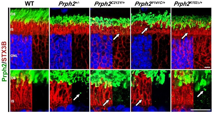
Figure 3. Mutated Prph2 protein traffics to the OS while a small pool is retained in the inner segment.
Retinal sections at P30 from the indicated genotypes were probed with antibodies against Prph2Cells 2020, 9, 784 12 of 25
(green) and syntaxin 3B (STX3B) (red). Arrows indicate regions of mislocalization of Prph2. OS, outer
segments; IS, inner segment; ONL, outer nuclear layer. Scale bar: 20 µm. Primary antibodies used for
immunostaining were polyclonal antibody against Prph2 C-terminus (Prph2-CT) [22] and
monoclonal antibody against STX3B [111] (inner segment marker) diluted at (1:1000). AlexaFluor
conjugated secondary antibodies (Alexa 488 Rabbit and Alexa 555 Mouse, Life
Technologies/ThermoFisher) were used at a dilution of 1:1000 for 2 hours at room temperature.
Images were captured on a ZEISS Confocal LSM 900 microscope equipped with a Zeiss Axiocam
(Zeiss, Jena, Germany) using a 63× (oil, 1.4 NA) objective. Images were then processed using ZEN
Image Analysis software (Zeiss, Jena, Germany). All images shown are orthogonally projected from
an eight slice confocal z‐stack.
3.2.4. Prph2K153Δ/+ and Prph2K153Δ/K153Δ
Another mutation in PRPH2 that leads to variable phenotypes among patients is the deletion of
codon 153 (K153Δ) that results in the elimination of the lysine at position 153 in the D2 loop of Prph2.
This mutation is found to associate with RP, pattern dystrophy and fundus flavimaculatus [80].
K153Δ-Prph2 knockin mouse model was generated and provided evidence that the mutant protein
cannot form the complexes required for OS formation [79]. The heterozygous knockin mice
(Prph2K153Δ/+) displayed a shortened OS with minor structural defects at P30 [79] (Figure 1). At P180,
these animals also exhibited a reduction in ONL thickness without further structural deterioration of
the OS [79]. The overall Prph2 protein level in these mice was around 80% compared to the level in
WT mice, thus demonstrating that the observed structural defects are most likely caused by the
dominant effect of the mutant protein rather than due to haploinsufficiency.
The heterozygous animals displayed a significant progressive reduction in scotopic responses
that started as early as P30 (Figure 2) and worsened with age. The photopic response in the Prph2K153Δ/+
was also significantly reduced at P30, albeit this reduction did not worsen as the animals aged (Figure
2). In the homozygous animals, the scotopic and photopic responses were minimal at P30.
Biochemical analysis showed that the formation of covalently linked Prph2 dimers in Prph2K153Δ/K153Δ
mice was abolished while Rom1 homodimers were present [79].
Due to the observed effects on cone function, Prph2K153Δ/+ animals were crossed into the Nrl-/-
background in order to assess the effects of the K153Δ mutation on cones. Photoreceptor cells in the
resulting Prph2K153Δ/+/Nrl-/- retina displayed a highly disrupted structure with many photoreceptors
having no lamellae what so ever. Interestingly, the mutant Prph2 was able to interact with Rom1 in
the rod-dominant Prph2K153Δ/K153Δ retinas, but this interaction was abolished in Prph2K153Δ/K153Δ/Nrl-/-
retina, indicating a defect in the Prph2/Rom1 interaction specific to cones [79]. Sucrose gradient
velocity sedimentation showed no significant alteration in complex formation in Prph2K153Δ/+ retina
when compared to WT while in the Prph2K153Δ/K153Δ retina, the formation of intermediate and higher
order complexes is abolished [79]. Here, both Prph2 and Rom1 are restricted to the tetramer fractions.
The same was observed in the Prph2K153Δ/K153Δ/Nrl-/- retinas. Sedimentation profile showed that the
amount of higher order complexes in the Prph2K153Δ/+/Nrl-/- retina was reduced, while unaffected in
Prph2K153Δ/+ retina [79]. This provides further evidence for a differential role of the lysine at position
153 in rods and cones, and hence emphasizes the notion for potential varied roles for Prph2 in rods
versus cones. Localization studies performed in Prph2K153Δ/+ mice demonstrated that most of Prph2
and Rom1 were successfully transported to the OS with some amount of Prph2 mislocalized to the IS
(arrows in Figure 3). Small amount of rhodopsin (Rho) and M-opsin were also found to be
mislocalized to the ONL and outer plexiform layer (OPL) in this model [79].
In the Prph2K153Δ/K153Δ retinas, the majority of Rho and Prph2, but not Rom1, were found to be
mislocalized to the ONL. As stated above, patients with the K153Δ mutation exhibit a highly variable
phenotype, ranging from rod dominant RP to more cone related defects in the macula [80]. The K153Δ
knockin mouse model displayed functional and structural defects in both rods and cones, and thus
mimics the phenotype seen in patients carrying this mutation. While the lack of a macula in the
murine retina made it impossible to observe the macular pattern dystrophy often found in patients
[80], the knockin mouse still showed funduscopic anomalies [79] which are characteristic of theCells 2020, 9, 784 13 of 25
pattern dystrophy in patients. Prph2K153Δ/+ mice show a flecking in the fundus at P180 which is more
severe in the Prph2K153Δ/K153Δ mice. This phenotype does not deteriorate further in P365 heterozygous
animals while the flecking was replaced by large splotches in the homozygous animals [79].
Gene supplementation using the NMP mouse that over-express Prph2 (NMP/Prph2K153Δ/+ and
NMP/Prph2K153Δ/K153Δ) rescued the structural defects but failed to rescue the functional decline seen in
scotopic and photopic ERGs [79]. This indicates that the presence of the mutant protein alone is
sufficient to deteriorate the photoreceptor function and suggest that gene silencing along with gene
augmentation is the best strategy for this model.
The knockin models for PRPH2 related patient mutations proved to be very useful in
highlighting dominant-effects of the Prph2 mutations. In general, the models were more successful in
mimicking patient phenotypes related to a decline in rod function. Patient phenotypes related to
functional defects in the cones or pattern dystrophies in the macula were more difficult to reproduce
in mice due to the lack of a macula and a lower overall percentage of cones in the murine retina.
Crossing the knockin mouse models with Nrl-/- mice, as done in the studies with the K153Δ model
[79], has proven to be a successful approach in studying the effects of the mutation on cones in detail.
While the knockin models could not reproduce the macular pattern dystrophy, they successfully
reproduced the funduscopic aberrations, which connect with the pattern dystrophy.
3.2.5. Prph2N229S/+ and Prph2N229S/N229
The knockin mouse that alters the N-linked glycosylation at asparagine 229 (N229S) in the D2
loop was used to study if Prph2 glycosylation plays a role in its interaction with Rom1. Heterozygous
mice (Prph2N229S/+) did not have any significant changes in structure or function of the OS [75].
However, homozygous mice (Prph2N229S/N229S) displayed a late onset thinning of the outer nuclear layer
(ONL) and occasional abnormal disc staking in cones and a slightly reduced photopic ERG at P180.
Since Prph2 could not be glycosylated, higher order complexes were decreased and there was an
increase of Prph2 and Rom-1 in the intermediate complexes [75]. Therefore, it was concluded that the
glycosylation plays a major part in regulating the interaction between Prph2 and Rom1, which is
critical for cone health.
Tables 1 and 2 summarize the phenotypes associated with the Prph2 knockin models,
highlighting rod and cone structure, Scotopic and Photopic ERG, complex formation and protein
localization. It also summarizes fundus observations, and patient’s phenotypes whether mainly rod-
or cone-specific defects or the combination of the two. Figures 1 and 2 show structural and functional
differences between Prph2 heterozygous knockin mutations, while Figure 3 is a representative IF
showing retinal localization of Prph2 among the models.
Table 1. Prph2 mutations and correlating phenotypes.
Genotype Prph2+/- Prph2-/- Prph2K153∆/+ Prph2K153∆/K153∆ Prph2Y141C/+ Prph2Y141C/Y141C
Small OS with
Short OS with
flattened whorls
Short OSs Short OSs Almost no Oss longer discs and
and vesicular
with whorl with whorl with rare whorl accumulation of
structures at P30,
Mouse Rod structures No OS structures structures and vesicular
no OS present at
Structure and ONL structures. and ONL ONL thinning structures, no
P180, ONL
thinning at thinning at at P30, more ONL thinning at
thinning at P30,
P180. P180. severe at P180. P30, ONL
more severe at
thinning at P180.
P180.
Occasional
whorl
Mouse Oss with Open OS Occasional COS Abnormal and Occasional COS
shaped OS,
Cone whorl with no seen but mostly short COS seen but mostly
mostly open
Structure structures lamella absent. structures. absent.
OS with no
lamella
57% and 96% and 90% and 89%
Scotopic 63% and 49% 54% and 27% 90% and 78%
33% 93% reduction in a-
ERG reduction in reduction in a- reduction in a-
reduction in reduction in and b-wave atCells 2020, 9, 784 14 of 25
a- and b- a- and b- a- and b- P30, and b-wave at and b-wave at
wave at P30. wave at P30, wave at P30. respectively. P30 P30.
74% and respectively. 83% and at 50% and 25% 95% and 94%
48% 60% reduction in a- reduction in a-
reduction in reduction in and b-wave at and b-wave at
a- and b- a- and b- P180, P180,
waves at wave at P180, respectively. respectively.
P180, respectively.
respectively.
Photopic b-
wave
comparable
24%
to WT at 91%
reduction in 64% reduction 10% reduction in 64% reduction in
Photopic P30. reduction in
b-wave P30 in b-wave at b-wave at P30 b-wave at P30
ERG 35% b-wave at
and 50% at P30. and P180. and 90% at P180.
reduction in P30.
P180.
photopic b-
wave at
P180.
No Prph2
dimers were Prph2 Prph2 almost
formed, while occasionally exclusively
Prph2 Rom1 dimers found in found in
complexes were still abnormal high abnormal high
and formed. molecular weight molecular weight
Prph2 distribution Prph2 aggregates. aggregates.
complexes unchanged, interacted with Abnormal Abnormal
and while Rom1 Rom1. Prph2 aggregates were aggregates were
distribution shifted and Rom1 held together by held together by
No Prph2
unchanged, towards restricted to intermolecular intermolecular
present,
50% less tetramers. tetramers. disulfide bonds. disulfide bonds.
Complex Rom1 still
Prph2 and On NRL-/- On NRL-/- Rom1 also Rom1 also
formation present but
Rom1. background, background, no present in present in
at lesser
On NRL-/- higher order Prph2 dimers abnormal abnormal
amount.
background, complexes were formed, aggregates. aggregates.
higher order decreased, while Rom1 Intermediate and Intermediate and
complexes shift of Prph2 dimers were higher order higher order
decreased. and Rom1 still formed. complexes complexes
towards Prph2 did not formed but reduced in favor
intermediate interact with abnormal high of the abnormal
complexes. Rom1. Prph2 molecular weight high molecular
and Rom1 aggregates also weight
restricted to present. aggregates.
tetramers.
Small
Huge amount
Some amount of
Rhodopsin of rhodopsin
rhodopsin rhodopsin
Protein mislocalized and Prph2
detected in and M-opsin NA NA
localization to IS and mislocalized in
the IS and mislocalized
ONL. the IS and
ONL. in the IS and
ONL.
ONL.
Flecking and Flecking at Severe flecking
No splotches at P180 and no at P180 and big
Fundus Flecking at P180. Flecking at P180.
abnormality P360 and change at splotches at
older. P365. P365.
Night blindness
Rod defect
NA NA RP NA and RP reported NA
in patients
in some patients.
Pattern Pattern
Cone
dystrophy dystrophy
defect in NA NA NA NA
and fundus changed fundus
patients
flavimaculs. in macula.
Reference [26,76,79] [26,76,79] [79] [79] [82] [82]Cells 2020, 9, 784 15 of 25
Table 2. Prph2 mutations and correlating phenotypes (continued).
Genotyp Prph2C213Y/ Prph2C213Y/C Prph2N229S/ Prph2N229S/N Prph2C150S/ Prph2C150S/C Prph23073
Prph2307/+
e + 213Y + 229S + 150S 07
Shortened
OS,
irregular Short OS Shortened
Modest ONL
structure, formed OS, ONL
elongatio thinning
some disc with highly elongated thinning
Modest n of discs, starting at
Rod structure disorganize Structure discs starting
ONL occasional P60. Rod
structure better d discs. unaffecte curving at P30
thinning at formation OS
mouse organized Severe d. into and rod
P180. of whorls. shortened,
than in ONL whorls. No OSs and
No ONL formation
Prph2+/-. thinning at ONL absent.
thinning. of whorls.
ONL P30. thinning.
thinning
at P30.
Shortened
Well Abnormall
Occasional OS,
organized y stacked
Cone Normal abnormal elongated Cone
lamella at lamella
structure COS disc NA discs NA OS are
P30 but with whorl
mouse structures. stacking at curving absent.
slightly shaped
P180 into
shorter. structures.
whorls.
60% and 60% and
47% 62%
reduction reduction
in a- and 50% and in a- and
96% and ERG a- 75% and
b-wave ERG a- and 30% b-wave at
94% and b- 56%
P30. b-wave reduction P180.
reduction wave reduction Scotopic
Scotopic 89% and comparable in a- and 80% and
in a- and b- comparab in a- and b- ERG
ERG 67% to WT at b-wave at 75%
wave P30, le to WT wave P30, absent
reduction P30 and P30, reduction
respectivel at P30 and respectivel
in a- and P180. respective in a- and
y. P180. y.
b-wave at ly. b-wave at
P180, P300,
respective respectivel
ly. y.
33% and 60% and
46% 79% ERG b- Normal at 80%
reduction reduction wave P30 but 29% 64% reduction
Photopi
Photopic in b-wave in b-wave comparab 22% reduction reduction in b-wave
c ERG
ERG at P30 at P30 le to WT reduction in b-wave in b-wave at P180
absent
and P180, ERG UV b- at P30 and in b-wave at P30. at P30. and P300,
respective wave. P180 at P180. respectivel
ly. y.
Mutant Mutant
Prph2 Prph2 Strong
Prph2
unable to unable to decrease in
could not
interact interact Prph2 and
be
with with Rom1 Prph2 Reduction Rom1
glycosylate
Rom1 in in rods. could not in Prph2 protein
d. Amount
rods. Protein be protein levels. No
of higher
Intermedi levels of glycosylat levels, less intermediat
order
Complex ate and Rom1 ed and pronounc e or higher
complexes
formatio higher decreased formed ed than in order
decreased,
n rods order No normal Prph2+/-. complexes
while the
complex intermediat Prph2/Ro Rom1 were
amount of
formation e or higher m1 protein formed.
intermediat
slightly order complexes levels not Prph2 and
e
impaired. complexes . affected. Rom1
complexes
Rom1 were restricted
was
occasional formed to
increased.
ly found Prph2 and tetramers.
in higher Rom1Cells 2020, 9, 784 16 of 25
order restricted to
complexes tetramers
.
Prph2
almost
completely
mislocalize
Prph2
d to IS and
partially
ONL. Small
mislocaliz
amount of
ed to IS
Protein Rom1
and ONL.
localizati mislocalize None None None None NA NA
Small
on d to IS. M-
amount of
and S-
Rom1
Opsin as
mislocaliz
well as
ed to IS.
rhodopsin
mislocalize
d to the IS
and ONL.
Flecking at
P180, more
Flecking pronounce
at P180, d than in
Fundus None None NA NA NA NA
persisted C213Y/+,
till P365. replaced by
splotches at
P365.
Rod No No
defect NA NA patient NA patient NA NA NA
patient model model
Cone No No
Pattern
defect NA patient NA patient NA NA NA
dystrophy
patient model model
Referenc
[76] [76] [75] [75] [78] [78] [107] [107]
e
4. Gene Therapy of PRPH2 Mutations
There is a vast variety of PRPH2 mutations associated with autosomal dominant retinal
degenerative diseases, including RP, several forms of macular dystrophy and cone rod dystrophies
[29]. Gene therapy seems to be a promising approach in the treatment of those PRPH2 associated
diseases. The small size of the Prph2 cDNA, which is approximately 1.1 kb, is advantageous since
many vectors used for gene therapy can only carry DNA of a limited size. Despite the variety of
feasible approaches available for the gene therapy of Prph2, thus far no treatment ready for clinical
trials has been developed.
The expression of the WT Prph2 in the background of pathogenic Prph2 mutations represents
one of the most promising approaches. Pioneer studies using transgenic mice expressing WT Prph2
under different promoters in a Prph2+/- or Prph2-/- background revealed a significant increase in both
structural and functional properties of rods and cones [105,112]. Replacing Prph2 in Prph2-/- mice
utilizing adeno-associated virus (AAV) carrying Prph2 regulated under the rhodopsin promoter
provided promising results [113]. Here the subretinal injection of the AAV resulted in a partial rescue
of ROS structure as well as scotopic ERG response. A follow up study was able to show that repeated
injections with the AAV resulted in an even more pronounced rescue of the phenotype with an
increase in the scotopic b-wave response observed in the injected mice [114]. In this study, several
time points after the injections were analyzed in order to validate whether the observed rescue was
long lasting. This analyses revealed that the rescue achieved by the AAV injection was lost after 15
weeks post injection [114]. In addition to the loss of the rescue with time, the amplitude of the scotopic
b-wave was significantly lower than in WT mice and the scotopic a-wave was not improved by theYou can also read