Conservation and divergence of meiotic DNA double strand break forming mechanisms in Arabidopsis thaliana - Oxford Academic Journals
←
→
Page content transcription
If your browser does not render page correctly, please read the page content below
Published online 30 August 2021                                                    Nucleic Acids Research, 2021, Vol. 49, No. 17 9821–9835
                                                                                                         https://doi.org/10.1093/nar/gkab715

Conservation and divergence of meiotic DNA double
strand break forming mechanisms in Arabidopsis
thaliana
Nathalie Vrielynck1,† , Katja Schneider2,† , Marion Rodriguez1 , Jason Sims2 ,
Aurélie Chambon1 , Aurélie Hurel1 , Arnaud De Muyt1 , Arnaud Ronceret1 , Ondrej Krsicka                                                                  2
                                                                                                                                                               ,
Christine Mézard1 , Peter Schlögelhofer 2,* and Mathilde Grelon 1,*

                                                                                                                                                                   Downloaded from https://academic.oup.com/nar/article/49/17/9821/6357737 by guest on 26 November 2021
1
 Institut Jean-Pierre Bourgin, INRAE, AgroParisTech, Université Paris-Saclay, 78000 Versailles, France and
2
 Department of Chromosome Biology, Max Perutz Labs, University of Vienna, Vienna Biocenter, Dr. Bohr-Gasse 9,
1030 Vienna, Austria

Received February 01, 2021; Revised July 16, 2021; Editorial Decision August 04, 2021; Accepted August 04, 2021

ABSTRACT                                                                          bling that occurs during fertilization. In most organisms,
                                                                                  correct separation of chromosomes at the first meiotic di-
In the current meiotic recombination initiation                                   vision relies on the formation of bivalents. These stable
model, the SPO11 catalytic subunits associate with                                structures are connected homologous chromosomes, held
MTOPVIB to form a Topoisomerase VI-like com-                                      together by crossovers (COs) and sister chromatid cohesion.
plex that generates DNA double strand breaks                                      COs, the reciprocal exchange of DNA between two homolo-
(DSBs). Four additional proteins, PRD1/AtMEI1,                                    gous chromosomes, are one of the outcomes of homologous
PRD2/AtMEI4, PRD3/AtMER2 and the plant spe-                                       recombination. At least one CO per homologous chromo-
cific DFO are required for meiotic DSB forma-                                     some pair is required for bivalent formation and subsequent
tion. Here we show that (i) MTOPVIB and PRD1                                      balanced chromosomal segregation (1).
provide the link between the catalytic sub-complex                                   The cascade of events leading to CO formation has been
and the other DSB proteins, (ii) PRD3/AtMER2,                                     well described (2–4). In most species, formation of the oblig-
                                                                                  atory meiotic COs is ensured at different levels of the mei-
while localized to the axis, does not assemble a
                                                                                  otic recombination pathway. One is the programmed induc-
canonical pre-DSB complex but establishes a di-                                   tion of a large number of CO precursors: the meiotic DNA
rect link between the DSB-forming and resection                                   double strand breaks (DSBs). DSB numbers exceed the final
machineries, (iii) DFO controls MTOPVIB foci for-                                 number of COs by several orders of magnitude (5). The cel-
mation and is part of a divergent RMM-like com-                                   lular toxicity of DNA lesions is well known, suggesting that
plex including PHS1/AtREC114 and PRD2/AtMEI4                                      very robust control mechanisms for meiotic DNA scission
but not PRD3/AtMER2, (iv) PHS1/AtREC114 is abso-                                  and repair must be in place to prevent any meiotic catas-
lutely unnecessary for DSB formation despite having                               trophe. These include the temporal and spatial coupling of
a conserved position within the DSB protein network                               meiotic DSB formation with DNA replication, DNA repair,
and (v) MTOPVIB and PRD2/AtMEI4 interact directly                                 homologous chromosome engagement and synapsis (6–10).
with chromosome axis proteins to anchor the meiotic                                  A number of components of the meiotic DSB forming
                                                                                  machinery have been identified in several model species.
DSB machinery to the axis.
                                                                                  These show variable levels of conservation in terms of
                                                                                  protein sequences and/or functions. It is now accepted
INTRODUCTION                                                                      that the catalytic activity responsible for meiotic DSB for-
During meiosis, maternal and paternal chromosomes re-                             mation evolved from an ancestral topoisomerase function
combine and segregate in two consecutive divisions, gen-                          still present in Archaea and some eukaryotes (Topoiso-
erating genetically distinct haploid cells. This halving of                       merase VI) (11). The catalytic part of the Topoisomerase
the genome content is mandatory to prepare for the dou-                           VI-like complex, active during meiosis, is composed of two

* To
   whom correspondence should be addressed. Tel: +33 1 30833308; Email: mathilde.grelon@inrae.fr
Correspondence may also be addressed to Peter Schlögelhofer. Tel: +43 1 427756240; Email: peter.schloegelhofer@univie.ac.at
†
 The authors wish it to be known that, in their opinion, the first two authors should be regarded as joint First Authors.
Present addresses:
Arnaud De Muyt, Institut Curie, PSL Research University, CNRS, UMR3244, Paris, France.
Arnaud Ronceret, Instituto de Biotecnologı́a / UNAM, Av. Universidad #2001, Col. Chamilpa C.P. 62210, Cuernavaca, Morelos, Mexico.

C The Author(s) 2021. Published by Oxford University Press on behalf of Nucleic Acids Research.
This is an Open Access article distributed under the terms of the Creative Commons Attribution-NonCommercial License
(http://creativecommons.org/licenses/by-nc/4.0/), which permits non-commercial re-use, distribution, and reproduction in any medium, provided the original work
is properly cited. For commercial re-use, please contact journals.permissions@oup.com
9822 Nucleic Acids Research, 2021, Vol. 49, No. 17

subunits of the A monomer, SPO11. They associate to trig-        COMPASS histone methyl transferase complex responsible
ger meiotic DSB formation through coordinated transes-           for histone methylation at DSB sites. Mer2 in S. cerevisiae
terification reactions that involve the catalytic tyrosine of    could therefore be directly involved in the tethering of the
each SPO11 monomer together with each DNA strand of              DSB sites to the chromosome axis, establishing the indis-
the double helix. In Arabidopsis thaliana (and probably in       pensable contacts between the DSB catalytic complex lo-
plants in general), it is not a homodimer of SPO11 pro-          cated at the loops and the other DSB proteins located at
teins that is active but more likely a heterodimer com-          the axis (44).
posed of the two SPO11-1 and SPO11-2 proteins (12–18).              In this study, we took advantage of the systematic screen-
In A. thaliana, a distant homolog of the archaeal TopoVI         ing for DSB proteins previously performed in A. thaliana
B subunit, the MTOPVIB protein, is likely to play a cru-         (45,46) to clarify the cascade of events that occur during
cial role in SPO11 catalytic dimer formation, since its in-      the steps of meiotic DSB formation. We methodically inves-
teraction with each of the SPO11 monomers is required            tigated the interactions among the DSB proteins and also
to promote the interaction between SPO11-1 and SPO11-            between the DSB proteins, axis proteins and the DSB pro-

                                                                                                                                 Downloaded from https://academic.oup.com/nar/article/49/17/9821/6357737 by guest on 26 November 2021
2 (15). The situation could be different in species with ho-     cessing machinery. We also compared the localization and
modimeric Spo11 catalytic complexes, as illustrated by the       the epistatic relationships between three of the DSB pro-
fact that, in Saccharomyces cerevisiae, the complex formed       teins (MTOPVIB, SPO11-1 and PRD3/AtMER2) during
between Spo11 and Rec102/Rec104 [related to MTOPVIB,             meiosis. Lastly, we revisited the role of PHS1/AtREC114
(19)] is monomeric (20). Once DSB formation has been             in order to clarify its involvement in meiotic recombination
triggered, the SPO11 proteins remain covalently attached         initiation.
to the 5’ ends of the broken DNA (21), blocking DSB re-
pair until they are removed by the action of the MRX/N
complexes (Mre11 Rad50 Xrs2/NBS1) in conjunction with            MATERIALS AND METHODS
SAE2/COM1/CtIP (22–27). How DSB formation is coor-               Plant material and growth conditions
dinated with DSB repair is a largely unanswered question.
   Together with the components of the meiotic TopoVI-           Plants were grown in greenhouses with 70% humidity. Ara-
like complex, additional proteins are essential for meiotic      bidopsis thaliana were grown under a 16 h/8 h day/night
DSB formation (3,5,28). In S. cerevisiae, Rec114, Mei4,          photoperiod with temperatures of 19 and 16◦ C for day and
Mer2, Ski8 and the components of the MRX complex are             night, respectively. Nicotiana benthamiana were grown un-
all required for successful DSB formation. These proteins        der a 13 h/11 h day/night photoperiod with temperatures
are all conserved across distant phyla, even if their con-       of 25 and 17◦ C for day and night, respectively. The mutant
servation in terms of primary sequence can be very weak,         alleles used in the study are listed in Supplementary Table
as is the case for Rec114, Mei4 and Mer2 (29–31). Their          S5.
role in meiotic DSB formation can also be quite divergent
from one species to another. For example, while the MRX
                                                                 Construction of vectors for yeast two-hybrid (Y2H) and bi-
complex is strictly required for meiotic DSB repair in all
                                                                 molecular fluorescence complementation (BiFC) experiments
organisms tested, its involvement in meiotic DSB forma-
tion is restricted to S. cerevisiae and Caenorhabditis elegans   Y2H and BiFC plasmids were constructed as described in
(28). Similarly, the mRNA decay protein Ski8/Rec103 is re-       (15) by amplifying a full-length or truncated version of the
quired for DSB formation only in fungi (32–34) but proba-        cDNAs with primers flanked by aatB1 and attB2 recombi-
bly not outside this kingdom (35). Last, some DSB proteins       nation sites (Supplementary Tables S6 and S7). These were
appear to be completely specific to a given phylum. This is,     designed to remove the STOP codon from the cDNA. The
for example, the case of the recently identified mammalian       amplification products were cloned into pDONOR207 (In-
protein ANKRD31 (36,37) and the A. thaliana DFO pro-             vitrogen). For Y2H experiments, these entry vectors were
tein (38). The function of these DSB proteins during meiotic     used to generate the appropriate expression vectors after
DSB formation is still largely unknown (5,8,28), but they        LR reactions with pDEST-GADT7 and pDEST-GBKT7
could act as regulators of either the formation or the activa-   (47) for N-terminal fusions, or pGADCg and pGBKCg
tion of the catalytic core complex, to trigger DSB formation     (48) for C-terminal fusions. The plasmids pBIFP1 to pB-
in a timely and accurately regulated manner.                     IFP4 (49) were used for BiFC assays. The complete list of
   During meiosis, sister chromatids are structured into         pEntry clones generated in this study is given in Supple-
chromatin loops emanating from a protein structure called        mentary Table S6. Cloning of the full-length cDNAs of
the axis (39). Most of the proteins involved in meiotic re-      MTOPVIB, SPO11-1 and SPO11-2 was described previ-
combination, including DSB proteins, are axis-associated         ously in (15), of ASY1, ASY3 and ASY4 in (50), of PRD1
whereas DSB sites are located in the loops (40,41). The cur-     in (51), of PRD3 in (45), of NBS1 and MRE11 in (52).
rent working hypothesis proposes that in order to intro-         Full-length SKI8, PHS1, RAD50 and COM1 cDNAs were
duce DSBs and promote recombination, some regions of             obtained from the Arabidopsis stock center (ABRC stock
the loops need to be temporarily tethered to the axis (40,41).   numbers U23481, PENTR221-AT1G10710, U22216 and
During these events in S. cerevisiae, Mer2 occupies a key po-    PENTR221-AT3G52115, respectively). DFO and PRD2
sition since it establishes a physical link between the chro-    full-length cDNAs were amplified from Col-0 flower buds.
mosome axis and the DSB machinery (42,43). Mer2 is an            The two DFO splicing variants described in (38) were am-
axis-associated DSB protein, which directly interacts with       plified using the primers attB1DFO with either attB2DFO.1
Mei4 and Rec114 as well as with Spp1, a member of the Set1       or attB2DFO.2. The PRD2 coding sequence was amplified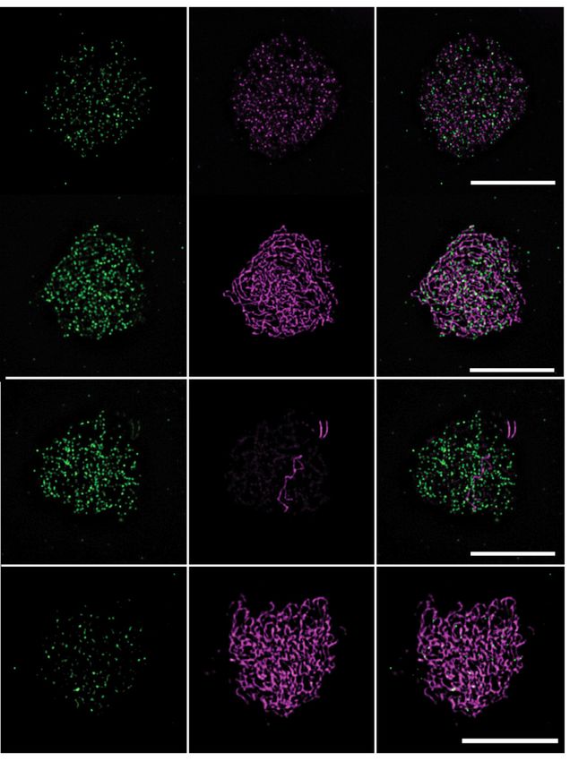
Nucleic Acids Research, 2021, Vol. 49, No. 17 9823
using primers MTI20-12#12 and MTI20-12#15 (see Sup- was introduced into a spo11-1-2 mutant background. Plant
plementary Table S7 for primer sequences). lines that were homozygous for both the AtSPO11-1-cMYC
transgene and the spo11-1-2 mutation were used for further
Yeast two-hybrid assays investigation.
Y2H assays were carried out using the Matchmaker™
GAL4 Two-Hybrid System 3 from Clontech as previously Cytology
described in (15). Briefly, the yeast plasmids were intro-
Seeds were counted after siliques clearing in 70% ethanol.
duced into AH109 or Y187 strains by lithium acetate trans-
Meiotic chromosome spreads were DAPI-stained as de-
formation. The appropriate pairwise combinations were
scribed in (54). Immunostaining was carried out either on
mated in non-selective media (YPD) and the resulting
spread male meiotic cells as described in (55) and (56) or on
diploid cells were selected on SD medium lacking the cor-
3D-preserved male meiocytes as described in (57). The pri-
rect combination of amino acids (SD-LW). Interactions
mary antibodies used were as follow: guinea pig ␣-ASY1
Downloaded from https://academic.oup.com/nar/article/49/17/9821/6357737 by guest on 26 November 2021
were then scored on selective media lacking leucine, tryp-
(1:250) (57), chicken ␣-ASY1 (1:50) (this study), guinea
tophan and histidine (SD-LWH) and adenine (SD-LWHA).
pig ␣-ASY1 (1:10 000) (58), rabbit ␣-DMC1 (1:20) (59),
All positive interactions were confirmed at least twice. West-
rat ␣-ZYP1 (1:250) (60), rabbit ␣-MLH1 (1:200) (56), rab-
ern blotting was used to verify expression in clones that re-
bit ␣-MTOPVIB (1:750; 1:400 for super-resolution experi-
sulted in negative interactions in all combinations tested as
ments) (15), rat ␣-REC8 (1:250) (61), rat ␣-RAD51 (1:100)
described in (15). The complete set of results is given in Sup-
(62), rabbit ␣-SCC3 (1:500) (63), rabbit ␣-ASY3 (1:300)
plementary Table S1.
(64), ␣-cMYC (abcam ab9106) (1:500), rat ␣-PRD3 (1:20)
(this study) and rabbit ␣-HEI10 (1:250) (65). Rat ␣-PRD3
Bimolecular fluorescence complementation assays
and chicken ␣-ASY1 antibodies productions are described
Protein interactions were tested in planta, using BiFC as- in Supplementary Materials and Methods. MTOPVIB,
says in N. benthamiana leaf epidermal cells expressing a nu- HEI10, DMC1 and RAD51 immunofluorescence studies
clear cyan fluorescent protein (CFP fused to histone 2B) were performed on 2D lipsol spread meiocytes as described
as previously described in (15). Briefly, for each target pro- in (55) together with either ␣-REC8 or ␣-ASY1 as axis
tein, four expression vectors were produced, generating in- staining markers or ␣-ZYP1 as a synapsis marker. Im-
active N- or C- moieties of YFP (YFPN , YFPC ), fused ages were taken using a Zeiss Axio Observer microscope.
with the target sequences at either their N- or C-termini. MTOPVIB, DMC1 and RAD51 foci number were quan-
Leaves were infiltrated with Agrobacterium tumefaciens cul- tified in Fiji, using a semi-automatized procedure as de-
tures transformed with the plasmids for the two candidates scribed in (66). MLH1 immunofluorescence studies were
to co-express the complementary YFP fusions (Supplemen- carried out on 3D preserved meiocytes as described in (57).
tary Table S1, BiFC detailed results). Results were scored Images were acquired using a Leica confocal microscope
for fluorescence 4 days after infiltration, using a LEICA TCS SP8 AOBS (Acousto-Optical Beam Splitter) (Leica
SP5 II AOBS Tandem HyD confocal laser scanning micro- Microsystems) with a 100× HCX PL APO, 1.4 NA im-
scope. The validity of the YFP signal was systematically mersion objective. Fluorescent signals were recorded using
checked by determining the fluorescence emission spectrum the Lightning mode of LASX software. Z-stacks with 0.13
of the signal (Supplementary Figure S1). A negative control m intervals were acquired and deconvolved using Light-
was included for each positive interaction detected consist- ning default parameters and the adaptative-vectashield op-
ing of each of the fusion proteins of interest expressed to- tion. MLH1 foci were counted using Imaris Spot tool.
gether with the complementary YFP moiety fused to unre- Slides for PRD3 and SPO11-1-cMYC immunofluorescence
lated proteins (i.e. DEFICIENS and GLOBOSA) (Supple- and for PRD3 or MTOPVIB STED nanoscopy were pre-
mentary Table S1 and Supplementary Figure S1). All inter- pared as described in (58,62,67). Before mounting the slides
actions were observed in at least two independent experi- with ProLong Glass medium (Invitrogen) they were washed
ments. twice in 2× SSC. The secondary antibodies were anti-rat
STARRED 1:100 (only for STED Abberior), anti-rabbit
Generation of CRISPR-Cas9 phs1 mutants Alexa 568 1:400 (Abcam, also used for STED), anti-rat
Alexa 568 (ThermoFisher) and anti-guinea pig Alexa 488
Single guide RNAs targeting the AtPHS1 gene (At1g10710)
(ThermoFisher). Immunostained nuclei detecting PRD3 or
were chosen using the CRISPOR selection tool (http://
SPO11-1-cMYC were imaged with a conventional fluores-
tefor.net/crispor/crispor.cgi) (53). The target locus was se-
cence microscope (Zeiss Axioplan2) and appropriate filters.
lected in the first PHS1 exon (sgRNA-PHS1#1: aaaccgc-
Z-stacks with 100 nm intervals were acquired, deconvolved
cgtagaaacgc) (Supplementary Figure S6). Details of the ex-
using AutoQuantX software and are presented as projec-
periments for generating phs1-2 and phs1-3 alleles are given
tions made with HeliconFocus software. Super-resolution
in Supplementary Materials and Methods.
images (PRD3 or MTOPVIB) were acquired using the Ab-
berior STEDYCON system. Protein foci were counted man-
Transgenic plant lines expressing AtSPO11-1-cMYC
ually with the help of the count tool in Adobe Photoshop.
An AtSPO11-1 expression construct was generated to ex- The distance of foci from the axis was measured with Adobe
press an 18x cMYC tagged genomic clone of AtSPO11- Photoshop.
1 under the control of its native promoter (see Supple- Scatter dot plots and statistical analyses were per-
mentary Materials and Methods for details). The construct formed using the GraphPad Prism 6 software. Statistical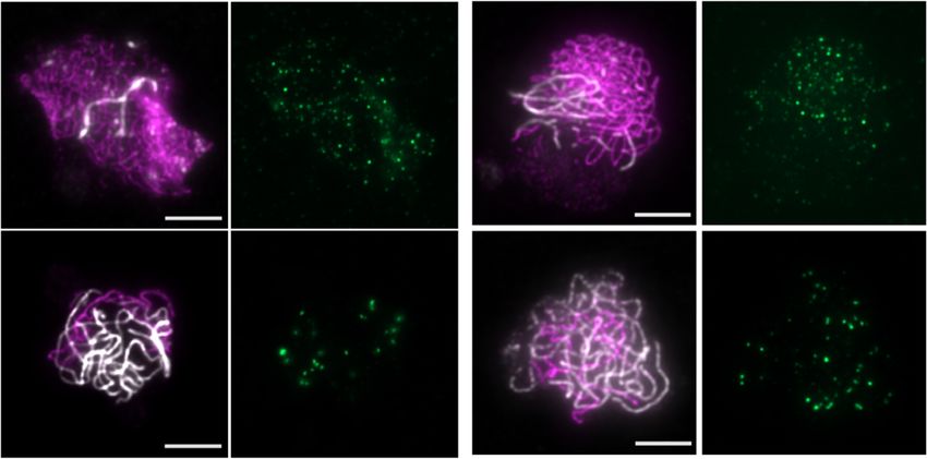
9824 Nucleic Acids Research, 2021, Vol. 49, No. 17

methods used were either two-sided Student’s t test or one-       vealed that the N-terminal domain of PRD1 is crucial for
way ANOVA with multiple comparison procedure. A sig-              most of the interactions (all but with AtMEI4/PRD2). In
nificance level of ␣ = 0.01 was chosen for all analyses.          the case of AtMEI4/PRD2, both the N- and C-terminal do-
                                                                  mains of PRD1 are involved (Figure 1D and Supplemen-
RESULTS                                                           tary Table S1).
                                                                     In S. cerevisiae and mouse, Rec114, Mei4 and Mer2
The A. thaliana DSB protein network and its association with
                                                                  (IHO1 in Mus musculus) are proposed to form a functional
the chromatin axis and DNA repair machinery
                                                                  entity [the RMM (Rec114-Mei4-Mer2) complex] based on
In order to gain insights into the function of the proteins in-   various evidence including a direct interaction among them
volved in meiotic DSB formation, we systematically tested         (see Discussion). Our study of the A. thaliana homologs
the interactions among them as well as with the axial ele-        revealed a direct interaction in Y2H between AtREC114
ment proteins ASY1, ASY3 and ASY4. We used a combi-               (PHS1) and AtMEI4 (PRD2). However, no direct interac-
nation of yeast two-hybrid (Y2H) and bimolecular fluores-         tion was detected between AtMER2 (PRD3) and the other

                                                                                                                                 Downloaded from https://academic.oup.com/nar/article/49/17/9821/6357737 by guest on 26 November 2021
cence complementation assays (BiFC or Split-yellow fluo-          two RMM-like components. Instead, our Y2H assay re-
rescent [YFP]) as described in (15). We also included Rec114      vealed strong interactions between the plant-specific DFO,
(PHS1) and Ski8 homologs (35,68) in the Y2H assays. A             AtREC114 (PHS1) and AtMEI4 (PRD2) proteins. Over-
summary of the results is shown in Figure 1 with the com-         all, in the DSB interaction network we found that AtMER2
plete set of data in Supplementary Table S1.                      (PRD3) establishes very few direct interactions with any of
   Overall, this interaction study revealed several important     the DSB proteins except with PRD1. A previous study (52)
features. First, the catalytic components of the meiotic DSB      reported that PRD3 interacts with MRE11 protein in A.
machinery (SPO11-1 and SPO11-2) show few direct interac-          thaliana. We therefore tested for possible interactions be-
tions with the DSB proteins other than MTOPVIB and no             tween AtMER2 (PRD3) and components of the resection
direct interactions could be detected with the axial proteins     machinery: MRE11, RAD50, NBS1 and COM1. We con-
ASY1/3/4. In contrast to the situation in S. cerevisiae, no       firmed the interaction between PRD3 and MRE11 and also
interaction could be detected between AtSKI8 and SPO11            detected an interaction with COM1 (Figure 1C and Supple-
proteins (Supplementary Table S1), consistent with previ-         mentary Table S1), showing that, in Arabidopsis, AtMER2
ous reports suggesting that in A. thaliana SKI8 is not in-        (PRD3) establishes a link between the DSB-forming and re-
volved in meiotic recombination (35).                             section machineries. MRE11 immunostaining was similar
   Second, we found that the MTOPVIB protein plays                in wild-type and prd3-3 backgrounds (Supplementary Fig-
a central role in the meiotic DSB protein network. We             ure S2) suggesting that interaction of PRD3 with the resec-
previously showed that the MTOPVIB interaction with               tion machinery per se is not necessary for MRE11 loading
SPO11-1 and SPO11-2 is necessary to promote the inter-            and/or stability.
action between the two SPO11 paralogs (15). MTOPVIB                  Finally, we explored the possible links between the DSB
has also been shown to interact with PRD1 (69). Here              forming proteins and the axis. We focused on the ASY pro-
we found that in Y2H assays, MTOPVIB establishes a di-            teins (ASY1, ASY3 and ASY4), the functional homologs of
rect interaction with most of the DSB proteins, with the          the mammalian HORMADs/SYCP2/SYCP3 respectively
notable exception of PRD3/AtMER2. Hence, it is likely             (Hop1/Red1 in S. cerevisiae). We and others have shown
that MTOPVIB establishes a connection between the cat-            previously that ASY1 interacts with ASY3 and that ASY3
alytic components of the complex and its regulatory fac-          interacts with ASY4 (50,64,70,71). Here we found that
tors. We previously showed that the last 149 amino acids of       among the DSB proteins, MTOPVIB and PRD2/AtMEI4
MTOPVIB (MTOPVIB345-493 ) were sufficient to establish            establish robust interactions with the three axis components
the interaction with the N- terminal regions of SPO11-1 and       ASY1, ASY3 and ASY4. In contrast, PHS1, DFO, PRD1
SPO11-2 (15). We refined this finding by showing that the         and PRD3, interacted with only some but not all of the
divergent C-terminal domain of MTOPVIB (aa 435 to 493)            tested axis components (Figure 1 and Supplementary Ta-
is dispensable for these interactions (Figure 1D and Sup-         ble S1).
plementary Table S1), restricting the interaction domain to
90 aa (MTOPVIB345-434 ) that correspond to the MTOPVIB
                                                                  Formation of SPO11-1-cMYC foci on meiotic chromatin de-
b4 conserved motif defined in (15). This restricted interac-
                                                                  pends on catalytic core complex partners
tion interface is specific to the interactions with the SPO11
proteins. The interactions with the other DSB proteins in-        In order to study SPO11-1 dynamics during Arabidopsis
volve much larger domains of MTOPVIB (Figure 1D and               meiosis we tagged the C-terminus of SPO11-1 with 18
Supplementary Table S1).                                          cMYC epitopes and complemented the spo11-1-2 mutant
   Third, our results combined with those of Tang et al. (69)     (Supplementary Figure S3). We then analyzed the spatial
reveal that the PRD1 protein also occupies a central posi-        and temporal localization of SPO11-1-cMYC during male
tion within the meiotic DSB protein network. PRD1 inter-          meiosis using a specific antibody against the cMYC epitope.
acts with all the DSB proteins, suggesting that it could act      We observed that SPO11-1-cMYC forms numerous foci on
as a platform, possibly supporting the confluence among           chromosomes from early leptotene (defined as nuclei with a
DSB proteins. Its direct interaction with AtMER2/PRD3             materializing chromosome axis characterized by a discon-
shows that one important function of PRD1 is to connect           tinuous ASY1 signal), zygotene and throughout pachytene
AtMER2 to the other components of the DSB forming ma-             (Figure 2A) (161 foci ± 30 [n = 13] in leptotene nuclei; 173
chinery. Truncation of PRD1 to various subdomains re-             foci ± 46 [n = 12] in zygotene nuclei; 239 foci ± 30 [n = 9]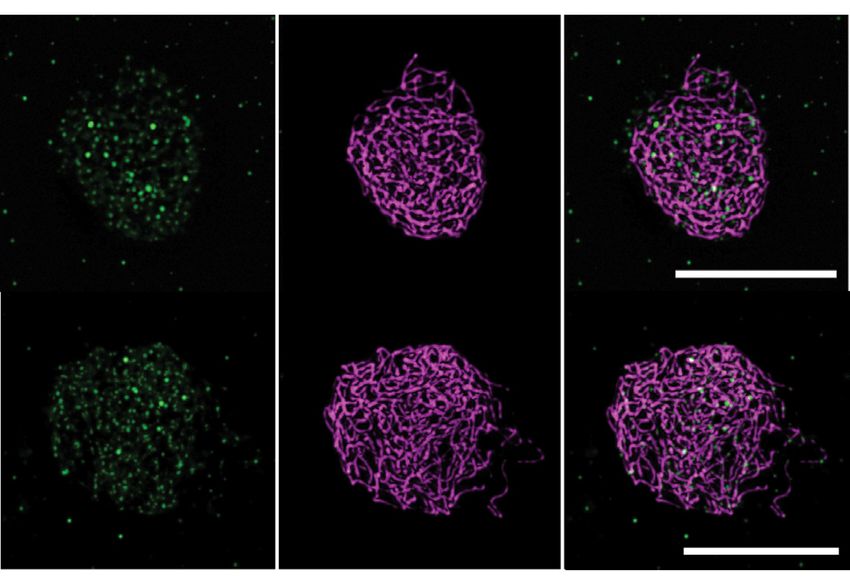
Nucleic Acids Research, 2021, Vol. 49, No. 17 9825

                                                                                                                                                                 Downloaded from https://academic.oup.com/nar/article/49/17/9821/6357737 by guest on 26 November 2021
Figure 1. The DSB protein interaction network. (A) Summary of the yeast two-hybrid assay results. Dark green indicates growth on SD-LWHA for at least
one of the combinations tested (involving either full-length or truncated versions of the tested proteins). Light green indicates growth on SD-LWH medium
for at least one of the combinations tested. Pink indicates that none of the combinations tested conferred auxotrophy. The complete set of results is given in
Supplementary Table S1. (B) Summary of the bimolecular fluorescence complementation (BiFC) assays. Green indicates that an interaction was detected
with at least one of the combinations tested. Pink indicates that none of the combinations tested conferred a YFP signal. Gray indicates that the interaction
was not tested. The complete set of results is given in Supplementary Table S1. Interactions of PRD1 with SPO11-1, SPO11-2, MTOPVIB, PRD3 and
DFO correspond to results reported by (69). (C) Schematic representation of the interaction network. Green arrows: Strong interactions between two
proteins. Bold letters: proteins essential for DSB formation. Gray shapes: Proteins that directly interact with axial proteins ASY1, ASY3 or ASY4. (D)
Detailed depiction of interaction domains defined in yeast two-hybrid assays. Schematic representation of the DSB proteins with their functional domains
(WHD for winged-helix domain, transd. for transducer domain, PH for pleckstrin homology domain). Blue vertical bars indicate structurally conserved
motifs. Below each protein, black bars represent the regions that interact with the proteins indicated on the right or below the bars.

in pachytene nuclei; Figure 2 and Supplementary Table S2).                        (when the ASY1 signal is not yet linear) from the rest of
These data are consistent with results published in (72).                         prophase (when the ASY1 signal is linear). In meiocytes
   SPO11-1-cMYC was detected on spreads of meiotic chro-                          of prd1, prd3 or dfo, SPO11-1-cMYC foci numbers were
matin in prd1, prd2, prd3, dfo, spo11-2 and mtopVIb mu-                           not significantly different to those in wild-type through-
tant lines (Figure 2, Supplementary Figure S4 and Supple-                         out prophase (Figure 2). While wild type-like SPO11-1-
mentary Table S2). All these mutants are synapsis-defective                       cMYC plants had on average 186 foci (±48, n = 34; all
therefore we could only discriminate early leptotene stages                       prophase stages), prd1 mutants had 156 (±33, n = 26), prd3
9826 Nucleic Acids Research, 2021, Vol. 49, No. 17
Downloaded from https://academic.oup.com/nar/article/49/17/9821/6357737 by guest on 26 November 2021
Figure 2. Immunolocalization of SPO11-1-cMYC on spreads of male meiocytes. (A) Co-immunolocalization of the axis protein ASY1 (magenta) and
SPO11-1-cMYC (green) in SPO11-1-cMYC expressing plant lines (containing the spo11-1-2–/– mutation) and wild-type (Col-0) (without SPO11-1-cMYC);
size bar: 10 m. (B) Detection of SPO11-1-cMYC foci in spo11-2-3 and mtopVIb mutants; size bar: 10 m. (C) Quantification of SPO11-1-cMYC foci in
Col-0 (Wt), spo11-1-2 (L: leptotene, Z: zygotene, P: pachytene), spo11-2-3, mtopVIb-2, prd1-2, prd2-1, prd3-4, dfo-2, asy1-2 and asy3-1 (see Supplementary
Figure S4). Statistical analysis was used to compare the mean of SPO11-1-cMyc in spo11-1-2 (all stages included) to the mean of all other plant lines
indicated (one-way ANOVA, with Dunnett correction for multiple comparisons, 99% confidence interval; ns, 0.01 < P; *** P < 0.0001; error bars represent
SD).
mutants 163 (±34, n = 30) and dfo mutants 186 (±45, (mean ± SD, n = 97). In prd1, prd2 and prd3 mutants, no
n = 32) foci (combined prophase stages). The prd2 mutant, significant differences in MTOPVIB foci numbers were ob-
however, showed a slight but significant reduction in foci served (Figure 3 and Supplementary Table S3). Mutation
numbers with an average of 129 (±49, n = 18) foci. of DFO does, however, modify MTOPVIB abundance. An
Remarkably, mutants related to factors of the DSB form- almost two-fold increase (353 ± 134, n = 32) in the number
ing core complex, SPO11-2 and MTOPVIB, had signifi- of foci was observed in dfo-1 mutants (Figure 3 and Supple-
cantly lower SPO11-1-cMYC foci numbers when compared mentary Table S3). These results reveal that among all the
to the wild type-like SPO11-1-cMYC plants (75 ± 31, n = 30 DSB proteins, DFO is a negative regulator of MTOPVIB
and 87 ± 58, n = 39 in spo11-2 and mtopVIb, respectively). loading and/or stabilization onto chromatin.
These findings suggest that normal association of SPO11-
1-cMYC with meiotic chromatin requires the presence of
SPO11-2, MTOPVIB and to some extent PRD2 but not of Foci formation of the core complex proteins SPO11-1-cMYC
PRD1, PRD3 or DFO. and MTOPVIB is largely independent of the axial proteins
We then investigated whether SPO11-1-cMYC and
MTOPVIB foci formation is dependent on the presence
MTOPVIB foci formation is independent of most of the DSB
of a fully intact axis. As shown in Figure 2 and Supple-
proteins with the exception of DFO and SPO11-1
mentary Figure S4, no changes to SPO11-1-cMYC foci
MTOPVIB forms foci associated with meiotic chromo- numbers were detected in either asy1 or asy3 (170 ± 21.5
somes from leptotene to pachytene. These foci are abol- and 170 ± 22, respectively). MTOPVIB foci were only
ished in spo11-1 but not in spo11-2 mutant backgrounds slightly modified by mutations affecting the axial elements
(15). Here, we investigated MTOPVIB dynamics during (Figure 3 and Supplementary Table S3). In wild-type,
male meiosis in more DSB-defective backgrounds. In wild- 192 ± 53 (n = 97) MTOPVIB foci can be detected (all
type, the number of MTOPVIB foci is on average 192 ± 53 prophase stages included). In the pch2 and asy1 mutants,
Nucleic Acids Research, 2021, Vol. 49, No. 17 9827
catalytic machinery core to chromatin but can affect its
abundance or stability.
PRD3/AtMER2 forms axis associated foci that depend on
ASY1 and ASY3
To analyze the dynamics of PRD3/AtMER2 during male
meiosis, we raised an antibody against its N-terminus (AA
1–125). We observed that PRD3/AtMER2 appears early
during prophase, before the axis signal is linear and forms
numerous foci on unsynapsed chromosome axes during lep-
totene and zygotene (171 ± 48, n = 28; Figure 4 and Sup-
plementary Figure S5). This number strongly decreases as
Downloaded from https://academic.oup.com/nar/article/49/17/9821/6357737 by guest on 26 November 2021
synapsis proceeds to an average level of 40 foci (±22, n = 12)
at pachytene. To analyze the localization of PRD3 rela-
tive to the meiotic axis, we obtained images using STED
nanoscopy. In leptotene, PRD3 localizes pre-dominantly
with foci or stretches of the meiotic chromosome axis pro-
teins ASY1 (Supplementary Figure S5) or ASY3 (Figure
4A). During zygotene and pachytene, PRD3 foci can still
be detected, but mostly detected co-localizing with the un-
synapsed parts of the chromosome axis (Figure 4A). The
distance of PRD3 foci to the axis was measured on four lep-
totene, five zygotene and two pachytene cells. This showed
a close association of PRD3 foci with axes of unsynapsed
chromosomes (56 nm ± 22, n = 80) and a distant associa-
tion with axes after synapsis (144 nm ± 72, n = 46; Figure
4A).
Having established that PRD3 colocalizes with the mei-
otic axis, we were interested in whether PRD3 localiza-
tion depends on meiotic axis proteins. In both asy1 and
asy3 mutant nuclei, PRD3 foci numbers drop strongly from
171 ± 48 (n = 28) in wild-type to 65 ± 33 (n = 31) and
39 ± 24 (n = 28) foci/cell respectively (Figure 4C, Supple-
mentary Figure S5 and Supplementary Table S4), suggest-
ing that PRD3 foci formation/stabilization on the axes is
largely dependent upon these two proteins.
To test whether PRD3 binding to meiotic chromatin de-
pends on further DSB promoting proteins, we analyzed its
localization and abundance in meiocytes of mutants de-
pleted of different members of the DSB forming complex
(Figure 4C, Supplementary Figure S5 and Supplementary
Table S4). In all mutant lines PRD3 foci numbers were sig-
nificantly reduced compared to wild-type (P value < 0.0001;
ANOVA). While 171 (±48, n = 28) foci were counted in
wild-type leptotene cells, only 96 (±38, n = 21), 85 (±34,
Figure 3. Immunolocalization of MTOPVIB on spreads of male meio- n = 21) and 95 (±29, n = 30) were detected in prd1, prd2
cytes. (A) Co-immunolocalization of an axis protein (ASY1 or REC8, ma- and dfo, respectively. Even less foci were observed in mu-
genta) together with MTOPVIB (green); scale bar: 5 m. (B) MTOPVIB
foci counts in various mutant backgrounds. Statistical analysis compares
tants lacking a member of the catalytic core subunit (spo11-
mean foci numbers in each mutant to wild-type mean (one-way ANOVA 1, 51 ± 34, n = 33; spo11-2, 66 ± 35, n = 34; mtopVIb,
with Dunnett correction for multiple comparison, 99% confidence interval; 77 ± 30, n = 15 and spo11-1 spo11-2 70 ± 37, n = 39).
ns, 0.01 < P; * 0.001 < P < 0.01; *** P < 0.0001; error bars represent SD). We also analyzed MTOPVIB localization relative to the
Alleles investigated were mtopVIb-2, prd1-2, prd2-1, prd3-3, dfo-1, asy1-4, axis by STED nanoscopy. We observed that as prophase
pch2-1 and rec8-3.
progresses and synapsis takes place, MTOPVIB foci are lo-
cated further away from the axes, with an average distance
of 70 nm (±31, n = 75) at unsynapsed axes to 104 nm
(±46, n = 80) at synapsed axes (Figure 4B). Lastly, we per-
the numbers increased moderately (238 ± 54, n = 30 and formed a colocalization study on MTOPVIB and PRD3
233 ± 68, n = 60, respectively), whereas in the rec8 mutant in male meiocytes. We observed that the windows of ex-
they decreased slightly (150 ± 72, n = 34). These findings pression of PRD3 and MTOPVIB are not completely sim-
show that an intact axis is not crucial for loading the DSB ilar, with PRD3 appearing and disappearing earlier than
9828 Nucleic Acids Research, 2021, Vol. 49, No. 17
Downloaded from https://academic.oup.com/nar/article/49/17/9821/6357737 by guest on 26 November 2021
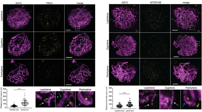
Figure 4. AtMER2/PRD3 and MTOPVIB form non-overlapping foci, more tightly associated with unpaired axes than paired axes. (A and B) Co-
immunolocalization of the axis protein ASY3 (magenta) with either PRD3 (A, yellow) or MTOPVIB (B, yellow) using STED nanoscopy. Enlargements
correspond to the boxes indicated with dashed white lines. White arrows indicate unpaired axes, and blue arrows indicate paired axes. Graphs represent the
distances measured from foci to the unpaired or paired meiotic axes. Differences between the mean values were evaluated (non-parametric Mann–Whitney
test; **** P < 0.0001; error bars represent SD); size bar: 2 m. (C) Quantification of PRD3 foci in Col-0 (Wt, L: leptotene, Z: zygotene, P: pachytene),
spo11-1-2, spo11-2-3, spo11-1-2 spo11-2-3, mtopVIb-2, prd1-2, prd2-1, prd3-4, dfo-2, asy1-2 and asy3-1 plants (see Supplementary Figure S5). Statistical
analysis was used to compare the mean of Col-0 (leptotene/zygotene) to the mean of each mutant (one-way ANOVA, with Dunnett correction for multiple
comparison, 99% confidence interval; ns, 0.01 < P; *** P < 0.0001; error bars represent SD). (D) Co-immunolocalization of the axis associated protein
ASY1 (magenta), PRD3 (yellow) and MTOPVIB (green). The reconstructed image shows the detected PRD3 (yellow spheres) and MTOPVIB foci (green
spheres) using the Imaris spot detection tool. Hardly any colocalization could be detected; size bar: 5 m.
Nucleic Acids Research, 2021, Vol. 49, No. 17 9829

MTOPVIB. At leptotene, when large numbers of foci re-             DISCUSSION
lated to both proteins are visible on chromosomes, hardly
                                                                  MTOPVIB and PRD1 occupy a central position within the
any colocalization could be detected between them (Figure
                                                                  DSB protein network
4D).
                                                                  Systematic investigation of the interactions among the DSB
                                                                  proteins revealed that MTOPVIB and PRD1 directly inter-
Is AtREC114/PHS1 involved in meiotic recombination initi-
                                                                  act with most of the other DSB proteins (Figure 1). We pre-
ation?
                                                                  viously showed that MTOPVIB is a key element of the DSB
Interaction studies revealed that AtREC114/PHS1 inter-            forming machinery since its interaction with SPO11-1 and
acts with several components of the meiotic DSB machin-           SPO11-2 is necessary to promote the interaction between
ery (see above). We therefore examined its role in meiotic        these two SPO11 paralogues (15), which likely promotes
DSB formation. Previously, Atphs1 mutants were reported           the assembly of the core catalytic complex. Here we showed
(68) to be required for RAD50 import into the nucleus. We         that in addition to this, MTOPVIB is also likely to establish

                                                                                                                                  Downloaded from https://academic.oup.com/nar/article/49/17/9821/6357737 by guest on 26 November 2021
therefore investigated the phenotype of phs1-1 mutants de-        a connection between the catalytic core complex (SPO11-1
scribed in (68) as well as additional publicly available in-      and SPO11-2) and most of its regulatory factors. In agree-
sertion lines (Figure 5A). Under our experimental condi-          ment with this, we observed that SPO11-1-cMYC foci for-
tions, none of these insertion alleles displayed phenotypes       mation is drastically perturbed in the mtopVIb mutant, a
related to fertility or chiasma formation (Supplementary          finding which suggests that MTOPVIB is not only promot-
Figure S6), raising the possibility that either these alleles     ing the assembly of the catalytic core complex but also its
are leaky or that the AtREC114/PHS1 protein plays no ma-          loading and/or stabilization on chromosomes. We found
jor role in reproduction. In order to clarify this, we gener-     that PRD3/AtMER2 is the only DSB protein that does
ated CRISPR-Cas9 null mutant alleles by targeting the first       not directly interact with MTOPVIB. This recapitulates the
exon in PHS1. We selected two independent lines, hereafter        situation in S. cerevisiae, where the Rec102/Rec104 dimer
termed phs1-2 and phs1-3, with either a deletion or insertion     [which is distantly related to the MTOPVIB/TopoVIBL
of a single nucleotide in position 41 of the cDNA (Supple-        protein family (74)] also interacts with all the other DSB
mentary Figure S6). These mutations introduce a frameshift        proteins except Mer2 (33). In A. thaliana, PRD3/AtMER2
at the beginning of the coding sequence of PHS1 (from             is connected to the other DSB proteins through a direct
codon 14, Supplementary Figure S6). To confirm that the           interaction with PRD1. PRD1 is a large protein of 1330
introduced mutations do not modify PHS1 splicing, we se-          amino acids showing clear sequence similarities with Mm-
quenced the PHS1 cDNA and did not find any differences            Mei1 but without any known homologues outside animals
between wild-type and the mutants. This indicates that both       and plants (51,75,76). We found that PRD1 directly inter-
alleles are likely to produce a modified variant of PHS1          acts with most of the other DSB proteins. This suggests
of 42 and 91 aa, respectively, sharing only 13 aa with the        that it could act as a platform to support the association
wild-type PHS1 protein (Supplementary Figure S6). There-          among DSB proteins and therefore likely to promote ei-
fore, it is very likely that phs1-2 and phs1-3 mutants are null   ther the assembly and/or the activation of the meiotic cat-
mutant alleles. Neither of the phs1-2 and phs1-3 homozy-          alytic complex. Its direct interaction with PRD3/AtMER2
gous mutants showed reduced fertility (seed count) or chi-        shows that one important function of PRD1 is to con-
asma formation (Supplementary Figure S6). We then an-             nect AtMER2 to the other components of the DSB form-
alyzed a number of markers of meiotic progression to de-          ing machinery, a role that could be conserved in mammals
cipher whether PHS1/AtREC114 mutation could have an               where PRD1 and MER2 homologs (MEI1 and IHO1, re-
impact on some steps of meiotic recombination. No change          spectively) colocalize with the other DSB factors REC114,
in MTOPVIB, RAD51, DMC1 or early HEI10 foci num-                  MEI4 and ANKRD31 (77).
bers was observed (Figure 5B and C, Supplementary Figure
S7). Last, we introduced the phs1-2 mutation into the DSB-
                                                                  Connection of the meiotic DSB machinery to the axis
repair-defective mutant mre11 (73). We observed that the
phs1-2 mutation was unable to suppress any of the fragmen-        An important result of the interaction analyses is that
tation defects observed in this background (Supplementary         several of the DSB proteins directly interact with one or
Figure S8). Taken together, these data show that meiotic          more of the components of the chromosome axial ele-
DSB formation and recombination initiation steps are not          ments (ASY1, ASY3 and ASY4). Among these, the in-
affected by loss of PHS1 in Arabidopsis. Nevertheless, when       teractions involving MTOPVIB and PRD2/AtMEI4 were
we quantified class I COs in two phs1 mutant alleles (phs1-2      the strongest (Figure 1). This confirms the key position of
and phs1-5), by detecting the MLH1 protein in male meio-          MTOPVIB in the DSB-forming machinery and suggests
cytes (Figure 5D), we observed a clear increase in MLH1           that MTOPVIB could play a direct role in connecting the
foci numbers in both mutants, from 10 ± 1.7 (n = 73) foci         DSB-forming machinery (including the catalytic compo-
per cell in wild-type to 11 ± 2 (n = 243) in phs1-2 and 14 ± 2    nents SPO11-1 and SPO11-2) to the axis. Further studies
(n = 148) in phs1-5 mutants. The increase was confirmed           are required to decipher whether MTOPVIB’s presence is
using another Class I CO marker (HEI10 staining at late           required for the stable axis association of all the other DSB
pachytene stage, Supplementary Figure S7). Taken together,        proteins with the axis. However, our finding that the number
these data reveal that REC114 (PHS1) in Arabidopsis is            of PRD3 and SPO11-1-cMYC foci is strongly reduced in the
clearly not needed for meiotic DSB formation but is nev-          mtopVIb mutant background (Figures 2 and 4) is heading in
ertheless a regulator of meiotic recombination outcomes.          this direction. Despite the direct interaction of MTOPVIB
9830 Nucleic Acids Research, 2021, Vol. 49, No. 17
Downloaded from https://academic.oup.com/nar/article/49/17/9821/6357737 by guest on 26 November 2021
Figure 5. AtREC114/PHS1 is not essential for meiotic DSB formation. (A) AtREC114/PHS1 gene structure. Blue rectangles represent exons. Colored
triangles indicate the position of the mutations (insertion lines in orange; CRISPR-Cas9 generated mutants in green). (B) Co-immunolocalization of the
axis protein ASY1 (magenta) and MTOPVIB (green) in wild-type and phs1-2 meiocytes. The graph shows the quantification of MTOPVIB foci numbers.
(C) Co-immunolocalization of the axis protein ASY1 (magenta), the central element protein ZYP1 (white) and RAD51 (green). Quantification of RAD51
foci according to the level of synapsis is shown. No or low ZYP1 signal, corresponding to leptotene or early zygotene were grouped together; extended
ZYP1 signal corresponding to late zygotene and pachytene stages were counted together. Quantification of DMC1 foci in wild-type and mutants is also
shown, all stages counted together. (D) Immunolocalization of MLH1 in wild-type, phs1-2 and phs1-5 meiocytes. The graph shows the quantification of
MLH1 foci number on meiocytes at diplotene and diakinesis stages (determined according to ZYP1 staining, not shown); scale bars = 5 m. Statistical
analyses compare mean foci numbers either between wild-type and mutant (MLH1 and RAD51 foci, unpaired Student’s t-tests) or between all means
(MTOPVIB and DMC1 foci numbers, one-way ANOVA, with Tukey correction for multiple comparison). All tests were analyzed using a 99% confidence
interval; ns 0.01 < P; *** P < 0.0001; error bars represent SD.Nucleic Acids Research, 2021, Vol. 49, No. 17 9831
with the axis, we observed that MTOPVIB foci formation be stabilized at the axis upon interaction of the axis- and
is not or only slightly modified in the axis mutants tested, loop-localized sub-complexes. The entire DSB complex
asy1 and rec8 (Figure 3), showing that an intact axis is not would be anchored at the axis relying on the various inter-
required for MTOPVIB loading. actions between DSB complex proteins and axis proteins,
So far, the only component of the DSB machinery that especially highlighting the central role of MTOPVIB. In
has been shown to be involved in the connection to the axis line with our observations, we propose that in the absence
is Mer2 (Rec15 and IHO1 in Schizosaccharomyces pombe of the other DSB factors, PRD3/AtMER2’s residence time
and mouse, respectively). In S. cerevisiae the axial element at the meiotic axis would be very limited. MTOPVIB and
components Red1 and Hop1 promote axis-localization of SPO11-1-cMYC (and most likely also SPO11-2) are highly
Mer2, which in turn recruits Rec114 and Mei4, to assem- abundant on meiotic chromatin and only a sub-fraction of
ble the RMM (Rec114, Mei4, Mer2) complex (41,78). In S. these proteins will be involved in generating meiotic DSBs.
pombe, Rec15 directly interacts with the axis proteins Rec10 The hyper-abundance of the core-complex components and
(SpRed1, ASY3) and Hop1 (SpASY1) (79,80) and in mam- their long-lasting chromatin association [until pachytene,
Downloaded from https://academic.oup.com/nar/article/49/17/9821/6357737 by guest on 26 November 2021
mals IHO1 interacts with HORMAD1 (MmHop1/ASY1) Figure 2 and (15)] suggests that the DSB core sub-complex
(81). In addition to these direct interactions of Mer2 with is inactive until it encounters all other partners of the
several axis components, converging studies revealed that DSB complex. This would include the axis-associated
most of the other DSB proteins are dependent upon Mer2 PRD3, apparently needed to activate the transesterification
for their association with the chromosome axis, and not vice reaction.
versa. This is, for example, the case in Sordaria macrospora
where SPO11::GFP foci are not formed in mer2Δ while
Minimal conservation of the RMM complex in A. thaliana
MER2::GFP foci are formed normally in spo11Δ and ski8Δ
(31). In S. cerevisiae, Rec114 and Mei4 foci formation are Rec114, Mei4 and Mer2 were first shown to form a sta-
absolutely dependent upon Mer2 but not the reverse. In ble complex in S. cerevisiae (33,84) and this was then con-
mammals, the association of IHO1 with the chromosome firmed in S. pombe (80). In mouse, the three proteins may
axis only depends on axial components and does not change also form a functional entity since direct interactions were
in the absence of MEI4 or REC114. However, as observed found between Mei4 and Rec114 (29). Our study revealed a
in S. cerevisiae, MEI4 foci formation is dependent on IHO1 direct interaction between AtREC114 (PHS1) and AtMEI4
(29,41,81,82). Based on these data, Mer2 was proposed to (PRD2) but none was detected between AtMER2 (PRD3)
play a conserved role promoting the assembly of the pre- and any of the other RMM-like components. Instead, we
DSB complex on the chromosome axes, through its dual revealed a strong interaction in yeast two-hybrid assays be-
interaction first with axial components, then with Rec114 tween the three proteins DFO, AtREC114 (PHS1) and At-
and Mei4. MEI4 (PRD2), suggesting that DFO but not MER2 tightly
In A. thaliana PRD3/AtMER2 interacts with ASY1 in associates with AtMEI4 and AtREC114 to regulate DSB
Y2H and BiFC assays (Figure 1 and Supplementary Table formation. DFO is a small protein of9832 Nucleic Acids Research, 2021, Vol. 49, No. 17
by DFO with AtREC114/PHS1 and AtMEI4/PRD2. In- cal RMM complex is not conserved in A. thaliana. How-
terestingly, MTOPVIB foci numbers are significantly higher ever, our study revealed that PRD3 interacts with the two
in dfo mutants, suggesting it plays an important role in reg- DNA processing and repair proteins MRE11 and COM1.
ulating the abundance of a key component of the meiotic These proteins are part of the meiotic DSB repair machin-
DSB complex. ery (MRN complex-MRE11, RAD50, NBS1 and COM1-
The involvement of the Arabidopsis REC114/PHS1 in a also known as SAE2/CtIP) and are both required for DSB
divergent RMM-like complex raised the question of its pre- processing, SPO11-oligo generation and correct DSB resec-
cise role in meiotic DSB formation. While the Rec114 pro- tion. While the prd3 mutant is defective in DSB formation,
tein sequence is poorly conserved (30,31) in all species in- mre11 and com1 are defective in DSB repair. Meiosis in
vestigated so far, Rec114 plays a key role in meiotic DSB mre11 and com1 mutants is characterized by strong chro-
formation (29,87–91). In contrast, we found that Arabidop- mosome fragmentation at the metaphase I/anaphase I tran-
sis phs1 mutants are completely fertile and show no ob- sition (25,73). Direct interaction between PRD3 and these
vious alteration of meiotic DSB formation with normal two components of the DSB resection machinery suggests
Downloaded from https://academic.oup.com/nar/article/49/17/9821/6357737 by guest on 26 November 2021
DMC1, RAD51, and early HEI10 foci formation and dy- that PRD3 has a role in either recruiting or activating the
namics (Figure 5 and Supplementary Figure S7). It has to DSB resection machinery, thereby allowing coordination
be noted that we cannot exclude the possibility that none between DSB formation and DSB repair. In S. cerevisiae,
of the phs1 allele investigated is null. But this appears very there is also a connection between Mer2 and the MRX com-
unlikely considering that we have analyzed seven indepen- plex since ScMer2 interacts in a yeast two-hybrid assay with
dent lines, corresponding to mutations covering the whole Xrs2 (ScNBS1) and MRE11 (33). However, since the MRX
coding sequence. In maize however, the phs1 mutant is com- complex is required for the DSB formation step itself in
pletely sterile, a phenotype associated with an absence of S. cerevisiae, a possible role in coordinating DSB forma-
RAD51 foci formation and occurrence of non-homologous tion and DSB repair has not been investigated. This poten-
synapsis (92). These phenotypes are also observed in the tial role of AtMER2/PRD3 in coordinating DSB formation
maize spo11-1 mutant (17), therefore we propose that, in and the initial resection steps of homologous recombination
maize, REC114 function is conserved and that PHS1 is can be put in parallel with the recent findings obtained in S.
required for meiotic DSB formation. Such discrepancies macrospora: Mer2 is needed for pre-DSB complex assembly
in the function of conserved meiotic genes have already and also for subsequent meiotic recombination, since it is
been described, notably concerning the mechanisms of mei- required to promote spatial pairing and synapsis of the ho-
otic DSB formation. For example, the two genes P31COMET mologous chromosomes (31). It would be interesting to test
and PCH2 are essential for meiotic DSB formation in rice whether these post-DSB functions of Mer2 in S. macrospora
(93,94) but not in A. thaliana (95–97). Rice together with depend on the interaction of Mer2 with the Mre11 complex.
maize and Arabidopsis belong to two distinct classes of flow-
ering plants (monocotyledons and dicotyledons) that di- CONCLUSIONS
verged approximately 150 Mya ago (98). Our results suggest
that the requirement for REC114 in DSB formation could Meiotic recombination, initiated by the induction of a large
have been lost in the dicotyledon lineage (Arabidopsis) but number of DNA DSBs, is an ancestral attribute of sexual
not in the monocotyledons (rice and maize). Nevertheless, reproduction. Comparison of the proteins required for mei-
we also demonstrate in this study that AtREC114 has re- otic DSB formation in distantly related organisms revealed
tained its place among the DSB protein network, suggest- a common phylogenetic origin. Our work reveals striking
ing that even if Arabidopsis REC114/PHS1 is unnecessary specificities of the meiotic DSB machinery in Arabidopsis
for meiotic DSB formation, it likely participates in regulat- compared to other eukaryotes, highlighting the diversity of
ing some aspect of meiotic recombination. Our finding that mechanisms that were selected during evolution towards the
MLH1 and late HEI10 foci numbers are increased in the achievement of a common purpose.
absence of PHS1 supports this idea (Figure 5 and Supple-
mentary Figure S7). A finding also in agreement with the SUPPLEMENTARY DATA
recent discovery that, In S. cerevisiae, the RMM proteins Supplementary Data are available at NAR Online.
form clusters on which the other DSB proteins as well as
additional regulatory proteins could be recruited, promot-
ACKNOWLEDGEMENTS
ing not only the assembly and the activation of the DSB
core complex but, possibly, also regulating further steps of We wish to thank Fabien Nogué (IJPB, Versailles) for
DSB repair (99). his help in CRISPR-Cas9 mutagenesis, Raphaël Mercier
(MPIPZ, Cologne), Eric Jenczewski (IJPB, Versailles) and
Marie-Therese Kurzbauer (Max Perutz Labs, Vienna) for
A possible role of AtMER2/PRD3 in coordinating meiotic
critical discussions on the manuscript. We also wish to
DSB formation with subsequent repair
thank Chris West (University of Leeds) for sharing NBS1
In all organisms studied so far, Mer2 is a central compo- yeast two-hybrid clones. The authors also thank Khalissa
nent of the meiotic DSB machinery. It mediates the forma- Bouchenine (IJPB, Versailles) for her help in Y2H and Lena
tion of the pre-DSB complexes by establishing a link be- Hess (Max Perutz Labs, Vienna) for helping with protein
tween the axis and the DSB machinery at least in yeasts, preparation for antibody production and the Max Perutz
mouse and Sordaria. As discussed above, our data suggest Labs light microscopy facility for technical support. The au-
that MER2/PRD3 proficiency in assembling in a canoni- thors thank ‘LKG Scientific editing’ for proofreading theNucleic Acids Research, 2021, Vol. 49, No. 17 9833
manuscript. This work has benefited from the support of Williams-Carrier,R. et al. (2020) Dynamic localization of SPO11-1
IJPB’s Plant Observatory technological platforms. and conformational changes of meiotic axial elements during
recombination initiation of maize meiosis. PLoS Genet., 16,
e1007881.
18. Da Ines,O., Michard,R., Fayos,I., Bastianelli,G., Nicolas,A.,
FUNDING Guiderdoni,E., White,C. and Sourdille,P. (2020) Bread Wheat
FWF Austrian Science Fund (P18036, I1468, F3408, TaSPO11-1 exhibits evolutionary conserved function in meiotic
recombination across distant plant species. Plant J., 103, 2052–2068.
I3685); Agence Nationale de la Recherche (ANR- 19. Robert,T., Nore,A., Brun,C., Maffre,C., Crimi,B., Guichard,V.,
ESSPOIR-17-CE12-0032); Saclay Plant Sciences-SPS Bourbon,H.M. and de Massy,B. (2016) The TopoVIB-Like protein
(ANR-17-EUR-0007). Funding for open access charge: family is required for meiotic DNA double-strand break formation.
ANR (ESSPOIR-17-CE12-0032). Science, 351, 943–949.
20. Claeys Bouuaert,C., Tischfield,S.E., Pu,S., Mimitou,E.P.,
Conflict of interest statement. None declared. Arias-Palomo,E., Berger,J.M. and Keeney,S. (2021) Structural and
functional characterization of the Spo11 core complex. Nat. Struct.
Downloaded from https://academic.oup.com/nar/article/49/17/9821/6357737 by guest on 26 November 2021
Mol. Biol., 28, 92–102.
REFERENCES 21. Keeney,S., Giroux,C.N. and Kleckner,N. (1997) Meiosis-specific
1. Wang,S., Zickler,D., Kleckner,N. and Zhang,L. (2015) Meiotic DNA double-strand breaks are catalyzed by Spo11, a member of a
crossover patterns: obligatory crossover, interference and homeostasis widely conserved protein family. Cell, 88, 375–384.
in a single process. Cell Cycle, 14, 305–314. 22. Neale,M.J., Pan,J. and Keeney,S. (2005) Endonucleolytic processing
2. Hunter,N. (2015) Meiotic recombination: the essence of heredity. of covalent protein-linked DNA double-strand breaks. Nature, 436,
Cold Spring Harb. Perspect. Biol., 7, a016618. 1053–1057.
3. Mercier,R., Mézard,C., Jenczewski,E., Macaisne,N. and Grelon,M. 23. McKee,A.H. and Kleckner,N. (1997) A general method for
(2015) The molecular biology of meiosis in plants. Annu. Rev. Plant identifying recessive diploid-specific mutations in Saccharomyces
Biol., 66, 297–327. cerevisiae, its application to the isolation of mutants blocked at
4. Zelkowski,M., Olson,M.A., Wang,M. and Pawlowski,W.P. (2019) intermediate stages of meiotic prophase and characterization of a new
Diversity and determinants of meiotic recombination landscapes. gene SAE2. Genetics, 146, 797–816.
Trends Genet., 35, 359–370. 24. Prinz,S., Amon,A. and Klein,F. (1997) Isolation of COM1, a new
5. de Massy,B. (2013) Initiation of meiotic recombination: how and gene required to complete meiotic double-strand break-induced
where? Conservation and specificities among eukaryotes. Annu. Rev. recombination in Saccharomyces cerevisiae. Genetics, 146, 781–795.
Genet., 47, 563–599. 25. Uanschou,C., Siwiec,T., Pedrosa-Harand,A., Kerzendorfer,C.,
6. Keeney,S., Lange,J. and Mohibullah,N. (2014) Self-organization of Sanchez-Moran,E., Novatchkova,M., Akimcheva,S., Woglar,A.,
meiotic recombination initiation: general principles and molecular Klein,F. and Schlögelhofer,P. (2007) A novel plant gene essential for
pathways. Annu. Rev. Genet., 48, 187–214. meiosis is related to the human CtIP and the yeast COM1/SAE2
7. Lukaszewicz,A., Lange,J., Keeney,S. and Jasin,M. (2018) Control of gene. EMBO J., 26, 5061–5070.
meiotic double-strand-break formation by ATM: local and global 26. Schaeper,U., Subramanian,T., Lim,L., Boyd,J.M. and
views. Cell Cycle, 17, 1155–1172. Chinnadurai,G. (1998) Interaction between a cellular protein that
8. Yadav,V.K. and Claeys Bouuaert,C. (2021) Mechanism and control binds to the C-terminal region of adenovirus E1A (CtBP) and a novel
of meiotic DNA double-strand break formation in S. cerevisiae. cellular protein is disrupted by E1A through a conserved PLDLS
Front. Cell Dev. Biol., 9, 642737. motif. J. Biol. Chem., 273, 8549–8552.
9. Kurzbauer,M., Janisiw,M.P., Paulin,L.F., Prusén Mota,I., 27. Fusco,C., Reymond,A. and Zervos,A.S. (1998) Molecular cloning
Tomanov,K., Krsicka,O., Haeseler,A. Von, Schubert,V. and and characterization of a novel retinoblastoma-binding protein.
Schlögelhofer,P. (2021) ATM controls meiotic DNA double-strand Genomics, 51, 351–358.
break formation and recombination and affects synaptonemal 28. Lam,I. and Keeney,S. (2014) Mechanism and regulation of meiotic
complex organization in plants. Plant Cell, 33, 1633–1656. recombination initiation. Cold Spring Harb. Perspect. Biol., 7,
10. Dereli,I., Stanzione,M., Olmeda,F., Papanikos,F., Baumann,M., a016634.
Demir,S., Carofiglio,F., Lange,J., de Massy,B., Baarends,W.M. et al. 29. Kumar,R., Oliver,C., Brun,C., Juarez-Martinez,A.B., Tarabay,Y.,
(2021) Four-pronged negative feedback of DSB machinery in meiotic Kadlec,J. and de Massy,B. (2018) Mouse REC114 is essential for
DNA-break control in mice. Nucleic Acids Res., 49, 2609–2628. meiotic DNA double-strand break formation and forms a complex
11. Robert,T., Vrielynck,N., Mézard,C., de Massy,B. and Grelon,M. with MEI4. Life Sci. Allian., 1, e201800259.
(2016) A new light on the meiotic DSB catalytic complex. Semin. Cell 30. Kumar,R., Bourbon,H.M. and De Massy,B. (2010) Functional
Dev. Biol., 54, 165–176. conservation of Mei4 for meiotic DNA double-strand break
12. Grelon,M., Vezon,D., Gendrot,G. and Pelletier,G. (2001) AtSPO11-1 formation from yeasts to mice. Genes Dev., 24, 1266–1280.
is necessary for efficient meiotic recombination in plants. EMBO J., 31. Tessé,S., Bourbon,H.M., Debuchy,R., Budin,K., Dubois,E.,
20, 589–600. Liangran,Z., Antoine,R., Piolot,T., Kleckner,N., Zickler,D. et al.
13. Stacey,N.J., Kuromori,T., Azumi,Y., Roberts,G., Breuer,C., Wada,T., (2017) Asy2/Mer2: an evolutionarily conserved mediator of meiotic
Maxwell,A., Roberts,K. and Sugimoto-Shirasu,K. (2006) recombination, pairing, and global chromosome compaction. Genes
Arabidopsis SPO11-2 functions with SPO11-1 in meiotic Dev., 31, 1880–1893.
recombination. Plant J., 48, 206–216. 32. Tessé,S., Storlazzi,A., Kleckner,N. and Gargano,S. Zickler, D. (2003)
14. Hartung,F., Wurz-Wildersinn,R., Fuchs,J., Schubert,I., Suer,S. and Localization and roles of Ski8p protein in Sordaria meiosis and
Puchta,H. (2007) The catalytically active tyrosine residues of both delineation of three mechanistically distinct steps of meiotic homolog
SPO11-1 and SPO11-2 are required for meiotic double-strand break juxtaposition. Proc. Natl. Acad. Sci. USA, 100, 12865–12870.
induction in Arabidopsis. Plant Cell, 19, 3090–3099. 33. Arora,C., Kee,K., Maleki,S. and Keeney,S. (2004) Antiviral protein
15. Vrielynck,N., Chambon,A., Vezon,D., Pereira,L., Chelysheva,L.A., Ski8 is a direct partner of Spo11 in meiotic DNA break formation,
De Muyt,A., Mézard,C., Mayer,C. and Grelon,M. (2016) A DNA independent of its cytoplasmic role in RNA metabolism. Mol. Cell,
topoisomerase VI-like complex initiates meiotic recombination. 13, 549–559.
Science, 351, 939–943. 34. Evans,D.H., Li,Y.F., Fox,M.E. and Smith,G.R. (1997) A WD repeat
16. Fayos,I., Meunier,A.C., Vernet,A., Sanz,S.N., Portefaix,M., protein, Rec14, essential for meiotic recombination in
Lartaud,M., Bastianelli,G., Périn,C., Nicolas,A. and Guiderdoni,E. Schizosaccharomyces pombe. Genetics, 146, 1253–1264.
(2020) Assessment of the roles of OsSPO11-2 and OsSPO11-4 in rice 35. Jolivet,S., Vezon,D., Froger,N. and Mercier,R. (2006) Non
meiosis using CRISPR/Cas9 mutagenesis. J. Exp. Bot., 71, conservation of the meiotic function of the Ski8/Rec103 homolog in
7046–7058. Arabidopsis. Genes Cells, 11, 615–622.
17. Ku,J.-C., Ronceret,A., Golubovskaya,I., Lee,D.H., Wang,C., 36. Boekhout,M., Karasu,M.E., Wang,J., Acquaviva,L., Pratto,F.,
Timofejeva,L., Kao,Y.-H., Gomez Angoa,A.K., Kremling,K., Brick,K., Eng,D.Y., Xu,J., Camerini-Otero,R.D., Patel,D.J. et al.You can also read