Mitochondrial dysfunction in inflammatory bowel disease alters intestinal epithelial metabolism of hepatic acylcarnitines - JCI
←
→
Page content transcription
If your browser does not render page correctly, please read the page content below

The Journal of Clinical Investigation                                                                                                                                 RESEARCH ARTICLE

Mitochondrial dysfunction in inflammatory bowel
disease alters intestinal epithelial metabolism of
hepatic acylcarnitines
Sarah A. Smith,1 Sayaka A. Ogawa,1 Lillian Chau,1 Kelly A. Whelan,2 Kathryn E. Hamilton,3 Jie Chen,4 Lu Tan,4 Eric Z. Chen,5,12
Sue Keilbaugh,1 Franz Fogt,6 Meenakshi Bewtra,1 Jonathan Braun,7 Ramnik J. Xavier,8,9 Clary B. Clish,8 Barry Slaff,10
Aalim M. Weljie,10 Frederic D. Bushman,11 James D. Lewis,1,12 Hongzhe Li,12 Stephen R. Master,4 Michael J. Bennett,4,6
Hiroshi Nakagawa,13 and Gary D. Wu1
Department of Medicine, Division of Gastroenterology, Perelman School of Medicine, University of Pennsylvania, Philadelphia, Pennsylvania, USA. 2Fels Institute for Cancer Research and Molecular Biology,
1

Department of Pathology and Laboratory Medicine, Lewis Katz School of Medicine, Temple University, Philadelphia, Pennsylvania, USA. 3Division of Gastroenterology, Hepatology and Nutrition, Children’s
Hospital of Philadelphia, Philadelphia, Pennsylvania, USA. 4Department of Pathology and Laboratory Medicine, Children’s Hospital of Philadelphia, Philadelphia, Pennsylvania, USA. 5Department of Informatics,
Dana-Farber Cancer Institute, Boston, Massachusetts, USA. 6Department of Pathology and Laboratory Medicine, Perelman School of Medicine, University of Pennsylvania, Philadelphia, Pennsylvania, USA.
7
    Inflammatory Bowel and Immunobiology Research Institute, Cedars-Sinai Medical Center, Los Angeles, California, USA. 8Broad Institute of Massachusetts Institute of Technology (MIT) and Harvard University,
Cambridge, Massachusetts, USA. 9Center for the Study of Inflammatory Bowel Disease, Massachusetts General Hospital, Boston, Massachusetts, USA. 10Department of Systems Pharmacology and Translational
Therapeutics, Perelman School of Medicine, University of Pennsylvania, Philadelphia, Pennsylvania, USA. 11Department of Microbiology, Perelman School of Medicine, University of Pennsylvania, Philadelphia,
Pennsylvania, USA. 12Department of Biostatistics, Epidemiology and Informatics, Perelman School of Medicine, University of Pennsylvania, Philadelphia, Pennsylvania, USA. 13Division of Digestive and Liver
Diseases, Department of Medicine, Herbert Irving Comprehensive Cancer Research Center, Columbia University Irving Medical Center, New York, New York, USA.

                As the interface between the gut microbiota and the mucosal immune system, there has been great interest in the
                maintenance of colonic epithelial integrity through mitochondrial oxidation of butyrate, a short-chain fatty acid produced
                by the gut microbiota. Herein, we showed that the intestinal epithelium could also oxidize long-chain fatty acids, and that
                luminally delivered acylcarnitines in bile could be consumed via apical absorption by the intestinal epithelium, resulting in
                mitochondrial oxidation. Finally, intestinal inflammation led to mitochondrial dysfunction in the apical domain of the surface
                epithelium that may reduce the consumption of fatty acids, contributing to higher concentrations of fecal acylcarnitines
                in murine Citrobacter rodentium–induced colitis and human inflammatory bowel disease. These results emphasized the
                importance of both the gut microbiota and the liver in the delivery of energy substrates for mitochondrial metabolism by the
                intestinal epithelium.

Introduction                                                                                                cell proliferation, as well as pathways involved in cellular metabo-
The intestinal epithelial barrier, along with specialized cells                                             lism that are critical for cell growth and proliferation, where glu-
responsible for the secretion of mucus, antimicrobial peptides, and                                         cose metabolism is essential to support rapid cellular proliferation
enteric hormones, is constantly renewed every 4–5 days, making                                              in both nontransformed and tumor cells (2).
the intestinal epithelium the most rapidly proliferating cell type                                               By contrast, fatty acid oxidation (FAO), principally of short-
in adult animals. This rapid turnover is controlled by an intestinal                                        chain fatty acids (SCFAs) produced by the gut microbiota, is
stem cell population at the base of the intestinal crypts, known as                                         widely believed to be the primary source of metabolic fuel for the
crypt base cells, giving rise to transit amplifying cells that rapidly                                      colonic epithelium. Acetate, propionate, and butyrate are SCFAs
divide 4–5 times before becoming terminally differentiated (1).                                             produced by gut microbiota through fermentation of undigested
The regeneration of the intestinal epithelium is particularly crit-                                         carbohydrates. They reach millimolar concentrations in the
ical in the setting of intestinal inflammation, where hypertrophy                                           colonic lumen. After they are absorbed by colonocytes via the
of the proliferating crypt compartment is observed. Wnt signaling                                           monocarboxylate transporter 1 (3) and passive diffusion, SCFAs
is a critical pathway that regulates intestinal epithelial progenitor                                       (particularly butyrate) are oxidized by the colonic epithelium,
                                                                                                            accounting for 70% to 90% of energy and 70% of oxygen con-
                                                                                                            sumption (4–7). The observation that colonocytes in germ-free
Authorship note: SAS, SAO, and LC are co–first authors who contributed equally to                           mice show evidence of autophagy pathway activation relative to
this work.                                                                                                  colonized mice supports the importance of SCFAs as a source of
Conflict of interest: The authors declare no conflict of interest.
                                                                                                            energy for the colonocyte (8).
Copyright: © 2021, American Society for Clinical Investigation.
Submitted: September 13, 2019; Accepted: October 9, 2020; Published: January 4, 2021.
                                                                                                                 Butyrate is not only a substrate for energy production, but it
Reference information: J Clin Invest. 2021;131(1):e133371.                                                  also has epigenetic effects through its ability to inhibit histone
https://doi.org/10.1172/JCI133371.                                                                          deacetylases (HDACs), leading to histone hyperacetylation and

                                                                                                                                                                                                          1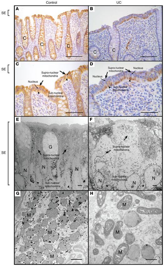
RESEARCH ARTICLE                                                                                        The Journal of Clinical Investigation

alterations in gene expression (9). In the colonic epithelium, these        chondrial oxidation. Reduction of intestinal epithelial mitochon-
effects are determined by the status of cellular differentiation (10)       drial number and function in C. rodentium colitis and in human
and whether or not the cells have undergone neoplastic transfor-            IBD may contribute to the observed higher levels of fecal acylcar-
mation (8). In the former, mitochondrial beta-oxidation of butyr-           nitines that serve as a biomarker for IBD in humans.
ate by the differentiated surface cells of the colonic epithelium               In total, these observations provide evidence for the dynamic
prevents the inhibition of HDACs and impairment of cellular pro-            regulation of colonocyte mitochondrial FAO in health and how it
liferation by stem cells in the colonic crypt (10). In the latter, aer-     is disrupted in the setting of colonic inflammation, where there
obic glycolysis in neoplastic colonic cells via the “Warburg effect”        can be a reduction in gut microbiota–produced energy substrates
reduces mitochondrial beta-oxidation of butyrate, making it avail-          for epithelial metabolism, such as butyrate, and in host-derived
able for HDAC inhibition, leading to epigenetic effects and the             metabolites like medium- and long-chain acylcarnitines. These
inhibition of cellular proliferation (11). Thus, because of the high        findings provide insights into the growing interest in the multi-
levels of butyrate production by the gut microbiota, mitochondrial          faceted interaction between the host- and gut microbiota–derived
beta-oxidation of this SCFA plays a unique tissue-specific role in          molecules for metabolic homeostasis at the intestinal epithelial
the biology of the colonic epithelium.                                      interface in general, and the role for epithelial mitochondrial func-
     Alterations in butyrate metabolism by the colonic epithelium           tion in the pathogenesis of IBD in particular.
are also associated with colonic inflammation. It has been more
than 2 decades since Roediger proposed that deficits in the oxi-            Results
dation of SCFAs by the colonic epithelium may play a role in the            Both long- and short-chain fatty acids are oxidized in the colon, where
pathogenesis of inflammatory bowel disease (IBD), particularly              there is an inverse relationship between the two regulated by the abun-
ulcerative colitis (UC) (5). Indeed, pharmacological inhibitors             dance of the SCFA butyrate. Long-chain FAO was quantified ex vivo
of FAO lead to the development of colitis (12), and reduction of            using an intestinal explant culture system whereby an intestinal
colonic SCFAs by diversion of the fecal stream lead to the devel-           segment was inverted, placing the intestinal epithelium in contact
opment of diversion colitis, an inflammatory disease that can be            with the assay medium to minimize the metabolic contribution
treated by instillation of SCFAs by enema (13). The identification          of nonepithelial cells to this assay. The oxidation of tritiated oleic
of the SLC22A5 gene encoding OCTN2, a sodium-dependent                      acid led to the time-dependent accumulation of tritiated water
L-carnitine transporter critical for FAO, as a Crohn’s disease–             that remained linear, indicating the viability of the explant tissues
associated (CD-associated) genomic locus provides support for               over several hours. FAO of oleic acid was very robust in the colon,
the notion that mitochondria may play a role in IBD pathogenesis            where it was approximately 5- to 10-fold greater than in the small
via alterations in FAO (14). Nevertheless, the mechanisms for this          intestine (Figure 1A). After transport into the mitochondria, fatty
deficit remain poorly defined, and various explanations have been           acids are conjugated to coenzyme A–generating acyl-CoAs that
proposed, such as reductions in the expression of specific oxida-           serve as substrates for beta-oxidation. Profiling of acyl-CoAs in
tive pathways (5, 15, 16).                                                  various tissues by tandem mass spectrometry has been used to
     Although the regulation of mitochondrial butyrate beta-                provide a static representation of FAO intermediates in the liver
oxidation by the colonic epithelium is central to all of these biolog-      (17). To broadly quantify the spectrum of FAO intermediates, acyl-
ical effects, a systematic characterization of mitochondrial meta-          CoAs were quantified in both the small and large intestine of mice.
bolic function in the colonic epithelium has not been previously            Short-chain acyl-CoAs predominated in the colon relative to the
described. Herein, we showed that the colonic epithelium could              small intestine (Figure 1B), reflecting the abundance of SCFAs
oxidize SCFAs and long-chain fatty acids (LCFAs), and the rela-             produced by the gut microbiota in the colon. By contrast, long-
tionship between the two was substrate-dependent and inversely              chain acyl-CoAs were lower in the colon compared with the small
related. Inhibition of SCFA oxidation in short-chain acyl-CoA               intestine (Figure 1C).
dehydrogenase–deficient (SCAD-deficient) mice led to greater                     The inverse relationship between short-chain and long-chain
long-chain FAO, whereas the addition of butyrate to colonic                 acyl-CoAs in the small versus large intestine might reflect alterna-
explant cultures led to a suppression of long-chain FAO. However,           tive fuel utilization by the intestinal epithelium due to substrate
both long- and short-chain FAO were reduced in the setting of               abundance. For example, such a functional effect would be consis-
colitis induced by infection with Citrobacter rodentium, providing          tent with the example of the reciprocal relationship between gly-
evidence for a more global mitochondrial dysfunction where both             colysis versus FAO regulated by cellular energy status via AMPK
the number and function of colonic mitochondria were reduced.               (18). Because of its juxtaposition with gut microbiota–dependent
Colonic mitochondria were dysmorphic and reduced in number                  production of SCFAs, the colonic epithelium may be the only cell
throughout the surface colonic epithelium in both C. rodentium–             type in the body that might demonstrate alternative fuel utiliza-
induced murine colitis and in human IBD.                                    tion based on the chain length of fatty acid substrate. To explore
     Concurrent with our observation that the colonic epithelium            this notion, we examined the spectrum of acyl-CoAs in the SCAD-
can oxidize SCFAs and LCFAs, we showed that high levels of                  deficient mouse. SCAD deficiency is the result of a mutation in the
medium- and long-chain acylcarnitines, fatty acid intermediates             Acads gene, leading to a block of the first step in the FAO pathway
needed for transport into mitochondria, were delivered at high              for C4–C6 SCFAs (19, 20). The BALB/cByJ mouse strain, a murine
concentrations into the gut lumen via biliary secretions. Based on          model of the SCAD deficiency (21), has an autosomal recessive
in vitro modeling, we showed that they were taken up along the              mutation of Acads and has recently been shown to have a reduction
apical side of the colonic epithelium and metabolized via mito-             in crypt cell proliferation (10). We observed a significant increase

   2                                    J Clin Invest. 2021;131(1):e133371 https://doi.org/10.1172/JCI133371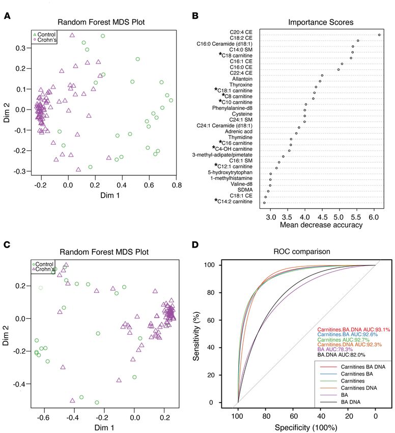
The Journal of Clinical Investigation                                                                          RESEARCH ARTICLE

                                                                                                         Figure 1. The reciprocal relationship
                                                                                                         between short- and long-chain fatty
                                                                                                         oxidation by the colonic epithelium.
                                                                                                         (A) Quantification of oleic acid oxi-
                                                                                                         dation using a tritiated water release
                                                                                                         assay in inverted colonic and small
                                                                                                         intestinal mouse explant cultures
                                                                                                         harvested from mice. n = 3, mean
                                                                                                         ± SEM, *P < 0.05. Representative
                                                                                                         results of at least 2–3 independent
                                                                                                         experiments. (B–E) Quantification of
                                                                                                         the fatty acid oxidation intermedi-
                                                                                                         ates, acyl-CoAs, by mass spectrom-
                                                                                                         etry in mouse intestinal tissues. (B)
                                                                                                         Short- and (C) long-chain acyl-CoAs
                                                                                                         in the small intestine and colon. (D)
                                                                                                         Short- and (E) long-chain acyl-CoAs
                                                                                                         in the colon of wild-type versus
                                                                                                         SCAD-deficient mice. Representative
                                                                                                         results of at least 3 experiments. n
                                                                                                         = 5, mean ± SEM, *P < 0.05, **P <
                                                                                                         0.01. (F) Quantification of oleic acid
                                                                                                         oxidation using the tritiated water
                                                                                                         release assay in colonic explants
                                                                                                         isolated from wild-type versus
                                                                                                         SCAD-deficient mice. n = 3, mean
                                                                                                         ± SEM, *P < 0.05. The assays were
                                                                                                         repeated in the presence or absence
                                                                                                         of 10 mM sodium butyrate
                                                                                                         in using colonic explants isolated
                                                                                                         from (G) wild-type or (H) SCAD-
                                                                                                         deficient mice. n = 3, mean ± SEM,
                                                                                                         *P < 0.05. Representative results of
                                                                                                         2 independent experiments. P values
                                                                                                         represent a 2-tailed Student’s t test
                                                                                                         and paired-sample t test.

                                  J Clin Invest. 2021;131(1):e133371 https://doi.org/10.1172/JCI133371                                    3
RESEARCH ARTICLE                                                                                               The Journal of Clinical Investigation

Figure 2. Quantification of acylcarnitine levels in bile (n = 3), colonic tissue (n = 4), and fecal pellets (n = 3) from conventionally housed mice. *Bile
compared to colon, *P < 0.05, **P < 0.01, ***P < 0.001; †Bile compared to fecal, †P < 0.05, ††P < 0.01, †††P < 0.005. P values represent a 2-tailed Student’s
t test and paired-sample t test.

in butyryl-CoA in the colon of SCAD-deficient mice (Figure 1D)                    in colonic explants and high levels of acyl-CoAs in colonic tissue
due to a defect in the first step of the FAO pathway, a finding sim-              (Figure 1), we postulated that the host can produce endogenous
ilar to that reported in liver tissue of these mice (17). There was               LCFAs that could also be utilized by colonic epithelium for energy
a general increase in long-chain acyl-CoAs in the colon of these                  production. Medium- and long-chain fatty acids are esterified to
mice relative to wild-type mice (Figure 1E). Based on this acyl-                  carnitine to produce acylcarnitines so that they can be transported
CoA profile, we quantified oleic acid oxidation via tritiated water               into the mitochondria (23). The cellular secretion of acylcarnitines
release in the colon of SCAD-deficient mice and their wild-type                   may not only be a detoxification mechanism: when excreted into
counterparts. We observed a significant increase in the oxidation                 the plasma, growing evidence, reviewed by McCoin et al., suggests
of oleic acid in the colonic explants of SCAD-deficient mice (Fig-                that they might also act as signaling molecules that regulate vari-
ure 1F). Since the bioenergetic cellular state regulates the choice               ous metabolic and inflammatory pathways (24). The liver can pro-
of energy substrates through mechanisms that involve AMPK (22),                   duce acylcarnitines that are exported from hepatocytes into the
we determined whether SCFA oxidation inhibits long-chain FAO                      plasma (25) as well as into the bile (26). Indeed, in mice, biliary
in the colonic epithelium. Indeed, there was an inhibitory effect                 levels of medium- and long-chain acylcarnitines were extremely
of butyrate short-chain FAO on oleic acid long-chain FAO in the                   high relative to levels found in either colonic tissue or stool (Fig-
colon of wild-type mice (Figure 1G) but not in the SCAD-deficient                 ure 2). Although much attention has been paid to plasma levels
mice (Figure 1H). These results demonstrated that FAO of SCFAs                    of acylcarnitines, the levels were significantly higher in human
and LCFAs in the colonic epithelium was inversely related.                        stool (Supplemental Figure 1A; supplemental material available
     High concentrations of acylcarnitines are delivered to the gut               online with this article; https://doi.org/10.1172/JCI133371DS1).
lumen via biliary secretions, where intestinal epithelial modeling shows          Although the gut microbiota played a role in the production of
that they can be absorbed apically and utilized for mitochondrial oxi-            many small-molecule metabolites, it did not play a significant
dative metabolism. Having provided evidence for long-chain FAO                    role in the production of acylcarnitines because fecal levels were

   4                                        J Clin Invest. 2021;131(1):e133371 https://doi.org/10.1172/JCI133371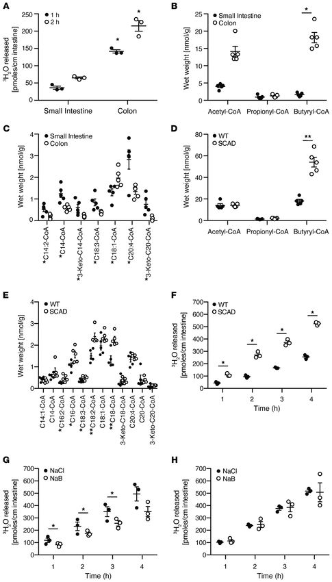
The Journal of Clinical Investigation                                                                               RESEARCH ARTICLE

                                                                                                              Figure 3. Consumption of palmi-
                                                                                                              toylcarnitine by intestinal cell
                                                                                                              lines. Supernatant concentrations
                                                                                                              of palmitoylcarnitine 24 hours after
                                                                                                              incubation with the intestinal cell
                                                                                                              lines (A) Caco2, (B) LS174t, and (C)
                                                                                                              T84. (D) Basolateral versus apical
                                                                                                              uptake of palmitoylcarnitine after
                                                                                                              24-hour incubation with T84 cell
                                                                                                              monolayers grown on Transwell
                                                                                                              plates to a high transepithelial
                                                                                                              resistance (TER). (E) Palmitoylcar-
                                                                                                              nitine uptake in intestinal cell lines
                                                                                                              24 hours after incubation with the
                                                                                                              mitochondrial inhibitor piericidin
                                                                                                              A or (F) the peroxisomal inhibitor
                                                                                                              thioridazine. n = 3, mean ± SEM, *P
                                                                                                              < 0.05, **P < 0.01, ***P < 0.001. P
                                                                                                              values represent a 2-tailed Student’s
                                                                                                              t test and paired-sample t test.

actually higher in germ-free mice compared with conventionally             mitochondrial inhibitor piericidin A, but not with the peroxiso-
housed mice (Supplemental Figure 1B). To the contrary, bacterial           mal inhibitor thioridazine (Figure 3, E and F, respectively). These
batch cultures using 3 different culture media showed that palmi-          results are consistent with the notion that medium- and long-chain
toylcarnitine could be consumed by the bacteria in the bacterial           acylcarnitines, delivered to the gut lumen via biliary secretion, can
batch cultures, where it had a modest growth effect under 2 of the         be taken up apically by the intestinal epithelium and used as an
3 culture conditions (Supplemental Figure 2).                              alternative source of energy via mitochondrial beta-oxidation.
    To determine whether acylcarnitines could be consumed by                   Based on this evidence, we then performed a comprehen-
the intestinal epithelium, we examined concentration-dependent             sive characterization of acylcarnitine metabolism using primary
consumption of the long-chain acylcarnitine, palmitoylcarnitine            human intestinal epithelial cells in culture. We showed that colo-
(spiked with 0, 0.1, and 1 mM), in 3 model intestinal epithelial cell      noids generated from a healthy human subject grown as high
lines: Caco-2, LS174T, and T84. All 3 showed significant consump-          resistance 2D monolayers could consume palmitoylcarnitine
tion over 24 hours (Figure 3, A–C). Consistent with much higher            apically and basolaterally (Supplemental Figure 3A). In fact, pal-
levels of acylcarnitines in stool than plasma (Supplemental Figure         mitoylcarnitine (C16) was consumed apically and basolaterally
1A), palmitoylcarnitine consumption was greater when delivered             to a greater degree than all other long-chain acylcarnitines (see
apically than basolaterally to the T84 polarized cell line (Figure         apical and basolateral controls, shown as CT, in Supplemental
3D). The consumption of palmitoylcarnitine was dependent upon              Table 1). We therefore examined the impact of apical and basolat-
mitochondrial but not peroxisomal oxidation because palmitoyl-             eral supplementation of palmitoylcarnitine (C16) on apical and
carnitine uptake was reduced in all 3 cell lines treated with the          basolateral consumption and production of a spectrum of acyl-

                                       J Clin Invest. 2021;131(1):e133371 https://doi.org/10.1172/JCI133371                                    5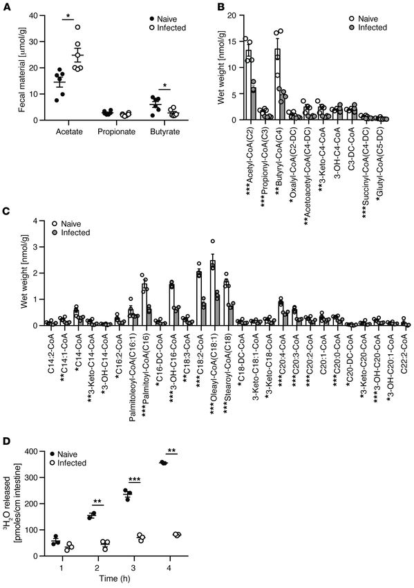
RESEARCH ARTICLE                                                                                      The Journal of Clinical Investigation

carnitines in these 2D colonoid cultures (Supplemental Table 1).          (29, 30), acyl-CoA profiling in the colon of C. rodentium–infected
In general, acylcarnitines were consumed from the basolateral             mice showed a significant reduction in short-chain acyl-CoAs
surface and secreted apically. A notable exception to this was the        (Figure 4B). Unexpectedly and inconsistent with our observa-
robust consumption of acetylcarnitine (C2) from the apical and            tions in healthy wild-type mice (Figure 1), a significant decrease
basolateral surfaces, where a reversal to secretion was induced           in long-chain acyl-CoAs in the colon of C. rodentium–infected
specifically into basolateral compartment by supplementation              mice was also observed (Figure 4C). We confirmed that long-
of palmitoylcarnitine (C16) at either the apical or basolateral           chain FAO was functionally reduced in C. rodentium infection
surface. This may be a reflection of cellular energy homeostasis,         using the tritiated oleic acid colonic explant assay (Figure 4D).
whereby excess nutrient is secreted rather than converted into            Collectively, these results suggest that C. rodentium–induced
triglyceride or glycogen for storage. Consistent with this notion,        colitis led to a global alteration in FAO.
basolateral palmitoylcarnitine (C16) supplementation led to a                  C. rodentium infection reduces both the number and function
generalized increase in apical secretion of several acylcarnitines        of colonic mitochondria. The reduction of both short- and long-
(see CT+BPAC in Apical Fraction, Supplemental Table 1). Thus,             chain FAO in C. rodentium–induced colitis may be due to a
in addition to being used for oxidative metabolism, acylcarni-            reduction in colonic epithelial mitochondria, functional abnor-
tines may also act as a signaling molecule that may alter cellular        malities, or both. To address this issue, isolated colonic epithe-
metabolic homeostasis, as has been proposed in other tissues              lial cells from naive and C. rodentium–infected mice were eval-
(24). Despite this effect, the addition of palmitoylcarnitine had         uated genomically, biochemically, and functionally (Figure 4
minimal effects on transepithelial resistance relative to control         and Supplemental Figure 5). Quantitative PCR of mitochondrial
conditions (Supplemental Figure 3B).                                      DNA and mitochondrial citrate synthase assays (Supplemen-
     These studies also showed that the metabolism of acylcarni-          tal Figure 5, A and B) showed that C. rodentium–induced colitis
tines by the intestinal epithelium may be selective, with the great-      reduced both the number and function of colonic mitochondria,
est levels of consumption being observed for C16, C14:2, C10:2,           respectively. These results were confirmed by flow cytome-
C3, C3:1, and C2 acylcarnitines both apically and basolaterally           try of mitochondria stained with MitoTracker Green and Deep
(see controls, CT, in Supplemental Table 1). Since we only stim-          Red (Supplemental Figure 5, C–F). MitoTracker probes contain
ulated the colonoids with palmitoylcarnitine, additional pharma-          chloromethyl moieties that diffuse across the plasma membrane
cokinetic studies will be needed to examine the capacity of the           into mitochondria and bind to sulfhydryls. MitoTracker Green is
intestinal epithelium to consume other acylcarnitines.                    not dependent on membrane potential and is therefore an indi-
     To examine the dynamics of palmitoylcarnitine metabolism             cator of mitochondrial mass, whereas MitoTracker Deep Red
ex vivo, similar to our studies quantifying oleic acid oxidation in       is a positively charged rosamine-containing probe dependent
explant cultures (Figure 1, G and H), colonic explants from wild-         on negative mitochondrial membrane potential (31, 32). The
type and SCAD mice were incubated with palmitoylcarnitine                 decrease in both MitoTracker Green and Deep Red staining indi-
in the presence of either sodium butyrate or NaCl (Supplemen-             cates a decrease in mitochondrial content as well as function in
tal Figure 4). Consistent with our observation that 2D colonoids          isolated epithelial cells from the colon of C. rodentium–infected
were able to secrete acylcarnitines apically under certain condi-         mice compared with naive mice.
tions (Supplemental Table 1), palmitoylcarnitine levels increased              C. rodentium infection and IBD lead to an alteration in number,
during the first 2 hours of incubation. However, after 4 hours in         structure, and position of colonic epithelial mitochondria. MTCO1
culture, the level of palmitoylcarnitine in the culture supernatant       (mitochondrially encoded cytochrome c oxidase I), also known
was significantly higher in the wild-type colonic tissue supple-          as COX-1 (cytochrome c oxidase), is one of 3 mitochondrial DNA-
mented with sodium butyrate relative to the other 3 conditions            encoded subunits of respiratory complex IV, the final enzyme of
(Supplemental Figure 4), consistent with the notion that oxidation        the electron transport chain of mitochondrial oxidative phosphor-
of SCFAs could reduce the oxidation of long-chain fatty acids, as         ylation (33). MTCO1 staining can serve as a biomarker for both
we described in Figure 1, E–H.                                            the presence of mitochondria and functional mitochondrial res-
     Colonic inflammation induced by C. rodentium infection inhib-        piration (33). In colonic tissue of naive mice, MTCO1 was most
its both SCFA and LCFA oxidation. Multiple studies have shown             prominent in epithelial cells, where a diffuse pattern of cytoplas-
that FAO of SCFAs is reduced in the setting of colonic inflam-            mic staining was observed (Figure 5, A and C). By contrast, there
mation in patients with UC, where this metabolic alteration has           was a decrease in MTCO1 staining in the colonic epithelium of C.
been proposed to play a role in disease pathogenesis (3, 27).             rodentium–infected mice that was most pronounced in the super-
Based on the reciprocal functionality of short-chain versus long-         ficial epithelium, where staining was more preserved in the sub-
chain FAO (Figure 1), we hypothesized that colonic inflamma-              nuclear than the supranuclear compartment (Figure 5, B and
tion would reduce short-chain but increase long-chain FAO. We             D). These observations were confirmed by electron microscopy
examined this notion using the C. rodentium model of mouse                comparing the number of mitochondria and their morphological
colitis in which the colonic epithelium remains relatively intact         characteristics in the superficial colonic epithelium of naive mice
with crypt hypertrophy and epithelial hyperproliferation (28).            (Figure 5, E and G) with C. rodentium–infected mice (Figure 5, F
Fecal SCFAs, quantified by proton nuclear magnetic resonance              and H), where the latter showed dysmorphic mitochondria (i.e.,
(1H NMR), showed that C. rodentium infection led to a reduction           swelling with loss of cristae abundance and organization) and a
in butyrate but increase in acetate (Figure 4A). Consistent with          reduction in the number of mitochondria that were most striking
this reduction in substrate and previous studies in human IBD             in the supranuclear compartment of the colonic epithelium.

   6                                  J Clin Invest. 2021;131(1):e133371 https://doi.org/10.1172/JCI133371
The Journal of Clinical Investigation RESEARCH ARTICLE
Figure 4. Quantification of the fatty acid oxidation intermediates,
acyl-CoAs, in the colon of mice infected with C. rodentium. (A) 1H
NMR quantification of SCFAs from fecal water of naive and C. roden-
tium–infected mice. Quantification of (B) short- and (C) long-chain
acyl-CoAs extracted from the colon of naive and C. rodentium–infected
mice. (D) Oleic acid oxidation quantified by tritiated water release
assay in colonic explants of naive and C. rodentium–infected mice. All
experiments n = 5, mean ± SEM, *P < 0.05, **P < 0.01. Representative
results of at least 2 independent experiments. P values represent a
2-tailed Student’s t test and paired-sample t test.
To determine whether these alterations are also associated relative to the subnuclear region in 56% of patients with CD and
with colonic inflammation in human disease, we examined 60% patients with UC (Figure 6, B and D). We found no differ-
MTCO1 staining in surgical tissue specimens obtained from ences in the rate of subnuclear staining between IBD types (CD
healthy subjects versus patients with IBD (Supplemental Table vs. UC, Fisher’s exact test P value 1.00). Electron microscopy
2). In healthy subjects, we observed diffuse prominent staining also confirmed a reduction in supranuclear mitochondria in IBD
throughout the colonic epithelium, where staining was uniformly as well as dysmorphic mitochondrial characteristics in the super-
distributed throughout the cytoplasm (Figure 6, A and C). By ficial epithelium relative to the images of normal colonic tissue
contrast, IBD samples showed a marked decrease in MTCO1 (Figure 6, E–H), similar to those characteristics observed in C.
staining in the supranuclear region of the superficial epithelium rodentium–induced colitis (Figure 5, E–H). In total, these results
J Clin Invest. 2021;131(1):e133371 https://doi.org/10.1172/JCI133371 7RESEARCH ARTICLE The Journal of Clinical Investigation
Figure 5. Comparative morpholog-
ical analysis of colonic epithelial
mitochondria in naive and C.
rodentium–infected mice. MTCO1
staining of colonic tissue isolated
from naive (A and C) and C. roden-
tium–infected mice (B and D).
Electron microscopy of the super-
ficial colonic epithelium in naive (E
and G) and C. rodentium–infected
mice (F and H). Scale bars: 200 μm
(A and B), 150 μm (C and D), 10 μm
(E and F), 500 nm (G and H). Sym-
bols: crypt (C), nucleus (N), surface
epithelium (SE), mitochondria
(M), endoplasmic reticulum (ER),
supranuclear mitochondria (black
arrows), subnuclear mitochondria
(black arrowheads).
8 J Clin Invest. 2021;131(1):e133371 https://doi.org/10.1172/JCI133371The Journal of Clinical Investigation RESEARCH ARTICLE
Figure 6. Comparative mor-
phological analysis of colonic
epithelial mitochondria in
healthy human subjects versus
patients with IBD. MTCO1 staining
of colonic tissue obtained from a
representative healthy (control)
human subject (A and C) and a
representative patient with IBD
(UC) (B and D). Electron micros-
copy of the superficial colonic
epithelium obtained from a repre-
sentative healthy human subject
(E and G) and a representative
patient with IBD (UC) (F and H).
Scale bars: 400 μm (A and B), 200
μm (C and D), 2 μm (E and F), 500
nm (G and H). Symbols: crypt (C),
goblet cell (G), nucleus (N), surface
epithelium (SE), mitochondria (M),
supranuclear mitochondria (black
arrows), subnuclear mitochondria
(black arrowheads).
J Clin Invest. 2021;131(1):e133371 https://doi.org/10.1172/JCI133371 9RESEARCH ARTICLE The Journal of Clinical Investigation
support the notion of C. rodentium–induced colitis as a model carbohydrates (10). Specifically, the oxidation of butyrate by the
for mitochondrial dysfunction in human IBD, where both show colonic epithelium prevents epigenetic alterations of the genome
similar morphological alterations in colonic epithelial mitochon- marked by histone hyperacetylation via the inhibition of HDACs
dria. The biochemical and functional characterization of mito- (11). The consequence of such alterations leads to the inhibition
chondrial dysfunction in C. rodentium–induced colitis (Figure of cellular proliferation, a phenotype that could reduce colon-
4, Figure 5, and Supplemental Figure 5) therefore provides an ic epithelial wound healing (10). The regulation of FAO by the
explanation for the reduction in butyrate oxidation observed in colonic epithelium may therefore play a critical role in disease
patients with IBD (5). states where the epithelial barrier is wounded, such as IBD, as well
Elevation of fecal acylcarnitines is associated with IBD in as in colonic neoplasia. A few reports in the literature have docu-
patients and C. rodentium colitis in mice. The inhibition of palmi- mented a reduction of butyrate oxidation by the colonic epithelium
toylcarnitine uptake by an intestinal epithelial cell line in vitro by in the setting of UC (12, 13). However, the mechanisms have
pharmacological mitochondrial inhibitors (Figure 3E), together remained poorly defined. Indeed, there are currently no studies
with functional and histological evidence of mitochondrial dys- that systematically characterize the overall regulation of FAO by
function in both C. rodentium–induced colitis and human IBD, the colonic epithelium in either health or disease.
suggests that the decrease in mitochondrial FAO might lead to Herein, we used a combination of technologies to characterize
an elevation of fecal acylcarnitine levels in intestinal inflamma- the FAO of a spectrum of fatty acids in the colonic epithelium both
tory disease. Indeed, the levels of a wide spectrum of acylcar- functionally using explant cultures and biochemically via tandem
nitines were elevated in mice with C. rodentium–induced colitis mass spectrometry. The results demonstrated that the colonic
(Supplemental Figure 7). epithelium could oxidize not only SCFAs, as have been previ-
This was also the case for patients with CD, a form of IBD ously described, but also LCFAs at levels actually exceeding that
where elevations were associated with increased disease activity observed in the small intestine. By studying the impact of butyr-
(quantified by fecal calprotectin levels), an increase in human yl-CoA oxidation on long-chain FAO using SCAD-deficient mice,
DNA in the feces, and dysbiosis, defined as “cluster 2” (34) (Sup- we showed that butyrate inhibited long-chain FAO. Although
plemental Figure 6). Two acylcarnitines, C3 carnitine and C20:4 additional studies will be needed to determine the mechanism(s)
carnitine, showed the opposite pattern with a decrease in abun- involved in the reciprocal relationship between short- and long-
dance, the former being consistent with the known reduction in chain FAO, it is plausible that AMPK might be involved, similar to
fecal propionate levels associated with intestinal inflammation the reciprocal relationship between glycolysis and beta-oxidation
(Figure 1B) (35). The association between dysbiosis in IBD and (36), given the primary role of mitochondria in maintaining cel-
bile acids as well as acylcarnitines in the feces has recently been lular energy homeostasis. Regardless of the mechanism, the high
reported (30), where a number of associations were described levels of butyrate produced by the gut microbiota as an energy
with bacterial taxa. An analysis of our cohort revealed that there is substrate for the colonic epithelium would suppress long-chain
a very robust separation that distinguishes the fecal metabolome FAO, supporting the conclusion that the SCFAs are the primary
of healthy subjects from those with CD in a multidimensional scal- source of metabolic fuel for colonocytes (4–7).
ing (MDS) plot (Figure 7A), where a random forest analysis showed The ability of the colonic epithelium to use both SCFAs and
that acylcarnitines were among the most important features LCFAs for oxidative metabolism suggests that, in addition to
responsible for this separation (Figure 7B). Using only the previ- microbiota-based substrates in the form of the SCFA butyrate,
ously described metabolomic features used as biomarkers for IBD the host might also contribute substrates for FAO in the form
and dysbiosis, bile acids (30) and human DNA (34), together with of medium- to long-chain fatty acids. Indeed, we provide evi-
acylcarnitines, showed good separation on the MDS plot between dence that acylcarnitines delivered to the gut lumen in bile can
healthy subjects and patients with IBD, where the IBD subjects be consumed apically in a polarized intestinal epithelial model
were more tightly clustered together on the right of the plot than system, where they undergo mitochondrial but not peroxisomal
the more widely dispersed healthy control subjects (Figure 7C). oxidation. This previously unappreciated mechanism by which
Among these 3 features, alone or in combination, acylcarnitines biliary secretions may have a physiological impact on the intes-
alone were the single strongest predictor of the presence of IBD, tinal tract is consistent with other interactions between the
having an AUC of 92.7% on a receiver operating characteristic liver and the gut, such as the enterohepatic circulation of bile
curve (ROC) analysis (Figure 7D). In total, these results support acids, as well as bile acid–mediated activation of the farnesoid
the notion that intestinal epithelial mitochondrial function plays a X nuclear hormone receptor in the terminal ileal epithelium
role in defining fecal acylcarnitines as a robust biomarker for IBD. (37). The mechanism by which luminal acylcarnitines are
transported apically into the intestinal epithelium is currently
Discussion unknown, but may involve solute carrier (SLC) membrane
It is well known that mitochondria play a critical role in the regula- transport proteins, such as SLC22A1, which is responsible for
tion of many cellular processes, such as energy generation, apop- the efflux of acylcarnitine from the liver into the circulation (25)
tosis, the unfolded protein response, and autophagy (15). Recently, and has been shown to be expressed on the apical domain of
it has become apparent that there are functional features of mito- the intestinal epithelium (38). Indeed, we showed that human
chondria that are unique to the colonic epithelium because of its 2D colonoids could be stimulated to secrete acylcarnitines
juxtaposition with the gut microbiota and its production of metab- apically when stimulated by palmitoylcarnitine basolaterally.
olites such as SCFAs by bacterial fermentation of indigestible Although the impact of polarized secretion in the setting of dis-
10 J Clin Invest. 2021;131(1):e133371 https://doi.org/10.1172/JCI133371The Journal of Clinical Investigation RESEARCH ARTICLE
Figure 7. Fecal acylcarnitines as a biomarker for IBD. (A) MDS plot of fecal metabolites from a previously described cohort of healthy pediatric subjects
and those with CD (34, 47). (B) Random forest analysis of fecal metabolites stratified by importance in distinguishing healthy subjects from patients
with IBD. * indicates acylcarnitines. (C) MDS plot of the same cohort in A focused on human DNA, bile acids, and acylcarnitines. (D) A receiver operating
characteristic curve (ROC) showing the discriminative ability of human DNA, bile acids, and acylcarnitines to individually or in combination predict a
condition of health versus IBD.
ease remains to be determined, our result may suggest an addi- metabolic function of the colonic epithelium, especially since
tional mechanism by which intestinal epithelial mitochondrial we provide evidence that these are reciprocally related.
dysfunction might lead to greater levels of fecal acylcarnitine We determined the impact of disease on epithelial mitochon-
levels, namely enhanced apical secretion due to reduced mito- drial structure and function through a series of genomic, bio-
chondrial consumption. Indeed, elevated plasma acylcarnitine chemical, and FACS-based analyses, revealing that a reduction
levels are often monitored clinically in patients with mitochon- in both short- and long-chain acyl-CoAs in colonic tissue of mice
drial disorders (39). Interestingly, our data in the 2D colonoid infected with C. rodentium was due to a decrease in the number
model also suggests that both apical and basolateral consump- and function of mitochondria in the colonic epithelium. MTCO1
tion may be restricted to acylcarnitines, a number of which are immunohistochemistry and electron microscopy confirmed and
found at higher levels in human stool than plasma. Ultimately, extended this finding, showing not only a dysmorphic appearance
it will be of interest to determine the impact of host-derived of mitochondria, but also a reduction in number that was partic-
acylcarnitines relative to microbiota-generated SCFAs on the ularly severe in the superficial surface epithelium of the colon,
J Clin Invest. 2021;131(1):e133371 https://doi.org/10.1172/JCI133371 11RESEARCH ARTICLE The Journal of Clinical Investigation
consistent with previously reported observations by Ma et al. (40). Figure 6). The performance of fecal acylcarnitine as a predictor
These mitochondrial alterations are in part due to a C. rodentium of IBD was impressive, having an AUC of over 92% in the ROC
effector protein known as mitochondrial-associated protein (40). analysis, which was better than previous fecal biomarkers for
Similar findings were observed in patients with IBD, providing an IBD, such as human DNA and bile acid composition. In addi-
explanation for previous studies reporting a reduction in SCFA tion to providing support for the notion that acylcarnitines are
metabolism by the colonic epithelium (29). consumed by the intestinal epithelium for mitochondrial oxi-
The reduction of mitochondria specifically in the supranu- dation, the elevation of these lipids may be a useful marker of
clear compartment of the superficial colonic epithelium is par- disease in patients with intestinal inflammation.
ticularly intriguing — not only may this histological character- Using FAO as a biomarker of mitochondrial function, we
istic be a biomarker for the intestinal inflammatory state, but it provide mechanistic insight into the use of energy substrates
may have functional implications for the cellular biology of the provided by the gut microbiota in the form of butyrate, as well
colonic epithelium. Differential mitochondrial positioning has as the host in the form of acylcarnitines — the latter being a pre-
been described in polarized cell types, such as budding yeast, viously unappreciated modality of enterohepatic communica-
neurons, and pancreatic acinar cells (41–43). Studies have shown tion. The most significant reduction in mitochondrial number
in these model systems that mitochondria localize to areas of and morphology restricted to the superficial colonic epithelium
the cell needed for metabolically intensive processes. This phe- in the supranuclear apical subcellular compartment may have
nomenon has not been previously described in the intestinal or significant effects on the host via the inhibition of stem cell pro-
colonic epithelium. We hypothesize that the polarized colono- liferation, as well as the gut microbiota with the expansion of
cyte would have a large number of mitochondria at the apical facultative anaerobes driven by aerobic respiration. The unique
side of the cell, where butyrate concentrations are high and a environment in which the superficial colonic epithelium resides,
high level of FAO is occurring. The loss of mitochondria would juxtaposed with an anaerobic environment high in fatty acid sub-
be consistent with the observed reduction in SCFA oxidation strates, emphasizes the importance of mitochondrial function
associated with IBD (3, 27), where it may have an impact on the via FAO in this cellular compartment.
cellular proliferation of stem cells located in the colonic crypt
via the inhibition of HDACs (10). It also provides an alterna- Methods
tive explanation by which colonic inflammation, induced by C. Animals. All experiments were approved by the IACUC at the Univer-
rodentium in mice or associated with IBD in humans, might lead sity of Pennsylvania Perelman School of Medicine. C57BL/6J, BALB/
to the expansion of aerotolerant, facultative anaerobic organ- cByJ (SCAD deficient) and BALB/cJ (wild-type) were obtained from
isms at the mucosal interface. C. rodentium might increase the the Jackson Laboratory. Germ-free mice were obtained from the Uni-
diffusion of oxygen into the luminal environment, not only by versity of Pennsylvania Gnotobiotic Mouse Facility. All animals used
reducing the consumption of oxygen via mitochondrial beta- in experiments were 7–10-week-old female mice. Animals were fed
oxidation by the superficial colonic epithelium due to a reduc- a standard chow diet and were kept on 12-hour light/12-hour dark
tion in gut microbiota butyrate production as proposed by Lopez cycles. For the C. rodentium model, conventionally housed, female,
et al. (44), but as our results suggest, a similar phenomenon 8–10-week-old, C57BL/6J mice were inoculated by oral gavage with 6
might also occur because of the impaired function and/or num- × 108 CFU of C. rodentium (strain DBS100, gift from David Artis, Weill
ber of mitochondria in this same cellular compartment. Cornell Medicine at Cornell University, New York, New York). Mice
Finally, we showed that alteration of epithelial mitochon- were euthanized by CO2 asphyxiation followed by cervical dislocation
drial function, effectively reducing FAO, may have an impact on on day 11 after infection.
substrate availability of fecal acylcarnitines, which were elevat- Human studies. Plasma and fecal samples assayed for acylcar-
ed in both C. rodentium–induced murine colitis and in patients nitine levels as well as rectal biopsies for colonoid primary cell line
with CD. Although there are bacterial taxonomic associations generation were obtained from the Food and Resulting Microbial
with the elevation of fecal acylcarnitines in human IBD, lead- Metabolite (FARMM) study (47). The FARMM study recruited
ing to the notion that the gut microbiota might be important healthy volunteers ages 18–60. Stool, urine, blood, and rectal biop-
for their production (30), our results in germ-free mice showing sies were collected throughout the 15-day inpatient study designed
high levels of fecal acylcarnitines strongly indicates that they to determine the impact of diet on the gut microbiome, fecal, and
are primarily of host and not of microbial origin. Nevertheless, plasma metabolome. The samples used for this study were obtained
we provide preliminary evidence that palmitoylcarnitine can be at the baseline timepoint at the beginning of the study. The fecal
consumed by the human gut microbiota in culture, where it has metabolomic analysis in pediatric CD, shown in Figure 7 and Supple-
a modest effect on growth. Previous studies have shown that mental Figure 6, was performed as part of the PLEASE study that has
acylcarnitines as well as carnitine itself can serve as a nitro- been previously described (34). Briefly, stool samples from pediatric
gen and carbon source for bacteria (45). Indeed, antibiotic patients with CD and healthy controls were used for a prospective
treatment of mice can increase fecal acylcarnitine levels (46), cohort study to assay metabolites such as acylcarnitines and measure
suggesting the interesting possibility that the IBD-associated calprotectin and human DNA. Both the FARMM and PLEASE studies
dysbiosis may play a role in the elevation of fecal acylcarnitines received approval from the University of Pennsylvania IRB.
due to reduced consumption. This would be consistent with our Bacterial batch culture. Bacteria were grown in lysogeny broth (LB,
observation that elevated fecal acylcarnitines in CD were pos- Thermo Fisher Scientific); brain heart infusion (BHI, Anaerobe Sys-
itively correlated with dysbiosis (“cluster 2” in Supplemental tems); gut microbiota medium (GMM, Anaerobe Systems); or yeast
12 J Clin Invest. 2021;131(1):e133371 https://doi.org/10.1172/JCI133371The Journal of Clinical Investigation RESEARCH ARTICLE
extract, casitone, and fatty acid (YCFA, Anaerobe Systems) culture Isolation of colonocytes. Animals were euthanized by CO2 asphyxi-
media shaken at 37°C. A fecal sample from a healthy 24-year-old male ation followed by cervical dislocation. Colons were dissected, flushed
subject (47) was grown in BHI, GMM, or YCFA with or without pal- with sterile PBS, and splayed open longitudinally and washed again
mitoylcarnitine, final concentration 1 μM. OD (600) was measured with PBS. Tissue was incubated on ice in PBS containing 30 mM EDTA
to determine growth rate in the presence or absence of palmitoylcar- and 1.5 mM DTT for 20 minutes with occasional inverting. This was
nitine. Culture media supernatants were taken at 0 and 24 hours for followed by shaking every 2 minutes in PBS containing 30 mM EDTA
acylcarnitine analysis by mass spectrometry. Control samples consist- for 10 minutes to remove crypts from the submucosa. The mesen-
ed of media with or without palmitoylcarnitine. chyme was removed, and cells were pelleted by centrifugation at 400
Human colon cancer cell lines. Human colon cancer cell lines g for 5 minutes at 4°C and washed in PBS.
T84, Caco-2, and LS174T (ATCC) were maintained at 37°C in 5% Quantification of mitochondrial DNA. Mitochondrial DNA was
CO2 in a humidified incubator. LS174T and Caco-2 cells were main- isolated using Qiagen Blood and Tissue Kit following the manufac-
tained in DMEM supplemented with penicillin-streptomycin and turer’s protocol (Qiagen). Primers for mitochondria D-loop (encod-
10% FBS. T84 cells were maintained in DMEM/Ham’s F12 sup- ed by mitochondrial DNA) and Ikb-β (encoded by nuclear DNA)
plemented with penicillin-streptomycin and 10% FBS. Cultures were utilized to determine relative mitochondrial DNA level, and
were used for experiments when grown to confluent monolayers on β-actin was used as an internal control; primers are shown in the for-
6-well plates unless otherwise noted. mat of gene, forward primer (F), reverse primer (R): mtD-loop, (F)
Healthy human colonoids. Healthy human colonoids (from ACTATCCCCTTCCCCATTTG, (R) TGTTGGTCATGGGCTGAT-
FARMM study) were generated and propagated in Matrigel (Corn- TA; Ikb-β, (F) GCTGGTGTCTGGGGTACAGT, (R) ATCCTTGGG-
ing, 356231) on 24-well polystyrene plates as previously described GAGGCATCTAC; β-actin, (F) TGTTACCAACTGGGACGACA, (R)
(48) with full growth medium comprising basal medium (advanced ACCAGAGGCATACAGGGACA.
DMEM supplemented with 10% FBS, 1× penicillin/streptomycin, Quantitative mass spectrometry. Acyl-CoAs from small intestinal
1× GlutaMAX, 10 mM HEPES, 1× N2 supplement, 1× B27 supple- (duodenum, 5 cm from the stomach) or colonic tissue were extracted
ment, all from Invitrogen), 1 mM N-acetyl-cysteine, 10 nM leu- with chloroform and methanol (2:1, v/v), together with stable iso-
gastrin (all from MilliporeSigma) and WRN cell conditioned medium tope-labeled and unlabeled standards, were measured by flow injec-
(WRN cells from ATCC; basal medium and WRN conditioned tion tandem mass spectrometry as previously described (17) and nor-
medium mixed at 1:1 ratio), and supplements (500 nM A83-01 malized to the wet tissue weight.
[Tocris], 10 μM SB-20210, 10 mM nicotinamide, 50 ng/mL human Mass spectrometry for acylcarnitines. Acylcarnitines were mea-
EGF [all from MilliporeSigma], and 2 μL/mL 1× Primocin [Invivo- sured as their butylated derivatives using tandem mass spectrom-
Gen]). For the first 48 hours of colonoid culture, growth medium etry. Stable isotope-labeled internal standards (Cambridge Isotope
was supplemented with 10 μM Y27632, 2.5 μM CHIR99021 (both Laboratories) were added to samples. Ethanol was added and the
from MilliporeSigma), and 2.5 μM thiazovivin (Reprocell). Media samples were dried under a stream of nitrogen at 60°C. After adding
was replaced every 48 hours. butanolic hydrochloric acid (Regis Technologies), the samples were
Transepithelial electrical resistance assay. Transepithelial electri- heated to 65°C for 15 minutes and dried under a stream of nitrogen.
cal resistance (TER) was measured by an epithelial volt/ohm meter The dried samples were reconstituted with acetonitrile/water (80:20)
(World Precision Instruments, EVOM2 300523) with colon cancer and injected into a Xevo TQ-XS tandem mass spectrometer (Waters
cell lines and healthy human colonoid-derived cells grown as mono- Corporation). Data were acquired to collect the parent compounds
layers on Transwell permeable supports (Costar, 3470). To grow colo- of mass m/z 85. Quantitation was against the nearest chain-length
noid-derived cells on the Transwell cell culture insert, established stable isotope labeled internal standard. The mass spectrometer was
colonoids were dislodged and resuspended in TrypLE (Invitrogen, operated in positive ion mode with resolving power of 140,000 at m/z
12605010) for 5–10 minutes in a 37oC water bath to disrupt colonoid 200, mass scanning range being m/z 140–600, and 5 μL of sample was
formation and generate single cells. The cell suspension was sup- delivered to mass spectrometer through direct flow injection. Mobile
plemented with 10% v/v of FBS and pipetted using a 100 μL pipette phase was acetonitrile/water (80:20, v/v). Flow rate was 0.1 mL/min-
tip by gentle strokes of approximately 50 times. Cells were counted ute. The source temperature was 150°C and desolvation temperature
using an automated Corning cell counter. Next, 100 μL of single-cell was 200°C, with gas flow of 550 L/hour. The cone and capillary volt-
suspension containing 2 × 10⁵ cells in growth medium were plated age was 50 V and 3.2 kV, respectively. Data were acquired in multiple
on 0.4 μm pore, 12-mm diameter polycarbonate membrane inserts reaction monitoring (MRM). The mass spectrometer was operated in
(Corning, 3413) coated with 33.3 μg/mL human collagen IV (Millipore, positive ion mode with resolving power of 140,000 at m/z 200, mass
C954D93). After 24 hours, the lower chamber medium was replaced scanning range being m/z 140–600.
with differentiation medium (growth medium supplemented with 2 NMR metabolomics. SCFAs were extracted and analyzed as
μg/mL hBMP4 [MilliporeSigma]) and the upper chamber medium described previously (49). An NMR spectrum for each fecal sample was
was supplemented with 100 μL of differentiation medium. Fresh acquired using NOESY-presaturation on a 4-channel Bruker Ascend
media were replaced every 48 hours. Cells were maintained at 37°C, 700 MHz spectrometer (Bruker Biospin) with a TXI 700 MHz 3 mm
5% CO2. TER was evaluated when filter-grown monolayers reached probe with triple-axis gradients. All spectra were acquired with a 14
full resistance (at least 800 Ω-cm2 of TER). ppm sweep width, 4-second acquisition time, 4 dummy scans, and 384
Chemical reagents. Palmitoylcarnitine (MilliporeSigma) was used transients. All spectra were zero filled to 128,000 data points, Fourier
at 1 μM. Piericidin A (Cayman Chemical) was used at 100 nM. Thiori- transformed with 0.1 Hz line broadening applied, manually phased,
dazine (Cayman Chemical) was used at 200 nM. and baseline-corrected using TopSpin NMR software. The SCFA
J Clin Invest. 2021;131(1):e133371 https://doi.org/10.1172/JCI133371 13RESEARCH ARTICLE The Journal of Clinical Investigation
concentrations were quantitatively determined using the 700 MHz fitted with a Hamamatsu digital camera and AMT Advantage
library from Chenomx NMR Suite 7.1 after acquiring an NMR spectrum imaging software. Micrographs were assessed by 2 investigators
from each fecal sample. The resulting quantified metabolites were blinded to clinical diagnosis.
used as input variables into subsequent statistical analysis and nor- Statistics. Results are expressed as the mean ± SEM. A 2-tailed
malized by fecal weight. Student’s t test and paired-sample t test were used for direct com-
FAO assay. FAO was measured with 3H-oleic acid (Ameri- parisons between 2 groups and within groups, respectively. A P value
can Radiolabeled Chemicals) and quantified using tritiated water of less than 0.05 was considered statistically significant. *P < 0.05,
released per centimeter length of intestine. Briefly, the colon or duo- **P < 0.01, ***P < 0.001. A Fisher’s exact test was used to calculate
denum (first 5 cm from the stomach) was isolated and then flushed P values for the MTCO1 scoring of subnuclear versus supranuclear
with oxygenated PBS to remove waste. The intestinal segment was staining. A 2-way ANOVA with Holm-Sidak’s post hoc test for multi-
everted on a gavage needle and filled with PBS and ligated at both ple comparisons was used for direct comparisons between palmitoyl-
ends to form a sac. The explant was incubated in a buffer containing carnitine-treated and untreated groups.
3
H-oleic acid and 100 mM carnitine for 4 hours at 37°C. At each hour, Metabolites with more than 80% missing values across all sam-
a 400 μL aliquot of assay buffer was removed from each sample and ples were removed from the analysis. For each metabolite, samples
replaced with 400 μL of fresh assay buffer. The collected aliquot was with the missing values were imputed with its minimum abundance
quenched with 400 μL of 10% (w/v) trichloroacetic acid and centri- across samples. For each sample, the metabolite abundances were
fuged at 6800 g for 10 minutes. The supernatant was neutralized with further normalized by dividing 90% cumulative sum of the abun-
6 N sodium hydroxide solution and applied to columns containing dances of all metabolites. The 90% cumulative sum, instead of total
AGX-1 resin (BioRad). The flow-through containing the unretained abundance, was chosen to avoid potential large outliers in the samples.
sample and the fraction eluted with 2 mL of water were pooled. Then, The normalized abundances were then log transformed and used in all
10 mL of scintillation fluid was added and radioactivity measured on analyses. The metabolite annotation was obtained from the Human
a scintillation counter. For the butyrate studies, explants were incu- Metabolome Database (HMDB). Two group comparisons were per-
bated with either 10 mM sodium chloride or 10 mM sodium butyrate. formed using Wilcoxon rank-sum test. P values were adjusted for mul-
Flow cytometry. Flow cytometry was performed using FACS- tiple comparisons with FDR control.
Canto or LSR II cytometers (BD Biosciences) and FlowJo software Study approval. All animal studies were performed under approv-
(Tree Star) for cells suspended in DPBS containing 1% BSA (Sig- al by the IACUC of the University of Pennsylvania. All study protocols
ma-Aldrich) to assess mitochondrial mass and membrane poten- involving human samples were collected according to IRB-approved pro-
tial (50). Single cell suspensions generated from murine intestinal tocols at the University of Pennsylvania for which written informed con-
crypts were incubated with 5 nM MitoTracker Green (Molecular sent was obtained and there was no access to identifiable information.
Probes; membrane potential insensitive) and 1 nM MitoTracker
Deep Red (Molecular Probes; membrane potential sensitive) in Author contributions
DPBS containing 10% FBS at 37°C for 30 minutes. DAPI (Molec- SAS, LC, SAO, KAW, KH, JC, BS, CBC, RJX, JB, and SAK performed
ular Probes) was used to determine cell viability. A suspension of experiments. FF and MB provided human colonic tissues and ana-
unstained cells pooled from all samples was utilized to establish lyzed them by electron microscopy and immunohistochemistry.
background fluorescence in each experiment. Geometric MFIs for AMW, MB, HN, FDB, JDL, and GDW designed the experiments.
MitoTracker Green and MitoTracker Deep Red were determined in EC and HL performed statistical analyses. SAS, LC, and GDW
the live-cell fraction (DAPI–) for each specimen after subtraction of generated the figures and wrote the manuscript. The order of the
background fluorescence. co–first authors, SAS, SAO, and LC, was based upon the amount of
Histology. Paraffin-embedded 4% paraformaldehyde-fixed sur- experiments performed as well as contributions to data analysis
gical colonic tissue sections from the Cooperative Human Tissue and drafting of the text for this manuscript.
Network and anonymized archival tissue from the Department of
Pathology, Penn Presbyterian Medical Center (expedited IRB approv- Acknowledgments
al) were stained with H&E. Immunohistochemistry was performed We acknowledge the following funding sources: K01DK103953 (to
with MTCO1 (Abcam, ab14705, clone 1D6E1A8). Slides were ana- KAW), R03DK114463 (to KH), and R01DK114436 (to HN) from
lyzed and scored by a blinded pathologist for disease activity, local- NIH/NIDDK; R01AA026297 (to HN) from NIAAA; R01GM103591
ization, and intensity of MTCO1 staining. In Supplemental Table 2, if (to GW) from NIGMS; Crohn’s and Colitis Foundation and
any biopsy section from an individual patient demonstrated a loss of P30DK050306 for the Human-Microbial Analytic and Reposito-
supranuclear staining in the superficial epithelium spanning at least ry Core of the Center for Molecular Studies in Digestive and Liver
5 crypts, the patient was classified as having “Sub-nuclear Staining.” Disease (to GDW) from NIH/NIDDK. The authors acknowledge the
If no sections for each patient showed a loss of supranuclear stain- PennCHOP Microbiome Program (to GDW) for assistance in metab-
ing in the superficial epithelium, the patient was classified as having olomic analyses and bacterial culture as well as Raymond Meade
“Diffuse Cytoplasmic Staining.” and Biao Zou for assistance with electron microscopy at the Electron
Transmission electron microscopy. Tissue samples were fixed Microscopy Resource Laboratory at the University of Pennsylvania
in cacodylate-buffered 2.5% (w/v) glutaraldehyde, postfixed in
2.0% osmium tetroxide, then embedded in epoxy resin. Ultrathin Address correspondence to: Gary D. Wu, 915 BRB II/III, 421
sections were poststained with uranyl acetate and lead citrate. Curie Blvd, Philadelphia, Pennsylvania 19104, USA. Phone:
Imaging was performed using a JEOL 1010 electron microscope 215.898.0158; Email: gdwu@pennmedicine.upenn.edu.
14 J Clin Invest. 2021;131(1):e133371 https://doi.org/10.1172/JCI133371You can also read