7 Control of Parasites and Fungal Infections in Small Pet Mammals - ESCCAP
←
→
Page content transcription
If your browser does not render page correctly, please read the page content below
Control of Parasites and Fungal
7     Infections in Small Pet Mammals
ESCCAP Guideline 07 First Edition – July 2017

                                                1
ESCCAP
                   Malvern Hills Science Park, Geraldine Road, Malvern,
                       Worcestershire, WR14 3SZ, United Kingdom

                             First Published by ESCCAP 2017

                                  © ESCCAP 2017– 2021

                                     All rights reserved

    This publication is made available subject to the condition that any redistribution or
     reproduction of part or all of the contents in any form or by any means, electronic,
         mechanical, photocopying, recording, or otherwise is with the prior written
                                     permission of ESCCAP.

    This publication may only be distributed in the covers in which it is first published
                   unless with the prior written permission of ESCCAP.

        A catalogue record for this publication is available from the British Library.

                                 ISBN: 978-1-907259-39-5

2
TABLE OF CONTENTS

INTRODUCTION                                                          4

CONSIDERATION OF PET HEALTH AND LIFESTYLE FACTORS                     5

CHAPTER 1:                                                            7
COMMON OR IMPORTANT PARASITES AND FUNGAL INFECTIONS OF RABBITS

CHAPTER 2:                                                           17
COMMON OR IMPORTANT PARASITES AND FUNGAL INFECTIONS OF RATS

CHAPTER 3:                                                           25
COMMON OR IMPORTANT PARASITES AND FUNGAL INFECTIONS OF MICE

CHAPTER 4:                                                           33
COMMON OR IMPORTANT PARASITES AND FUNGAL INFECTIONS OF GERBILS

CHAPTER 5:                                                           39
COMMON OR IMPORTANT PARASITES AND FUNGAL INFECTIONS OF GUINEA PIGS

CHAPTER 6:                                                           49
COMMON OR IMPORTANT PARASITES AND FUNGAL INFECTIONS OF HAMSTERS

CHAPTER 7:                                                           59
COMMON OR IMPORTANT PARASITES AND FUNGAL INFECTIONS OF CHINCHILLAS

CHAPTER 8:                                                           65
COMMON OR IMPORTANT PARASITES AND FUNGAL INFECTIONS OF FERRETS

APPENDIX 1 — BACKGROUND                                              73

IMAGE ACKNOWLEDGEMENTS                                               73

        Control of Parasites and Fungal
 7      Infections in Small Pet Mammals
 ESCCAP Guideline 07 First Edition – July 2017

                                                                          3
INTRODUCTION

    In common with larger mammals, parasites can cause considerable disease and suffering in small pet
    mammals. This guideline provides information on common or important parasites and fungal infections
    known to affect small pet mammals in Europe. It examines the risks to the host and provides guidance on
    control, which often depends on a combination of management and drug treatment.

    The list of parasites and fungal infections included in this guideline is not exhaustive but does include the
    most common and those that are pathogenic in Europe.

    Few licensed treatments exist for parasitic and fungal infections in small pet mammals therefore many
    preparations are used off-label. In some European countries, exemptions for the use of non-licensed products
    may apply. In countries where such exemptions are not in place, it is a veterinary decision which drugs to use
    if there are no licensed treatments available.

    Where possible, a licensed treatment is mentioned, however many of the suggested prophylactic or
    therapeutic treatments are unavoidably based on medicines unlicensed for small pet mammals. In contrast,
    lists of specific treatments available for cats and dogs in individual European countries are available on the
    ESCCAP national association websites.

    Where there are zoonotic implications, they have been discussed in the text. It is important to minimise human
    exposure to potentially contaminated environments and to implement good hygiene practices. Anyone who
    is immunocompromised or suffering from an existing illness should be advised of the health risks of such
    human–animal contact.

    A number of specialised publications are available on diseases affecting small pet mammals including parasitic
    and fungal diseases. Further reading suggestions may be available through ESCCAP national associations.

4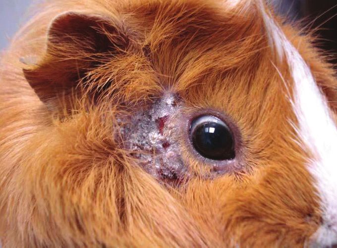
CONSIDERATION OF PET HEALTH AND LIFESTYLE FACTORS

Animals require care tailored to their individual needs. Certain factors may dictate more intensive monitoring
and/or treatment, while others may suggest a less aggressive approach.

Animal
The age and health status of the animal are important, including its history and origin. Some small pet animal
species have a greater susceptibility to some diseases, while other concomitant infections may predispose to
or aggravate existing parasitic or fungal diseases.

Environment
Animals kept in groups or those living outdoors may be at greater risk of acquiring infections than individual
animals living indoors. Also there may be cross-infection of some parasites and dermatophytes between small
mammals living in households with other domestic animal pets. The risk of transmission may also depend on
various local conditions such as geographical areas where certain parasitic diseases are endemic. Owners
should practise good husbandry and ensure that cage sizes are adequate and bedding quality is appropriate
for the species. Animals should be housed in a well-ventilated, dry and draught-free area protected from
temperature extremes.

Hygiene
Maintenance of good hygiene standards is important as is treatment of the environment in some cases. This
includes keeping cages or hutches clean and frequently changing bedding to eliminate possible sources of
reinfestation. Most small pet mammals kept in unhygienic conditions may be susceptible to blowfly strike and
maggots.

Nutrition
Poor nutrition may contribute to an animal’s susceptibility to many diseases including parasitic and fungal
infections. A good quality diet and vitamin and mineral supplementation is recommended and considered
important in aiding recovery.

Location and Travel
Animals living in, or travelling to, endemic areas are at a higher risk of acquiring certain infections. Extra care
should be taken when taking animals on holiday, to shows or if they are re-homed or placed in boarding
facilities.

                                                                                                                      5
6
1: Rabbits
Common or important parasites and fungal infections

                                                      7
8
CHAPTER 1:
COMMON OR IMPORTANT PARASITES AND
FUNGAL INFECTIONS OF RABBITS

This chapter does not deal with rabbits kept for food production where regulations are in place concerning
management and treatment.

                INTERNAL
                Roundworms          Passalurus ambiguus, Obeliscoides cuniculi, Graphidium strigosum,
                                    Trichostrongylus retortaeformis
                Adult Tapeworms     Cittotaenia ctenoides, Mosgovoyia pectinata

                Tapeworm Larvae     Alveolar hydatid cysts (Echinococcus multilocularis), larval stage of Taenia pisiformis
                                    (cysticercus pisiformis), larval stage of Taenia serialis (coenurus serialis)
                Protozoa            Eimeria spp., Giardia spp., Toxoplasma gondii
 PARASITES
                EXTERNAL
                Fleas               Spilopsyllus cuniculi, Ctenocephalides spp.
                Flies               Lucilia sericata and others
                Lice                Haemodipsus ventricosus
                Mites               Cheyletiella parasitivorax, Psoroptes cuniculi, Leporacarus gibbus, Demodex cuniculi,
                                    Sarcoptes scabiei, Notoedres cati, Ornithonyssus bacoti
                Ticks               Ixodes spp. and other Ixodidae
                INTERNAL

 FUNGAL         Systemic            Encephalitozoon cuniculi, Pneumocystis oryctolagi
 INFECTIONS     EXTERNAL
                Dermatophytes       Trichophyton mentagrophytes (complex species), Microsporum canis

INTERNAL PARASITES

Roundworms
Rabbits can become infected with a variety of worms
and in many cases the adult worms are found in the
gastrointestinal tract.

Nematodes known to infect rabbits include
Passalurus ambiguus (Figure 1). This is an oxyurid
(or pinworm) commonly found in the caecum and
large intestine of (domestic) rabbits. The presence
of even relatively large numbers of pinworms is
nonpathogenic. The adult worms measure up to
1 cm in length. Occasionally, some infections by
P. ambiguus can cause rectal, anal and perianal
irritation, rectal prolapse, restlessness and decreased
weight gain. Diagnosis is by adhesive tape method
or (rarely) coproscopy. Eggs are typically flattened              Figure 1: Passalurus ambiguus egg seen with Eimeria spp.
along one side as for most oxyurid species.

Obeliscoides cuniculi, Graphidium strigosum and Trichostrongylus retortaeformis are the most common
species of gastrointestinal nematodes in wild rabbits, all with direct life cycles. In domestic rabbits, they are
rarely found.

                                                                                                                              9
Tapeworms
The adult stage of several species of tapeworm can occur in the intestine of rabbits including Cittotaenia
ctenoides and Mosgovoyia pectinata. They occur predominantly in wild rabbits but can occasionally be
found in domestic rabbits. All have an indirect life cycle with free-living mites and other invertebrates as
intermediate hosts.
Rabbits can harbour the cystic stages of several adult tapeworms of dogs, the most common being Taenia
pisiformis and Taenia serialis. The former causes liver, peritoneal or retrobulbar cysts (cysticercus pisiformis)
and the latter cystic lesions in muscle and subcutaneous tissue (coenurus serialis). These cysts rarely cause
health problems but can be surgically removed if necessary. Domestic rabbit infections originating from egg
contamination from the faeces of urban foxes and other carnivores have been increasingly reported.
Protozoa
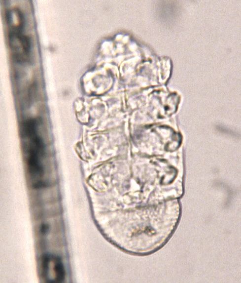
There are a number of Eimeria species that infect
rabbits. Of them, Eimeria intestinalis (Figure 2) and
Eimeria flavescens are the most pathogenic intestinal
species. Eimeria stiedai infects bile duct epithelial
cells causing hepatic coccidiosis. In all cases,
infection is transmitted by environmentally-resistant
oocysts passed in the faeces of infected rabbits.
Eimeria may be mistaken for the yeast Cyniclomyces
guttulatus (Figure 3).
Infection with Eimeria species is most likely to be
a problem where large numbers of rabbits are kept
in close proximity, however, infection may also
occur in pet rabbits. In conditions favourable for
oocyst survival, high levels of infection can build
up. Infection is most likely to cause clinical signs in Figure 2: Eimeria intestinalis oocyst (27 x 18 µm)
young rabbits but after an initial infection they often
acquire immunity to subsequent infection.
Intestinal coccidiosis may result in chronic diarrhoea,
weight loss and reduced appetite. Consequences of
hepatic coccidiosis include diarrhoea, weight loss,
jaundice, hepatomegaly and ascites. The severity
of these clinical signs will depend on the Eimeria
species involved, level of infection and the immune
status of the animal. Infections can occur if a pet
rabbit comes into contact with an Eimeria species
for which it has no immunity. Patent infections can
be diagnosed by the detection of oocysts in the
faeces.
Giardia spp. infect the small intestine of rabbits and
Giardia cysts (8–10 µm) can be found in the faeces.
The clinical significance of infection is unknown and,
at present, it is unclear whether Giardia spp. from Figure 3: Cyniclomyces guttulatus (8–10 μm in length)
rabbits belong to a zoonotic assemblage.
Rabbits can act as intermediate hosts for Toxoplasma gondii. Infection is usually asymptomatic but can also
cause granulomatous inflammation in a wide variety of organs including the central nervous system (CNS).
Contact with infected rabbits does not represent a risk for pet owners.
10EXTERNAL PARASITES
Fleas
Wild rabbits and domestic rabbits that live in close contact with wild rabbits may be infested with the rabbit
flea Spilopsyllus cuniculi. These fleas attach around the pinnae and tend not to move even when handled.
Spilopsyllus cuniculi fleas are vectors of myxomatosis (as are mosquitoes).
Rabbits, particularly those living in a household with dogs and cats, may become infested with the dog or
cat flea (Ctenocephalides spp.). These fleas can be found on the body of the rabbit and infestation can be
associated with considerable irritation. As with cats and dogs, diagnosis is based on the demonstration of
fleas or flea faeces using a flea comb.
Flies
Lucilia sericata and other flies may cause fly strike (myiasis) in hot summer months. Female blow flies lay their
eggs in wounds or soiled areas of the coat; they are particularly attracted to areas soiled by urine or faeces.
Once hatched, the larvae or maggots begin to feed in the skin and within a relatively short time they may
penetrate subcutaneous tissues. Affected rabbits rapidly become depressed and infection can prove fatal
unless treated promptly. Diagnosis is based on the demonstration of maggots on the skin.
Lice
Haemodipsus ventricosus lice are rare parasites in domestic rabbits. Animals can become infested by lice if
kept under poor husbandry conditions and may show irritation, suffer slight hair loss and/or become anaemic.
Diagnosis is based on the demonstration of lice on the coat or egg cases (nits) in fur samples.
Mites
The fur mites Cheyletiella parasitivorax and Leporacarus
gibbus can be well tolerated in rabbits but may also
be associated with skin irritation, slight hair loss and a
scaly dermatitis, usually along the back of the animal.
These non-burrowing mites are relatively large, visible
to the naked eye and may cause movement of skin
scales. This can be seen on close examination of the
coat as ‘walking dandruff’. Diagnosis is based on
microscopic identification of mites on fur samples
(Figure 4). Cheyletiella parasitivorax can be transferred
to humans by handling infected rabbits causing
irritation and skin lesions.
Psoroptes cuniculi is a surface mite that occurs most
commonly in the external ear canals and the pinnae
of rabbits causing a thick, scaly lesion known as an Figure 4: Cheyletiella parasitivorax
ear canker (Figure 5). The lesions are pathognomonic
of disease and mites may be seen on microscopic
examination.
Demodex cuniculi is a relatively rare parasite of
the domestic rabbit. Infestations cause moderate
pruritus and scaly skin lesions — signs similar to
those seen in cheyletiellosis.
The burrowing mites Sarcoptes scabiei and
Notoedres cati can cause scabies in rabbits.
Affected animals experience mild pruritus and show
typical skin changes including hyperkeratosis,
excoriations and scaly crusts on the head, ears,
distal limbs and interdigital areas. Both species are
zoonotic and can cause skin irritation to the handler,
however the life cycle of these mites is self-limiting
as it cannot be completed in humans. This is also Figure 5: Psoroptic mange
true for Cheyletiella.
11The tropical rat mite (Ornithonyssus bacoti) is worldwide in distribution and primarily affects wild rodents
such as rats and mice (Figure 15). However, small, domestic mammals including rabbits can also be possible
reservoirs. The blood-feeding mites, which can cause skin irritation and anaemia, are active at night and seek
dark hiding places during the daytime. A definitive diagnosis requires the detection of the parasite, which is
more likely to be found in the environment (e.g. in cages, in litter and in corners or cracks of the living area)
than on the host’s skin itself. In the case of close human–pet contact, mites can occasionally cause pruritic
dermatitis in humans.
Ticks
Ixodid ticks can affect rabbits if they live outside. These ticks will feed for several days before they drop off
naturally. They may be removed with a tick removal tool.
INTERNAL FUNGAL INFECTIONS
Encephalitozoon cuniculi is an intracellular
microsporidial parasite. Infection can be
asymptomatic but mild to severe neurological
consequences may result. The CNS, kidney and
eye are predilection sites for the organism. There
is evidence to suggest that up to 50% of rabbits
are seropositive. Neurological signs, for example
head tilt (Figure 6), ataxia and paralysis, or other
signs such as uveitis, symptoms of nephritis and
emaciation resulting in death may be seen in infected
rabbits. Transmission occurs via spores which are
passed in the urine from approximately one month
after initial infection. A tentative diagnosis is based
upon history, clinical signs, serology and, although
rarely detected, the demonstration of spores in the
urine. E. cuniculi is considered to be one of the
most virulent microsporidial organisms to infect
humans although disease accompanied by clinical Figure 6: Head tilt in a rabbit suspected of an
signs is rare in healthy individuals but can occur in Encephalitozoon cuniculi infection
immunocompromised patients.
Pneumocystis spp. may be commensal inhabitants of the lungs of rabbits. These atypical fungal organisms
are highly host-specific. Pneumocystis oryctolagi has been described in rabbits. Secondary interstitial
pneumonia may occur when the animals are immunosuppressed or debilitated owing to concurrent disease.
Pneumocystosis may also be observed at weaning in rabbits.
12EXTERNAL FUNGAL INFECTIONS Most cases of ringworm in rabbits are due to infection with dermatophytes which belong to the complex species Trichophyton mentagrophytes, although some (
Other Ectoparasites
     Permethrin may be used to treat/control flies, ticks and lice. The macrocyclic lactones ivermectin (0.2–0.4 mg/
     kg bodyweight subcutaneously every 10–14 days), doramectin (0.2–0.3 mg/kg bodyweight subcutaneously/
     intramuscularly as a single dose), moxidectin (0.2–0.3 mg/kg orally/subcutaneously, repeated after 10 days
     if needed) and selamectin are suitable for the treatment and control of lice and fur, ear and mange mites
     in rabbits. In some countries, ivermectin spot-on is available for use in rabbits and other small mammals.
     Dosage and treatment frequency should be in accordance with the manufacturer’s instructions. In the case of
     Psoroptes cuniculi infection, the abundant cerumen should be removed and the ears must be cleaned with a
     mild antiseptic prior to the administration of these drugs.

     Nitenpyram, reported by veterinarians and wildlife rehabilitators to kill blowfly maggots in mammals and birds,
     has anecdotally been reported as effective in rabbits. To alleviate the shock associated with blowfly strike,
     pain relief is recommended.

     Fungal Infections
     Fenbendazole is recommended for the treatment of Encephalitozoon cuniculi infection when administered at
     an oral dosage of 20 mg/kg bodyweight daily for 28 days. It can assist in reducing the severity of the clinical
     signs but elimination of the infection is not possible.

     For fungal infections, a combination of systemic and topical treatment is recommended. Systemic treatment
     relies on the daily administration of an oral antifungal drug: griseofulvin (25–50 mg/kg bodyweight – this can
     be given in two daily doses), itraconazole (2.5–10 mg/kg bodyweight) or terbinafine (8–20 mg/kg bodyweight).
     The decision to use topical therapy with enilconazole or miconazole should be based upon the owner’s ability
     and willingness to pour or sponge the product over the entire coat of the infected animal. The frequency of
     topical treatment should be at least twice a week. When sponging or bathing, the owner needs to consider
     hypothermia and the animal should be dried carefully. Ointments, creams, lotions or shampoos containing
     miconazole can also be used on localised lesions, but on a daily basis.

     Owners are advised to stop antifungal administration after two negative mycological cultures. Hygiene
     measures are extremely important, especially treatment of the environment. For large groups of affected
     rabbits, the environment can be sprayed with an enilconazole solution of 50 mg/m2 twice weekly for 4 months.
     Additionally, the rabbit owner should use disposable gloves and thoroughly wash/disinfect clothes and shoes
     after every treatment and/or animal manipulation.

     PREVENTION OF PARASITE AND FUNGAL INFECTIONS
     Prevention of parasite infection generally involves a combination of good environmental management and
     prophylactic drug treatments.

     For example, quarantine should be initiated for animals of unknown history before mixing with resident
     animals and high standards of husbandry should be maintained with particular attention to feed and bedding.
     Dogs with access to areas grazed by rabbits should be regularly treated for tapeworms to avoid infecting the
     rabbits with tapeworm cysts.

     Although good environmental management strategies will prove beneficial, the number of licensed drugs
     available for either the prophylactic or therapeutic treatment of rabbits remains limited.

     Coccidiosis is unlikely to be a problem for well-managed, small-scale rabbit keepers. Daily cleaning of cages
     and materials is advised to prevent environmental contamination with oocysts. Mixing rabbits which may be
     carrying different strains should be avoided.

     Fenbendazole may be used for the prevention of Encephalitozoon cuniculi infection when administered at an
     oral dose of 20 mg/kg bodyweight for 7–14 days 4 times a year. This preventative treatment may be used at
     times of increased risk of exposure to infection such as exhibitions or shows. Stressful situations can also
     induce shedding and flare-ups of an existing infection. Prolonged use of fenbendazole in rabbits can be
     associated with bone marrow suppression.

14
Various compounds marketed for the prevention of blowfly strike in sheep may be used in rabbits e.g.
dicyclanil and cyromazine. These can prevent fly strike for up to 16 weeks after application. Protection from
blowfly strike may be achieved by bringing rabbits indoors and keeping their coats clean, especially during
periods of warm, humid weather.
Disclaimer:
Every effort has been taken to ensure that the information in the guideline, which is based on the authors’
experience, is accurate. However the authors and publishers take no responsibility for any consequence
arising from the misinterpretation of the information herein nor is any condition or warranty implied. ESCCAP
emphasises that national, regional and local regulations must be borne in mind at all times before following
ESCCAP advice. All dosages and indications are provided for guidance. However, vets should consult
individual data sheets for details of locally approved treatment regimen.
1516
2: Rats
Common or important parasites and fungal infections

                                                      17
18
CHAPTER 2:
COMMON OR IMPORTANT PARASITES
AND FUNGAL INFECTIONS OF RATS

                INTERNAL
                Roundworms          Heligmosomoides polygyrus (syn. Nematospiroides dubius), Nippostrongylus spp.,
                                    Trichostrongylus spp., Heterakis spumosa, Calodium hepaticum (syn. Capillaria hepatica),
                                    Trichosomoides crassicauda, Syphacia muris, Aspiculuris tetraptera, Moniliformis
                                    moniliformis, Trichuris muris
                Adult Tapeworms     Rodentolepis nana (syn. Hymenolepis nana), Hymenolepis diminuta, Cataenotaenia pusilla

                Tapeworm Larvae     Larval stage of Taenia taeniaeformis (cysticercus fasciolaris)
                Protozoa            Giardia spp., Chilomastix spp., Trichomonas spp., Entamoeba muris, Trypanosoma lewisi,
 PARASITES                          Spironucleus muris, Eimeria spp., Cryptosporidium spp., Sarcocystis spp.,
                                    Toxoplasma gondii, Babesia microti
                EXTERNAL
                Fleas               Nosopsyllus fasciatus, Xenopsylla spp., Ctenocephalides spp.
                Lice                Polyplax spinulosa
                Mites               Myobia musculi, Myocoptes musculinus, Radfordia spp., Notoedres muris,
                                    Trixacarus diversus, Demodex ratticola, Psorergates simplex, Ornithonyssus bacoti,
                                    Liponyssoides sanguineus
                Ticks               Ixodes spp. and other Ixodidae
                INTERNAL

 FUNGAL         Systemic            Encephalitozoon cuniculi, Pneumocystis spp.
 INFECTIONS     EXTERNAL
                Dermatophytes       Trichophyton mentagrophytes (complex species), Microsporum spp.

INTERNAL PARASITES

Roundworms
Pinworms (Syphacia muris, Aspiculuris tetraptera) have a direct life cycle; they feed on bacteria in the intestinal
tract and are usually non-pathogenic, even in large numbers. Occasionally, Syphacia can cause rectal, anal
and perianal irritation, rectal prolapse and decreased weight gain. Debilitated animals are more susceptible
to other infections. Diagnosis is by adhesive tape method or (rarely) coproscopy. Eggs are typically flattened
along one side.

Trichosomoides crassicauda is a nematode found in the epithelium and lumen of the urinary bladder of wild
rats. Capillaria-like eggs are shed in the urine. Clinical signs are associated with the larvae which migrate
through the lungs and kidneys causing inflammation and granulomatous reactions.

Trichuris muris is a nematode of the large intestine, commonly known as whipworm. It is mostly associated
with wild rats and mice but can also be found in domestic equivalents. Clinical signs can include mucoid
diarrhoea with blood staining. Eggs can be isolated by centrifugal/flotation techniques. They are barrel or
lemon-shaped, light brown in colour and show bipolar plugs.

                                                                                                                               19
Tapeworms
The small tapeworms (Rodentolepis nana,
Hymenolepis diminuta) may be found in the lumen of
the small intestine of many mammalian hosts, including
rats. The parasite is transmitted directly via eggs or
through ingestion of an intermediate host. With the
auto-infective cycle, eggs mature within the intestinal
lumen without leaving the host. Infection levels depend
on the quality of husbandry. Eggs in a contaminated
environment may cause zoonotic infections, especially
in children. Strict hygiene, such as thorough cleaning
and sterilisation, is needed. Normally the infection
causes little harm in rats. Diagnosis is made by faecal
examination and by the demonstration of typical thick-
walled, round eggs containing an embryo with six Figure 8: Rodentolepis/Hymenolepis egg (60 x 80 µm)
hooklets (Figure 8).
Protozoa
Flagellates of the genus Giardia are common intestinal parasites in rodents and rats may be infected by
Giardia intestinalis or G. muris. Trophozoites attach to the mucous membrane of the intestinal villi. Giardia
cysts (8–10 µm) can be seen in faecal samples. Infections are often subclinical but can result in diarrhoea and
weight loss.
The coccidia of the genus Eimeria are often considered non-pathogenic or secondary pathogens in rodents
and the rat is the host of several species, two of which are found in the small intestine (E. nieschulzi and
E. miyairii) and one in the caecum (E. separata). Eimeria nieschulzi is the most commonly found and potentially
the most pathogenic. It affects mostly young animals in which heavy infection may be fatal. Diagnosis is
by faecal examination (typical oocysts can be detected) or by post-mortem examination showing enteric
thickening and petechial haemorrhages.
Cryptosporidiosis occurs in a large variety of hosts including rodents, frequently causing diarrhoea.
Polymerase chain reaction-based genotyping (PCR) and subtyping tools have allowed the identification of
several Cryptosporidium species (including C. parvum, C. muris, C. andersoni and C. wrairi) and nearly 20
genotypes of uncertain species status in rodents worldwide. Mixed Cryptosporidium species/genotypes
are also sometimes detected. The organisms are found within epithelial cells of the stomach or intestine.
Clinical signs are associated with subsequent villous atrophy and enteritis, which may lead to unthriftiness,
weight loss, diarrhoea and death. Cryptosporidium oocysts are very small (approximately 4–5 µm) but can be
detected by modified acid-fast staining of fresh faecal samples.
Rats can act as intermediate hosts for Toxoplasma gondii. The infection is acquired through the ingestion
of sporulated oocysts from cats or through vertical transmission via the placenta during pregnancy. The
pathogenicity of the infection depends on the number and virulence of the infecting organisms. Infections are
usually asymptomatic but can cause granulomatous inflammation in a wide variety of organs. Contact with
infected rats does not represent a risk for pet owners.
EXTERNAL PARASITES
Fleas
Wild rats are the preferred hosts of fleas of the
genera Nosopsyllus (Figure 9) and Xenopsylla,
whereas fleas of the genus Ctenocephalides are
often found infesting pet rats housed near cats and/
or dogs. Flea infestation is associated with a dull
coat, alopecia and pruritus. Secondary bacterial
infection, hypersensitivity and anaemia are common
complications in cases of severe flea infestation.
Figure 9: Nosopsyllus fasciatus 100x
20Lice
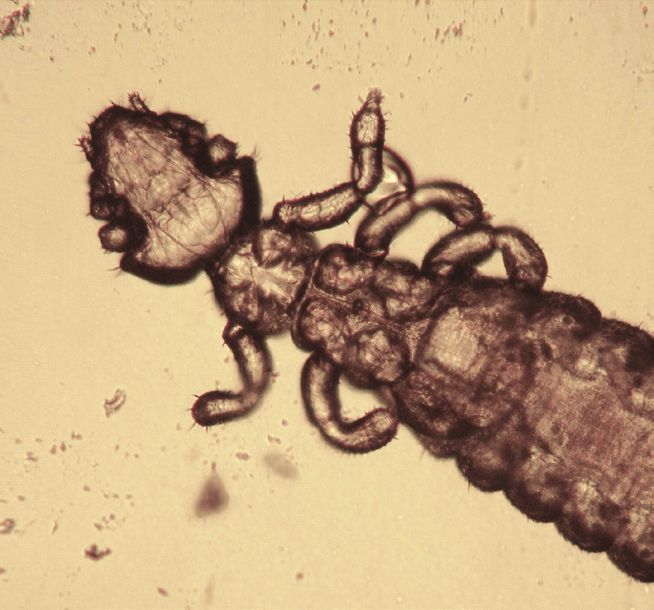
Blood-sucking lice (Polyplax spinulosa) may be
detected in large numbers, particularly in ageing,
sick animals. They can cause pruritus, restlessness
and anaemia. They may also transmit pathogenic
agents (bacteria). Normally lice are host-specific
and do not leave their host. Adults (Figure 10) and
eggs (nits) can be quite easily detected by careful
examination of the fur.
Mites Figure 10: Polyplax spinulosa 100x
Fur-dwelling mites (Myobia musculi, Myocoptes
musculinus, Radfordia affinis, Radfordia ensifera)
are pearly white mites seen near the base of the
hair. These mites have a markedly characteristic
body shape with the lateral margins extending
between the limbs (Figures 11 and 12). The eggs
are cemented to the base of hairs. Transmission
is by direct contact between rats (or other rodents
such as mice). Clinical signs include pruritus, hair
loss, erythema and thickening of the skin. Lesions
are usually present around the head and shoulders.
Figure 11: Myobia musculi Figure 12: Myocoptes
Secondary bacterial infections associated with self- 400x musculinus 400x
trauma may occur. Skin scrapings, hair plucks or
adhesive tape strips can be used to demonstrate the
presence of fur-dwelling mites.
Burrowing mites such as Notoedres muris (Figure 13)
and Trixacarus diversus are responsible for mange.
Notoedres muris prefers the epidermal tissue of the
ears and face (Figure 14). Wart-like, papular lesions
appear, usually accompanied by yellow crusts. Skin
scrapings are recommended to demonstrate the
presence of these mites.
Figure 13: Notoedres muris
The tropical rat mite (Ornithonyssus bacoti) is
worldwide in distribution and primarily affects wild
rodents such as rats and mice (Figure 15). However,
small, domestic mammals can also be possible
reservoirs. The blood-feeding mites, which can
cause skin irritation and anaemia, are active at night
and seek dark hiding places during the daytime. A
definitive diagnosis requires the detection of the
parasite, which is more likely to be found in the
environment (e.g. in cages, in litter and in corners
or cracks of the living area) than on the host’s skin
itself. In the case of close human–pet contact, mites
can occasionally cause pruritic dermatitis in humans. Figure 14: Notoedres muris infection on a rat
Ticks
Ixodid ticks can affect rats if they live outside.
These ticks will feed for several days before they
drop off naturally. They may be removed with a tick
removal tool.
Figure 15: Ornithonyssus bacoti 400x
21INTERNAL FUNGAL INFECTIONS
As with many mammals, Encephalitozoon cuniculi infection seldom occurs in rats.
Pneumocystis spp. are commensal inhabitants of the lungs of most rodents. These atypical fungal species are
highly host-specific. Pneumocystis carinii and P. wakefieldiae are present in domestic rats. Secondary interstitial
pneumonia may occur when the animals are immunosuppressed or debilitated owing to concurrent disease.
EXTERNAL FUNGAL INFECTIONS
Rats can become infected by dermatophytes
which belong to the complex species Trichophyton
mentagrophytes. In most cases, rats remain
asymptomatic but they may be a source of infection
for other animals, including humans. When lesions
are present, they usually include areas of circular or
diffuse alopecia with scaling of the head, neck and
tail (Figure 16). Pruritus is absent. Diagnosis is made
by microscopic demonstration of arthrospores in skin Figure 16: Ringworm infection in a rat
scrapings (KOH method) or by mycological cultures.
TREATMENT OF PARASITE AND FUNGAL INFECTIONS
When considering treatments for parasitic infections in rats, only a few suitable drugs are available and most
are used off-label. In the case of topical application, the risk of toxicity is high due to the small size of the
animals being treated and increased grooming activity which often accompanies pathological skin conditions.
Roundworms
Ivermectin has been used to treat infections in rats caused by small intestinal worms and pinworms. Various
dosage regimes for individual animals or those kept in groups have been recommended e.g. an oral dose at 0.2
mg/kg bodyweight daily for 5 consecutive days. Doramectin has also been effective administered in the food at
a rate of 0.2 mg/kg bodyweight daily for 4 days. Other anthelmintics such as fenbendazole and pyrantel have
been shown to be effective against pinworms in various domestic animals. Oxantel (25 mg/kg bodyweight) or
mebendazole (50 mg/kg bodyweight) administered twice are recommended for T. muris control. A combination
spot-on containing imidacloprid 10% and moxidectin 1% as a single dose can also be effective.
Tapeworms
Praziquantel (5–10 mg/kg bodyweight) orally or subcutaneously administered twice with an interval of 10 days
is the most effective treatment for adult tapeworms in the intestine; an oral dosage of fenbendazole (20 mg/
kg bodyweight) has also been used.
Protozoa
Metronidazole (2.5 mg/ml of drinking water) and dimetridazole (1 mg/ml of drinking water) for 7–14 days
have been recommended for many protozoal infections. Direct oral dosing with metronidazole is also
possible with dosages of 10–40 mg/kg bodyweight every 24 hours. Coccidiosis can be treated with toltrazuril
(10 mg/kg bodyweight orally using a 3 days on/3 days off schedule or at 25 ppm/l drinking water), sulfamerazine
(1 mg/ml drinking water), sulfamethazine (1–5 mg/ml drinking water) or sulfaquinoxaline (1 mg/ml drinking
water for 14–21 days).
Fleas
Rats can be treated with an insecticide (fipronil 7.5 mg/kg bodyweight or imidaclopid 20 mg/kg bodyweight)
topically every 30–60 days or an appropriate dose (15–30 mg/kg, repeated after 14 days) of selamectin pour-
on onto the neck. It is also important to treat the environment.
22Lice
Ivermectin can be used at 0.2–0.4 mg/kg bodyweight subcutaneously or orally every 7–14 days for 3
treatments. The successful use of selamectin and fipronil (one drop applied topically onto the neck) and
fipronil (applied as a spray over the whole body) has also been reported.
Mites
Ivermectin can be administered subcutaneously or orally (0.2–0.4 mg/kg bodyweight) every 7–14 days for
2–3 treatments. Moxidectin (0.5 mg/kg bodyweight topically or 2 mg/kg bodyweight orally), doramectin (0.2–
0.4 mg/kg bodyweight), selamectin, fipronil and permethrin have also been used orally, subcutaneously or
topically. All affected and in-contact animals must be treated. For large groups of animals, ivermectin can
be administered in the food. Interestingly, doramectin has been shown to have anxiolytic and anticonvulsant
properties in rats. These properties may contribute to a reduction in grooming activity which may aid the
resolution of cutaneous lesions.
Ticks
Ticks can be eliminated with the application of fipronil (spray).
Fungal Infections
Fenbendazole is recommended for the treatment of Encephalitozoon cuniculi infection and can assist in
reducing the severity of the clinical signs, but has not been proven to eliminate infection.
As with rabbits, dermatophytes should be treated with a combination of systemic and topical treatments.
Systemic treatment relies on daily oral administration of antifungal drugs: griseofulvin (25–50 mg/kg bodyweight
– this can be given in two daily doses), itraconazole (2.5–10 mg/kg bodyweight) or terbinafine (10–30 mg/kg
bodyweight). The decision to use topical therapy (with enilconazole or miconazole) should be based upon the
owner’s ability and willingness to pour or sponge the product over the entire coat of the infected animal. The
frequency of topical treatment should be at least twice a week. When sponging or bathing, the owner needs
to consider hypothermia and the animal should be dried carefully. Miconazole ointment can also be used on
localised lesions but on a daily basis.
Hygiene measures are extremely important, especially treatment of the environment. For large groups of
affected rodents, the environment can be sprayed with an enilconazole solution of 50 mg/m2 twice weekly
for 4 months. Additionally, the rat owner should use disposable gloves and thoroughly wash/disinfect clothes
and shoes after every treatment and/or animal manipulation.
PREVENTION OF PARASITE AND FUNGAL INFECTIONS
Prevention of parasite infection generally involves a combination of good environmental management and
prophylactic drug treatments.
For example, quarantine should be initiated for animals of unknown history before mixing with resident
animals and high standards of husbandry should be maintained with particular attention to feed and bedding.
Dogs with access to areas frequented by rats should be regularly treated for tapeworms to avoid infecting the
rats with tapeworm cysts.
Quarantine should be initiated for animals of unknown health history. Feed, cages and bedding should be
kept clean and dogs and cats that have contact with rats should be regularly treated for parasites including
fleas and tapeworms.
Disclaimer:
Every effort has been taken to ensure that the information in the guideline, which is based on the authors’
experience, is accurate. However the authors and publishers take no responsibility for any consequence
arising from the misinterpretation of the information herein nor is any condition or warranty implied. ESCCAP
emphasises that national, regional and local regulations must be borne in mind at all times before following
ESCCAP advice. All dosages and indications are provided for guidance. However, vets should consult
individual data sheets for details of locally approved treatment regimens.
2324
3: Mice
Common or important parasites and fungal infections

                                                      25
26
CHAPTER 3:
COMMON OR IMPORTANT PARASITES
AND FUNGAL INFECTIONS OF MICE

                INTERNAL
                Roundworms          Nematospiroides dubius, Nippostrongylus spp., Trichostrongylus spp.,
                                    Syphacia obvelata, Aspiculuris tetraptera, Trichuris muris
                Adult Tapeworms     Rodentolepis nana (syn. Hymenolepis nana), Hymenolepis diminuta, Cataenotaenia pusilla

                Tapeworm Larvae     Alveolar hydatid cysts (Echinococcus multilocularis), larval stage of Taenia taeniaeformis
                                    (cysticercus fasciolaris)
                Protozoa            Giardia spp., Chilomastix spp., Trichomonas muris, Spironucleus muris (syn. Hexamita
 PARASITES                          muris), Entamoeba muris, Eimeria spp., Cryptosporidium spp., Klossiella muris,
                                    Sarcocystis spp., Toxoplasma gondii
                EXTERNAL
                Fleas               Leptospylla segnis, Ctenocephalides spp.
                Lice                Polyplax serrata
                Mites               Myobia musculi, Radfordia affinis, Myocoptes musculinus, Psorergates spp.,
                                    Liponyssoides sanguineus, Ornithonyssus bacoti
                Ticks               Ixodes spp. and other Ixodidae
                INTERNAL

 FUNGAL         Systemic            Pneumocystis murina
 INFECTIONS     EXTERNAL
                Dermatophytes       Trichophyton spp.

INTERNAL PARASITES

Roundworms
Pinworms (Syphacia obvelata, Aspiculuris tetraptera) are normally considered to be non-pathogenic nematodes
that feed on bacteria inhabiting the intestinal tract of mice. They can, however, cause rectal, anal and perianal
irritation, rectal prolapse and decreased weight gain. Debilitated animals are more susceptible to infection.
Diagnosis is by coproscopy or the adhesive tape method. Eggs are typically flattened along one side.

Trichuris muris is a nematode of the large intestine, commonly known as whipworm. It is mostly associated
with wild rats and mice but can also be found in domestic equivalents. Clinical signs can include mucoid
diarrhoea with blood staining. Eggs can be isolated by centrifugal/flotation techniques. They are barrel or
lemon-shaped, light brown in colour and show bipolar plugs.

Tapeworms
The small tapeworms (Rodentolepis nana, Hymenolepis diminuta) may be found in the lumen of the small
intestine of many mammalian hosts, including mice. Transmission of these parasites can be directly via
ingestion of eggs, or indirectly through the ingestion of an intermediate host. Auto-infection is also possible as
the eggs are capable of maturing within the intestinal lumen without leaving the host. Eggs which are present
in the environment are also infective to humans. Strict hygiene and precautionary measures are needed e.g.
cleaning and sterilisation to reduce the risk of transmission. Tapeworm infections are not normally clinically
significant but weight loss, diarrhoea and death have been reported in heavily infected young mice. Diagnosis
is made by faecal examination and the demonstration of thick-walled, round eggs containing an embryo with
typical hooklets.

Mice are the intermediate hosts for Taenia taeniaeformis, an important tapeworm of cats. The larval stage
(cysticercus fasciolaris) develops in the liver of mice but infections are usually asymptomatic. It has been
suggested that liver neoplasia may be associated with this infection in mice. Mice with access to the external
environment may also act as intermediate hosts of the fox and dog tapeworm Echinococcus multilocularis,
an important zoonosis, but the larval infection (alveolar hydatid cysts) in mice represents no risk to humans.

                                                                                                                                 27
Protozoa
Flagellates of the genus Giardia are common intestinal parasites of rodents; mice are usually infected with
Giardia muris. Infections are often subclinical but can result in diarrhoea and weight loss. Trophozoites attach
to the mucous membrane of the intestinal villi and Giardia cysts (8–10 µm) can be detected in faecal samples.
Spironucleus (Hexamita) muris is a small piriform flagellate (2–3 x 7–9 µm) which inhabits the middle to lower
part of the small intestine in mice. Infection has been associated with clinical signs such as weight loss and
diarrhoea but only in certain strains of laboratory mice.
The coccidia of the genus Eimeria are often considered to be either non-pathogenic or secondary pathogens
in rodents. Several host-specific species have been described in mice. Eimeria pragensis develops within the
crypts of the caecum and colon and may be more pathogenic than the others. Clinical signs include profuse
and often bloody diarrhoea, weight loss and death. Diagnosis of coccidiosis is by faecal examination (typical
oocysts are detected) or by post-mortem examination showing enteric thickening and petechial haemorrhage.
Cryptosporidiosis occurs in a very large variety of rodent hosts. PCR-based genotyping and subtyping tools
have allowed the identification of several Cryptosporidium species (including C. parvum, C. muris, C. andersoni
and C. wrairi) and nearly 20 genotypes of uncertain species status in rodents worldwide. These organisms
are found within the epithelial cells of the stomach or the intestines and it is possible for mixed infections with
Cryptosporidium species/genotypes to occur. Cryptosporidiosis is often non-pathogenic in mice, although
clinical signs associated with villous atrophy and enteritis may lead to weight loss, unthriftiness and death.
Due to the small size of Cryptosporidium oocysts (approximately 4–5 µm), modified acid-fast staining of fresh
faecal samples is needed to aid detection by microscopy.
Infection with the renal coccidian parasite Klossiella spp. has been reported in mice although this is often as an
incidental finding. Whilst infections are usually asymptomatic, heavy parasite burdens can result in focal tubular
necrosis. Diagnosis of klossiellosis is based on histopathology or the detection of sporocysts in the urine.
Mice are natural intermediate hosts of Toxoplasma gondii and acquire the infection via the ingestion
of sporulated oocysts from cats or through vertical transmission via the placenta during pregnancy. The
pathogenicity of the infection depends on the number and virulence of the infecting organisms; infections are
usually asymptomatic, but can cause granulomatous inflammation in a wide variety of organs. Infected mice
do not represent a direct risk for pet owners.
EXTERNAL PARASITES
Ectoparasites are frequently reported in mice.
Fleas
The ‘blind’ mouse flea (Leptopsylla segnis) may be observed in wild mice whereas fleas of the genus
Ctenocephalides are often found infesting pet mice within the same household as cats and/or dogs. Flea
infestation is associated with a dull coat, alopecia and pruritus. Secondary bacterial infection, hypersensitivity
and anaemia are common complications in cases of severe infestation.
Lice
Sucking lice (Polyplax serrata) are rarely observed in mice. When present, they can cause irritation and
anaemia. Polyplax serrata also carries the rickettsial agent Eperythrozoon coccoides which causes murine
eperythrozoonosis. Lice are host-specific and do not normally leave their host. Adults and eggs (nits) can be
quite easily detected by careful examination of the fur.
28Mites
Fur-dwelling mites (Myobia musculi, Radfordia affinis, Myocoptes musculinus) are pearly white mites seen
near the base of the hair. The genera Myobia and Radfordia have a markedly characteristic body shape with
the lateral margins extending between the limbs (Figure 11). Myocoptes mites display heavily chitinised legs
which are adapted for clasping (Figure 12). The eggs are cemented to the base of hair. Transmission occurs
by direct contact between mice (or other rodents for non-host-specific mites). A healthy mouse can tolerate
a heavy infestation without apparent clinical signs. The increase in numbers and the occurrence of cutaneous
lesions are usually associated with age or stress factors including pregnancy. Clinical signs include pruritus,
hair loss, erythema and thickening of the skin. Secondary bacterial infection associated with self-trauma may
occur. Skin scrapings, hair plucks or adhesive tape strips are recommended to demonstrate the presence of
fur-dwelling mites.
Follicle-dwelling mites (Psorergates spp.) are small and spherical. Infestation is usually asymptomatic but
sometimes small, white nodules may appear on ear pinnae and the rest of the body.
Infestation with the house mouse mite (Liponyssoides sanguineus) is usually asymptomatic unless large
numbers of mites are present. These haematophagous mites may be detected on the animals but also in the
environment. The house mouse mite may transmit Rickettsia akari (the agent responsible for rickettsial pox)
to humans.
The tropical rat mite (Ornithonyssus bacoti) is worldwide in distribution and primarily affects wild rodents such
as rats and mice (Figure 15). However, small, domestic mammals can also be possible reservoirs. The blood-
feeding mites, which can cause skin irritation and anaemia, are active at night and seek dark hiding places
during the daytime. A definitive diagnosis requires the detection of the parasite, which is more likely to be
found in the environment (e.g. in cages, in litter and in corners or cracks of the living area) than on the host’s
skin itself. In the case of close human–pet contact, mites can occasionally cause pruritic dermatitis in humans.
Ticks
Ixodid ticks can affect mice if they live outside. These ticks will feed for several days before they drop off
naturally. They may be removed with a tick removal tool.
INTERNAL FUNGAL INFECTIONS
Pneumocystis spp. are commensal inhabitants of the lungs of most rodents. These atypical fungal species
are highly host-specific such as the mouse-adapted Pneumocystis murina. Secondary interstitial pneumonia
may occur if an animal is immunosuppressed or debilitated.
EXTERNAL FUNGAL INFECTIONS
Mice are usually infected by dermatophytes which belong to the complex species Trichophyton mentagrophytes
and, in most cases, the infection is asymptomatic. However, transmission to other animals including humans
may still occur. Lesions, when present, usually include areas of circular or diffuse alopecia with scaling on the
head, neck and tail. Pruritus is absent. In the case of infection by Trichophyton quinckeanum, cup-shaped
crusts may be seen grouped in patches or “favus”. Diagnosis is made by microscopic determination of
arthrospores in skin scrapings (KOH method) or mycological cultures.
29TREATMENT OF PARASITE AND FUNGAL INFECTIONS
Few treatment options are available for parasitic infections in mice and most of these are used off-label. In the
case of topical application, the risk of toxicity is high due to the very small size of the animals and increased
grooming activity associated with pathological skin conditions.
Roundworms
Ivermectin eliminates pinworms (with the same protocols as those recommended below for mites). Fenbendazole
20–50 mg/kg bodyweight orally for 5 days or 0.3% through the feed for 14 days may also be used. In all cases,
it is imperative that disinfection of the environment occurs concurrently. Oxantel (25 mg/kg bodyweight) or
mebendazole (50 mg/kg bodyweight) administered twice are recommended for T. muris control. A combination
spot-on containing imidacloprid 10% and moxidectin 1% as a single dose can also be effective.
Tapeworms
Praziquantel (30 mg/kg bodyweight twice or three times orally or subcutaneously at 10–14 day intervals) may
be used. Treatment should be accompanied by husbandry changes to prevent reinfection.
Protozoa
Metronidazole (2.5 mg/ml of drinking water for 5 days), dimetridazole (1 mg/ml drinking water) and
ronidazole have been recommended for the treatment of Giardia spp. and Spironucleus muris infections.
Hygienic measures are important for control of giardiosis. Coccidiosis can be treated with toltrazuril (10 mg/kg
bodyweight orally using a 3 days on/3 days off schedule or at 25 ppm/l drinking water), sulfamerazine
(1 mg/ml drinking water), sulfamethazine (1-5 mg/ml drinking water) or sulfaquinoxaline (1 mg/ml drinking
water).
Fleas
Mice can be treated topically with an insecticide such as fipronil 7.5 mg/kg bodyweight every 30–60 days (spray
the pump into a gloved hand and spread on to the rodent avoiding the mouth, ears and eyes) or imidacloprid
(20 mg/kg bodyweight). Alternatively, selamectin can be used (15–30 mg/kg bodyweight topically). It is also
important to treat the environment.
Lice
Ivermectin can be administered subcutaneously or orally (0.2–0.4 mg/kg bodyweight) every 7–14 days for 2–3
treatments.
Mites
Several protocols have been tested, but in colonies eradication is always much more difficult to achieve
than in individual animals. For mite-infected mice, the ‘micro-dot’ dermal delivery technique with undiluted
ivermectin can be used. Two treatments (5 µL of 1% ivermectin solution) on the skin between the scapulae
may be recommended at 10-day intervals. For large groups of mice, the total dose may be calculated based
on group bodyweight and the ivermectin solution can be sprayed on the group and the cages. One part 1%
ivermectin (10 mg/ml) should be mixed with 10 parts tap water and sprayed once weekly for three weeks.
Please note that ivermectin is poorly soluble in water therefore using a lipid carrier such as propylene glycol
is recommended. No more than 1 ml should be administered for each animal. For large groups of animals,
ivermectin can be administered in the food. In mice, moxidectin (0.5 mg/kg bodyweight topically or 2 mg/kg
bodyweight orally), selamectin (15–30 mg/kg bodyweight) and fipronil may also be used.
Mite infections can also be markedly reduced by dusting adult and weanling mice and their bedding with
permethrin powder at weekly intervals. Cotton wool balls containing permethrin have also been used as
bedding and nesting material to treat mice with mite infections.
Ticks
Fipronil 7.5 mg/kg bodyweight every 30–60 days can be used for both prophylaxis and/or therapy (spray the
pump into a gloved hand and spread on to the rodent avoiding the mouth, ears and eyes).
30Fungal Infections
A combination of systemic and topical treatment should be recommended. Systemic treatment relies on
oral antifungal drugs: griseofulvin (25–50 mg/kg bodyweight daily – this can be given in two daily doses),
itraconazole (2.5–10 mg/kg bodyweight daily) or terbinafine (10–30 mg/kg bodyweight daily). The decision to
use topical therapy (with enilconazole or miconazole) should be based upon the owner’s ability and willingness
to pour or sponge the product over the entire coat of the infected animal. The frequency of topical treatment
should be at least twice a week. When sponging or bathing, the owner needs to consider hypothermia and
the animal should be dried carefully. Miconazole ointment or cream can also be used on localised lesions but
on a daily basis.
Hygiene measures are extremely important, especially treatment of the environment. For large groups of
affected rodents, the environment can be sprayed with an enilconazole solution of 50 mg/m2 twice weekly for
4 months. Additionally, the mouse owner should use disposable gloves and thoroughly wash/disinfect clothes
and shoes after every treatment and/or animal manipulation.
PREVENTION OF PARASITE AND FUNGAL INFECTIONS
Prevention of parasite infection generally involves a combination of good environmental management and
prophylactic drug treatments.
For example, quarantine should be initiated for animals of unknown history before mixing with resident
animals and high standards of husbandry should be maintained with particular attention to feed and bedding.
Dogs with access to areas frequented by mice should be regularly treated for tapeworms to avoid infecting
the mice with tapeworm cysts.
Quarantine should be initiated for animals with an unknown health history. Feed, cages and bedding should be
kept clean and cats and dogs that have contact with rats or mice should be regularly treated for tapeworms.
Caesarean section followed by cross-fostering onto mite-free dams has been proposed as a means of
eliminating mite infestation from commercial mouse colonies.
Disclaimer:
Every effort has been taken to ensure that the information in the guideline, which is based on the authors’
experience, is accurate. However the authors and publishers take no responsibility for any consequence
arising from the misinterpretation of the information herein nor is any condition or warranty implied. ESCCAP
emphasises that national, regional and local regulations must be borne in mind at all times before following
ESCCAP advice. All dosages and indications are provided for guidance. However, vets should consult
individual data sheets for details of locally approved treatment regimens.
3132
4: Gerbils
Common or important parasites and fungal infections

                                                      33
34
CHAPTER 4:
COMMON OR IMPORTANT PARASITES
AND FUNGAL INFECTIONS OF GERBILS

                INTERNAL
                Roundworms          Dentostomella translucida, Syphacia spp., Aspiculuris tetraptera
                Adult Tapeworms     Rodentolepis nana (syn. Hymenolepis nana), Hymenolepis diminuta

 PARASITES      Tapeworm Larvae     Larval stage of Taenia taeniaeformis (cysticercus fasciolaris)
                Protozoa            Entamoeba muris, Tritrichomonas caviae, Giardia spp., Eimeria spp., Toxoplasma gondii
                EXTERNAL
                Mites               Demodex spp., Liponyssoides sanguineus, Notoedres muris, Trixacarus diversus,
                                    Tyrophagus castellanii, Ornithonyssus bacoti

 FUNGAL         EXTERNAL
 INFECTIONS     Dermatophytes       Trichophyton mentagrophytes (complex species), Microsporum gypseum

INTERNAL PARASITES

Roundworms
Dentostomella translucida is the most common cause of pinworm infection in gerbils. The male pinworm is
about 10 mm long and the female 20 mm. Transmission occurs by the ingestion of embryonated eggs which
originate from around the perianal region and contaminate food and drinking water. Retrograde infections by
penetration of liberated larvae from the perianal region back into the colon or caecum can also occur. The
eggs are typical for oxyurid helminths, flattened along one side. Infected animals may show few or no clinical
signs, however in heavy infections, gerbils may lose weight or show poor growth rates. Occasionally, these
pinworms have been known to cause intestinal obstruction and intussusception.

Syphacia muris, S. obvelata and Aspiculuris tetraptera are other oxyurid worms seen in gerbils. The latter two
are most likely transmitted through contact with infected mice in the immediate environment.

Tapeworms
Rodentolepis nana and Hymenolepis diminuta are common small intestinal tapeworms of rodents. Infection
may be transmitted directly via eggs (R. nana) or through ingestion of an arthropod intermediate host such as
a flea or grain beetle. The clinical consequences of infection for the host are negligible. Diagnosis is made by
faecal examination demonstrating the thick-walled round eggs containing a larva, typical hooklets and polar
filaments. Note that the eggs of R. nana can cause infections in humans, especially children.

The larval stage of the tapeworm Taenia taeniaeformis (cysticercus fasciolaris) is found in the liver of rodent
intermediate hosts. The definitive host is the cat and occasionally the fox. Gerbils infected with these cysticerci
do not seem to display any clinical signs.

Protozoa
Entamoeba muris is regularly observed in gerbils through the detection of cysts during coprological
examinations. This species of Entamoeba appears to be non-pathogenic.

Tritrichomonas caviae may be observed in fresh faecal samples as motile protozoa with flagellae. Tritrichomonas
caviae is not considered as a pathogen.

                                                                                                                            35
EXTERNAL PARASITES
External parasites are uncommon in gerbils unless, for example, there are fleas such as Ctenocephalides spp.
affecting dogs, cats or rabbits in the same household.
Mites
Direct contact between animals or with infected skin matter (e.g. crusts from infected animals) may be an
important route of infection with Demodex spp. At first, the clinical signs may resemble bite wounds. Dry hair,
alopecia, crust formation and erythema of the skin with occasional ulceration can be caused by Demodex
in immunosuppressed, young or elderly animals. Diagnosis is made by skin scrapings treated with KOH and
examined microscopically.
The mite Liponyssoides sanguineus does not cause irritation unless present in large numbers.
Trixacarus diversus is a sarcoptic mite which can occasionally cause mange in gerbils. There is a higher risk of
infection in breeding colonies than in animals kept individually. This mite can also be transmitted to humans
causing lesions.
Notoedres muris is the other burrowing mite that can be found in gerbils causing irritation, pruritus and
thickened skin. This mite may also cause lesions in humans.
In the case of Tyrophagus castellani (copra itch mite), handling of contaminated faeces by humans may cause
itchy skin.
The tropical rat mite (Ornithonyssus bacoti) is worldwide in distribution and primarily affects wild rodents
such as rats and mice (Figure 15). However, small, domestic mammals including gerbils can also be possible
reservoirs. The blood-feeding mites, which can cause skin irritation and anaemia, are active at night and seek
dark hiding places during the daytime. A definitive diagnosis requires the detection of the parasite, which is
more likely to be found in the environment (e.g. in cages, in litter and in corners or cracks of the living area)
than on the host’s skin itself. In the case of close human–pet contact, mites can occasionally cause pruritic
dermatitis in humans.
EXTERNAL FUNGAL INFECTIONS
In gerbils, most dermatomycoses are caused by Microsporum species, especially M. gypseum. Infections
with Trichophyton species have also been described and of these, T. mentagrophytes (complex species) is the
most commonly found in gerbils. Most animals do not show clinical signs but some individuals can develop
circular areas of alopecia with erythema and crust formation particularly around the eyes, ears and nose.
The skin can also appear dry and thickened. Infection may spread within a group and very young, stressed
or immunosuppressed gerbils are the most susceptible. Diagnosis is made by detection via microscopy of
arthrospores in skin scrapings (with KOH) or mycological cultures. Dermatophyte infections of animals are a
major cause of zoonotic infection in humans.
36You can also read