Role of Melatonin in the Synchronization of Asexual Forms in the Parasite Plasmodium falciparum - MDPI
←
→
Page content transcription
If your browser does not render page correctly, please read the page content below
biomolecules

Review
Role of Melatonin in the Synchronization of Asexual
Forms in the Parasite Plasmodium falciparum
Maneesh Kumar Singh 1 , Bárbara Karina de Menezes Dias 2 and Célia R. S. Garcia 1, *
 1   Department of Clinical and Toxicological Analysis, Faculty of Pharmaceutical Sciences,
     University of São Paulo, São Paulo, SP 05508-000, Brazil; maneesh@usp.br
 2   Department of Parasitology, Institute of Biomedical Sciences, University of São Paulo,
     São Paulo, SP 05508-000, Brazil; bkmdias@gmail.com
 *   Correspondence: cgarcia@usp.br; Tel.: +55-11-3091-8536

 Received: 15 July 2020; Accepted: 26 August 2020; Published: 27 August 2020

 Abstract: The indoleamine compound melatonin has been extensively studied in the regulation of the
 circadian rhythm in nearly all vertebrates. The effects of melatonin have also been studied in Protozoan
 parasites, especially in the synchronization of the human malaria parasite Plasmodium falciparum
 via a complex downstream signalling pathway. Melatonin activates protein kinase A (PfPKA)
 and requires the activation of protein kinase 7 (PfPK7), PLC-IP3 , and a subset of genes from the
 ubiquitin-proteasome system. In other parasites, such as Trypanosoma cruzi and Toxoplasma gondii,
 melatonin increases inflammatory components, thus amplifying the protective response of the host’s
 immune system and affecting parasite load. The development of melatonin-related indole compounds
 exhibiting antiparasitic properties clearly suggests this new and effective approach as an alternative
 treatment. Therefore, it is critical to understand how melatonin confers stimulatory functions in
 host–parasite biology.

 Keywords: melatonin; Apicomplexa; rhythm; signalling

1. Introduction
      Malaria is a disease associated with a remarkably high mortality rate in its endemic areas, which
have subtropical climates. Data collected by the World Health Organization in 2019 using various
methods, such as epidemiological, geographic, and demographic, reported an estimated 228 million
cases and approximately 4.05 × 10⁵ deaths in more than 80 countries [1]. An estimated 93% of the
casualties were associated with sub-Saharan Africa alone, especially those in pregnant women and
children below 5 years of age. Although these numbers have consistently fallen over the years,
additional and reinvigorated efforts are needed for preventive measures. Most malaria fatalities are
associated with cerebral P. falciparum infection, but other Plasmodium parasites, viz. P. vivax, P. ovale,
P. malariae and the monkey malaria parasite P. knowlesi, can pose opportunistic threats to humans [2].
      The life cycle of the human malaria parasite P. falciparum spans an insect vector for meiosis and
a vertebrate host for mitosis. A female Anopheles mosquito injects few sporozoites subcutaneously
into the host during blood feeding. These sporozoites primarily establish infection in hepatocytes,
remain hidden from the host’s immune system, and divide mitotically into liver-stage merozoites [3].
After their release from the hepatocytes, the merozoites infect red blood cells (RBCs or erythrocytes) to
initiate asexual replication. This asexual intraerythrocytic development (IED) occurs when parasites
transform through a series of developmental stages, termed ring, trophozoite, and schizont, and at later
stages, the progeny merozoites are released to infect more RBCs. Notably, a few parasites transform
into sexual gametocyte forms that guarantee the beginning of the sexual cycle after being ingested
by the mosquito. The clinical manifestations and pathology of malaria are associated with periodic

Biomolecules 2020, 10, 1243; doi:10.3390/biom10091243                        www.mdpi.com/journal/biomolecules
Biomolecules 2020, 10, 1243 2 of 16
or recurring fever as a result of blood-stage parasite proliferation, when mature schizonts burst the
host’s RBCs. The periodic rupture of the RBCs releases cytosolic debris and parasite metabolites
that prompt host responses, causing malarial symptoms. Plasmodium-infected patients experience
chills and fever, often accompanied by nausea, headache, and fatigue [4]. More importantly, the
periodicity of malarial seizures follows a 24 h cycle, i.e., P. knowlesi (24 h), P. vivax and P. falciparum
(48 h), and P. malariae (72 h) [5]. Four decades ago, researchers found that the IED of the parasites
coincided with the host’s body temperature. A study by Hawking et al. shows that the schizogony
of P. knowlesi or P. cynomolgi changed to midnight instead of midday when an infected monkey was
subjected to continuous lowering of body temperature for 2–3 days [6]. The same study pointed
out that no alteration in cell division was observed in P. lophure because it does not exhibit 24 h IED,
indicating the rhythmic sensitivity of the host. Similarly, inversion of the nycthemeral cycle of mice
changed the schizogony of P. chabaudi to midday [7]. It was also shown that the schizogonic cycle of
P. chabaudi adjusted to the circadian rhythm of the infected mice after 10 days, even after changing
the timing of parasite inoculation time [8]. This evidence suggests that the circadian rhythm of the
host plays a role in the synchronous IED of parasites. In this review, we focus on the rhythm of
parasitism, especially that of the human malaria parasite P. falciparum. Over the years, many studies
have suggested that the host hormone melatonin is integral in synchronous parasite proliferation. We
will also discuss the efforts that have been made to develop indole-derivative compounds showing
antagonistic effects on parasite proliferation. Briefly, we will discuss the effect of melatonin in other
unicellular protozoan parasites, such as Toxoplasma gondii (toxoplasmosis), Leishmania (leishmaniases),
and Trypanosomes species (Chagas’ and sleeping sickness).
2. Melatonin-Dependent Rhythm in Parasites
Melatonin (N-acetyl-5-methoxytryptamine) is an indoleamine compound that was first extracted
from the bovine pineal gland and has also been reported in the peripheral nerves of other mammals,
including humans [9]. Later, endogenous production of melatonin was also reported in plants, bacteria,
unicellular organisms, and invertebrates [10–12]. It was speculated that melatonin was primarily
present in primitive bacteria (cyanobacteria and α-proteobacteria); over the course of evolution, these
bacteria cohabited endosymbiotically in early prokaryotes and later became universally distributed
in other organisms (reviewed in [13]). The melatonin level in the blood is controlled by day/night
exposure; it is high at night when it is secreted from the pineal gland, and its level drops during the
day. In animals, melatonin biosynthesis begins after tryptophan, a necessary precursor present in the
bloodstream, enters the pineal gland. Once tryptophan reaches the pineal gland, it is hydroxylated to
form 5-hydroxytryptophan (5-HTP) and then decarboxylated to produce serotonin. Arylalkylamine
N-acetyltransferase (AANAT), which is active during the night, acetylates serotonin and converts it to
N-acetylserotonin (NAS). Then, the enzyme hydroxyindole-o-methyltransferase (HIOMT) methylates the
hydroxyl group of NAS to produce melatonin. The whole melatonin synthesis process is regulated by
β-adrenergic receptor (β-AR) activation by norepinephrine (NE), which activates protein kinase A (PKA)
via cAMP elevation. The suprachiasmatic nucleus (SCN) perceives light/dark information to control
the amount of NE in the day via proteasomal degradation [14]. The primary function of melatonin
was thought to be nullifying the adverse effects of free radicals and reducing receptor-independent
oxidative stress, possibly for two potential reasons. First, it can easily cross cellular membranes due to
its hydrophobic nature; second, it can interact with various ROS and donate an electron or a hydrogen
atom [15]. Melatonin is known to regulate the circadian rhythm through the measurement of the
day/night interval [16]. Its metabolites have been found to control various physiological processes,
such as sleep, vasomotor regulation, anti-excitatory activities, immunomodulation and antioxidant
properties [17]. The role of the circadian rhythm has been extensively studied in various organisms,
including bacteria, fungi, plants, and animals [18–20]. It has been estimated that approximately 10% of
the genomes of higher vertebrates, such as mammals, are under circadian control [21].Biomolecules 2020, 10, 1243 3 of 16
As in higher animals, it has been found that circadian rhythm plays crucial roles in several
parasites to support transmission, an important phenomenon that enables the parasites to acquire
a new host. This process is more important for Plasmodium parasites, considering their short time
window from erythrocyte rupture to invasion, because after RBC rupture, the parasites are exposed to
the host defence system. To avoid prolonged exposure to the host immune system, parasite egress is
very synchronous; to accomplish this, Plasmodium spp. follow cycles that are multiples of 24 h [22].
The proliferation of asexual malaria parasites and their host-transmission efficiency are severely
affected when the rhythm of the host and parasite during IED is perturbed; however, parasites quickly
adapt to changes in the new environment [23,24]. Despite the periodic febrile waves generated in
Plasmodium-infected hosts, the driving cues related to this periodicity are largely unknown, and if there
is a host cue, then how it regulates periodicity during IED is unknown. Interestingly, it is known that
Plasmodium parasites match the host’s rhythm after establishing an infection but become asynchronous
when grown in vitro, indicating a host-derived signal. The role of the circadian rhythm was overlooked
until Hotta et al. for the first time studied the role of melatonin in the synchronization of IED, both
in vitro and in vivo, in Plasmodium [25]. In this study, when asynchronous P. falciparum parasites were
grown in vitro with 100 nM melatonin, the parasite cycle was accelerated towards a more mature
schizont stage. Similarly, murine P. chabaudi parasites lost synchrony in mice with surgically removed
pineal glands or mice injected with the melatonin receptor antagonist luzindole, and synchrony was
restored by administering melatonin [25]. Interestingly, two murine parasites, P. berghei and P. yoelii,
do not display melatonin-dependent synchronization in vivo [26]. The difference in the parasites
showing melatonin response is the ability of melatonin to mobilize cytosolic Ca2+ ([Ca2+ ]cyt ). We will
discuss the molecular mechanism of melatonin-induced signalling in the next section. Additionally,
melatonin modulates the expression of genes related to the ubiquitin/proteasome system (UPS) in
both mammals and P. falciparum [27–29]. It is evident that the UPS is fundamental in regulating the
cell cycle and transcriptional activity of cells [30,31]. Not only melatonin, but also its precursors
5-hydroxytryptophan, serotonin and N-acetylserotonin modulate IED in P. falciparum, increase mature
schizont percentage and mobilize [Ca2+ ]cyt [32] (Table 1). Apart from that, the melatonin catabolic
product N1-acetyl-N2-formyl-5-methoxykynuramine (AFMK) accelerates P. falciparum and P. chabaudi
synchronization via [Ca2+ ]cyt mobilization [33]). This leads to a new question of whether other
compounds having an indole ring are also capable of affecting IED in these parasites. To this end, the
indole-derivative plant hormone indole-3-acetic acid (IAA) was studied, and it was found that IAA
modulates neither the IED of the parasites nor UPS gene expression [34]. In addition, few synthetic
indole derivatives can regulate the cell cycles of parasites [35], suggesting that not all indole derivatives
confer cell cycle modulation. More recently, RNA-seq analysis of melatonin-treated P. falciparum
parasites revealed 38 differentially expressed genes, among which several genes were related to
post-translational modification (PTM), Zn-binding proteins and nucleic acid-binding proteins [36].
Another important aspect of melatonin is its synthesis in the mitochondria [37], cellular
powerhouses that produce ATP, generate free radicals and induce apoptosis. The production of
melatonin in the mitochondria provides a buffer against oxidative stress to prevent the mitochondria
from collapsing. Additionally, evidence supports the presence of the melatonin receptors MT1 and
MT2 in the mitochondria, which may provide protection against ischaemia-induced brain injury [38,39].
Mitochondria also act as Ca2+ reservoirs, along with the ER. It has been shown that mitochondria take
up Ca2+ very efficiently due to their proximity to the ER, so they can easily sense the Ca2+ microdomain
as a result of IP3 hydrolysis [40]. Although the role of melatonin in Ca2+ homeostasis in relation to
mitochondria has mostly been explored in the mammalian system, how melatonin affects parasite
mitochondria is still unknown. The genome sequence of Plasmodium has not revealed any melatonin
receptor, making it difficult to investigate melatonin signalling in relation to Ca2+ homeostasis in
mitochondria. However, our group has shown that mitochondria participate in Ca2+ homeostasis
through the reversible uptake of ER-released [Ca2+ ]cyt [41]. It would be interesting to investigate how
melatonin modulates mitochondrial Ca2+ and whether it has beneficial effects in parasites.Biomolecules 2020, 10, 1243 4 of 16
Table 1. Effects of indole-related compounds on Plasmodium parasites.
Compounds Action
P. falciparum-infected erythrocytes exhibit higher tryptophan uptake [42].
Tryptophan Inhibition of tryptophan catabolism during P. yoelii infection partially protects mice
against lethal infection [43]
Activates PCL/IP3 pathway to increase cytosolic Ca2+ and promotes cell cycle
5-Hydroxytryptophan
acceleration to increase schizont percentage in vitro in P. falciparum [32]
Activates PCL/IP3 pathway to increase cytosolic Ca2+ and promotes cell cycle
Serotonin
acceleration to increase schizont percentage in vitro in P. falciparum [32]
Activates PCL/IP3 pathway to increase cytosolic Ca2+ and promotes cell cycle
N-Acetylserotonin
acceleration to increase schizont percentage in vitro in P. falciparum [32,44]
Activates PCL/IP3 pathway to increase cytosolic Ca2+ and promotes cell cycle
acceleration to increase the schizont percentage in vitro in P. falciparum [25,32,44].
Modulates neither parasite maturation nor cytosolic Ca2+ in P. yoelii and P. berghei [26].
Melatonin Induces cAMP-dependent kinase PfPKA [45].
Upregulates UPS genes [46].
Promotes differential gene expression in P. falciparum trophozoite stage parasites [36].
Activates mitochondrial genes PfFIS1, PfDYN1, and PfDYN2 [47].
Indole-3-acetic acid (IAA) Modulates neither the IED of the parasites nor UPS gene expression [34].
3. Melatonin Triggers a Signalling Cascade in Plasmodium Parasites
The molecular mechanism by which melatonin controls the signalling cascade to regulate the
parasite cell cycle is still not fully understood. When Hotta et al. showed that melatonin accelerates
the IED, they also noticed that melatonin triggered the release of Ca2+ from the internal stores of
Plasmodium and activated the PLC-IP3 signalling pathway. The generation of IP3 was triggered
by the parasite’s phospholipase C (PLC) enzyme, and blocking PfPLC with U73122 obliterated the
melatonin-induced Ca2+ increase [25] (Figure 1). To further strengthen the melatonin hypothesis, it has
been shown that parasites that are not sensitive to melatonin-induced synchronization are not able
to elicit [Ca2+ ]cyt release from the internal stores [26]. In another study, Vaid et al. showed that the
PLC blocking compound U73122 inhibited Ca2+ release and blocked PfPKB activation, suggesting
that Ca2+ acts upstream of the PfPKB signalling cascade in P. falciparum [48]. The dispensable role of
phosphoinositide-specific PLC in Ca2+ generation was further suggested by Raabe et al., who performed
a transcriptional analysis of PLC in the blood stage of P. falciparum. They found steady PfPLC expression
in the blood stage and showed a transient increase in expression in late schizonts [49]. The first direct
study by Alves et al. reported a relationship between melatonin and the PLC-IP3 pathway, where
melatonin triggers IP3 production by activating PfPLC and opens ER-localized IP3 -sensitive Ca2+
channels [50]. Alves et al. used cell-permeant caged IP3 in intact P. falciparum-infected RBCs that releases
IP3 upon UV light exposure. They found that UV light exposure of the parasites caused photolysis of
caged IP3 and elicited a rapid and transient increase in [Ca2+ ]cyt , but melatonin stimulation before the
flash photolysis of the caged IP3 abolished the [Ca2+ ]cyt increase [50]. It was also reported that both P.
falciparum and P. chabaudi sustain intracellular Ca2+ stores, and isolated and permeabilized P. chabaudi
showed IP3 -dependent Ca2+ discharge [51]. It is interesting to consider that either a putative melatonin
receptor or a putative IP3 -receptor (IP3 R) has remained unidentified in P. falciparum. However, IP3 R
has been characterized in two different protozoan parasites, T. cruzi [52] and the food vacuoles of T.
brucei [53]. These studies independently showed that either disrupting or inhibiting the IP3 receptor
resulted in impaired growth and infection rates.
At the molecular level, melatonin has been shown to establish cross-talk between cAMP and
Ca . A membrane-permeant 30 ,50 -cyclic monophosphate N6-benzoyl/protein kinase A activator
2+
(6BZ-cAMP) exhibited dual specificity, activated protein kinase A (PfPKA) and increased [Ca2+ ]cyt
levels. Melatonin-induced cAMP was blocked by the PLC inhibitor U73122 or by blocking PfPKA with
a peptide inhibitor of PKA (PKI) that prevents increases in [Ca2+ ]cyt , indicating that PKA is required for
Ca2+ induction [45] (Figure 1). To elucidate how melatonin signalling occurs in parasites, Beraldo et al.
found that the IP3 R antagonist 2-aminoethoxydiphenyl borate (2-APB) nullifies the effects of melatonin
on the cell cycle and [Ca2+ ]cyt . They have also reported that Ca2+ influx is linked to sustained Ca2+sensor during parasite egress that triggers the release of [Ca2+]cyt and enables the parasite to adapt
under stress conditions [61]. It is not clear how PfSR25 triggers the signalling cascade, since GPCRs
are coupled to trimeric G-proteins that are absent in the Plasmodium genome. Interestingly, an RBC
protein, Gαs, along with β-adrenergic receptor (β-AR), is recruited on the vacuolar membrane of the
Biomolecules
parasite. 2020, 10, 1243 of β-AR activates Gαs by cAMP production and increases the infection
Stimulation 5 of 16
percentage, while competitive inhibition of Gαs with peptides reduces P. berghei parasitemia [62]. This
study indicates
mobilization fromthat parasites require
intracellular GPCR-mediated
organelles upon treatmentsignalling,
with theand this area iscAMP
cell-permeant central for the
analogue
development of new therapies against malaria. 2+Another Plasmodium GPCR-like
8BrcAMP, which is referred to as capacitative Ca entry (CCE) [54]. This store-operated Ca entry protein, PfSR10,
2+
exhibitswas
(SOCE) a circadian transcription
also blocked profilederivatives,
by two 2-APB with peak expression
DPB162-AEatand twoDPB163-AE,
time points butduring
thisthe IED, the
occurred at
first after 8 h and the second after 32 h post-invasion. Disrupting its orthologous
high concentrations in the presence of melatonin, suggesting a differential activation mechanism ofgene in P. chabaudi,
PcSR10,
SOCE shortens [55].
in parasites the IED by 2–3
Pecenin et al.h have
in mice
also [63].
shownSimilarly, it has been
that P. falciparum reported
failed thatinPlasmodium
to survive vitro when
parasites exhibit intrinsic rhythm in more than 75% genes during IED, increasing
transfected with IP3 -sponge or IP3 binding domain (IRIS)-containing plasmids [55]. These findings the complexity of
host–parasite periodicity [64,65].
provide additional evidence that melatonin-induced signalling is associated with the IP /Ca pathway 2+
3
and critical for parasite IED.
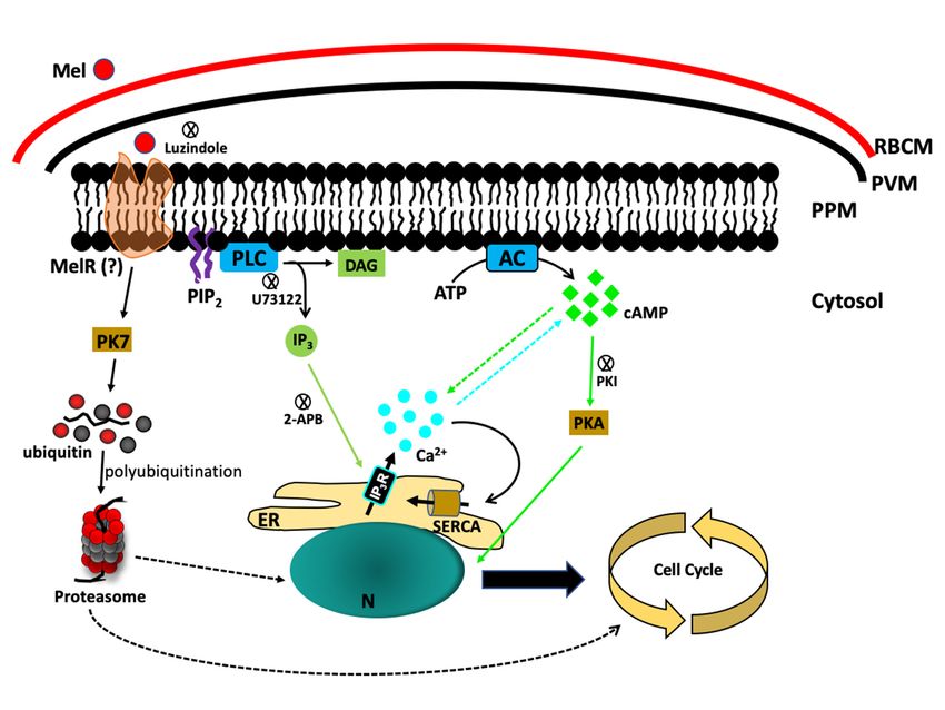
Figure 1. Melatonin signalling in P. Falciparum. Melatonin stimulates the cleavage of PIP2
by phospholipase
Figure 1. Melatonin C (PLC) to produce
signalling IP3 , whichMelatonin
in P. Falciparum. activates IP 3 R to release
stimulates Ca2+ in the
the cleavage cytosol.
of PIP 2 by
Simultaneously,
phospholipase Cmelatonin
(PLC) toalso activates
produce IP3the production
, which of cAMP,
activates IP3R towhich triggers
release Ca2+ the
in downstream
the cytosol.
PfPKA signallingmelatonin
Simultaneously, cascade. also
Onactivates
the other
the hand, the orphan
production of cAMP, kinase
whichPfPK7 is the
triggers upregulated
downstream by
melatonin and is linked with the parasite’s proteasomal activation. RBCM—RBC
PfPKA signalling cascade. On the other hand, the orphan kinase PfPK7 is upregulated by melatonin membrane;
PVM—Parasitophorous
and is linked with the vacuole membrane;
parasite’s PPM—Parasite
proteasomal activation.plasma membrane;
RBCM—RBC Mel—Melatonin;
membrane; PVM—
MelR—Melatonin receptor
Parasitophorous vacuole (hypothetical);
membrane; AC—Adenylyl
PPM—Parasite cyclase; Mel—Melatonin;
plasma membrane; PLC—Phospholipase MelR— C;
PIP 2 —Phosphatidylinositol-4,5-biphosphate;
Melatonin receptor (hypothetical); AC—Adenylyl IP3 —Inositol-1,4,5-triphosphate;
cyclase; PLC—PhospholipaseDAG—Diacylglycerol;
C; PIP2—
ER—Endoplasmic reticulum; N—Nucleus;
Phosphatidylinositol-4,5-biphosphate; SERCA—sarco/endoplasmic reticulum
IP3—Inositol-1,4,5-triphosphate; Ca2+ —ATPase.
DAG—Diacylglycerol; ER—
Endoplasmic reticulum; N—Nucleus; SERCA—sarco/endoplasmic reticulum Ca2+—ATPase.
Another piece of evidence came to light during a study of the orphan kinase PfPK7, which has
C-terminal homology with MEK and N-terminal homology with fungal PKA [56]. The PfPK7 knockout
(PfPK7− ) parasites were not able to exhibit melatonin-induced synchronization and [Ca2+ ]cyt rise, but
complementation with a functional copy of PfPK7 reverted the parasites towards melatonin sensitivity.
Additionally, the upregulation of 14 UPS genes, including E1, E2, E3, ubiquitin-like, deubiquitinase,
and proteasome subunits, by melatonin has been shown in wild-type P. falciparum and abolished in
PfPK7− parasites [28]. Further evidence that PfPK7 is pivotal for melatonin signalling came from two
studies that performed RNA-seq analysis and examined mitochondrial genes related to mitochondrial
fission [36,47]. Lima et al., in the RNA-seq study, compared the effects of 5 h melatonin treatment on
differentially expressed genes in wild-type P. falciparum 3D7 and in PfPK7− parasites. They foundBiomolecules 2020, 10, 1243 6 of 16
that 5 h melatonin treatment at the trophozoite stage resulted in 38 differentially expressed genes
in wild-type parasites but not in PfPK7− or untreated parasites. Additionally, 6 h cAMP treatment
differentially modulated 75 genes in rings, 101 genes in trophozoites, and 141 genes in schizonts [36].
A study by Scarpelli et al., however, showed the relevance of PfPK7 to the alteration of mitochondrial
fission genes PfFIS1, PfDYN1, and PfDYN2 upon melatonin treatment. The relative expression of these
genes was significantly altered in the presence of melatonin in wild-type parasites, but the effect was
abolished in PfPK7− parasites [47]. Increased transcription of UPS genes, especially ubiquitin-activating
enzyme E1 and ubiquitin ligase E3, along with the expression of the transcription factor PfNF-YB,
was observed in P. falciparum parasites in the presence of melatonin [57]. This evidence suggests
the pivotal role of melatonin in the signalling cascade that controls the gene expression leading to
parasite maturation. Considering all the above mentioned factors, it became important to investigate
the physiological and molecular aspects of Ca2+ signalling in these parasites, including how these
parasites sense external cues and activate signalling cascades mediated by a transient increase in
secondary messengers, viz. cAMP and/or [Ca2+ ]cyt , from their internal stores. It would be interesting
to investigate genes that modulate P. falciparum growth and are related to circadian rhythm.
In mammalian systems, melatonin acts through specific receptors to confer downstream effects in
cells. Receptors for melatonin (MT1 and MT2) have been identified in mammalian systems as G-protein
coupled receptors (GPCRs) [58] that are associated with downstream signalling events to affect the
levels of cytosolic secondary messengers, generally decreasing cAMP or cGMP and increasing IP3
or Ca2+ [59]. To identify melatonin receptors in the Plasmodium genome, a genome-wide search was
performed. Madeira et al. identified four GPCR-like or serpentine receptor-like (SR) genes, PfSR1,
PfSR10, PfSR12, and PfSR25, in P. falciparum, which are expressed mostly during the IED of this
parasite [60]. The functional characterization of these GPCR-like proteins is still under investigation.
Among these GPCR-like proteins, PfSR25 has been characterized as an external K+ sensor during
parasite egress that triggers the release of [Ca2+ ]cyt and enables the parasite to adapt under stress
conditions [61]. It is not clear how PfSR25 triggers the signalling cascade, since GPCRs are coupled to
trimeric G-proteins that are absent in the Plasmodium genome. Interestingly, an RBC protein, Gαs , along
with β-adrenergic receptor (β-AR), is recruited on the vacuolar membrane of the parasite. Stimulation
of β-AR activates Gαs by cAMP production and increases the infection percentage, while competitive
inhibition of Gαs with peptides reduces P. berghei parasitemia [62]. This study indicates that parasites
require GPCR-mediated signalling, and this area is central for the development of new therapies
against malaria. Another Plasmodium GPCR-like protein, PfSR10, exhibits a circadian transcription
profile with peak expression at two time points during the IED, the first after 8 h and the second after
32 h post-invasion. Disrupting its orthologous gene in P. chabaudi, PcSR10, shortens the IED by 2–3 h in
mice [63]. Similarly, it has been reported that Plasmodium parasites exhibit intrinsic rhythm in more
than 75% genes during IED, increasing the complexity of host–parasite periodicity [64,65].
4. Melatonin Confers Protective Immunity against Parasitic Infection
Early pathogenic infection must be recognized rapidly by the host innate immune system to clear
invasive microbes. The innate immune response provides the first line of defence against pathogens
and is based on pattern recognition receptors (PRRs) that recognize pathogen-associated molecular
patterns (PAMPs). PRRs are classified into membrane-associated toll-like receptors (TLRs), C-type
lectin receptors (CLRs), cytoplasmic non-membranous NOD-like receptors (NLRs) and RIG-I-like
receptors (RLRs) (reviewed in [66]). PAMPs consist of a different range of molecules, including
lipids, carbohydrates, nucleic acids or their combinations. Once PAMPs are recognized by PRRs,
innate immune cells activate specific PAMP-related signalling cascades via cytokine and chemokine
production. More importantly, these features are unique to pathogens and essential for survival.
However, the majority of the information available is restricted to viral and bacterial infection, but a few
studies have revealed how PAMPs work during parasitic infections. However, recent advancements in
this area have revealed the PAMPs associated with Apicomplexan and Trypanosomatid protozoanBiomolecules 2020, 10, 1243 7 of 16
parasites. In the case of Plasmodium parasites, glycosylphosphatidylinositol (GPI) expressed on the
merozoite surface is recognized by the TLR1-TLR2 heterodimer [67,68] and activates MAPK and NF-κB
pathways [69]. Similarly, growing malaria parasites digest haemoglobin to produce inert haemozoin
moieties, which are recognized by TLR9 by an unusual process where haemozoin forms a complex
with the parasite DNA and is presented to TLR9 [70]. Plasmodium-derived haemozoin and DNA also
activate cytosolic NLRP3 through Lyn and Syk kinases and induce the production of the cytokines IL-1
and IL-18 [71,72]. In T. gondii, GPI anchors and profilin-like proteins (PFTGs) are recognized by TLR2,
TLR4 and TLR11, respectively [73,74]. TLR11 recognition of PFTG induces IL-12 production that is
dependent on myeloid differentiation factor 88 (MyD88) [74]. Despite this evidence, we have very
elementary knowledge of whether the hormone melatonin plays any protective role in the host during
parasitic infection and enhancement of innate immunity. Therefore, we directed our focus towards
studies that reveal how melatonin induces or enhances the host response during parasitic infection.
Similar to Plasmodium, other protozoan parasites, such as Trypanosoma spp., T. gondii, and
Leishmania spp. respond to the host hormone melatonin. It is apparent that melatonin modulates
the immune system via receptor recognition. Melatonin receptors have been identified on CD4+
and CD8+ cells and B lymphocytes [75]. CD4+ cells induce delayed-type hypersensitivity (DTH)
that activates proinflammatory cytokine production, while CD8+ cells exert a cytotoxic effect; both
protect against intracellular protozoa [reviewed in [76]. In mice infected with T. gondii, melatonin
prompts the cellular immune response by galvanizing CD4+ and CD8+ lymphocyte production,
causing a surge in proinflammatory cytokines [77]. T. gondii-infected mice show increased nitric
oxide (NO) levels in the plasma, which have both immunoprotective and immunomodulatory roles
in chronic infection. However, higher NO production has a neurotoxic effect and promotes central
nervous system (CNS) degeneration in infected mice [77]. Melatonin can counteract the NO level by
reducing induced NOS (iNOS) activity to provide beneficial support during toxoplasmosis. In this
regard, it was shown that NO levels increased after T. gondii infection, especially in pinealectomized
rats [78]. Subsequently, it was found that cellular infiltration of lymphocytes and CD cells increased in
melatonin- and zinc-supplemented rats, and an adjunctive therapy to treat Toxoplasma retinochoroiditis
in immunocompromised patients was proposed [79]. In a recently published report on a monkey
kidney epithelial cell line, LLC-MK2 showed protection against T. gondii with melatonin treatment.
Despite a lack of alteration in cell viability, Machado et al. found that melatonin treatment altered the
invasive tachyzoite shape, which was associated with ruptured plasma membrane and cytoplasmic
leakage [80].
Similarly, another vector-borne parasite, Trypanosoma cruzi, has serious effects if left untreated.
It was found that proinflammatory cytokines play protective roles against T. cruzi infection and activate
macrophages during acute infection. Activated macrophages trigger two responses: first, they increase
NO production to protect against early infection, and second, they enhance the cellular response
by activating Th1 cells [81,82]. These responses involve the production of various proinflammatory
cytokines, such as Interferon-γ (IFN-γ), tumour necrosis factor-α (TNF-α), and interleukins (IL-2, IL-12),
that help in parasite clearance. The role of melatonin in T. cruzi infection, in which it stimulates the
host immune response, has been studied. Concomitant treatment with 5 mg/kg body weight melatonin
in rats infected with T. cruzi enhances the rat immune response and elevates IFN-γ, TNF-α, IL-2,
and IL-12. along with increasing peritoneal macrophage numbers [83,84]. In infected rats, melatonin
administration also reduced parasite burden, tissue destruction and inflammatory cells [84]. Increased
cytokine levels and reduced parasitaemia were also detected in infected rats when melatonin was
administered along with the anti-inflammatory drug meloxicam [85] and dehydroepiandrosterone
(DHEA), a secretory substance from the adrenal cortex [86]. Oxidative stress-induced pathophysiology
during T. cruzi infection was observed through lipid peroxidation (LPO) and in the myocardium of
infected animals [87,88]. Melatonin treatment of infected rats protects against parasites by counteracting
NO and TNF-α levels [89] and enhances the immune response by increasing the production of the
intracellular cytokines IFN-γ, TNF-α, IL-4, IL-10 and IL-17 [87,90]. Another Trypanosoma parasite,Biomolecules 2020, 10, 1243 8 of 16
T. brucei, causes sleeping sickness and exhibits an intrinsic circadian clock, and approximately 10%
of its genes are in periodic rhythm during in vitro growth [91]. However, alteration in the melatonin
secretion rhythm and its binding to melatonin receptors have been observed in T. brucei-infected mice,
suggesting the clinical pathology associated with the disease, where the sleep cycle of the host is
affected [92].
Consistent with the above-described phenomena, melatonin could also regulate the infectivity
of leishmaniases caused by protozoan parasites of Leishmania spp. It was shown that the melatonin
rhythm was unaffected in L. amazonensis-infected hamsters, and melatonin provided protection against
parasites injected in the night. Similarly, administering melatonin during light reduces parasite burden
and lesion progression. In another study, the authors showed that melatonin reduces arginine uptake
and polyamine availability, which are essential for parasite replication [93]. Leishmania parasites
proliferate inside phagocytic cells such as macrophages and use polyamines for their growth [94].
Arginine uptake in macrophages is facilitated by a cationic amino acid transporter (CAT-2B) and then
converted to either polyamine by arginase I or NO by NOS2, where NO is cytotoxic for Leishmania.
Interestingly, this study showed that Leishmania parasites can upregulate CAT-2B expression to
increase arginine uptake by macrophages, which can play either protective or proliferative roles [95].
Moreover, melatonin has recently been shown to modify the transcriptomic profile of miRNAs in
Leishmania-infected macrophages. In this study, Fernandes et al. showed that melatonin impaired
L. amazonensis infection by altering miRNA expression, which in turn modulated Nos2, Tnf, Mcp-1/Ccl2,
and Rantes/Ccl5 expression, correlating with the activation of macrophages and cell recruitment to the
inflammation site in infected mice [96].
All the above pieces of evidence suggest that melatonin provides positive feedback during parasitic
infection and protects the host against severe parasitic load by increasing proinflammatory cytokines
and counteracting oxidative stress.
5. Indole-Derivative Compounds as Antimalarials
Very limited numbers of drugs are available to treat malaria, and the emerging drug-resistant
strains of P. falciparum in Southeast Asia are rapidly compromising existing malaria prevention
strategies [97]. Based on their sites of action, most antimalarial drugs are divided into six classes,
viz. 4-aminoquinolines (amodiaquine, chloroquine), 8-aminoquinolines (primaquine), artemisinin,
antifolates, arylamino alcohols, and respiratory chain inhibitors [98]. Chloroquine (CQ) is the most
widely used gold standard drug for treating uncomplicated malaria, but its prolonged use has resulted
in the emergence of resistant parasites, making it less effective [99–101]. Understanding the biology of
the parasite is essential to develop new drugs to counteract parasite growth, since the current therapy
is rapidly failing due to the emergence of resistant parasite strains [102,103]. We already know that
melatonin has strong anti-inflammatory, antioxidative and immunoregulatory properties and thus
maintains the balance between innate and adaptive immunity. Alongside CQ, melatonin is gaining
strong support to mitigate mild symptoms of the current global COVID-19 pandemic caused by the
novel coronavirus SARS-CoV-2 [104]. It is very interesting that CQ, hydroxychloroquine (HCQ) and
melatonin share very similar chemical structures and a common interaction site for human quinone
reductase 2 (hQR2, or NQO2), also known as melatonin receptor 3 (MT3) (reviewed in [105]). Evidence
suggests an interaction between CQ and melatonin in vitro, where CQ blocked melatonin-induced
autophagy [106]. However, no such data are available in the malarial model to implicate the direct
role of melatonin and CQ in either synergistic or antagonistic effects. It would be interesting to study
the kinetic behaviour of these molecules to further investigate their application in malaria treatment.
Indole compounds have shown antimalarial activity in vitro, representing a potential new class of
antimalarials and raising the interest of multiple groups to study the effects of this class of compounds
and to elucidate the mechanism by which these indoles impair the development of parasites.
Agarwal et al. synthesized a series of 24 substituted indole derivatives and tested the antimalarial
activity of each compound. These synthetic compounds presented an indole fraction and aBiomolecules 2020, 10, 1243 9 of 16
pyrimidine fraction with cyclic amine substituents in the pyrimidine ring. The results indicated
that substitution at the 2-position of the pyrimidine ring is important for antimalarial activity and
that the substituent N-methyl piperazine presented better efficiency than piperidine, morpholine and
pyrrolidine. Six compounds showed an MIC50 of 1 µg/mL [107]. Chierrito et al. investigated the
antimalarial potential of Aspidosperma olivaceum, a plant used to treat human diseases in the tropics,
focusing on monoterpene indole alkaloids present in the plant. The authors tested extracts from the
bark and leaf and monoterpene indole alkaloids isolated from stem bark and leaf of A. olivaceum
(aspidocarpine, uleine, apparacine and N-methyl-tetrahydrolivacine) against the chloroquine-resistant
Plasmodium falciparum strain W2 in vitro. The results obtained showed that aspidocarpine was the
most promising compound, presenting IC50 values of 5.4 µµg/mL by the [3 H]-hypoxanthine method
and 4.4 µg/mL by HRPII assays. This compound also presented the highest selection index (SI) of
56 and presented low toxicity against HepG2 cells. In addition, the authors also showed that an
acidic extract from A. olivaceum leaves reduced parasitaemia in mice infected with P. berghei [108]. In
another study, of an Aspidosperma plant by Dolabela et al., extracts from the trunk bark of Aspidosperma
parvifolium were tested against chloroquine-sensitive (3D7) and chloroquine-resistant (W2) strains of
P. falciparum in vitro. Uleine, a monoterpene indole alkaloid obtained from an ethanol extract of A.
parvifolium trunk bark, was the most promising compound tested, with IC50 values of 0.75 µg/mL in P.
falciparum W2 and 11.90 µg/mL in 3D7 obtained by the microscopic method, 8.78 µg/mL obtained by
the [3 H]-hypoxanthine method, and 2.95 µg/mL obtained by HPRII assay in W2 parasites [109].
Shuck et al. investigated the ability of a series of ten indole compounds analogous to melatonin to
impair the synchronization of the P. falciparum erythrocytic cycle triggered by melatonin and parasite
growth. The authors showed that eight out of ten compounds were able to impair the melatonin
effect on synchronicity at a concentration of 500 nM of each compound combined with 100 nM of
melatonin. Furthermore, the authors identified three compounds with antimalarial activity against
P. falciparum in vitro [35]. The compound with the highest activity (Compound 14) showed an IC50 of
2.93 µM. In another study, Luthra et al. tested five indole-based C2 -arylimino tryptamine derivative
compounds in the melatonin pathway. The authors identified compound 2a as the most potent, since
it inhibited 47% of P. falciparum growth at 5 µM. Fourteen new compounds were then synthesized
based on the lead compound of the study (2b). Five compounds (2g, 2i, 2j, 2k and 2p) presented
antimalarial activity against P. falciparum in vitro and presented IC50 values of 4.28 µM, 0.89 µM,
0.74 µM, 2.73 µM and 0.73 µM, respectively. In addition, these compounds also showed efficacy against
the chloroquine-resistant strain RKL9 [110]. To elucidate whether these compounds act in the pathway
triggered by melatonin in P. falciparum, they were tested against an asynchronous culture of parasites,
and compound 2j was able to impede the effect of the hormone and bind strongly to the mammalian
melatonin receptor MT1 [110]. In a study of structure and activity, Lunga and colleagues synthesized
a series of indole compounds with substituents in C5, C2, and C3. The authors pointed out that
methylation at C2 decreased the activity of the compounds. For the C5 substitutions, hydrophobicity
was more important than electron-withdrawing capacity, and substitutions with chloride, fluorine,
methyl, methoxy, or nitrile moieties pointed to the existence of an optimal substituent size, with the
best result obtained with a chloride radical in C5 [111]. The most promising compounds were further
tested against P. falciparum NF54 and strain K1, which are resistant to chloroquine. Compound 14 was
the most active compound against the NF54 and K1 strains of the parasite. Pasaje et al. identified
two tryptophanyl-tRNA synthetases in P. falciparum, one in the apicoplast (TrpRSApi ) and the other in
the cytoplasm (TrpRSCyt ). Tryptophanyl-tRNA synthetase is essential for protein translation, since
it combines tRNA with tryptophan. Interestingly, the authors tested the natural indole compound
indolmycin, a tryptophan analogue, and two indolmycin analogues against P. falciparum in vitro.
The results show that indolmycin elicits delayed death in P. falciparum, inhibiting parasite growth
through the second cell cycle after treatment with an IC50 of 1.7 µM. The results obtained indicated that
indolmycin acts against the parasite by targeting TrpRSApi and impairing apicoplast function [112].
In another study, Dangi et al. tested a library of indole compounds based on the natural productsBiomolecules 2020, 10, 1243 10 of 16
usambarine and aspidocarpine. The authors identified two compounds (6 and 7) able to inhibit
parasite growth at 80% with 50 µM and obtained IC50 values of 32.4 µM and 21.8 µM for compounds 6
and 7, respectively. Furthermore, the results showed that the parasite life cycle was arrested at the
trophozoite stage by compound 7. In addition, the authors showed that compound 7 affects the Na+
balance in the parasite and that compounds 6 and 7 fit into the binding pocket from PfATP4, a P-type
cation translocation ATPase, and interacts with amino acid residues in the active site of the enzyme in
silico [113].
6. Concluding Remarks and Future Perspectives
Vector-borne parasitic diseases still pose a major threat to developing nations despite global
research efforts to increase preventive measures. Malaria has emerged as a major problem causing severe
setbacks to the global economy. Studies suggest that coinciding with host rhythm is advantageous for
parasite proliferation in the host. Periodicity ensures a greater survival efficiency and growth rate of
the parasites, especially by allowing them to avoid the host immune response. Recent studies show
that malaria parasites have an intrinsic periodic rhythm for more than 80% of the genes expressed
during IED [64,65] but do not deny the possibility of host cues. Melatonin, an ancient indolamine
compound, has very complex physiological properties in all organisms and has also been implicated
in host–parasite interactions. It plays a fundamental role in controlling parasite replication and
host survival. However, malaria parasites adapt well to the host circadian rhythm, and melatonin
modulates the synchrony of parasites both in vivo and in vitro. This phenomenon implicates the
evolutionary adaptability of the parasite, allowing it to escape from host immune surveillance given
that free merozoites have a low survival rate if they do not invade immediately; hence, synchronous
rupture gives them a better opportunity to invade circulating RBCs. Melatonin-related analogues
show antagonistic properties against Plasmodium parasites, suggesting that they may have great
importance as a novel therapeutic approach against these malaria parasites, which affect millions of
people worldwide.
Author Contributions: Conceptualization, M.K.S. and C.R.S.G.; writing—original draft preparation, M.K.S. and
B.K.d.M.D.; writing—review and editing, M.K.S. and C.R.S.G.; funding acquisition, C.R.S.G. All authors have
read and agreed to the published version of the manuscript.
Funding: This research was funded by a Fundação de Amparo à Pesquisa do Estado de São Paulo (FAPESP)
through awards to C.R.S.G. (2017/08684-7) and M.K.S. (2019/09490-7).
Conflicts of Interest: The authors declare no conflict of interest. The funders had no role in the design or writing
of this review.
References
1. WHO. World Malaria Reports 2019; World Health Organization: Geneva, Switzerland, 2019.
2. Nadjm, B.; Behrens, R.H. Malaria: An update for physicians. Infect. Dis. Clin. N. Am. 2012, 26, 243–259.
[CrossRef] [PubMed]
3. Sturm, A.; Amino, R.; van de Sand, C.; Regen, T.; Retzlaff, S.; Rennenberg, A.; Krueger, A.; Pollok, J.M.;
Menard, R.; Heussler, V.T. Manipulation of host hepatocytes by the malaria parasite for delivery into liver
sinusoids. Science 2006, 313, 1287–1290. [CrossRef] [PubMed]
4. White, N.J.; Pukrittayakamee, S.; Hien, T.T.; Faiz, M.A.; Mokuolu, O.A.; Dondorp, A.M. Malaria. Lancet 2014,
383, 723–735. [CrossRef]
5. Garcia, C.R.; Markus, R.P.; Madeira, L. Tertian and quartan fevers: Temporal regulation in malarial infection.
J. Biol. Rhythm. 2001, 16, 436–443. [CrossRef] [PubMed]
6. Hawking, F.; Worms, M.J.; Gammage, K. Host temperature and control of 24-hour and 48-hour cycles in
malaria parasites. Lancet 1968, 1, 506–509. [CrossRef]
7. David, P.H.; Hommel, M.; Benichou, J.C.; Eisen, H.A.; da Silva, L.H. Isolation of malaria merozoites: Release
of Plasmodium chabaudi merozoites from schizonts bound to immobilized concanavalin A. Proc. Natl. Acad.
Sci. USA 1978, 75, 5081–5084. [CrossRef]Biomolecules 2020, 10, 1243 11 of 16
8. Gautret, P.; Deharo, E.; Tahar, R.; Chabaud, A.G.; Landau, I. The adjustment of the schizogonic cycle of
Plasmodium chabaudi chabaudi in the blood to the circadian rhythm of the host. Parasite 1995, 2, 69–74.
[CrossRef]
9. Lerner, A.B.; Case, J.D.; Mori, W.; Wright, M.R. Melatonin in peripheral nerve. Nature 1959, 183, 1821.
[CrossRef]
10. Paredes, S.D.; Korkmaz, A.; Manchester, L.C.; Tan, D.X.; Reiter, R.J. Phytomelatonin: A review. J. Exp. Bot.
2009, 60, 57–69. [CrossRef]
11. Rodriguez-Naranjo, M.I.; Torija, M.J.; Mas, A.; Cantos-Villar, E.; Garcia-Parrilla Mdel, C. Production of
melatonin by Saccharomyces strains under growth and fermentation conditions. J. Pineal Res. 2012, 53,
219–224. [CrossRef]
12. Roopin, M.; Levy, O. Melatonin distribution reveals clues to its biological significance in basal metazoans.
PLoS ONE 2012, 7, e52266. [CrossRef] [PubMed]
13. Zhao, D.; Yu, Y.; Shen, Y.; Liu, Q.; Zhao, Z.; Sharma, R.; Reiter, R.J. Melatonin synthesis and function:
Evolutionary history in animals and plants. Front. Endocrinol. 2019, 10, 249. [CrossRef] [PubMed]
14. Schomerus, C.; Korf, H.W. Mechanisms regulating melatonin synthesis in the mammalian pineal organ.
Ann. N. Y. Acad. Sci. 2005, 1057, 372–383. [CrossRef] [PubMed]
15. Galano, A.; Tan, D.X.; Reiter, R.J. Melatonin: A versatile protector against oxidative DNA damage. Molecules
2018, 23, 530. [CrossRef]
16. Reiter, R.J. The melatonin rhythm: Both a clock and a calendar. Experientia 1993, 49, 654–664. [CrossRef]
17. Silvestri, M.; Rossi, G.A. Melatonin: Its possible role in the management of viral infections—A brief review.
Ital. J. Pediatrics 2013, 39, 61. [CrossRef]
18. Cassone, V.M.; Natesan, A.K. Time and time again: The phylogeny of melatonin as a transducer of biological
time. J. Biol. Rhythm. 1997, 12, 489–497. [CrossRef]
19. Edgar, R.S.; Green, E.W.; Zhao, Y.; van Ooijen, G.; Olmedo, M.; Qin, X.; Xu, Y.; Pan, M.; Valekunja, U.K.;
Feeney, K.A.; et al. Peroxiredoxins are conserved markers of circadian rhythms. Nature 2012, 485, 459–464.
[CrossRef]
20. McClung, C.R. Plant circadian rhythms. Plant Cell 2006, 18, 792–803. [CrossRef]
21. Storch, K.F.; Lipan, O.; Leykin, I.; Viswanathan, N.; Davis, F.C.; Wong, W.H.; Weitz, C.J. Extensive and
divergent circadian gene expression in liver and heart. Nature 2002, 417, 78–83. [CrossRef]
22. Hawking, F.; Gammage, K.; Worms, M.J. The asexual and sexual circadian rhythms of Plasmodium vinckei
chabaudi, of P. berghei and of P. gallinaceum. Parasitology 1972, 65, 189–201. [CrossRef] [PubMed]
23. O’Donnell, A.J.; Mideo, N.; Reece, S.E. Disrupting rhythms in Plasmodium chabaudi: Costs accrue quickly
and independently of how infections are initiated. Malar. J. 2013, 12, 372. [CrossRef] [PubMed]
24. O’Donnell, A.J.; Schneider, P.; McWatters, H.G.; Reece, S.E. Fitness costs of disrupting circadian rhythms in
malaria parasites. Proc. Biol. Sci. 2011, 278, 2429–2436. [CrossRef] [PubMed]
25. Hotta, C.T.; Gazarini, M.L.; Beraldo, F.H.; Varotti, F.P.; Lopes, C.; Markus, R.P.; Pozzan, T.; Garcia, C.R.
Calcium-dependent modulation by melatonin of the circadian rhythm in malarial parasites. Nat. Cell Biol.
2000, 2, 466–468. [CrossRef]
26. Bagnaresi, P.; Alves, E.; da Silva, H.B.; Epiphanio, S.; Mota, M.M.; Garcia, C.R. Unlike the synchronous
Plasmodium falciparum and P. chabaudi infection, the P. berghei and P. yoelii asynchronous infections are not
affected by melatonin. Int. J. Gen. Med. 2009, 2, 47–55. [CrossRef]
27. Cho, J.W.; Kim, C.W.; Lee, K.S. Modification of gene expression by melatonin in UVB-irradiated HaCaT
keratinocyte cell lines using a cDNA microarray. Oncol. Rep. 2007, 17, 573–577. [CrossRef]
28. Koyama, F.C.; Ribeiro, R.Y.; Garcia, J.L.; Azevedo, M.F.; Chakrabarti, D.; Garcia, C.R. Ubiquitin proteasome
system and the atypical kinase PfPK7 are involved in melatonin signaling in Plasmodium falciparum. J. Pineal
Res. 2012, 53, 147–153. [CrossRef]
29. Sung, J.H.; Cho, E.H.; Kim, M.O.; Koh, P.O. Identification of proteins differentially expressed by melatonin
treatment in cerebral ischemic injury—A proteomics approach. J. Pineal Res. 2009, 46, 300–306. [CrossRef]
30. Benanti, J.A. Coordination of cell growth and division by the ubiquitin-proteasome system. Semin. Cell Dev.
Biol. 2012, 23, 492–498. [CrossRef]
31. Frescas, D.; Pagano, M. Deregulated proteolysis by the F-box proteins SKP2 and beta-TrCP: Tipping the
scales of cancer. Nat. Rev. Cancer 2008, 8, 438–449. [CrossRef]Biomolecules 2020, 10, 1243 12 of 16
32. Beraldo, F.H.; Garcia, C.R. Products of tryptophan catabolism induce Ca2+ release and modulate the cell
cycle of Plasmodium falciparum malaria parasites. J. Pineal Res. 2005, 39, 224–230. [CrossRef] [PubMed]
33. Budu, A.; Peres, R.; Bueno, V.B.; Catalani, L.H.; Garcia, C.R. N1-acetyl-N2-formyl-5-methoxykynuramine
modulates the cell cycle of malaria parasites. J. Pineal Res. 2007, 42, 261–266. [CrossRef]
34. Koyama, F.C.; Carvalho, T.L.; Alves, E.; da Silva, H.B.; de Azevedo, M.F.; Hemerly, A.S.; Garcia, C.R. The
structurally related auxin and melatonin tryptophan-derivatives and their roles in Arabidopsis thaliana and
in the human malaria parasite Plasmodium falciparum. J. Eukaryot. Microbiol. 2013, 60, 646–651. [CrossRef]
[PubMed]
35. Schuck, D.C.; Jordao, A.K.; Nakabashi, M.; Cunha, A.C.; Ferreira, V.F.; Garcia, C.R. Synthetic indole and
melatonin derivatives exhibit antimalarial activity on the cell cycle of the human malaria parasite Plasmodium
falciparum. Eur. J. Med. Chem. 2014, 78, 375–382. [CrossRef] [PubMed]
36. Lima, W.R.; Tessarin-Almeida, G.; Rozanski, A.; Parreira, K.S.; Moraes, M.S.; Martins, D.C.; Hashimoto, R.F.;
Galante, P.A.F.; Garcia, C.R.S. Signaling transcript profile of the asexual intraerythrocytic development cycle
of Plasmodium falciparum induced by melatonin and cAMP. Genes Cancer 2016, 7, 323–339. [CrossRef]
37. Manchester, L.C.; Coto-Montes, A.; Boga, J.A.; Andersen, L.P.; Zhou, Z.; Galano, A.; Vriend, J.; Tan, D.X.;
Reiter, R.J. Melatonin: An ancient molecule that makes oxygen metabolically tolerable. J. Pineal Res. 2015, 59,
403–419. [CrossRef]
38. Suofu, Y.; Li, W.; Jean-Alphonse, F.G.; Jia, J.; Khattar, N.K.; Li, J.; Baranov, S.V.; Leronni, D.; Mihalik, A.C.;
He, Y.; et al. Dual role of mitochondria in producing melatonin and driving GPCR signaling to block
cytochrome c release. Proc. Natl. Acad. Sci. USA 2017, 114, 7997–8006. [CrossRef]
39. Ahluwalia, A.; Brzozowska, I.M.; Hoa, N.; Jones, M.K.; Tarnawski, A.S. Melatonin signaling in mitochondria
extends beyond neurons and neuroprotection: Implications for angiogenesis and cardio/gastroprotection.
Proc. Natl. Acad. Sci. USA 2018, 115, 1942–1943. [CrossRef]
40. Rizzuto, R.; Brini, M.; Murgia, M.; Pozzan, T. Microdomains with high Ca2+ close to IP3-sensitive channels
that are sensed by neighboring mitochondria. Science 1993, 262, 744–747. [CrossRef]
41. Gazarini, M.L.; Garcia, C.R. The malaria parasite mitochondrion senses cytosolic Ca2+ fluctuations. Biochem.
Biophys. Res. Commun. 2004, 321, 138–144. [CrossRef]
42. Ginsburg, H.; Krugliak, M. Uptake of L-tryptophan by erythrocytes infected with malaria parasites
(Plasmodium falciparum). Biochim. Biophys. Acta 1983, 729, 97–103. [CrossRef]
43. Tetsutani, K.; To, H.; Torii, M.; Hisaeda, H.; Himeno, K. Malaria parasite induces tryptophan-related immune
suppression in mice. Parasitology 2007, 134, 923–930. [CrossRef] [PubMed]
44. Hotta, C.T.; Markus, R.P.; Garcia, C.R. Melatonin and N-acetyl-serotonin cross the red blood cell membrane
and evoke calcium mobilization in malarial parasites. Braz. J. Med. Biol. Res. 2003, 36, 1583–1587. [CrossRef]
[PubMed]
45. Beraldo, F.H.; Almeida, F.M.; da Silva, A.M.; Garcia, C.R. Cyclic AMP and calcium interplay as second
messengers in melatonin-dependent regulation of Plasmodium falciparum cell cycle. J. Cell Biol. 2005, 170,
551–557. [CrossRef] [PubMed]
46. Koyama, F.C.; Azevedo, M.F.; Budu, A.; Chakrabarti, D.; Garcia, C.R. Melatonin-induced temporal
up-regulation of gene expression related to ubiquitin/proteasome system (UPS) in the human malaria
parasite Plasmodium falciparum. Int. J. Mol. Sci. 2014, 15, 22320–22330. [CrossRef] [PubMed]
47. Scarpelli, P.H.; Tessarin-Almeida, G.; Vicoso, K.L.; Lima, W.R.; Borges-Pereira, L.; Meissner, K.A.; Wrenger, C.;
Raffaello, A.; Rizzuto, R.; Pozzan, T.; et al. Melatonin activates FIS1, DYN1, and DYN2 Plasmodium falciparum
related-genes for mitochondria fission: Mitoemerald-GFP as a tool to visualize mitochondria structure.
J. Pineal Res. 2019, 66, e12484. [CrossRef]
48. Vaid, A.; Sharma, P. PfPKB, a protein kinase B-like enzyme from Plasmodium falciparum: II. Identification of
calcium/calmodulin as its upstream activator and dissection of a novel signaling pathway. J. Biol. Chem.
2006, 281, 27126–27133. [CrossRef]
49. Raabe, A.; Berry, L.; Sollelis, L.; Cerdan, R.; Tawk, L.; Vial, H.J.; Billker, O.; Wengelnik, K. Genetic and
transcriptional analysis of phosphoinositide-specific phospholipase C in Plasmodium. Exp. Parasitol. 2011,
129, 75–80. [CrossRef]
50. Alves, E.; Bartlett, P.J.; Garcia, C.R.; Thomas, A.P. Melatonin and IP3-induced Ca2+ release from intracellular
stores in the malaria parasite Plasmodium falciparum within infected red blood cells. J. Biol. Chem. 2011, 286,
5905–5912. [CrossRef]Biomolecules 2020, 10, 1243 13 of 16
51. Passos, A.P.; Garcia, C.R. Inositol 1,4,5-trisphosphate induced Ca2+ release from chloroquine-sensitive and
-insensitive intracellular stores in the intraerythrocytic stage of the malaria parasite P. chabaudi. Biochem.
Biophys. Res. Commun. 1998, 245, 155–160. [CrossRef]
52. Hashimoto, M.; Enomoto, M.; Morales, J.; Kurebayashi, N.; Sakurai, T.; Hashimoto, T.; Nara, T.; Mikoshiba, K.
Inositol 1,4,5-trisphosphate receptor regulates replication, differentiation, infectivity and virulence of the
parasitic protist Trypanosoma cruzi. Mol. Microbiol. 2013, 87, 1133–1150. [CrossRef] [PubMed]
53. Huang, G.; Bartlett, P.J.; Thomas, A.P.; Moreno, S.N.; Docampo, R. Acidocalcisomes of Trypanosoma brucei
have an inositol 1,4,5-trisphosphate receptor that is required for growth and infectivity. Proc. Natl. Acad. Sci.
USA 2013, 110, 1887–1892. [CrossRef] [PubMed]
54. Beraldo, F.H.; Mikoshiba, K.; Garcia, C.R. Human malarial parasite, Plasmodium falciparum, displays
capacitative calcium entry: 2-aminoethyl diphenylborinate blocks the signal transduction pathway of
melatonin action on the P. falciparum cell cycle. J. Pineal Res. 2007, 43, 360–364. [CrossRef] [PubMed]
55. Pecenin, M.F.; Borges-Pereira, L.; Levano-Garcia, J.; Budu, A.; Alves, E.; Mikoshiba, K.; Thomas, A.;
Garcia, C.R.S. Blocking IP3 signal transduction pathways inhibits melatonin-induced Ca2+ signals and
impairs P. falciparum development and proliferation in erythrocytes. Cell Calcium 2018, 72, 81–90. [CrossRef]
[PubMed]
56. Dorin, D.; Semblat, J.P.; Poullet, P.; Alano, P.; Goldring, J.P.; Whittle, C.; Patterson, S.; Chakrabarti, D.;
Doerig, C. PfPK7, an atypical MEK-related protein kinase, reflects the absence of classical three-component
MAPK pathways in the human malaria parasite Plasmodium falciparum. Mol. Microbiol. 2005, 55, 184–196.
[CrossRef]
57. Lima, W.R.; Moraes, M.; Alves, E.; Azevedo, M.F.; Passos, D.O.; Garcia, C.R. The PfNF-YB transcription
factor is a downstream target of melatonin and cAMP signalling in the human malaria parasite Plasmodium
falciparum. J. Pineal Res. 2013, 54, 145–153. [CrossRef]
58. Dubocovich, M.L.; Markowska, M. Functional MT1 and MT2 melatonin receptors in mammals. Endocrine
2005, 27, 101–110. [CrossRef]
59. Reiter, R.J.; Tan, D.X.; Galano, A. Melatonin: Exceeding expectations. Physiology 2014, 29, 325–333. [CrossRef]
60. Madeira, L.; Galante, P.A.; Budu, A.; Azevedo, M.F.; Malnic, B.; Garcia, C.R. Genome-wide detection of
serpentine receptor-like proteins in malaria parasites. PLoS ONE 2008, 3, e1889. [CrossRef]
61. Moraes, M.S.; Budu, A.; Singh, M.K.; Borges-Pereira, L.; Levano-Garcia, J.; Curra, C.; Picci, L.; Pace, T.;
Ponzi, M.; Pozzan, T.; et al. Plasmodium falciparum GPCR-like receptor SR25 mediates extracellular K+ sensing
coupled to Ca2+ signaling and stress survival. Sci. Rep. 2017, 7, 9545. [CrossRef]
62. Harrison, T.; Samuel, B.U.; Akompong, T.; Hamm, H.; Mohandas, N.; Lomasney, J.W.; Haldar, K. Erythrocyte
G protein-coupled receptor signaling in malarial infection. Science 2003, 301, 1734–1736. [CrossRef] [PubMed]
63. Subudhi, A.K.; O’Donnell, A.J.; Ramaprasad, A.; Abkallo, H.M.; Kaushik, A.; Ansari, H.R.;
Abdel-Haleem, A.M.; Ben Rached, F.; Kaneko, O.; Culleton, R.; et al. Malaria parasites regulate
intra-erythrocytic development duration via serpentine receptor 10 to coordinate with host rhythms.
Nat. Commun. 2020, 11, 2763. [CrossRef] [PubMed]
64. Rijo-Ferreira, F.; Acosta-Rodriguez, V.A.; Abel, J.H.; Kornblum, I.; Bento, I.; Kilaru, G.; Klerman, E.B.;
Mota, M.M.; Takahashi, J.S. The malaria parasite has an intrinsic clock. Science 2020, 368, 746–753. [CrossRef]
[PubMed]
65. Smith, L.M.; Motta, F.C.; Chopra, G.; Moch, J.K.; Nerem, R.R.; Cummins, B.; Roche, K.E.; Kelliher, C.M.;
Leman, A.R.; Harer, J.; et al. An intrinsic oscillator drives the blood stage cycle of the malaria parasite
Plasmodium falciparum. Science 2020, 368, 754–759. [CrossRef]
66. Brubaker, S.W.; Bonham, K.S.; Zanoni, I.; Kagan, J.C. Innate immune pattern recognition: A cell biological
perspective. Annu. Rev. Immunol. 2015, 33, 257–290. [CrossRef]
67. Krishnegowda, G.; Hajjar, A.M.; Zhu, J.; Douglass, E.J.; Uematsu, S.; Akira, S.; Woods, A.S.; Gowda, D.C.
Induction of proinflammatory responses in macrophages by the glycosylphosphatidylinositols of Plasmodium
falciparum: Cell signaling receptors, glycosylphosphatidylinositol (GPI) structural requirement, and regulation
of GPI activity. J. Biol. Chem. 2005, 280, 8606–8616. [CrossRef]
68. Zhu, J.; Wu, X.; Goel, S.; Gowda, N.M.; Kumar, S.; Krishnegowda, G.; Mishra, G.; Weinberg, R.;
Li, G.; Gaestel, M.; et al. MAPK-activated protein kinase 2 differentially regulates Plasmodium falciparum
glycosylphosphatidylinositol-induced production of tumor necrosis factor-{alpha} and interleukin-12 in
macrophages. J. Biol. Chem. 2009, 284, 15750–15761. [CrossRef]You can also read