Novel Therapeutics for Epstein-Barr Virus - MDPI
←
→
Page content transcription
If your browser does not render page correctly, please read the page content below
molecules
Review
Novel Therapeutics for Epstein–Barr Virus
Graciela Andrei *, Erika Trompet            and Robert Snoeck
 Laboratory of Virology and Chemotherapy, Department of Microbiology and Immunology, Rega Institute for
 Medical Research, KU Leuven, 3000 Leuven, Belgium; erika.trompet@kuleuven.be (E.T.);
 robert.snoeck@kuleuven.be (R.S.)
 * Correspondence: graciela.andrei@kuleuven.be; Tel.: +32-16-321-915

 Academic Editor: Stefano Aquaro

 Received: 15 February 2019; Accepted: 4 March 2019; Published: 12 March 2019

 Abstract: Epstein–Barr virus (EBV) is a human γ-herpesvirus that infects up to 95% of the adult
 population. Primary EBV infection usually occurs during childhood and is generally asymptomatic,
 though the virus can cause infectious mononucleosis in 35–50% of the cases when infection occurs later
 in life. EBV infects mainly B-cells and epithelial cells, establishing latency in resting memory B-cells
 and possibly also in epithelial cells. EBV is recognized as an oncogenic virus but in immunocompetent
 hosts, EBV reactivation is controlled by the immune response preventing transformation in vivo.
 Under immunosuppression, regardless of the cause, the immune system can lose control of EBV
 replication, which may result in the appearance of neoplasms. The primary malignancies related to
 EBV are B-cell lymphomas and nasopharyngeal carcinoma, which reflects the primary cell targets
 of viral infection in vivo. Although a number of antivirals were proven to inhibit EBV replication
 in vitro, they had limited success in the clinic and to date no antiviral drug has been approved for the
 treatment of EBV infections. We review here the antiviral drugs that have been evaluated in the clinic
 to treat EBV infections and discuss novel molecules with anti-EBV activity under investigation as
 well as new strategies to treat EBV-related diseases.

 Keywords: Epstein–Barr virus; antivirals; nucleoside analogues; nucleotide analogues; cellular targets

1. Introduction
     The human γ-herpesviruses Epstein–Barr virus (EBV or human herpesvirus 4, HHV-4) is one
of the most commonly contracted herpesvirus, infecting up to 95% of the adult human population.
Primary EBV infection generally occurs during childhood and is usually asymptomatic. However, EBV
infection in adolescence and early adulthood may lead to infectious mononucleosis in 35% to 50% of
the cases. The symptoms of infectious mononucleosis (fatigue, fever, inflamed throat, swollen lymph
nodes in the neck, enlarged spleen, swollen liver, rash) typically subside in 1 to 2 months, although the
rates of chronic fatigue symptoms in adolescents following resolution of infectious mononucleosis are
of 13%, 7%, and 4%, at 6, 12, and 24 months, respectively [1].
     Similar to other herpesviruses, EBV establishes lifelong latency following primary infection.
Most of the persons who carry EBV for a lifetime do not suffer from the viral infection because
EBV infection is controlled by the immune system. However, EBV can also cause severe acute
diseases and a range of life-threatening malignancies of lymphoid and epithelial cell origin under
immunosuppressed conditions (congenital, in the context of HIV infections, or linked to the use of
immunomodulatory drugs in transplantation and in autoimmune diseases). EBV is associated with the
development of B-cell malignancies, such as Burkitt’s lymphoma and other non-Hodgkin’s lymphomas,
Hodgkin’s lymphoma, central nervous system lymphomas, and post-transplant lymphoproliferative
disorder (PTLD), AIDS-associated lymphoma as well as natural killer (NK), and T-cell lymphomas [2,3]

Molecules 2019, 24, 997; doi:10.3390/molecules24050997                     www.mdpi.com/journal/molecules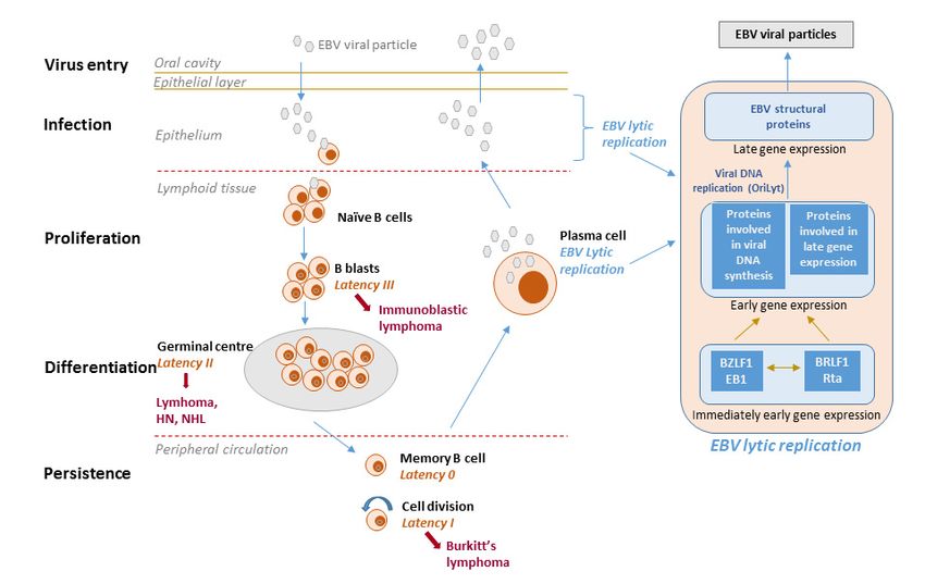
Molecules 2019, 24, 997 2 of 20
(Table 1). It is also found in 100% of non-keratinizing nasopharyngeal carcinomas and has been linked
sporadically to cancers of the gastrointestinal tract [4,5].
Table 1. Epstein–Barr (EBV)-associated lymphomas in immunocompetent and immunocompromised hosts.
Immunocompetent Host Immunocompromised Hosts
Lymphoma EBV Association Latency Program Lymphoma EBV Association Latency Program
BL (endemic) 100% I or Wp- restricted PTLD, B-cell >90% III
BL (sporadic) 15–85% I BL (HIV) 25–35% I
Classical HL 40% II HL (HIV) >80% II
DLBCL associated with chronic inflammation ~70% II PEL (primary effusion lymphoma) >80% I
EBV-positive DLBCL of the elderly 100% II Plasmablastic lymphoma ~70% I or II
Lymphomatoid granulomatosis 100% II Plasmablastic lymphoma, oral type (HIV) 100% I
Angioimmunoblastic T-cell lymphoma * >90% II Primary CNS lymphoma (HIV) 100% III
Extranodal NK/T-cell lymphoma, nasal type * 100% II NHLs with primary immune disorders >90% III
Iatrogenic immunodeficiency lymphoma 40–50% III
Aggressive NK-cell leukemia * >90% II
PTLD, NK/T-cell * >70% III
Adapted from [2–5]. * EBV-associated T- and NK-cell lymphomas. DLBCL: diffuse large B-cell lymphoma.
For sporadic Burkitt’s lymphoma (BL), the strength of the EBV association varies with geographical location,
hence the wide percentage range reported.
The EBV life cycle displays two distinct phases, i.e., lytic and latent (Figure 1). EBV preferentially
infects B-cells via the CD21 receptor but also infects epithelial cells as well as T- or NK-lineage cells
at a lower frequency. The virus undergoes lytic replication in epithelial cells and establishes lifelong
latency in circulating memory B-lymphocytes, reactivating periodically from latency [4,6]. During lytic
infection, the full repertoire of viral gene expression takes place and progeny virus is produced.
The two key EBV immediate-early lytic genes (i.e., BZLF1 and BRLF1) encode transactivators that
activate viral and certain cellular promoters, leading to an ordered cascade of viral gene expression:
early gene expression and genome replication followed by late gene expression. Virions produced
during lytic replication in permissive epithelial cells allow the dissemination of viral particles within
and among hosts.
In the course of latency, the virus expresses only a limited number of genes required for the
maintenance of the viral genome (as an episome in the nucleus) and evasion of the host immune system.
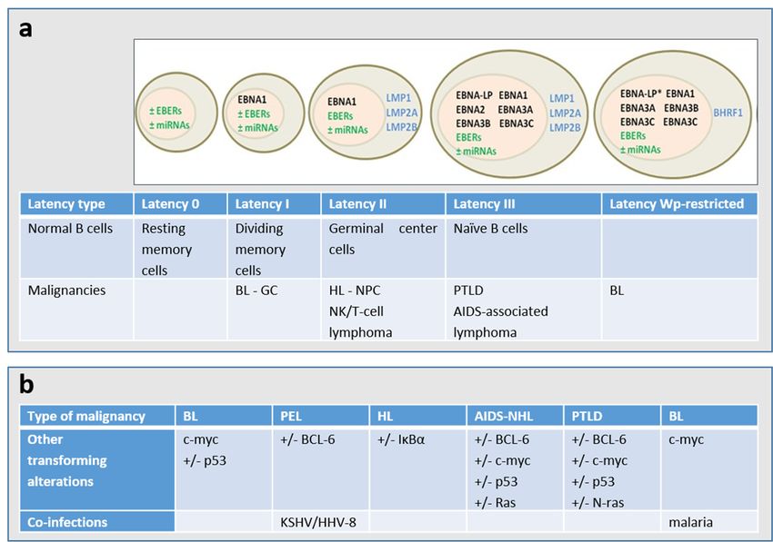
Based on the expression of latent genes, EBV latency is classified in different types (Figure 2A) [7].
It is worth noting that the Latency 0, I, II, III nomenclature represents a snapshot of gene expression.
EBV latency ranges from latency 0 (without EBV antigen expression, as observed in circulating memory
B-cells in healthy persons) to latency III (in which all nuclear proteins (EBNAs -1,-2, -3A, -3B, -3C,
and -LP) and 2 membrane proteins (LMP1 and LMP2) are expressed together with 2 small RNAs
(EBERs), as found in PTLD. Some tumors may not fall into one of these patterns of latency and
furthermore, immunohistochemistry may indicate heterogeneity of expression within a single biopsy.
In addition, cellular genetic alterations and/or co-infections occur in EBV-associated malignancies
(Figure 2B). Although latency programs predominate in EBV-driven tumors, lytic viral replication is
also of pathogenic importance [8,9].Molecules 2019, 24, 997 3 of 20
Molecules 2019, 24, x FOR PEER REVIEW 3 of 20
Figure1.1. EBV
Figure EBVlife
lifecycle,
cycle,latency
latencystages
stagesand andderived
derivedlymphomas.
lymphomas.The The viral
viral lifelife cycle
cycle includes
includes at least
at least five
different stages (virus entry, infection, proliferation, differentiation and persistence), and fourfour
five different stages (virus entry, infection, proliferation, differentiation and persistence), and of
of them
them
are are associated
associated with EBVwithdiseases.
EBV diseases.
The virusThe is
virus is transmitted
transmitted through through
the salivathe saliva and infects
and infects naïve naïve
B-cells
B-cells
in in the oropharyngeal
the oropharyngeal mucosa.mucosa. During primary
During primary infection, infection,
EBV-infectedEBV-infected
naïve B-cells naïveexpress
B-cells the
express
entire
the entire
latency latency
gene gene(10
complex complex (10 proteins:
proteins: EBV nuclear EBVantigens
nuclear antigens
(EBNAs), (EBNAs), latent membrane
latent membrane proteinprotein
(LMPs))
(LMPs))
as well asasEBV-encoded
well as EBV-encoded
small RNAs small (EBERs)
RNAs (EBERs) and microRNAs.
and microRNAs. This This
is calledis called
typetype III latency
III latency and
and this form of latency activates the resting B-cells and drives them
this form of latency activates the resting B-cells and drives them to proliferation and transformation. to proliferation and
transformation.
However, these However, these cells
cells are highly are highly and
immunogenic immunogenic
are rapidly and are rapidly
eliminated by eliminated
EBV-specific by TEBV-cells.
specific
The virusTiscells. The
able to virus in
survive is B-cells
able tobecause
surviveitindownregulates
B-cells because itsitimmunogenic
downregulates its immunogenic
proteins. EBV mimics
proteins.
antigen EBV mimics
driven antigen driven
B-cell responses B-celltoresponses
and similar and similar
antigen-stimulated to antigen-stimulated
blasts, the EBV-infected B-cells blasts, enter
the
EBV-infected B-cells enter the follicles, expand, and form germinal centers
the follicles, expand, and form germinal centers where they express only three viral proteins (type II where they express only
three viral
latency). proteins
Finally, they(type II latency).
exit the lymph node Finally, they exit
expressing thealymph
only single node expressing
viral protein only awhich
(EBNA1, singleensures
viral
protein (EBNA1, which ensures that the viral genome divides with the
that the viral genome divides with the cellular genome) (type I latency). The entry of EBV-infected cells cellular genome) (type I
latency).
into The entry blood
the peripheral of EBV-infected
results in the cells into the peripheral
shutdown of all viralblood
genesresults
encoding in theforshutdown
proteins; this of all
is viral
called
latency 0 or latency program where no viral proteins are expressed. Resting memory cells, in whichare
genes encoding for proteins; this is called latency 0 or latency program where no viral proteins the
expressed.
virus Resting are
is quiescent, memory cells, in which
not attacked by the the
host virus
immuneis quiescent,
systemare andnotare attacked
likely the by the
siteshost immune
of long-term
system and Memory
persistence. are likelyB thecellssites of long-term
occasionally divide persistence.
to maintain Memory B cells occasionally
stable number of cells and when dividea cell
to
maintain stable number of cells and when a cell that is carrying the virus
that is carrying the virus divides, the viral EBNA1 protein is expressed to allow the viral genome to divides, the viral EBNA1
protein isalong
replicate expressed
with theto allow the viral B-cells
cell. Memory genomemay to replicate alongterminal
also undergo with the differentiation
cell. Memory B-cells may
into plasma
also undergo terminal differentiation into plasma cells and secrete antibodies.
cells and secrete antibodies. If such a cell contains the virus, the EBV lytic program is activated and the If such a cell contains
the virus, virus
infectious the EBV lytic program
released from the isplasma
activated
cellsand
canthe infectious
infect epithelialvirus released
cells, wherefrom the plasma
the virus cells
can replicate
can infect epithelial cells, where the virus can replicate and be shed at
and be shed at high amounts and then be transmitted to other hosts. With the exception of latency type high amounts and then be
transmitted to other hosts. With the exception of latency type
0, each latency state is found in specific types of EBV-associated malignancies. 0, each latency state is found in specific
types of EBV-associated malignancies.Molecules 2019, 24, 997 4 of 20
Molecules 2019, 24, x FOR PEER REVIEW 4 of 20
Figure 2.
Figure 2. (a)
(a) Patterns
Patterns of
of gene
geneexpression
expressionduring
duringEBVEBVlatency.
latency. The
Themajority
majorityof ofthe
theendemic
endemicBL BLpresents
presents
a latency I type and carry a wild-type transformation-competent EBV genome and expressonly
a latency I type and carry a wild-type transformation-competent EBV genome and express onlythethe
Epstein–Barr nuclear
Epstein–Barr nuclear antigen
antigen 11 (EBNA1)
(EBNA1) fromfrom the
the EBNA1-specific
EBNA1-specific latent
latent promoter
promoterQp, Qp,non-coding
non-coding
EBERs (Epstein–Barr
EBERs (Epstein–Barrvirus-encoded
virus-encodedsmall smallRNAs)
RNAs)and and several microRNAs
several microRNAs (miRNAs).
(miRNAs). Around
Around 15%15%of
BL BL
of endemic
endemic tumors,
tumors,the the
so called Wp-restricted
so called BLs, BLs,
Wp-restricted carrycarry
an EBNA2 gene-deleted
an EBNA2 genome
gene-deleted and
genome
express
and EBNA1,
express -3A, -3B,
EBNA1, -3A,and-3C and theand
-3B, and-3C viralthe
Bcl2 homologue
viral BHRF1 from
Bcl2 homologue BHRF1the Wp
from latent promoter
the Wp latent
[2,6]. * The EBNA-LP gene is partially deleted in the Wp-restricted latency. A major
promoter [2,6]. * The EBNA-LP gene is partially deleted in the Wp-restricted latency. A major type type of latency in
EBV-associated
of malignanciesmalignancies
latency in EBV-associated is latency II, iniswhich
latencytheII,latent membrane
in which proteins
the latent LMP1,
membrane LMP2A,LMP1,
proteins and
LMP2B are
LMP2A, and expressed
LMP2B arein addition
expressed to the Latency Itogenes.
in addition The entire
the Latency EBV latency
I genes. geneEBV
The entire complex,
latencywhich
gene
consists ofwhich
complex, several EBNAof
consists proteins,
several LMP1,
EBNA LMP2A,
proteins,LMP2B, EBERs, and
LMP1, LMP2A, miRNAs
LMP2B, are expressed
EBERs, and miRNAs in the
are
type III latency. (b) The cellular genetic alterations and/or co-infections are known
expressed in the type III latency. (b) The cellular genetic alterations and/or co-infections are known toto occur in the
different
occur types
in the of EBV-associated
different malignancies.
types of EBV-associated PEL: primary
malignancies. PEL:effusion
primarylymphoma; HL: Hodgkin
effusion lymphoma; HL:
lymphoma;
Hodgkin BL: Burkitt
lymphoma; lymphoma;
BL: Burkitt lymphoma;NHL:NHL: non-Hodgkin
non-Hodgkin lymphoma;
lymphoma; PTLD:
PTLD:post-transplant
post-transplant
lymphoproliferative disorder; NPC: nasopharyngeal carcinoma; carcinoma; GC:
GC: gastric
gastric carcinoma.
carcinoma.
2. Why Is
2. Why Is There
There No
No Antiviral
Antiviral Drug
Drug Approved
Approved for
for the
the Treatment
Treatment of
of EBV
EBV Infections?
Infections?
Nucleoside (i.e., acyclovir
Nucleoside (i.e., acyclovir (ACV),
(ACV), penciclovir
penciclovir(PCV),(PCV),ganciclovir
ganciclovir(GCV),
(GCV),andanditsitsoral
oralprodrugs;
prodrugs;
valacyclovir (VACV), famciclovir valganciclovir (VGCV),
famciclovir (FAM), and valganciclovir (VGCV),respectively),
respectively),nucleotide
nucleotide(i.e.,
(i.e.,
cidofovir (CDV)), and pyrophosphate (i.e., (i.e., foscavir
foscavir(foscarnet
(foscarnetsodium,
sodium,PFA))
PFA))analogues
analoguesare areapproved
approved
for the
thetreatment
treatmentofofherpes
herpes simplex
simplexvirus 1 (HSV-1)
virus and and
1 (HSV-1) 2 (HSV-2), varicella-zoster
2 (HSV-2), virus (VZV),
varicella-zoster and/or
virus (VZV),
human cytomegalovirus
and/or human (HCMV)(HCMV)
cytomegalovirus [10,11]. In some In
[10,11]. European countries,countries,
some European brivudin brivudin
(BVDU) is(BVDU)
approvedis
for the therapy of HSV-1 and VZV associated diseases. Although some of these
approved for the therapy of HSV-1 and VZV associated diseases. Although some of these antiviral antiviral agents proved
to be effective
agents proved toinhibitors
be effectiveof EBV replication
inhibitors of EBVinreplication
vitro and were used
in vitro andexperimentally [11–13], none
were used experimentally [11–of
them received
13], none approval
of them received byapproval
the FDAby (Food
the FDA and Drug
(FoodAdministration) or EMA (European
and Drug Administration) Medicines
or EMA (European
Agency)
Medicines forAgency)
treatment forof EBV infections.
treatment of EBV infections.
In 2005, Gershburg and and Pagano
Pagano proposed
proposed threethree main
main explanations
explanationsforforthe
thelack
lackofofan
ananti-EBV
anti-EBV
drug [14].
[14]. First,
First,
thethe difficulty
difficulty in diagnosing
in diagnosing infectious
infectious mononucleosis
mononucleosis may may
be, atbe, at in
least least in
part,
part, responsible
responsible for theforlackthe
of lack of success
success in the development
in the development of a drug of a drug
to treat to treat EBV-associated
EBV-associated infections.
While EBV infects most persons at the age of 30, only a few of them suffer from infectiousMolecules 2019, 24, 997 5 of 20
infections. While EBV infects most persons at the age of 30, only a few of them suffer from
infectious mononucleosis (usually those who acquired the infection in the twenties). The infectious
mononucleosis symptoms are subtle in onset and the disease has a long incubation time (4–6 weeks),
resulting in a late diagnosis, in contrast to infections caused by the α-herpesviruses HSV (i.e., herpes
labialis) or VZV (i.e., chickenpox).
Second, antivirals should be achieving high concentrations in the oropharynx where EBV is
released at high titers. Although acyclovir was shown to significantly reduced EBV shedding in
the oropharynx when administered intravenously and orally, virus release resumed at the initial
level within 3 weeks of cessation of the treatment [15,16]. Maybe the most important reason for
the failure of antivirals for infectious mononucleosis therapy can be ascribed to the fact that the
symptoms and signs of the disease are not the consequences of viral replication but the immunological
response to EBV-infected B-cells that circulate in the blood and infiltrate the tissues of different organs.
Infectious mononucleosis is characterized by atypical lymphocytosis due to the massive cell-mediated
immune response against viral-infected B-lymphocytes. Thus, antivirals in combination with
immunomodulatory drugs (such as corticosteroids, used empirically by physicians to treat infectious
mononucleosis) might be effective. However in a multicenter, double-blind, placebo controlled
study, prednisolone administered with acyclovir for treatment of infectious mononucleosis inhibited
oropharyngeal EBV replication without affecting duration of clinical symptoms or development of
EBV-specific cellular immunity [16].
3. Medical Need for Anti-EBV Therapeutics Targeting Lytic Replication
Primary EBV infection is usually asymptomatic but some patients develop infectious
mononucleosis, which can have mild symptoms (i.e., fever, sore throat and lymphoadenopathy) or be
fatal in the immunocompromised hosts. Furthermore, primary EBV infection with or without infectious
mononucleosis may lead to complications (such as autoimmune hemolysis, airway obstruction
from enlarged tonsils, splenic rupture, encephalitis, severe hepatitis and myocarditis), which are
primarily a consequence of the immunopathological responses to the virus. Other rare but serious
complications (such as agranulocytis and aplastic anemia) may occasionally arise in healthy patients.
The mainstay for treatment of complications related to infectious mononucleosis are corticosteroids.
Although the role of antivirals in the management of severe infectious mononucleosis complications
is debatable based on case series, physicians may consider the use of antiviral agents in severe
manifestations of EBV infections. Even though the pathogenesis of infectious mononucleosis is
primarily immune mediated, the severity of EBV-associated hepatitis was shown to be related to a
high viral burden [17,18]. Therefore, the use of specific antivirals is expected to alleviate the symptoms
of EBV-related complications found in infectious mononucleosis.
EBV lytic replication is not only directly associated with infectious mononucleosis but also
with chronic active EBV infection (CAEBV) and oral hairy leukoplakia. CAEBV is rare in USA and
Europe but occurs more frequently in Asia and South America. CAEBV is a lymphoproliferative
disorder with markedly elevated levels of EBV-specific antibodies and high viral loads. It is often
a fatal disorder characterized by chronic or recurrent infectious mononucleosis-like symptoms
persisting for a long time and by an unusual pattern of anti-EBV antibodies. Life-threatening
CAEBV complications include virus-associated hemophagocytic syndrome, leukemia and lymphoma
of T/NK-cell lineages [19]. Intragenic EBV deletions that reactivate the lytic cycle by upregulating
the expression of immediately early genes were linked to avert viral production and promotion
of lymphomagenesis [20]. Immunomodulatory agents (such as interferon-α and interleukin-2),
antivirals (including acyclovir, ganciclovir and vidarabine), chemotherapeutic agents, cell therapy
using EBV-specific cytotoxic T lymphocytes, and hematopoietic stem cell transplantation have been
used for treatment of CAEBV but with limited success [21].
Post-transplant lymphoproliferative disorder (PTLD) is a serious and often fatal complication
following solid organ transplantation, being EBV a major risk factor for PTLD in this group of patientsMolecules 2019, 24, 997 6 of 20
as 30% to 50% of EBV-naïve patients that seroconvert are diagnosed with PTLD [22,23]. PTLD is also a
life threatening disorder following allogeneic hematopoietic stem cell transplant (HSCT), caused by
drug-induced reduced immune surveillance leading to an uncontrolled proliferation of lymphocytes.
PTLD incidence has increased significantly during the last two decades due to several reasons,
including a growing number of transplant activities, increasing age of donors and recipients, use of new
and potent immunosuppressive agents, introduction of haplo-identical HSCT, increased awareness of
the disorder and improved diagnostic tools [24]. A sustained persistence of high viral load and an
elevated viral load have been associated with increased risk of PTLD [25,26]. Antiviral therapy has not
been regarded as useful in PTLD because the virus is latent. However, prophylactic administration
of antiviral drugs resulted in reduce incidence of PTLD [27–29]. Antiviral agents may have a role in
preventing PTLD as high level of EBV DNA in the blood of solid organ transplant recipients has been
shown to predict PTLD. Most cases of early onset PTLD (occurring during the first year following
transplantation) are associated with recent EBV infection. Late-onset lymphomas occurring after the
first year of transplantation are less likely to be associated with EBV.
Treatment strategies aimed at suppressing the expression of lytic proteins should be helpful
for controlling early stages of EBV-associated malignancies as EBV lytic infection was shown to
contribute to lymphoproliferative disease [8,30]. The EBV IE proteins BZLF1 and BRLF1 contribute
to IL-6 secretion in lytically infected cells promoting early lymphoproliferative disease [31]. IL-6 is
a cytokine known to play a crucial role in the maintenance of immune functions, stimulation of
differentiation of hematopoietic cells and perpetuation of inflammation but it is an important factor
in a variety of hematological and epithelial malignancies. IL-6 acts through paracrine and autocrine
mechanisms to promote cell survival and induces the signal transducer and activator of transcription 3
(STAT3). Therefore, it is not surprising that viruses (such as EBV) capable of infecting both epithelial
and lymphoid cells, have mechanisms to induce IL-6 expression. Also, lytically infected cells induce
the expression of cellular and viral IL-10 [32], allowing B-cells to grow more efficiently, and of VEGF
contributing to angiogenesis in both B-cell and epithelial malignancies [33].
4. Antivirals Against EBV Evaluated in The Clinic
4.1. Nucleoside Analogues (Acyclovir, Valacyclovir, Ganciclovir, and Valganciclovir)
As acyclovir and ganciclovir inhibit EBV in vitro [11,34,35], these drugs and their oral prodrugs
were evaluated for suppression of EBV reactivation during immunosuppression. PTLD incidence
in lung and heart-lung transplant EBV-seronegative recipients was reduced by antiviral prophylaxis
with acyclovir, valacyclovir or ganciclovir [28]. The incidence of this lymphoproliferative disorder
was analyzed before 1996 (historic group) and between 1996 and 2000 (group receiving antiviral
prophylaxis) to compare the impact of long-term antiviral prophylaxis on PTLD development in
EBV-seronegative recipients. None of the EBV-seronegative recipients receiving continuous antiviral
prophylaxis developed PTLD while in the historic group, PTLD developed in 4.2% of the patients.
The effects of ganciclovir and valganciclovir prophylaxis on EBV viral load were evaluated
in a group of EBV-naïve pediatric renal transplant recipients (R-) who had received a graft from an
EBV-positive donor (D+) and therefore at risk to develop EBV primary infection [27]. Over the first year
post-transplantation, antiviral prophylaxis with ganciclovir or valganciclovir resulted in a significant
decreased incidence of EBV primary infection: 9/20 (45%) in the prophylaxis group had a primary
EBV infection versus 8/8 (100%) in the non-prophylaxis group. Antiviral prophylaxis afforded a
significantly lower EBV viral load while the type or intensity of immunosuppressive therapy did not
affect the incidence of EBV primary infection or the level/persistence of viral load.
The impact of antiviral drugs used to prevent HCMV disease was investigated in a monocentric
retrospective cohort of 73 adult kidney or kidney-pancreas EBV-seronegative recipients, transplanted
between January 2000 and January 2016 [36]. Thirty-seven (50.7%, prophylaxis group) received
valacyclovir or valganciclovir for 3–6 months and 36 (49.3%, no-prophylaxis group) received noMolecules 2019, 24, 997 7 of 20
antivirals with mean follow-up times of 69 months (prophylaxis group) and 91 months (no-prophylaxis
group). Prophylaxis delayed primary infection at 100 days (43% versus 84%) as determined by
monitoring EBV viral load. Early PTLD incidence did not differ between groups but EBV-related
neoplasia incidence was significantly lower in treated patients (no cases observed) than in the
no-prophylaxis group (six neoplasia cases reported). Despite a weak level of evidence, antiviral
prophylaxis could prevent late onset PTLD.
In a cohort of pediatric liver transplant recipients, treatment with intravenous ganciclovir did not
change the proportion of patients with reduction in EBV load at 8 weeks and 1 year after detection
of EBV viremia [37]. This retrospective study performed in Norway from 2002 until 2015 included
38 patients with EBV viremia and 32 of them were treated with intravenous ganciclovir for a median
of 22 (21–38) days. Short time from transplantation to viremia, younger age at transplantation and lack
of EBV seroconversion prior to transplantation were significant predictors of chronic EBV viremia.
Valganciclovir suppressed EBV reactivation in a group of 29 patients under immunosuppression
with alemtuzumab (a humanized monoclonal antibody against CD52 expressed on all B- and
T-lymphocytes), which predisposes to HCMV and EBV reactivation [38]. Plasma EBV DNA load
was quantified in 29 patients (258 samples with a median of seven specimens per patient). In 24 of the
patients, no quantifiable EBV DNA was detected while five patients (17%) had EBV reactivation that
dropped spontaneously in four cases. One patient, who had also received previously another potent
T-cell suppressing drug (fludarabine), developed EBV-positive Hodgkin lymphoma.
The efficacy and safety of valganciclovir without immunosuppression decrease was assessed
in a group of children undergoing liver transplantation who showed sustained EBV DNA in their
blood [29]. Undetectable viral load was observed in 20/42 (47.6%) of patients under prolonged antiviral
therapy (median 8 months), 60% of whom maintained response to therapy. However, the results of
this study should be interpreted with cautious because of the lack of a control group.
The effects of valganciclovir on oral EBV shedding were evaluated in a randomized, double blind,
placebo-controlled study [39]. Twenty-six men were included and all participants self-identified as
men who have sex with men, and 16 participants (62%) were HIV-1 infected. They received oral
valganciclovir or daily placebo for 8 weeks, followed by a 2-week “washout period” and then 8 weeks
of the alternative treatment. Valganciclovir significantly reduced the proportion of days with EBV
detected from 61.3% to 17.8% and the quantity of virus detected by 0.77 logs, which warrants further
investigations into the impact of valganciclovir on EBV-associated diseases.
4.2. Nucleotide Analogues
The nucleotide analogue cidofovir (CDV) is a broad spectrum anti-DNA virus agent.
The compound is approved for the treatment of retinitis in AIDS patients but is used off-labeled
to treat several infections caused by DNA viruses [40]. Besides its recognized antiviral properties,
the drug is also known for its antiproliferative effects [41].
Successful treatment of locally recurrent EBV-associated nasopharyngeal carcinoma using the
antiviral agent cidofovir was reported in two patients [42]. Further, injection of cidofovir into the
tumor tissue of EBV-positive nasopharyngeal carcinoma xenografts in nude mice suppressed tumor
growth [43]. In combination with the ribonucleotide reductase inhibitors hydroxyurea and didox
(3,4-dihydroxybenzohydroxamic acid), cidofovir-induced apoptosis in EBV-transformed epithelial cells
and in EBV-positive nasopharyngeal carcinoma xenografts was augmented [44]. Cidofovir decreased
EBV oncoproteins and enhanced the radiosensitivity in EBV-related malignancies (Burkitt’s lymphoma
and nasopharyngeal carcinoma) [45].
4.3. Pyrophosphate Analogues
Foscarnet, an inorganic pyrophosphate analogue, is a direct inhibitor of herpesvirus DNA
polymerases. It blocks the pyrophosphate-binding site and prevents cleavage of pyrophosphate from
deoxynucleoside triphosphates. Foscarnet, a non-competitive inhibitor of viral DNA polymerases,Molecules 2019, 24, 997 8 of 20
does not incorporate into the growing viral DNA and is ~100-fold more active against viral than
cellular enzymes. Although the drug has activity against all human herpesviruses, including EBV,
foscarnet is approved for treatment of HCMV retinitis in AIDS patients and for the therapy of
acyclovir-resistant HSV infections in immunocompromised patients. It has also been used for therapy
of ganciclovir-resistant HCMV infections due to mutations in the UL97 protein kinase (PK). Its safety
and efficacy for the treatment of other herpesvirus infections has not yet been established [10].
The successful use of foscarnet to manage a persistent EBV infection was occasionally reported.
Foscarnet in combination with immunoglobulins was successful to control the persistent EBV infection
in a lung transplant patient that showed clinical improvement of PTLD following reduction in
immunosuppression intensity [46]. This patient required treatment with an antiviral drug and
immunoglobulins since restoration of the cellular immunity improved PTLD but was ineffective
against controlling the EBV infection. Regression of EBV-associated lymphoproliferative disorders in
two AIDS patients during therapy with foscarnet has also been described [47].
5. Anti-EBV Compounds Under Investigation
5.1. Inhibitors of EBV Protein Kinase BGLF4
Maribavir (MBV) is an investigational oral benzimidazole L-riboside with significant activity
against both HCMV and EBV but no other human herpesviruses [48,49]. Maribavir has fewer adverse
side effects and is more specific compared to anti-HCMV drugs that target the viral DNA polymerase.
Unlike nucleoside and nucleotide analogues, maribavir’s inhibitory effects are mainly produced
through inhibition of the HCMV and EBV PKs [50]. This compound selectively inhibits the HCMV
UL97 PK (as determined by direct inhibition of kinase activity) and single point mutations in the
UL97 gene confer maribavir resistance in HCMV [51]. UL97 is a serine/threonine-specific PK playing
an important role in HCMV egress. UL97 is necessary for the phosphorylation of several viral
and cellular proteins in HCMV infected cells [50,52]. In vitro and in vivo UL97 mutations selected
under ganciclovir and maribavir were found to be distinct and to confer no cross-resistance [53,54],
while partial cross-resistance between ganciclovir and cyclopropavir, a methylenecyclopropane
nucleoside analog active against HCMV was observed [51,55]. However, Chou and colleagues [56]
reported UL97 kinase mutations either found in ganciclovir-treated subjects or after propagation
under cyclopropavir in vitro with moderate- to high-level resistance to all three drugs. Low levels of
resistance (two- to three-fold) to maribavir can also arise due to mutations in the HCMV UL27 gene.
Although the function of UL27 is unknown, it does not appear to be a direct target for maribavir.
The efficacy of maribavir prophylaxis for prevention of HCMV disease in recipients of allogeneic
stem-cell transplants was evaluated in phase three: double blind, placebo-controlled, randomized
trials [57,58]. The clinical development of maribavir for the management of HCMV infections is currently
on hold because the drug failed to meet the primary endpoint (prevention of HCMV disease) in recipients
of allogeneic stem-cell transplants although several critics on the study design were raised [59].
Maribavir exhibits also marked activity against EBV, having a unique dual effect against
EBV: inhibition of viral DNA replication and of virus transcription [14,60]. In contrast to HCMV,
the activity of maribavir against EBV could not be ascribed to direct inhibition of the EBV PK
BGLF4. In fact, maribavir treatment was shown to inhibit the phosphorylation of the EBV DNA
polymerase processivity factor BMRF1 [49]. Unlike acyclovir that has little effect on EBV RNAs,
maribavir inhibits the expression of multiple RNAs. Furthermore, the inhibitory profile of maribavir
transcripts appeared to be similar to that produced by an EBV mutant in which PK expression and
activity were knocked out [61], suggesting that maribavir largely affects EBV transcript levels through
inhibition of BGLF4 although the drug does not directly affects the EBV PK [62]. Considering that EBV
BGLF4 has at least 20 viral targets, maribavir may also affect downstream targets indirectly.Molecules 2019, 24, 997 9 of 20
5.2. Inhibitors of EBV DNA Polymerase
The novel l-dioxolane thymidine analog, 1-[(2S,4S-2-(hydroxymethyl)-1,3-dioxolan-4-yl]5-
vinylpyrimidine-2,4(1H,3H)-dione, or HDVD, proved active against HSV-1, KSHV, EBV, as well
as murine herpesvirus 68 (MHV-68) [35]. The compound weakly inhibited replication of HSV-2,
VZV and herpesvirus saimiri (HVS), and no antiviral activity was found against HCMV and rhesus
rhadinovirus (RRV). Thus, its antiviral activity spectrum differed from that of the related compound
brivudin (which is known for its potent activity against HSV-1 and VZV). However, similar to brivudin,
characterization of HDVD-resistant viruses indicated that the viral thymidine kinases (TKs) of HSV-1,
MHV-68, and HVS were required for activation of the compound. Oral treatment with HDVD and
brivudin was assessed in an intranasal model of MHV-68 infection in BALB/c mice. HDVD treatment,
in contrast to brivudin treatment, resulted in a reduction in viral DNA load and diminished viral gene
expression during acute viral replication in the lungs compared to untreated controls. The valyl ester
prodrug of HDVD (USS-02-71-44) was more effective in preventing the latent infection in the spleen
than HDVD [35]. Studies on mechanisms of resistance of various nucleoside derivatives indicated that
pyrimidine nucleoside derivatives are phosphorylated by the γ-herpesvirus TK and purine nucleosides
are preferentially activated by the γ-herpesvirus PK [13], consistent with previous findings showing
that the EBV-encoded PK, but not the TK, is required for ganciclovir and acyclovir inhibition of lytic
viral production [63].
Two thionucleoside derivatives, KAY-2-41 and KAH-39-149, displayed effective in vivo
(MHV-68 mouse model) antiviral efficacy and potent and selective in vitro anti-EBV activity [34].
The compounds also proved active against HSV and VZV. KAY-2-41- and KAH-39-149-resistant HSV
and MHV-68 harbored mutations in the viral TK though these mutations conferred only low levels of
resistance to KAY-2-41 and KAH-39-149 compared to other TK-dependent drugs. Antiviral assays in
HeLa TK-deficient cells showed a lack of KAY-2-41 and KAH-39-149 activities against HSV TK-deficient
mutants. Furthermore, enzymatic assays showed the ability of HSV-1 and VZV TK, and cellular
TK1 and TK2 to recognize and phosphorylate KAY-2-41 and KAH-39-149, demonstrating that the
compounds depend on both viral and host TKs to exert antiviral activity. KAH-39-149 proved superior
to KAY-2-41 in a mouse model of γ-herpesvirus infection, highlighting their potential as antiviral
therapeutics against EBV.
Various methylenecyclopropane nucleoside (MCPN) analogues proved active against several
herpesviruses in cell culture and animal models [64–68]. The first series of MCPN analogues
had a single hydroxymethyl group on the cyclopropane ring mimicking the 50 hydroxyl moiety
in deoxyribonulceosides. Compounds from this series, similar to acyclovir, block further strand
elongation once incorporated into DNA because of the lack of a counterpart to the 30 hydroxy group.
The second generation of MCPN analogues are dihydroxymethyl derivatives, and similar to ganciclovir,
have a second hydroxyl group that can be recognized by the DNA polymerase allowing further strand
elongation. Cyclopropavir, the most active compound of this class, displays good antiviral activity
against HCMV, murine CMV, EBV, HHV-6 and HHV-8 and its prodrug 6-deoxycyclopropavir has
shown good activity when administered orally [69]. The mechanism of action of cyclopropavir against
CMV is complex involving both the inhibition of DNA synthesis and the UL97 PK [70]. Analogues of
the first series with a single hydroxymethyl group, such as (S)-synguanol exhibit a broader spectrum
of antiviral activity encompassing hepatitis B and HIV. More recently, monohydroxymethyl and
dihydroxymethyl analogues with 6-ether and –thioether moieties have also been reported to be
active against several herpesviruses, including EBV. Some of these analogues exhibit a broader
spectrum of activity than cyclopropavir inhibiting also HSV-1, HSV-2 and VZV [66]. The activity
of these compounds was dependent on the HCMV UL97 PK but was relatively independent from
HSV TK activity. These data point to a different mechanism of action of these analogues from that of
cyclopropavir and suggest that they can eventually be used as broad-spectrum anti-herpesvirus agents.
N-methanocarbathymidine, a conformational locked nucleoside analogue, proved active against
α-herpesviruses, γ-herpesviruses and orthopoxviruses [71]. The antiviral activity of this compound isMolecules 2019, 24, 997 10 of 20
dependent on the activation by viral TKs. The drug inhibits viral DNA synthesis once it is activated
by the viral TKs. The compound proved also effective in animal models of orthopoxvirus and HSV
infection [72,73] and there is an ongoing Phase I trial in healthy volunteers [10] to evaluate its safety.
Several acyclic nucleoside phosphonates (ANPs), including cidofovir derivatives, inhibited with
high potency and selectivity the replication of EBV and other γ-herpesviruses [12] Notable, cyclic
prodrugs of ANPs exhibited reduced activities against the EBV strain P3HR-1, but not against the
EBV strain Akata. Metabolism studies with cidofovir and its cyclic form (cyclic-cidofovir) revealed
that these differences were attributable to an altered drug metabolism in P3HR-1 cells after EBV
reactivation, i.e., to a reduced hydrolysis of cyclic-cidofovir by cyclic CMP phosphodiesterase [12].
Altered cyclic AMP levels in P3HR-1 cells implied a competitive inhibition of the phosphodiesterase
by this cyclic nucleotide. Cidofovir and its 5-aza derivative (HPMP-5azaC) emerged as highly effective
inhibitors of murine γ-herpesvirus replication and dissemination in a mouse model [12].
Brincidofovir (CMX-001) is the orally bioavailable form of cidofovir. This alkoxyalkyl ester
prodrug of cidofovir has the same in vitro broad-spectrum antiviral activity as cidofovir but with
an activity up to 1000-fold higher compared with cidofovir because of higher intracellular levels of
cidofovir-diphosphate [74]. Besides its enhanced antiviral activity, brincidofovir is not nephrotoxic,
because, in contrast to cidofovir, brincidofovir is not a substrate of the human organic anion transporter
1 enzyme located in the proximal renal tubule. Despite the promising preclinical data reported for
brincidofovir, a Phase III trial evaluating its use for the prevention of HCMV disease in seropositive
allogeneic hematopoietic stem cell transplant patients delivered disappointing results slowing down
its progress to the market. In this trial, named SUPPRESS, increased HCMV disease was reported in the
brincidofovir group compared to the placebo arm possibly related to the misdiagnosis of brincidofovir
gastrointestinal disease as graft-versus host disease, which was treated with corticosteroids [75].
Following these findings, HCMV trials were suspended but adenovirus trials are ongoing and an
IV formulation is in development. No trials are foreseen for evaluating this drug for EBV-associated
diseases [10].
5.3. Inhibitors of EBV Nuclear Antigen 1 (EBNA1)
The EBV-encoded nuclear antigen 1 (EBNA1) is a versatile protein with functions in the maintenance,
replication, and segregation of the EBV genome and represents an attractive therapeutic target to treat
EBV-associated malignancies. This protein is express in all EBV latency types except for latency 0.
The replication and persistence of the EBV episomal genome in latently infected cells primarily depend on
the binding of EBV-encoded nuclear antigen 1 (EBNA1) to the cognate EBV oriP element.
Considerable efforts have been done the last years in the design or identification of inhibitors
of EBNA-1 to decrease its expression or interfere with its functions. The salient features of EBNA-1,
its functional domains and advances in the development of EBNA-1 inhibitors have been recently
reviewed in detail [76]. For example, Lee and colleagues [77] characterized EBNA1 small molecule
inhibitors (H20, H31) and their underlying inhibitory mechanisms. H20 fits into a pocket in
the EBNA1 DNA binding domain (DBD) as predicted by in silico docking analyses but H20 did
not significantly affect EBNA1 binding to its cognate sequence. A limited structure-relationship
study of H20 allowed the identification of H31, a hydrophobic compound, as an EBNA1 inhibitor.
H31 inhibited EBNA1-dependent oriP sequence-specific DNA binding activity, but did not affect
sequence-nonspecific chromosomal association. H31 repressed the EBNA1-dependent transcription,
replication, and persistence of an EBV oriP plasmid, consistent with the inhibition of EBNA1 binding
activities. Importantly, H31 produced gradual loss of EBV episome and selectively delayed the
growth of EBV-infected lymphoblastoid cell lines or Burkitt’s lymphoma cells. Thus, inhibition of
EBNA1-dependent DNA binding by H31 decreased EBNA1-dependent transcription and persistence
of EBV episome in EBV-infected cells. Screening approaches also identified molecules that could block
EBNA1-DNA binding, EBNA1-oriP transactivation, EBNA1 linking regions. Also, inhibitors based onMolecules 2019, 24, 997 11 of 20
truncated peptides from EBNA1 dimeric interface were described confirming the “druggability” of
EBNA1 for the treatment of EBV-related cancers [78].
Computational identification and structural characterization of EBNA1 binding pockets, likely to
accommodate ligand molecules (i.e., “druggable” binding sites) were validated by docking against
a set of compounds previously tested in vitro for EBNA1 inhibition (PubChem AID-2381) [79].
Assessments of pocket druggability were performed by induced fit docking and molecular dynamics
simulations paired with binding affinity predictions by Molecular Mechanics Generalized Born Surface
Area calculations for a number of hits belonging to druggable binding sites. These investigations
established EBNA1 as a target for drug discovery, and provided the computational evidence that active
AID-2381 hits disrupt EBNA1:DNA binding upon interacting at individual sites. Cullinan Oncology is
developing a novel EBNA1 inhibitor, VK-2019 (that binds to EBNA1 and inhibits EBNA1 DNA binding
activity), discovered by the Wistar Institute. There is currently a Phase 1–2a clinical trial (https://
clinicaltrials.gov/ct2/show/NCT03682055), open-label, dose escalation and expansion, first-in-human
clinical study to evaluate the safety and tolerability, pharmacokinetics, pharmacodynamics and
preliminary efficacy of VK-2019.
6. Cellular Targets
An alternative strategy to direct acting antivirals designed to target a step of the viral replicative
cycle, cellular proteins that are indispensable for viral replication may serve as novel targets to
specifically hamper virus replication. Classical antiviral agents are active against a small number of
viruses and resistance development is considered a hallmark of their specificity. In contrast, antivirals
targeting cellular proteins essential for viral replication are expected to be active against a broader
spectrum of viruses because replication of various unrelated viruses may involve the same cellular
proteins. Further, antivirals targeting cellular events are expected to select less rapidly drug-resistant
viral mutants than antivirals acting on viral proteins. Besides, they should remain active against viral
mutants resistant to conventional antiviral agents. Yet, one of major drawbacks of targeting cellular
proteins might be increased cytotoxicity and side effects.
As cellular topoisomerases I and II (Topo I and II) are essential for γ-herpesvirus lytic DNA
replication [80,81], certain Topo I and II inhibitors may be considered as potential antivirals against
EBV infection [82]. Topo II inhibitors are classified in two categories: Topo II poisons that target the
topoisomerase-DNA intermediate and Topo II catalytic inhibitors that disrupt the turnover of the
enzyme [83]. Topoisomerase II poisons include etoposide and doxorubicin, which are used as antitumor
drugs and although they were shown to inhibit KSHV replication and virion production, as expected,
they exhibited considerable toxicities [83]. In contrast, Topo II catalytic inhibitors, encompassing
novobiocin, merbarone and rutamarin, exhibited antiviral activities against human γ-herpesviruses
with minimal toxicities [83]. In particular, (+)rutamarin showed the highest selectivity (SI > 63) among
the Topo II inhibitors tested and was able of inhibiting EBV DNA replication and virus production
with little adverse effects on cell proliferation [82]. Therefore, rutamarin may be considered as a safe
drug with potential for the treatment of human diseases associated with EBV infection.
Verdinexor belongs to a new class of novel small molecules known as SINE (Selective Inhibitors of
Nuclear Export) compounds. These compounds covalently bind and block the nuclear export protein
XPO1, leading to sequestration of XPO1-dependent proteins in the cell nucleus. Verdinexor showed
various levels of efficacy against opportunistic viruses affecting immunocompromised individuals [84].
It was effective in inhibiting EBV replication in Akata cells, with 50% effective concentrations (EC50 ) of
50 nM and a selectivity index of 7. The efficacy of verdinexor could be explained by the dependence of
the viral protein SM (adaptor protein involved in the nuclear-cytoplasmatic export of EBV mRNAs
during lytic replication) on XPO1-mediated nuclear export. By blocking nuclear export, verdinexor
prevents shuttling of EBV mRNAs to the cytoplasm for translation.
Several cellular protein kinase inhibitors have been tested for anti-herpesvirus efficacy as there is
abundant evidence that host cellular protein kinases, and the downstream pathways that they control,Molecules 2019, 24, 997 12 of 20
play a crucial role in herpesvirus infections [85]. The success of mammalian target of rapamycin
(mTOR) inhibitors in reducing HCMV disease in transplant patients may encourage further studies on
the potential of cellular protein kinase inhibitors for therapy of herpesvirus-associated diseases [86–88].
Recently, everolimus was shown to delayed and suppress DNA synthesis, spread of the infection,
and alleviated cytomegalovirus infection [89].
7. Medicinal Plants
A number of compounds isolated from medicinal plants are known to inhibit EBV lytic replication.
Among them, Andrographis paniculata, commonly used to treat a range of illnesses, including bacterial
infections, inflammations and high blood pressure, was shown to inhibit transcription of EBV IE genes
and the production of EBV virions [90]. The diterpenoid andrographolide present in A. paniculata is
important because of its anti-inflammatory, antithrombotic, anticancer and anti-immunostimulatory
activities. Furthermore, andrographolide showed antiviral activity not only against EBV but also against
other viruses including HIV [91,92], influenza virus, SARS [93] and HSV-1 [92,94]. It is currently unknown
how andrographolide inhibits the transcription of EBV BRLF1 and BZLF1 genes but it is plausible that
andrographolide inhibits signaling pathways that activate the transcription of EBV IE genes [90].
The ethyl acetate subfraction F3 obtained from Polygonum cuspidatum roots and its major
component (i.e., emodin) were shown to inhibit EBV lytic cycle [95]. P. cuspidatum is a Chinese
herbal medicine commonly used for the treatment of atherosclerosis and also of cancer, asthma,
hypertension and coughs. F3 and emodin reduced the expression of EBV IE proteins, Rta (R
transactivator, the product of BZLF1 gene), Zta (Z EB replication activator, the product of BZLF1)
and EA-D (Early Diffuse Protein) in a dose-dependent manner, suggesting that they interfere with
an early step of the EBV replication cycle [95]. F3 and emodin inhibited also the BRLF1 and BZLF1
mRNA expression, which in turn, affected viral lytic proteins expression and EBV DNA replication.
As previous studies have shown that emodin inhibits the activation of p38, MAPK, ERK and JNK
signaling and affects the activation of the promoters that are activated by AP-1 and ATF1 [96–98],
it was suggested that the inhibition of emodin on activation of signaling pathways may be involved
in the inhibition of F3 and emodin on the EBV lytic cycle [95]. Inhibition of spontaneous EBV lytic
infection by (−)-Epigallocatechin-3-gallate (EGCG), the major green tea catechin, appears to involve
also the suppression of activation of MEK/ERK1/2 and PI3-K/Akt signaling [99]. (−)-Green tea is
characterized by the presence of high amounts of polyphenolic compounds known as flavanols or
catechins and EGCG appears to be the primary active ingredient responsible for the biological effects
of green tea, including inhibition of EBV lytic cycle [100].
Recently, the anti-EBV lytic replication activity of lignans isolated from the roots of Saururus
chinensis was reported [101]. Lignans are the main active constituents of S. chinensis and are known
to display a broad spectrum of biological activities including NF-kB [102] and HIV protease [103]
inhibitor activities and cardiovascular effects [104]. Among 19 new and nine known lignans isolated
following fractionation of ethanol extracts of S. chinensis, manassantin B exhibited the most promising
inhibition with an EC50 of 1.72 µM and low toxicity (CC50 > 200 µM) and a SI > 116.
Moronic acid, a triterpenoid keto acid, is found in galls of Rhus chinensis and Brazilian propolis.
Moronic acid was demonstrated to have activity against HIV [105] and EBV [106] in vitro and against
HSV-1 in mice [107]. Chang and coworkers reported that moronic acid inhibits the expression of Rta,
Zta and EA-D [106]. Furthermore, moronic acid inhibited the capacity of Rta to activate promoters
that contain an Rta-response element, indicating that moronic acid interferes with the function of
Rta. On the other hand, moronic acid was found to influence the transactivation function of Zta.
Hence, the lack of expression of Zta and EA-D following moronic acid treatment could be attributed
to the inhibition of the transactivation functions of Rta which results in a substantial reduction in the
number of EBV particles produced during the lytic cycle. In contrast, protoapigenone, a flavonoid
present in Thelypteris torresiana, was reported to inhibit the transactivation function of Zta preventing
the virus lytic cycle and to have no impact on the functions of the Rta promoter [108].Molecules 2019, 24, 997 13 of 20
Following screening of a laboratory collection of 116 compounds isolated from diverse natural
products, angelicin showed antiviral activity against MHV-68 and the two human γ-herpesviruses EBV
and KSHV [109]. Angelicin, a furocoumarin present in the seeds of Psoralea corylifolia and the roots of
Angelica archangelica, belongs to the class of psoralens (photo-synthesizers used for the treatment of
various skin diseases together with long wavelength UV irradiation). Although detailed molecular
mechanisms regarding the mode of action of angelicin against γ-herpesviruses is missing, angelicin
was found to inhibit autoactivation of the RTA promoter resulting in inhibition of the early steps of the
replicative lytic cycle [109]. Furocoumarins have also proven active against influenza virus [110] and
retroviruses [111].
8. Use of Antivirals in Lytic Induction Therapy
The development of strategies that reactivate viral lytic replication in latently infected tumor cells
(lytic induction therapy) are of increasing interest as lytic replication promotes the death of tumor cells.
In addition, tumor cells carrying the virus may also be killed by antiviral drugs (e.g., ganciclovir) which
are activated by viral kinases expressed during the lytic cycle. The combination of antiviral agents with
inducers of the lytic cycle is being considered as a promising strategy to treat EBV- and KSHV-driven
tumors [112–115]. Further research is required to improve the efficiency of induction to lytic cycle as
some cell lines are particularly resistant to lytic activation by external stimuli and even in cell lines that
are responsive to lytic induction stimuli, a subpopulation of cells remain unresponsive to lytic cycle
activation [112,116].
9. Conclusions and Perspectives
Anti-EBV therapy remains a major unmet medical need, in particular for patients with an
impaired immune system. Antivirals approved for other herpesviruses that have been evaluated for
EBV-associated diseases have delivered disappointing results. A few candidate anti-EBV drugs are
available but much work remains to be done to show their efficacy. Further research is needed to
develop therapeutic strategies for EBV-associated diseases as well as molecules that could be used
in prophylaxis among immunosuppressed patients to avoid complications related to EBV disease.
Although an EBV vaccine should be of high benefit to reduce the substantial burden due to primary
EBV infection and to diminish the incidence of certain human malignancies, the development of an
EBV vaccine has been extremely slow.
A novel strategy that could potentially be used to combat both productive and latent EBV infections is
the targeting of viral genetic elements required for viral fitness by CRISPR/Cas9 genome editing techniques.
Lebbink’s group demonstrated that by simultaneous targeting of EBV genome with multiple guided RNAs
(gRNAs), almost complete clearance of the virus from latently infected EBV-transformed cells was achieved.
This opens new avenues for the development of therapeutic approaches to manage pathogenic human
herpesviruses by means of novel genome-engineering technologies [117].
Funding: This research received no external funding.
Conflicts of Interest: The authors declare no conflict of interest.
References
1. Katz, B.Z.; Shiraishi, Y.; Mears, C.J.; Binns, H.J.; Taylor, R. Chronic fatigue syndrome after infectious
mononucleosis in adolescents. Pediatrics 2009, 124, 189–193. [CrossRef] [PubMed]
2. Cesarman, E. Gammaherpesvirus and lymphoproliferative disorders in immunocompromised patients.
Cancer Lett. 2011, 305, 163–174. [CrossRef] [PubMed]
3. Cesarman, E. Gammaherpesviruses and lymphoproliferative disorders. Annu. Rev. Pathol. 2014, 9, 349–372.
[CrossRef] [PubMed]
4. Murata, T.; Tsurumi, T. Switching of EBV cycles between latent and lytic states. Rev. Med. Virol. 2014, 24,
142–153. [CrossRef]You can also read