ATCC VIROLOGY GUIDE Tips and techniques for propagating virus in tissue culture and embryonated chicken eggs
←
→
Page content transcription
If your browser does not render page correctly, please read the page content below
ATCC® VIROLOGY GUIDE
  Tips and techniques for propagating virus in
tissue culture and embryonated chicken eggs

                                THE ESSENTIALS OF
                            LIFE SCIENCE RESEARCH
                               GLOBALLY DELIVERED™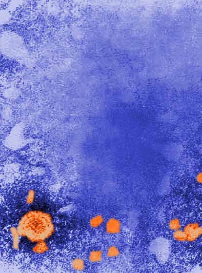
CoolCell
LX Container
                                                                        ®

We’ve taken cryopreservation
    to the next level of “cool”
    The CoolCell® LX container is the definitive
    cryopreservation tool, providing:
      • Ample sample space to create
        working stocks
      • Durable, alcohol-free construction
      • Dependable freezing rate
    An easy-to-use device designed to support
    cell, viral, bacterial, protozoan, and fungal
    cultures using a slow freezing rate of -1˚C
    per minute in a standard -80˚C freezer.
    Trust the leader in cryopreservation
    techniques, with over 85 years of
    experience, and order your CoolCell® LX
    Container (ATCC® No. ACS-6000™) today!

        www.atcc.org/CoolCell

               CoolCell® is a registered trademark of BioCision, LLC.
               ATCC® is a registered trademark of the American
               Type Culture Collection.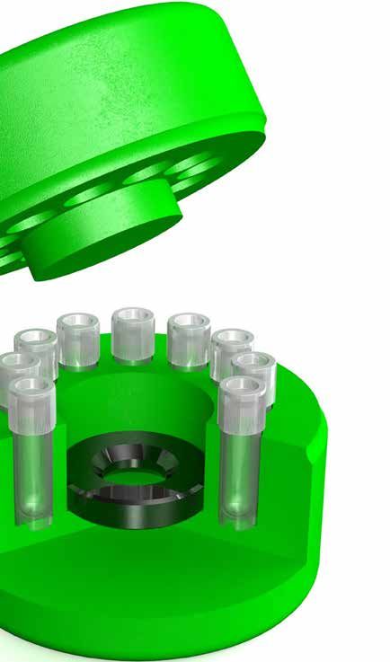
Table of Contents
This guide contains general technical information for viral growth, propagation, preservation, and
application. Additional information on viral culturing can be requested from ATCC Technical Services at
tech@atcc.org or can be found in A Manual of Basic Virological Techniques¹.

Getting Started with an ATCC Viral Strain....1                                                    Biosafety and Disposal..................................21
            Product Sheet..............................................................1                       Biosafety..................................................................... 21
            Viral Taxonomy ...........................................................1                        Disposal of Infectious Materials......................... 21
            Viral Replication..........................................................1
            Preparation of Propagation Host and                                                   Viral Authentication and Viability Testing.22
            Reagents........................................................................2                  Viability Testing........................................................ 22
            Opening Glass Ampoules Containing Frozen                                                           Viral Authentication............................................... 23
            Material..........................................................................2
            Initiating Frozen Cultures........................................2                   Viral Applications...........................................24
            Initiating Lyophilized Cultures..............................3                                     Cell Transformation................................................ 24
                                                                                                               Phage Therapy.......................................................... 24
Viral Replication and Propagation................4                                                             Nanotechnology...................................................... 24
            Propagation Host Range.........................................4                                   Recombinant Vectored Vaccines....................... 25
            Viral Propagation........................................................5
            Propagation of Common Viruses and                                                     Glossary............................................................26
            Chlamydia........................................................... 7
            Viral Titering.................................................................9      Appendix..........................................................27

Growth Media for Tissue Culture-Adapted                                                           References.......................................................28
Viruses 12                                                                                        ATCC Reagents for Virus Expansion ...........31
            Media for Culturing Propagation Hosts.......... 12
            Media Formulations............................................... 13
            Media Supplements............................................... 13

Preservation....................................................15
            Cryopreservation..................................................... 15
            Lyophilization........................................................... 18
            Preservation of Specific Strains.......................... 20
            Special Hazards........................................................ 20

Phone 800.638.6597                                                                    www.atcc.org
Getting Started with an ATCC Viral Strain Getting Started with an ATCC Viral Strain ATCC viral strains are predominantly shipped either frozen on dry ice in plastic cryopreservation vials, or as lyophilized materials within glass ampoules or serum vials. Upon receipt of frozen material, either thaw and transfer the viral agent to an appropriate propagation host, or briefly store the frozen material between -70°C and -80°C to allow time to seed host cells. However, please note that the viability of some materials may decline at temperatures above -120°C. Upon receipt of freeze-dried strains, reconstitute cultures with sterile, double-distilled water and add the rehydrated material to an appropriate propagation host. If this is not possible, store the vials in liquid nitrogen vapor phase (below -120°C). Product Sheet ATCC viral strains are shipped with a product sheet that contains information on the production host and recommendations for infection. The product sheet and additional information can be found on the ATCC website or can be requested from the ATCC Technical Service Department. Viral Taxonomy Viruses are placed into taxonomic groups based on characteristics including morphology, genome type, and host organism. Viral agents can significantly vary in size, often ranging between 20 and 300 nanometers in diameter. They also vary in structure, including helical, icosahedral, prolate, enveloped, and complex morphologies. In addition to unique morphological structures, viruses also vary in genomic structure. Unlike other microorganisms that have double stranded DNA as genomic material, viral genomes can be composed of double-stranded DNA, single-stranded DNA, double-stranded RNA, or single-stranded RNA. Single-stranded RNA viruses can be further described as positive sense, negative sense, or ambisense. Influenza ultra-structure courtesy of Jordan For more detailed information on the various morphological and Douglas, CDC genomic types, please refer to the glossary. Changes in taxonomy or further analysis of viral strains may lead to a change in nomenclature. Taxonomic nomenclature as well as the common name can be found on the product sheet. Further information on viral nomenclature is available online at http://ictvonline.org/. Viral Replication Viruses are pathogenic intracellular organisms requiring living cells in order to multiply. The virus life- cycle can be divided into three major steps: attachment, assembly, and release. Generally, infection is established when the virus binds to a specific cellular receptor and enters the host cell. Upon cellular entry, host proteins are recruited to assist with viral replication. Once viral structural proteins are generated, new viruses assemble within the cell. Depending on the nature of the viral agent, the replication and assembly process can vary in cellular location and process. Following viral assembly, new infectious particles either remain cell-associated or exit the cell via lysis or virus shedding. page 1 www.atcc.org Email tech@atcc.org
Getting Started with an ATCC Viral Strain
Preparation of Propagation Host and Reagents
In advance, prepare the propagation host and associated reagents necessary for viral propagation.
Information for the preparation of these products is available on the provided product sheet.

Opening Glass Ampoules Containing                                                     FREEZE-DRIED PREPARATIONS
Frozen Material
                        Tip
                            Insulator
Overview                    Cotton plug
All cultures should be          considered
                            Outer                potentially hazardous
                                  vial (soft glass)                                            Borosilicate glass
                            Inner vial
and should be opened        Freeze-driedby     individuals trained in
                                          pellet                                               Freeze-dried virus
microbiological techniques. Cotton           Work should only be
                            Desiccant with indicator
carried out in facilities         with containment requirements
appropriate for the biosafety level of the cultures. The
handling or opening of glass ampoules containing                         These preparations may be enclosed in a thin skin of
virusHeat
       material      must
             the tip of      be performed in a biological safety
                        the outer                                        cellulose; this skin must be removed (either with a sharp
      vial in a flame                                                    blade or by soaking in water for a few minutes). Score the
cabinet.    Ensure that all empty vials are sterilized before            ampule once briskly with a sharp file about one inch
disposal.                                                                from the tip.

1.   Disinfect the outside of the ampoule with freshly
     prepared 70% ethanol or dip it into a beaker of
     freshly prepared 70% ethanol.
2.   To recover the material from the glass ampoule,
     score
      Squirt athe   neckofofwater
               few drops       the ampoule with a sterile, small
      on the hot tip to crack glass
     file.
3.   Wrap the ampoule within several folds of a sterile
     towel or gauze to dry residual ethanol.                             Disinfect the ampule with alcohol-dampened gauze

4.   Working in a biological safety cabinet, hold the vial
     upright and snap open the vial. Ensure that your
     gauze does not become too wet with ethanol, or
     ethanol could be sucked into the culture when the
      Strike withis
     vacuum       filebroken.
                       or      Propagate the virus immediately.
      pencil to remove tip

Initiating Frozen Cultures
Tissue Culture-Adapted Strains                                           Wrap gauze around the ampule, and break at the scored area.
                                                                         Care should be taken not to have the gauze too wet, or
1. In advance, prepare the cell growth medium for                        alcohol could be sucked into the culture when the vacuum is
    growing the host cell line. Additionally, prepare                    broken. Rehydrate material at once.

    the   virus
     Remove      growth
              insulation andmedium        for virus propagation as
                              inner vial with
     forceps, gently raise cotton plug
    noted     on the product sheet. Viral growth medium
    is usually supplemented with a lower percentage
    of serum than cell growth medium, often ranging
    between 2-10% depending on the virus (See NOTE
    1). Ensure that both the cell and viral growth media
    are equilibrated for temperature and pH.
2. Initiate the recommended production host, and

Phone 800.638.6597                                        www.atcc.org                                                        page 2
Getting Started with an ATCC Viral Strain
grow the monolayer to an appropriate confluency.
NOTE 1:
3. Prior to thawing the frozen virus stock, check the virus titer listed on Viral growth medium prepared
the lot specific certificate of analysis to determine the amount of virus for tissue culture-adapted strains
of Influenza should not be
needed. Thaw the vial of frozen virus via gentle agitation in a water supplemented with FBS.
bath set at 37°C. Thawing will be rapid (approximately 2 minutes or
until all ice crystals have melted).
4. Remove the vial from the water bath and decontaminate the outer surface using 70% ethanol. Follow
strict aseptic conditions in a biological safety cabinet for all further manipulations.
5. Prepare the viral stock in base medium (without serum) unless specified.
6. Remove the cell growth medium from the cell culture, wash the monolayer of cells once or twice with
PBS, and inoculate with the prepared viral stock to provide an optimal multiplicity of infection (MOI) as
indicated on the product sheet, if applicable.
7. Propagate the virus according to the recommended infection conditions as described on the product
sheet. For additional information, refer to the chapter Viral Growth and Propagation.
Strains Propagated in Chicken Eggs
1. In advance, prepare chicken eggs by incubating eggs under the recommended temperature and
atmospheric conditions. Additionally, all eggs should be candled to ensure viability and properly
developed embryos. Healthy embryos will have an air sac, well-developed blood vessels, and
observable movement.
2. Prior to thawing the frozen viral preparation, check the certificate of analysis for lot specific titer
information. Thaw the frozen virus vial via gentle agitation in a water bath set at 37°C. Thawing will be
rapid (approximately 2 minutes or until all ice crystals have melted).
3. Remove the vial from the water bath and decontaminate the outer surface using 70% ethanol. Follow
strict aseptic conditions in a biological safety cabinet for all further manipulations.
4. Inoculate eggs using the inoculation procedure described in the chapter Viral Growth and Propagation.
Initiating Lyophilized Cultures
1. Open vial according to enclosed instructions. Instructions are also available on the ATCC website, www.
atcc.org, within the technical bulletin, “How to Revive Cultures”.
2. Aseptically add an appropriate volume of sterile double-distilled water at room temperature. Mix well.
3. Dilute the sample in base medium (without serum) unless specified, and inoculate host cells to provide
an optimal MOI as indicated on the product sheet, if applicable.
page 3 www.atcc.org Email tech@atcc.orgViral Replication and Propagation
Viral Replication and Propagation
Propagation Host Range
For tissue culture-adapted strains, the appropriate selection and processing of cell cultures is important
for successful viral isolation, titer, and infectivity². Typically, viruses can only infect a limited number of
hosts, known as the host range. This is best explained by a “lock and key” mechanism as certain proteins
on the viral surface must fit specific receptor sites on the host cell surface. When using ATCC viral strains,
the recommended host is indicated on the provided product sheet. For customer convenience, ATCC Cell
Biology holdings include cell lines for the propagation of viruses (Table 1, Appendix).
Table 1: Examples of propagation hosts for tissue culture-adapted viruses
ATCC® No. Product Name ATCC® No. Propagation host
53592™ Chlamydophila pneumonia* CCL-23™ HEp-2
VR-129B™ Encephalomyocarditis CCL-81™ Vero
Human herpesvirus 4
VR-1492™ N/A B95-8
(Epstein-Barr virus)
VR-260™ Humans herpesvirus 1 CCL-81™ Vero
VR-734™ Human herpesvirus 2 CCL-81™ Vero
VR-1367™ Human herpes 3 CCL-171™ MRC-5
VR-538™ Human herpesvirus 5 CCL-171™ MRC-5
VR-93™ Human parainfluenza CCL-7.1™ LLC-MK2 Derivative
Human respiratory
VR-26™ CCL-23™ HEp-2
syncytial virus
Human respiratory
VR-1540™ CCL-23™ HEp-2
syncytial virus
Human respiratory
VR-1580™ CCL-23™ HEp-2
syncytial virus
VR-283™ Human rhinovirus 16 CRL-1958™ H1-HeLa
VR-95™ Influenza A virus (H1N1) N/A SPF CE
VR-897™ Influenza A virus (H1N1) N/A SPF CE
VR-1469™ Influenza A virus (H1N1) CCL-34™ MDCK
VR-1520™ Influenza A (H1N1) CCL-34™ MDCK
VR-544™ Influenza A virus (H3N2) N/A SPF CE
VR-1679™ Influenza A virus (H3N2) CCL-34™ MDCK
VR-1535™ Influenza B virus CCL-34™ MDCK
VR-1583™ JC polyomavirus CRL-1651™ COS-7
*For historical reasons and because of similar requirements for handling, Chlamydia and Rickettsia are included in this guide
Phone 800.638.6597 www.atcc.org page 4Viral Replication and Propagation
Viral Propagation
Propagation in Cell Culture
A number of ATCC viruses are propagated in cell culture. Typically, propagation hosts are grown in tissue
culture vessels (such as T flasks) using media and reagents specified for the host cell line. Most cell lines are
seeded the day prior to setting up an infection and should not be seeded more than two days in advance,
nor passaged more than 9 times prior to infection. In addition to setting up cells for infection, negative
control cells should also be set up to monitor cellular health.
1. Identify the recommended propagation host as indicated on the product sheet. Plan to use the
propagation host.
2. Prepare the cell growth medium for growing the host cell line.
3. Prepare the virus growth medium as recommended on the product sheet.
4. One to two days prior to inoculation seed the host cells. Be sure to include a vessel that will not be
inoculated with virus to serve as a negative control.
5. Allow cells to reach the appropriate confluency. (See NOTE 2) NOTE 2:
The required confluency of the host
6. Prior to thawing the frozen virus stock check the virus titer listed on cell line will differ between viruses.
the certificate of analysis. Quickly thaw the virus in a 37˚C water bath.
7. Dilute the virus stock in the appropriate volume of viral growth medium.
8. Remove the cell growth medium from the cell culture flasks.
9. Inoculate the diluted virus to provide an optimal MOI as indicated on the product sheet.
10. Incubate tissue cultures under the appropriate temperature and atmospheric conditions for the
recommended incubation period.
11. After the recommended incubation time period, check the flask for cytopathic effects (CPE) when
applicable.
Propagation of Feline infectious peritonitis virus (ATCC® No. VR-2004™) in CRFK cells (ATCC®
No. CCL-94™). The panel on the left are uninfected CRFK cells. The panel on the right are
infected CRFK cells exhibiting CPE.
Propagation in Chicken eggs
Several ATCC virus holdings, such as influenza, are propagated in embryonated chicken eggs. There are
many advantages to culturing in eggs, including easy care, quick viral replication, and an inherently aseptic
environment. When culturing in embryonated eggs, ensure that eggs are viable, have properly developed
blood vessels and air sacs and are obtained from pathogen-free flocks. Depending on the virus, different
inoculation routes and/or organs may be used to cultivate the agent. These include the allantoic cavity, the
amniotic cavity, the chorioallantoic membrane (CAM), and the yolk sac (Table 2). Below, we describe the
procedures for allantoic cavity, amniotic cavity, and CAM inoculation. For the following procedures, ATCC
page 5 www.atcc.org Email tech@atcc.orgViral Replication and Propagation
recommends performing viral inoculation and harvest Chorioallantoic Amniotic
cavity
within a biological safety cabinet. For additional membrane Chorioallantic
membrane inoculation
information on the inoculation or propagation of Shell
embryonated eggs, contact ATCC technical services.
Amniotic
inoculation
A. Allantoic Cavity Inoculation Allantoic
Air
inoculation
1. For allantoic cavity inoculation, most sac
Yolk sac
viruses require 10-day old embryos. While
candling the eggs, draw a pencil line
Shell
around the air sac. Using an 18.5 gauge membrane Yolk sac
inoculation
needle, poke a hole into the air sac. Albumin Allantoic
cavity
2. Using a 1.0 mL syringe fitted with a 22.5
gauge needle, inject the viral inoculum through the hole, into the allantoic cavity, being
careful not to stick the embryo.
3. Seal the hole with tape or wax.
4. Incubate the inoculated eggs under conditions recommended for viral replication. Candle
eggs 12-18 hours after inoculation and discard eggs that are non-viable.
5. To harvest allantoic fluid, refrigerate eggs for at least 2 hours post-incubation. Once the
embryo is no longer viable, open the egg by tapping on the shell just above the air sac until
the shell breaks.
6. Use sterile scissors to cut away the shell around the air sac.
7. Aspirate the allantoic fluid with a syringe or a pipette. Usually 5 mL can be harvested from each
egg; this is considered infectious material.
B. Amniotic Cavity Inoculation
1. While candling the eggs, make a small hole in the side of the egg. Create the hole slowly as it
is easy to accidently stick the embryo.
2. Using a 1.0 mL syringe, insert the needle and syringe containing the viral inoculum into the
egg until the amniotic sac moves slightly. The needle is then thrust through the membrane
and the fluid is injected slowly.
3. Immediately seal the hole in the shell with tape or wax.
4. Incubate the inoculated eggs under conditions recommended for viral replication.
5. Candle eggs 12-18 hours after inoculation and discard eggs that are non-viable.
6. To harvest amniotic fluid, refrigerate eggs for at least 2 hours post-incubation. Once the
embryo is no longer viable, open the egg by tapping on the shell just above the air sac until
the shell breaks.
7. A needle and syringe must be used to aspirate the fluid. The needle may be inserted directly
into the amniotic sac, or the embryo may be carefully removed from the egg and placed in a
petri dish. Typically, one embryo will yield about 1.0 mL of fluid.
C. Chorioallantoic Membrane (CAM) Inoculation
1. Mark an area free from large blood vessels on the side where the embryo is located and the
area over the air sac.
2. Make one hole on the side and one over the air sac. Puncture the shell taking care not to
damage the CAM.
3. Gently apply suction to the hole over the air sac. Do this over an egg candler to see the CAM
drop and the new air sac form.
Phone 800.638.6597 www.atcc.org page 6Viral Replication and Propagation
4. Add 1.0 mL of the viral inoculum on the dropped membrane and rotate the egg to distribute
the inoculum over the membrane.
5. Seal the hole with nail polish.
6. Candle inoculated eggs daily. Any embryos that die within 24 hours should be discarded.
7. To harvest, refrigerate eggs for at least 2 hours. Once the embryo is no longer viable, make a
tape handle over the area of inoculation. To do so, cut a piece of tape, without joining either
end, join the middle of the piece together leaving both ends free to adhere to the egg shell
with a middle piece that is no longer sticky, forming a handle sticking up from the egg shell.
8. Cut off the top half of the eggshell, including the infected area, and gently remove the CAM,
which is attached to the shell.
9. Place the infected CAM in a tissue culture dish with PBS, spread the membrane flat against the
dish, and place the dish on a dark surface to facilitate counting of pocks.
Table 2: Examples of ATCC viruses propagated in embryonated chicken eggs
Optimal
Product Name Egg Age (Days) Inoculation Route Incubation (Days) Death of Embryo
Temperature (˚C)
Influenza A virus 10-11 Allantoic 2-3 33-35 -
Influenza B virus 10-11 Allantoic 2-3 33-35 -
Sendai virus 10-11 Allantoic 2-3 35-37 +
Rabies virus 7 Yolk 9-10 36.5 -
Propagation of Common Viruses and Chlamydia
Human herpesvirus 4 (Epstein-Barr Virus)
Epstein-Barr virus (EBV) is a universal human pathogen commonly transmitted via saliva³. Primary infections
among adolescents and young adults result in mononucleosis, a condition characterized by fever and the
swelling of lymph tissues. Following infection, EBV remains in a latent stage in most adults. In rare cases, the
virus may reactivate and contribute to neoplastic disorders such as Burkitt’s lymphoma or post-transplant
lymphoproliferative disorder (PTLD)⁴. To propagate EBV, the virus is often harvested from tumors growing
on patients with Burkitt’s lymphoma, or obtained from a biological resource center such as ATCC, and
further grown in human lymphoblastoid cells.
ATCC® No. VR-1492™, VR-603™, VR-602™
Recommended Host: Human lymphoblastoid cells
Preparation: Infected tissue culture with DMSO and FBS
Incubation: 5-15 days at 37°C
Atmosphere: 5% CO₂ in an air atmosphere
Effect: Polykaryocyte formation and cell swelling
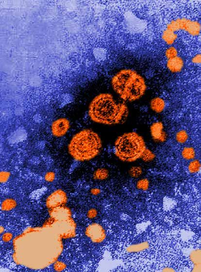
Influenza
Influenza viruses are highly contagious, enveloped, airborne pathogens that cause acute febrile illness,
fatigue, and respiratory infection. There are three types of influenza including influenza A, B, and C, which
are distinguished by the presence of specific, core nucleoproteins. Of these viral types, influenza A is
considered the most pathogenic. To propagate influenza, the virus is often cultured in the allantoic fluid of
page 7 www.atcc.org Email tech@atcc.orgViral Replication and Propagation
embryonated chicken eggs. However, some strains are propagated
in amniotic fluids or have been adapted for growth in tissue culture.
ATCC® No. VR-1469™, VR-95™, VR-1520™, VR-544™, VR-897™,
VR-1679™, VR-1807™, VR-1811™, VR-1813™
Recommended Host: Pathogen-free embryonated
chicken egg, 10-11 days old, or MDCK cells (ATCC® No.
CCL-34™)
Image of Influenza courtesy of Dr. Erskine L
Preparation: Infected chicken egg allantoic fluid, infected Palmer and ML Martini, CDC
tissue culture
Incubation: 2-3 days at 34°C for influenza propagated in chicken eggs (CE), 33-35˚C for influenza
propagated in tissue culture (TC). Harvest when CPE has progressed throughout the monolayer.
Atmosphere: An air atmosphere for CE, 5% CO₂ atmosphere for TC. CO2 level is determined by
media formulation.
Effect: Hemagglutination of chicken red blood cells, cell rounding sloughing in tissue culture.
Respiratory Syncytial Virus
Respiratory syncytial virus (RSV) is a major cause of respiratory
illness in young children, resulting in pneumonia and bronchiolitis.
In adults, RSV often manifests symptoms similar to the common
cold. This virus is commonly transmitted when droplets containing
the virus are aerosolized by coughing or sneezing.
ATCC® No. VR-1540™, VR-1540P™, VR-26™, VR-1580™, VR-
1803™
Recommended Host: HEp-2 (ATCC® No. CCL-23™) and
Image of Respiratory Syncytial Virus courtesy of
HeLa (ATCC® No. CCL-2™) cells Dr. Craig Lyerla, CDC
Preparation: Infected tissue culture fluid and cell lysate
Incubation: 5-12 days at 37˚C
Atmospherte: 5% CO₂ in an air atmosphere
Effect: Syncytia formation
Chlamydia
Species within the Chlamydia genus are obligate intracellular bacterial pathogens commonly transmitted
through sexual activity. Common symptoms associated with Chlamydia species include conjunctivitis,
pelvic inflammatory disease, and infection of the reproductive organs. These bacterial species are
commonly categorized with viruses as they have similar propagation properties. Often it is recommended
that the Chlamydia stock be sonicated first before inoculating cells, and infection may be enhanced by
centrifugation after inoculation. Follow the specific instructions on the product sheet for preparing the
cells.
ATCC® No. VR-123™, VR-346™, VR-347™, VR-573™, VR-879™, VR-885™, VR-886™, VR-887™
Recommended host: McCoy cells (ATCC® No. CRL-1696™)
Preparation: Infected tissue culture fluid and cell lysate
Incubation: 2-3 days at 35-37˚C
Atmosphere: 5% CO₂ in an air atmosphere
Effect: Intracellular inclusion bodies visualized by fluorescent staining with genus or species
specific conjugated monoclonal antibodies.
Phone 800.638.6597 www.atcc.org page 8Viral Replication and Propagation
Viral Titering
Plaque Assay
Calculating viral titer is necessary in order to determine viral infectivity. This
can be determined by a plaque assay or through calculating the infectious
dose. The plaque assay was initially developed to count and measure the
infectivity level of bacteriophages. This technique has since been modified
for use in animal virology, and has been a reliable determination of titer
for a number of tissue culture-adapted viruses. The basis of this assay is
to measure the ability of a single infectious virus to form a plaque on a
cell culture monolayer. A plaque is developed as part of the viral infection
cycle, where following viral replication, the host cell dies⁵.
1. Grow the host cells in wells with the recommended growth medium
for the cell line. Allow the cells to reach the appropriate confluency. Plaque assay of a tissue culture-
adapted viral strain.
2. Remove the growth medium and wash with Dulbecco's Phosphate-
Buffered Saline (DPBS). Add diluted virus to each well, using multiple wells per dilution.
3. Incubate dishes for 1-2 hours to allow for viral adsorption.
4. Remove the inoculum and wash with basal medium, if applicable.
5. Overlay the cells with overlay medium. Incubate for a length of time
appropriate for infection. Remove overlay, if applicable. NOTE 3:
This is a general procedure used
6. Observe the cell monolayers daily for the presence of foci or plaques to determine the potency of a
7. Count the number of plaques on plates with 20 or more plaques. virus. Please refer to the following
references for more details on
The average amount of plaques per Petri dish multiplied by the virus this procedure: A Manual of Basic
dilution gives the number of plaque forming units (PFU) per volume Virological Techniques, published
of inoculum. This number may be expressed as the titer of the virus. by Prentice-Hall Inc. , or Virology
1
Methods Manual, published by
(See NOTE 3) Academic Press. (1996) 5.
Tissue Culture Infectious Dose (TCID)
Viral titer can be determined in vitro by calculating the infectious dose. For tissue culture-adapted strains,
this calculation is ascertained through an endpoint dilution assay in cell culture. The most reproducible
endpoint of the dilution assay is the dilution of the virus that will produce a pathological change in 50%
of the cell cultures inoculated. This number is expressed as 50% the infectious dose, or TCID₅₀, which is
analogous to the calculation for lethal dose 50. The accuracy of this method is related to the number of
replicates at each dilution⁵.
1. Prepare a cell culture plate with the recommended cell line for viral propagation (preferably the same
cells that the virus being tested was grown in).
2. Incubate the plate under the appropriate conditions for cell growth until cells reach an optimum
density for infection.
3. Prepare viral dilutions in base medium.
4. Remove cell growth medium from plate, the monolayer may be washed with remove any inhibitory
agents.
5. Inoculate at least 3 wells with each dilution. Be sure to inoculate wells with base medium to serve as
negative controls. Use a fresh, sterile pipette for each dilution.
6. Allow the plate to incubate for 1 to 2 hours under conditions suitable for virus adsorption.
page 9 www.atcc.org Email tech@atcc.orgViral Replication and Propagation
7. Add viral growth medium and incubate the plate under conditions suitable for the virus and observe
all wells daily. Record results for each well daily.
8. The endpoint is determined when the CPE or immunofluorescence assay (IFA) read-out appear the
same per dilution for 3 separate readings.
9. The titer is calculated using the method of Reed and Muench⁶. A titer expressed as 10(3.0)TCID₅₀/0.2 mL
in 3 days in XXXX cell line may be translated as: 0.2 mL of virus diluted at 1:1000 will infect 50% of the
cells in 3 days when using XXXX cell line. (See NOTE 3)
The TCID₅₀ can be converted to plaque forming units (PFU) using the Poisson distribution. This conversion
is an estimate based on the rationale that the limiting dilution, which would infect 50% of the cell layers
challenged, would be expected to produce a single plaque in a cell monolayer. However, ATCC recommends
that the actual number of PFUs be determined empirically.
To estimate PFU from TCID₅₀, the Poisson distribution can be applied; P(o) is the proportion of negative tubes
and ‘m’ is the mean number of infectious units per volume (PFU/mL), P(o)=e(-m). For any titer expressed as
TCID₅₀, P(o)=0.5. Thus, e(-m)=0.5 and m= -ln 0.5, which is ≈0.7. Therefore, one could multiply the TCID₅₀ titer
(per mL) by 0.7 to predict the mean number of PFU/mL. For example, one can assume that material with
a TCID₅₀ of 1x10⁵ TCID₅₀/mL will produce approximately 0.7x10⁵ PFU/mL. When applying this calculation,
remember that the estimated mean will only be valid if the changes in the protocol required to visualize
plaques do not alter viral expression as compared to conditions used to determine TCID₅₀.
Chicken Embryo Infectious Dose (CEID)
For viruses normally propagated in chicken eggs, such as influenza virus, viral titer is calculated as the chicken
embryo infectious dose (CEID). Viral cultures are serially diluted and are used to inoculate embryonated
chicken eggs. Following incubation, allantoic fluid is harvested from each egg at all dilutions and virus titer
is determined by the appearance of hemagglutination. A positive hemagglutination reaction indicates the
virus is present at that dilution.
1. Order embryonated chicken eggs of the proper age, 8-12 days old depending on the virus. When the
eggs arrive, place them in an incubator at an appropriate temperature with a dish of water close by to
provide moisture to prevent the eggs from drying out.
2. When you are ready to inoculate, candle the eggs to locate the air sac
NOTE 4:
and embryo. Using a pencil, outline the air sac area and indicate where A viable egg has a series of small
the embryo’s eye is located. In a biological safety cabinet, punch a hole blood vessels within the allantoic
into the shell within the defined air sac area, on the opposite side of membrane, which can be seen
during candling. Additionally, the
the egg from the embryo’s eye. Inoculate the allantoic fluid of each embryo’s eye will look like a black
egg with the serial dilution of virus. Seal the hole with nail polish and spot, which will move.
incubate the eggs near a pan of water at the proper temperature as
recommended on the provided product sheet. (See NOTE 4)
3. Candle the eggs 24 hours post-inoculation to check for viral-induced death. Place any of the dead eggs
into a biohazard bag, seal the bag with autoclave indicator tape and refrigerate to await autoclaving.
Return the rest of the eggs to the incubator.
4. Following the recommended incubation period, harvest the virus. Depending upon the virus, death of
the embryo may have occurred. By this time, a viable embryo will have grown much larger, will be more
active, and the network of blood vessels will be more visible. A dead embryo will not move and the
egg may or may not be as transparent and the blood vessels will have mostly disappeared. For viable
embryos, sacrifice the embryo prior to viral harvesting by placing the egg at 4°C for at least 2 hours.
5. Prepare a 0.5% suspension of chicken red blood cells using phosphate buffered saline (PBS) while the
infected eggs are chilling in a biological safety cabinet. Use sterile forceps to break the top of the shell
Phone 800.638.6597 www.atcc.org page 10Viral Replication and Propagation
open and pull back the allantoic membrane. Place all egg fragments, including parts of the shell into
a double biohazardous materials bag. Harvest the allantoic fluid from each egg by using a pipette to
collect the fluid into a different well of a rounded-bottom 96 well plate for each egg, being sure to use
a new pipette for each dilution.
6. Add a 1:1 ratio of the 0.5% red blood cell suspension to allantoic fluid in each tube. Mix by gently
tapping and allow the red blood cells to settle to the bottom of the wells. After 30-45 minutes at room
temperature, read the hemagglutination assay by recording
the presence of a button of cells or the presence of a lattice
formation at the bottom of the tube. A negative result is seen
by the button of red blood cells at the bottom of the rounded
well. A positive result is seen by the formation of the lattice
formation at the bottom of the rounded well. Once the results
are properly recorded, place the plate in a biohazard bag to
await proper decontamination.
7. The end point is taken to be the highest dilution of virus
suspension that produces a positive result. HA = 1:640 means Hemagglutination assay depicting Influenza B
that the virus was titered by a hemagglutination assay (HA) virus (ATCC® No. VR-295™) at dilutions varying
and the endpoint for the assay was a dilution of 1:640. (See from 10¯⁹ to 10¯¹.
NOTE 3 ) Left column = Negative control
Columns 2-10 = Dilutions 10¯⁹ to 10¯¹
Column 11 = Positive control
page 11 www.atcc.org Email tech@atcc.orgGrowth Media for Tissue Culture-Adapted Viruses Growth Media for Tissue Culture-Adapted Viruses Media for Culturing Propagation Hosts For the propagation of tissue culture-adapted viruses, it is important to grow and maintain the host cell line in an appropriate medium. The recommended media for all host cells is indicated on the product sheet for the host cell line and can also be found online at www.atcc.org. Cell culture media are complex mixtures of salts, carbohydrates, vitamins, amino acids, metabolic precursors, growth factors, hormones, and trace elements. The requirements for these components vary among cell lines. Carbohydrates are supplied primarily in the form of glucose. In some instances, glucose is replaced with galactose to decrease lactic acid build-up, as galactose is metabolized at a slower rate. Other carbon sources include amino acids (particularly L-glutamine) and pyruvate. In addition to nutrients, the medium helps maintain the pH and osmolality in a culture system. The pH is maintained by one or more buffering systems; CO₂/sodium bicarbonate, phosphate, and HEPES (4-(2-hydroxyethyl)-1-piperazineethanesulfonic acid) are the most common. Sera will also buffer a complete medium. Phenol red, a pH indicator, is added to the medium to colorimetrically monitor changes in pH. Media commonly used in the propagation of tissue culture-adapted strains include the following: Eagle’s Minimum Essential Medium (EMEM) ATCC’s modification of EMEM (ATCC® No. 30-2003) contains Earle’s balanced salt solution, non-essential amino acids, L-glutamine, and sodium pyruvate. It is formulated with a reduced sodium bicarbonate concentration (1,500 mg/L) for use with 5% CO₂. Because EMEM is a simple medium, it is often fortified with additional supplements or higher levels of serum. Dulbecco’s Modified Eagle’s Medium (DMEM) has roughly twice the concentration of amino acids and four times the amount of vitamins as EMEM, as well as ferric nitrate, sodium pyruvate, and some supplementary amino acids (though not all nonessential amino acids). The original formulation contained 1,000 mg/L of glucose, but in the more commonly used variations this amount was increased to 4,500 mg/L. ATCC DMEM (ATCC® No. 30-2002) has 4,500 mg/L of glucose and a reduced sodium bicarbonate concentration (1,500 mg/L) for use with 5% CO₂. RPMI-1640 ATCC’s RPMI-1640 (ATCC® No. 30-2001) was modified to contain higher amounts of glucose (4,500 mg/L), sodium pyruvate, and HEPES buffer. It also contains a reduced concentration of sodium bicarbonate (1,500 mg/L) for use with 5% CO₂. Leibovitz’s L-15 Medium (ATCC® No. 30-2008) is formulated for free gas exchange with atmospheric air. The standard sodium bicarbonate/CO₂ buffering system is replaced by a combination of phosphate buffers, free-base amino acids, higher levels of sodium pyruvate, and galactose. A CO₂ and air mixture is detrimental to cells when using this medium for cultivation. However, cell cultures can be grown in CO₂ incubators with L-15 medium provided there is no exchange between the air in the culture vessel with that of the incubator (i.e., caps of flasks are tightly closed). VeroPlus SFM (ATCC® No. ACS-4001) is a defined, serum-free, animal component-free medium that contains inorganic salts, vitamins, amino acids, glucose, and phenol red. VeroPlus SFM should be supplemented with L-glutamine (ATCC® No. 30-2214) prior to use. Phone 800.638.6597 www.atcc.org page 12
Growth Media for Tissue Culture-Adapted Viruses
Media Formulations
The formulations for media used by ATCC can be found online. Please note that there are cell lines in the
collection used for viral propagation that may require media that is not currently sold by ATCC.

Media Supplements
The growth media recommended for host propagation may require the addition of components not
already available in the base medium. These components may include hormones, growth factors, or serum.
After supplements have been added to the base medium, the shelf life of the medium should be determined
on a case-by-case basis. Media containing serum, antibiotics, and/or antimycotics tend to degrade faster
than base media alone. Media containing supplements should not be frozen as this may cause certain
compounds to precipitate out of solution; media should be stored at 2°C to 8°C. For additional information
regarding the preparation, storage, or usage of specific additives, contact your local supplier or consult with
the manufacturer’s product information sheet.
Serum
Serum can serve as a source of growth factors, proteins, vitamins, hormones,
carbohydrates, lipids, amino acids, minerals, and trace elements. The exact
composition of serum is unknown and varies from lot to lot, although lot-
to-lot consistency has improved in recent years.
Sera from fetal bovine sources are commonly used to maintain cell cultures
in preparation for viral infection. Fetal serum is a rich source of growth
factors and is appropriate for the growth of fastidious cells. It is often
supplied at a concentration of 2-10%. (See NOTE 5)
Unfortunately, naturally derived products from animals, such as sera, may
contain adventitious microorganisms. All reputable suppliers routinely
test their products for infectious viruses by several methods including            NOTE 5:
                                                                                   Fo r v i r a l i n o c u l a t i o n , ATCC
fluorescent antibody labeling, cytopathic effect, and hemadsorption.               recommends decreasing the
These products are also screened for the standard microbial contaminants           percentage of serum to 2% as it can
such as bacteria, fungi, and mycoplasma. To reduce the risk of any possible        interfere with viral attachment. This
                                                                                   can vary from strain to strain, and is
contamination, ATCC recommends that all serum should be triple filtered            not applicable for influenza viruses.
through 0.1 µm sterile filters before use.
ATCC offers the following types of animal sera:
•   Fetal Bovine Serum (also known as fetal calf ) – ATCC® No. 30-2020
•   Fetal Bovine Serum qualified for embryonic stem cells – ATCC® No. SCRR-30-2020
•   Iron-supplemented Calf Bovine Serum – ATCC® No. 30-2030
•   Horse Serum – ATCC® No. 30-2040
These products are rigorously tested for adventitious infective agents and sourced only from U.S. herds.
Further, each lot is tested for its ability to support cell culture growth and is the same sera used in ATCC labs.
    A.    Storage
          Do not store serum at temperatures above -20°C for any length of time. Avoid any repeated freeze-
          thaws by dispensing and storing sera in aliquots. Additionally, ensure that sera are stored away
          from direct light.

page 13                                           www.atcc.org                                  Email tech@atcc.org
Growth Media for Tissue Culture-Adapted Viruses
B. Thawing
The following procedure is used to thaw serum:
1. Place the frozen serum in a refrigerator at 2°C to 8°C overnight.
2. Put the bottles in a 37°C water bath and gently agitate occasionally to mix the solutes that
tend to concentrate at the bottom of the bottle.
Do not keep the serum at 37°C any longer than necessary to thaw it, and do not thaw the serum
at higher temperatures. Thawing serum in a bath above 40°C without mixing may lead to the
formation of a precipitate inside the bottle.
C. Turbidity and precipitates
All sera may retain some fibrinogen. Because external factors may initiate the conversion of
fibrinogen to fibrin, flocculent material or turbidity may be observed after the serum is thawed.
The presence of this material does not alter the serum’s performance. If the presence of flocculent
material or turbidity is a concern, it can be removed by filtration through a 0.45 µm filter.
A precipitate can form in serum when incubated at 37°C or higher for prolonged periods. This
is often mistaken for microbial contamination. This precipitate may include crystals of calcium
phosphate, but this does not alter the performance of the serum as a supplement. Heat inactivation
of sera can also cause the formation of precipitates.
Additives and Cryoprotectants
To preserve viral strains by cryopreservation, ATCC may use a mixture of FBS and DMSO (ATCC® No. 4-X) or
glycerol. Some viruses require a specialized mixture of cryoprotectants or additives. For more information,
refer to the cryopreservation section of the chapter entitled Preservation.
Phone 800.638.6597 www.atcc.org page 14Preservation
Preservation
There is no single procedure that is optimal for the preservation of all animal viruses. Both the type of agent
and recommended growth conditions of the culture influence the method of cryopreservation, and the
determination of these ideal conditions may require some experimentation. There are many advantages of
preservation that far outweigh the required investment in equipment and reagents for continuous in vitro
expansion. These advantages include:
• Overall safety of viral stocks against loss due to equipment failure or contamination by other microbial
organisms.
• Elimination of time, energy, and material costs associated with the maintenance of viral strains not
currently in use.
• Insurance against phenotypic drift due to genetic instability and/or selective pressures.
• Creating a standard reagent that can be used for a series of experiments.
The methodologies used at ATCC have proven successful for a large number of organisms. Currently, ATCC
only uses cryopreservation methods to preserve viral strains. However, several viral strains in the collection
are available as freeze-dried (lyophilized) cultures. The basic methodology for the cryopreservation and
lyophilization of viruses are described in the subsequent sections. For historical reasons and because of
similar requirements for handling and preservation, Chlamydia and Rickettsia species are also included here.
Cryopreservation
Overview
Freezing a viral suspension often results in a decrease in viability and titer¹¹, ¹². For cell-associated pathogens,
such as Chlamydia and Rickettsia, as the suspension is cooled below the freezing point, ice crystals begin to
form and the concentration of solutes in the suspension increases resulting in damage of host cell structures.
This can be minimized if water within the cells is allowed to escape by osmosis during the cooling process;
a slow cooling rate, generally -1˚C to -10˚C per minute, facilitates this progression. However, as host cells
lose water, they shrink in size and will quickly lose viability if they surpass a minimum threshold volume.
The addition of cryoprotectant agents, such as glycerol or dimethylsulfoxide (DMSO), will mitigate these
effects¹³, ¹⁴.
The viability of viruses that are not cell-associated is best maintained by rapid freezing. In this method,
samples are quickly frozen in a dry ice slurry and stored in liquid nitrogen vapor or within a mechanical
freezer at -80°C.
Overall, there are numerous factors that can affect the viability of recovered viruses. These critical parameters
can include the composition of the cryoprotectant and the viral titer. For cell-associated viruses, obtain
optimal cell viability upon recovery by modifying the cryopreservation procedure for each viral strain.
Contact ATCC for more information on the cryopreservation of viral strains.
Freeze Medium
Glycerol and DMSO are the most common cryoprotectant agents. To preserve viral strains, ATCC commonly
uses a mixture of FBS and DMSO or glycerol. When employing these cryoprotectants, use only reagent-
grade DMSO or glycerol. Store both in aliquots protected from light. ATCC offers DMSO (ATCC® No. 4-X) that
has been thoroughly tested for use.
page 15 www.atcc.org Email tech@atcc.orgPreservation
Some viruses require specialized preservation methods. Several representative strains with unique
preparations are listed in Table 3; however, these examples and methods may not be applicable to all
members of the group. Overall, the optimum formulations for individual viral strains must be determined
empirically.
Table 3: Examples of Frozen Preparations
ATCC® No. Product Name Preparation
VR-1™ Human adenovirus 1 Infected culture medium
VR-343™ Japanese encephalitis virus Infected culture medium diluted 1:1 with calf serum
VR-977™ Human herpesvirus 5 Infected culture medium (EMEM + 10% FBS + 7% DMSO)
VR-897™ Influenza A virus (H1N1) Infected allantoic medium
VR-955™ Human respiratory syncytial virus Infected culture medium (EMEM + 2% FBS)
VR-129B™ Encephalomyocarditis virus Infected culture medium (MEM +2% FBS)
VR-838™ Raccoonpox virus Infected culture medium (EMEM + 2% FBS)
VR-156™ Vaccinia virus Infected culture medium (L-15 + 2% FBS + L-glut)
VR-659™ Rous sarcoma virus Infected culture medium (M199)
VR-137™ Rabies street virus 10% infected mouse brain suspension in PBS + 10% horse serum
VR-612™ Rickettsia akari Infected yolk sac diluted 1:1 in sucrose-phosphate glutamate
Equipment
A. Cryopreservation vials
There are two materials to choose from for cryopreservation vials: glass or plastic. Glass vials are
more difficult to work with: they need to be sterilized before use, they need to be sealed with a
hot flame, and they can be difficult to open. However, they are considered fail-safe once properly
sealed.
If cryopreservation in glass ampoules is not possible, plastic vials can be used. Plastic vials come in
two varieties: those with an internal thread and silicone gasket, and those with an external thread.
Vials with an internal-thread were the first commercially available, but have some disadvantages
over the external-thread version. For example, while the silicone gasket provides an excellent seal,
it needs to be tightened just right; the vial will leak if the seal is too tight or too loose.
B. Controlled-rate freezing chambers
For cell-associated viruses, there are several means to achieve a cooling rate of -1˚C per minute.
The best method involves the use of a computer controlled, programmable electronic freezing
unit (such as Thermo Scientific* CryoMed Freezers), which rigorously maintains this rate of cooling.
This is the method used exclusively at ATCC. Such equipment is relatively expensive and necessary
for only the most sensitive strains.
A less costly approach is to place the cryopreservation vials into an insulated chamber and cool
for 24 hours in a mechanical freezer at -70˚C or colder. There are several commercially available
freezing chambers, which achieve a cooling rate very close to the ideal -1˚C per minute (CoolCell®
LX; ATCC® ACS-6000). Alternatively, the vials can be placed into a polystyrene box, with 15 mm
(3/4 inch) thick walls and 1 L capacity that is packed with paper, cotton wool, or foam peanuts for
insulation.
Phone 800.638.6597 www.atcc.org page 16Preservation
Liquid Nitrogen Freezing Storage
The ultra-low temperatures (below -120˚C) required for long-term
storage can be maintained by specialized electric freezers, or more
commonly, by liquid nitrogen freezers. There are two basic types
of liquid nitrogen storage systems: immersing vials in the liquid or
holding vials in the vapor phase above the liquid. The liquid-phase
system holds more nitrogen and thus requires less maintenance.
However, there is always a chance that some liquid will enter
improperly sealed vials, which may then explode when retrieved.
For this reason, ATCC strongly recommends storage in the vapor-
phase.
Vapor-phase storage systems create a vertical temperature gradient within the container. The temperature
in the liquid nitrogen at the bottom will be -196˚C, whereas the temperature at the top will vary depending
upon the amount of liquid nitrogen at the bottom and the length of time the container is opened. To ensure
the safe storage of cells, maintain sufficient levels of liquid nitrogen in the container so that the temperature
at the top is -120˚C or colder. All storage systems should be equipped with temperature alarms.
Cryopreservation Procedure
A. Uncontrolled Freezing
The viability of most viruses is best maintained by fast freezing after harvesting. Dispense the
material into ampoules, place the ampoules in a rack, and dip in a dry ice/ethanol bath. Store the
frozen material in liquid nitrogen vapor or in a mechanical freezer at -80°C.
B. Controlled Freezing
To preserve cell-associated pathogens, such as Chlamydia and Rickettsia, a controlled freeze is
preferred.
1. Incubate the viral agent under the recommended conditions until a maximum yield is obtained
(maximum cytopathic effect if applicable).
2. Add a cryoprotective agent to the viral suspension. For many viral strains, this will be a mixture
of FBS and DMSO or glycerol. Some strains may require specialized conditions, examples of
these can be found in Table 1.
3. Within a biological safety cabinet, dispense 0.5 to 1.0 mL of the above mixture into sterile
plastic cryovials or glass ampoules. Store the filled glass ampoules on ice prior to sealing. Glass
ampoules can be sealed using a gas-oxygen torch, pulling the neck of the ampoules as it is
rotated in the flame. Following sealing, disinfect glass ampoules by spraying with disinfectant.
4. Place the vials into a pre-cooled (4°C), controlled-rate freeze chamber and place the chamber
in a mechanical freezer at -70°C (or colder) for at least 24 hours. Alternately, use a pre-cooled
(4°C) programmable freezer unit set to cool the vials at -1°C per minute until a temperature
below -40°C is achieved and then set the temperature to abruptly drop to -120°C.
5. Quickly transfer the vials to a liquid nitrogen or -120°C freezer. Frozen material will warm up
above -50°C.
6. Record the location and details of the freeze.
7. After 24 hours at -120°C, remove one vial, appropriately restore the viral strain, and determine
the viability and sterility.
Recovery of Cryopreserved Viruses
page 17 www.atcc.org Email tech@atcc.orgPreservation
1. In advance, grow the recommended production host in a prepared culture vessel that contains growth
medium equilibrated for both temperature and pH.
2. Remove the vial of frozen virus from the liquid nitrogen freezer and thaw by gentle agitation in a 37°C
water bath.
3. Thaw the strain rapidly until all ice crystals have melted (approximately 2 minutes).
4. Remove the vial from the water bath and decontaminate it by dipping in or spraying with 70% ethanol.
Follow strict aseptic conditions in a biological safety cabinet for all further manipulations.
5. Unscrew the top of the vial and inoculate the propagation host with the recommended infection
conditions as described on the supplied product sheet.
6. Examine the cultures after an appropriate length of time.
Lyophilization
Overview
Freeze-drying is a process where water and other solvents are removed from a frozen product via
sublimation¹⁵. Sublimation occurs when a frozen liquid goes directly to a gaseous state without entering a
liquid phase. The freeze-drying process results in a stable, readily rehydrated product. This process consists
of three steps: pre-freezing the product to form a frozen structure, primary drying to remove most water,
and secondary drying to remove bound water.
During the initial freezing process, ice crystals begin to form and the concentration of solutes in the
suspension increases. The method used during freezing can greatly affect the ability to freeze-dry the
material. Slow cooling rates are recommended as this will result in the formation of vertical ice crystal
structures and allow for more efficient water sublimation from the frozen product.
Freeze-dried products are hygroscopic and must be protected from moisture during storage. Additionally,
these products are sensitive to other factors including oxygen and temperature, which can significantly
decrease the shelf life. It is important to store freeze-dried material in a manner that protects the product
from exposure to moisture and oxygen and at refrigerated temperatures (4°C).
Generally, the lyophilization of viruses is not currently used by ATCC. This is because the methods for
lyophilization vary depending on the type of virus, and some viruses cannot be successfully preserved by
freeze-drying due to loss of viability. Strongly cell-associated viruses, including certain members of the
herpes virus family, such as varicella-zoster, lose viability outside the host. Therefore, these viruses cannot
be preserved by this method. Though ATCC does not currently freeze-dry any virus preparations, it was
done fairly extensively in the past. The methods of lyophilization that were previously used by ATCC are
described below. For additional information, contact ATCC technical services.
Equipment
A. Lyophilization Vials
For the storage of lyophilized viruses, ATCC uses sterile funnel-tipped glass ampoules (Wheaton)
and glass serum vials. Generally, during the lyophilization process, material is freeze-dried in a
glass ampoule, disinfected, and sealed.
B. Lyophilization Apparatuses
For the lyophilization of viruses, ATCC previously employed a commercial freeze-dryer. In this
freeze-drying procedure, samples are mixed with a suitable preservative, dispensed into the
appropriate ampoule, and allowed to slowly freeze into a solid mass. The preservative used for
lyophilization can vary between viral agents; several examples are listed below in Table 4. Once
Phone 800.638.6597 www.atcc.org page 18Preservation
frozen, samples are lyophilized within a freeze-drying system (Virtis Genesis®, Millrock® Max 85,
and LD 85).
During the primary drying phase, water is removed from the frozen product via sublimation. This is
accomplished through the use of a vacuum pump, which allows water molecules to migrate from
the frozen product and condense on a moisture trap called a condenser. For this to be possible
the temperature of the condenser must be colder than the product temperature; the difference
in these temperatures will affect the rate of sublimation. When primary drying is complete, all
residual moisture is removed by directly heating the product. During this secondary drying phase,
water must be desorbed to a residual moisture content of 1% or less. This process requires a low
pressure, low condenser temperature system. Once dried, the ampoules are properly sealed and
stored at refrigerated temperatures (4˚C).
Table 4: Examples of Freeze-Dried Preparations
ATCC® No. Product Name Preparation
VR-343™ Japanese encephalitis virus Infected primary hamster kidney cells + 50% calf serum
Infected mouse brain (10% suspension in normal saline) diluted
VR-90™ Colorado tick fever virus
1:1 in rabbit serum
VR-897™ Influenza A virus Infected allantoic fluid + 10% sucrose
Storage and Viability of Lyophilized Strains
To maximize the recovery of viable cells, viral cultures must be in optimum condition before the lyophilization
process¹⁶. Viruses should be propagated under the recommended conditions for infection as indicated on
the product sheet.
Because lyophilized products are hygroscopic, they must be stored under moisture-free conditions.
Additionally, other factors such as oxygen content and temperature can affect the shelf-life of freeze-dried
strains. Oxygen can chemically react with the product, negatively affecting culture viability. This reactivity
is directly proportional to storage temperature. Therefore, lyophilized products should be stored long-term
in liquid nitrogen.
Lyophilization Procedure
1. Grow the virus according to usual procedures
2. For freeze-drying viral suspensions, glucose, skim milk, or
Sucrose-Phosphate-Glutamate-Albumin (SPGA) should be
added before dispensing. The lyophilization of some viruses
may require specific preservation preparations; examples are
listed in Table 4.
3. In a biological safety cabinet, dispense the suspension into
either sterile funnel-tipped ampoules or serum vials. Plug the
ampoules loosely with slotted silicon rubber stoppers, and
place the ampoules in a freeze-dryer tray. Set an acrylic plate
on top of the stoppers.
4. Freeze-dry the material in a commercial freeze-dryer. Hold the
material at -30°C for 18-48 hours, depending on the additive. Then raise the shelf temperature to 10°C
per hour until the product temperature is 25°C.
5. When the cycles are complete, backfill the system with sterile nitrogen and press the caps into place.
Disinfect the outside of the ampoules and return them to the biological safety cabinet.
page 19 www.atcc.org Email tech@atcc.orgYou can also read