Effects of Melatonin on Anterior Pituitary Plasticity: A Comparison Between Mammals and Teleosts
←
→
Page content transcription
If your browser does not render page correctly, please read the page content below
REVIEW
                                                                                                                                         published: 11 January 2021
                                                                                                                                   doi: 10.3389/fendo.2020.605111

                                           Effects of Melatonin on Anterior
                                           Pituitary Plasticity: A Comparison
                                           Between Mammals and Teleosts
                                           Elia Ciani 1, Trude M. Haug 2, Gersende Maugars 3, Finn-Arne Weltzien 3, Jack Falcón 4
                                           and Romain Fontaine 3*
                                           1 Department of Pharmacy, Faculty of Mathematics and Natural Sciences, University of Oslo, Oslo, Norway, 2 Department of

                                           Oral Biology, Faculty of Dentistry, University of Oslo, Oslo, Norway, 3 Physiology Unit, Faculty of Veterinary Medicine,
                            Edited by:
                                           Norwegian University of Life Sciences, Oslo, Norway, 4 Laboratoire Biologie des Organismes et Ecosystèmes Aquatiques
                    Vance L. Trudeau,
                                           (BOREA), MNHN, CNRS FRE 2030, SU, IRD 207, UCN, UA, Paris, France
         University of Ottawa, Canada

                       Reviewed by:
                       Hélène Volkoff,    Melatonin is a key hormone involved in the photoperiodic signaling pathway. In both
Memorial University of Newfoundland,       teleosts and mammals, melatonin produced in the pineal gland at night is released into the
                              Canada
                      Hana Zemkova,
                                           blood and cerebrospinal fluid, providing rhythmic information to the whole organism.
  Academy of Sciences of the Czech         Melatonin acts via specific receptors, allowing the synchronization of daily and annual
          Republic (ASCR), Czechia
                                           physiological rhythms to environmental conditions. The pituitary gland, which produces
                 *Correspondence:
                                           several hormones involved in a variety of physiological processes such as growth,
                     Romain Fontaine
            romain.fontaine@nmbu.no        metabolism, stress and reproduction, is an important target of melatonin. Melatonin
                                           modulates pituitary cellular activities, adjusting the synthesis and release of the different
                   Specialty section:      pituitary hormones to the functional demands, which changes during the day, seasons
         This article was submitted to
             Neuroendocrine Science,       and life stages. It is, however, not always clear whether melatonin acts directly or indirectly
               a section of the journal    on the pituitary. Indeed, melatonin also acts both upstream, on brain centers that control
            Frontiers in Endocrinology
                                           the pituitary hormone production and release, as well as downstream, on the tissues
      Received: 11 September 2020
      Accepted: 12 November 2020
                                           targeted by the pituitary hormones, which provide positive and negative feedback to the
        Published: 11 January 2021         pituitary gland. In this review, we describe the known pathways through which melatonin
                               Citation:   modulates anterior pituitary hormonal production, distinguishing indirect effects mediated
       Ciani E, Haug TM, Maugars G,        by brain centers from direct effects on the anterior pituitary. We also highlight similarities
Weltzien F-A, Falcón J and Fontaine R
(2021) Effects of Melatonin on Anterior    and differences between teleosts and mammals, drawing attention to knowledge gaps,
     Pituitary Plasticity: A Comparison    and suggesting aims for future research.
    Between Mammals and Teleosts.
        Front. Endocrinol. 11:605111.      Keywords: melatonin, adenohypophysis, photoperiod, melatonin receptors, seasonal reproduction, plasticity,
    doi: 10.3389/fendo.2020.605111         endocrinology, light

Frontiers in Endocrinology | www.frontiersin.org                                 1                                      January 2021 | Volume 11 | Article 605111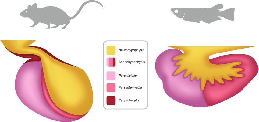
Ciani et al.                                                                                                                              Melatonin Effects on the Pituitary

INTRODUCTION                                                                                   the different enzymes differs between mammals and fish, as a
                                                                                               consequence of whole genome duplications that occurred in the
Our environment is constantly changing. While some variations are                              vertebrate lineage. Indeed, after the two successive whole genome
fast and unpredictable (e.g. meteorological phenomena), others,                                duplications (referred to as 1R and 2R) which occurred at the
such as solar cycles, moon phases, and seasons follow regular                                  base of the vertebrate lineage (10–12), a third one (3R) occurred
patterns. Photoperiod, the alternation of light and darkness, is the                           at the base of the teleost fish lineage (13), and a fourth one (4R)
most reliable (noise-free, characterized by predictable rhythms over                           occurred independently in both the cyprinid and salmonid
a long period of time) signal, allowing animals to synchronize their                           lineages (14, 15). Following a genome duplication, one of the
biological rhythms with both daily and seasonal changes.                                       paralogous genes may be lost or duplicated paralogues may
Photoperiod is conveyed by two types of signal: a neural message                               acquire differential specialized functions over time, and an
from photoreceptive structures to specific signaling centers in the                             increase in the number of paralogues, expands the hormone-
brain, and a hormonal message (1, 2).                                                          receptor combinations (16). In contrast to mammals, all
    Melatonin is the key hormone that conveys rhythmic                                         actinopterygians, including the teleosts, possess at least two
information from the environment, including photoperiod and                                    aanat genes (aanat1 and aanat2) (9, 17), resulting probably
temperature, to the organism. Circulating blood levels of melatonin                            from the whole genomic duplications that occurred in the
exhibit a daily rhythm with higher levels during night than during                             vertebrate lineage (18). Additionally, aanat1 and aanat2 have
day, and a seasonal rhythm with longer duration of the high level                              also been duplicated during the 3R (18). While one of the aanat2
period during winter, as a consequence of the longer dark phase                                paralogues was lost early after the 3R, this was not the case for the
(Figure 1). Additionally, variations in temperature fine-tune those                             aanat1 paralogues and, to date, some fish possess two Aanat1
rhythms by modulating the amplitude of melatonin production.                                   isoforms (aanat1a and aanat1b) or either one of them. While
Duration and amplitude of melatonin release therefore provide                                  aanat1 genes are mostly expressed in the retina, brain, and
clear information regarding time of the day and the year, and allow                            peripheral tissues, aanat2 expression is specific to the pineal
the synchronization of metabolic, physiological, and behavioral                                gland (19, 20), the site of production of circulating melatonin in
events, including growth, reproduction, and migration (3, 4).                                  both mammals (21) and teleosts (22, 23). Melatonin is then
    Melatonin is synthesized from tryptophan in four enzymatic steps                           released from the pineal gland into the blood and cerebrospinal
(4, 5). Tryptophan is first converted into 5-hydroxytryptophan by the                           fluid to be transported to its target organs.
tryptophan hydroxylase, then converted into serotonin by the 5-                                    Melatonin acts through several different receptors (MTNR),
hydroxy-tryptophan decarboxylase. Afterwards, serotonin is                                     belonging to the G‐protein coupled receptor superfamily (24). Four
acetylated by the arylalkylamine N-acetyltransferase (AANAT),                                  sub‐groups of Mtnr, arising from the 1R and 2R, have been
producing N-acetylserotonin, which is finally converted into                                    characterized in vertebrates: MTNR1A (Mel1a or MT1), MTNR1B
melatonin by the hydroxyindole-O-methyl transferase. AANAT                                     (Mel1b or MT2), MTNR1C (Mel1c or GPR50), and MTNR1D
has been reported to be the limiting enzyme driving the rhythm of                              (Mtnr1A-like or Mel1d) (25–28). In mammals, melatonin action is
melatonin production (6). It has been hypothesized that the                                    mediated only through two MTNR paralogues, MTNR1A and
functional shift of AANAT from amine detoxification to melatonin                                MTNR1B, since the Mtnr1d gene was lost in the mammalian
synthesis played a critical role in the evolution of melatonin as a night-                     lineage and MTNR1C lost its ability to bind melatonin (28).
time signal (7–9).                                                                             Teleosts may possess up to 7 Mtnr paralogues (excluding the
    While the general mechanism of melatonin synthesis is                                      polyploid cyprinids), arising from the 3R and 4R (25, 28). MTNR
conserved across vertebrates, the number of genes encoding                                     affects different intracellular signaling pathways, including cAMP/
                                                                                               PKA, via Gi proteins (MTNR1A and MTNR1B) (29, 30), PLC/PKC
                                                                                               via Gq‐proteins (MTNR1A and MTNR1C) (31) and cGMP via Gi/o
Abbreviations: 1R to 4R, 1st to 4th whole genome duplication; AANAT,                           proteins (MTNR1B) (32, 33). In medaka (Oryzias latipes), all four
Arylalkylamine N-acetyltransferase; AC, Adenylyl cyclase; ACTH, Adrenocorticotropic            Mtnr subtypes are functional and decrease cAMP in response to
hormone; ARC, Arcuate nucleus; ATP, Adenosine 5’-triphosphate; cAMP, Cyclic                    melatonin exposure (27). Interestingly, melatonin receptors in
adenosine 5’-monophosphate; cGMP, Cyclic guanosine monophosphate; CREB,                        Atlantic salmon (Salmo salar) increase cAMP when activated by
Calcium/cAMP response element binding protein; Cry1, Cryptochrome1; DIO2/3,
Deiodinase 2/3; ER, Endoplasmatic reticulum; EYA, Eyes absent homologue; FSH/
                                                                                               melatonin (25). The broad distribution of MTNR expression in the
FSHB, Follicle-stimulating hormone/FSH beta subunit; GH, Growth hormone; GnIH,                 central nervous system (including the pituitary) and peripheral
Gonadotropin inhibiting hormone; GnRH, Gonadotropin releasing hormone; GnRHR,                  tissues suggests melatonin can have widespread effects (28, 34).
Gonadotropin releasing hormone receptor; LP/SP, Long photoperiod/Short photoperiod;                The pituitary is a key endocrine gland in all vertebrates, involved
LH/LHB, Luteinizing hormone/LH beta subunit; LL/DD, Constant light/Constant                    in the regulation of many important physiological processes (35).
darkness; ME, Median eminence; MSH, Melanocyte-stimulating hormone; MTNR,
Melatonin receptors; PD, Pars distalis; Per1, Period1; PI, Pars intermedia; PKA, Protein
                                                                                               These include growth, puberty, seasonal sexual maturation,
kinase A; PKC, Protein kinase C; PLC, Phospholipase C; POA, Preoptic area; POMC,               metabolism, and homeostasis, which exhibit cycling components
Pro-opiomelanocortin; PRL, Prolactin; PT, Pars tuberalis; RFRP, RFamide related peptide;       over the day, the year and the life cycle. Located below the
SCN, Suprachiasmatic nucleus; SL, Somatolactin; T3, Triiodothyroxine; T4, Thyroxine;           hypothalamus, the pituitary is composed of two main parts with
TEF, Thyrotroph embryonic factor; TH, Thyroid hormone; TRH, Thyrotropin-releasing
                                                                                               different developmental origins (36): the anterior pituitary
hormone; TSH/TSHB, Thyroid stimulating hormone/TSH beta subunit.
Gene and protein nomenclature: The present review follows the ZFIN nomenclature                (adenohypophysis) and the posterior pituitary (neurohypophysis)
conventions for protein and gene names in mammals and fish (e.g. Mammalian                      (Figure 2). The neurohypophysis originates from a down-growth of
protein: LHB; Mammalian gene: Lhb; fish protein: Lhb; fish gene: lhb).                           the diencephalon and is mainly composed of nerve terminals from

Frontiers in Endocrinology | www.frontiersin.org                                           2                                   January 2021 | Volume 11 | Article 605111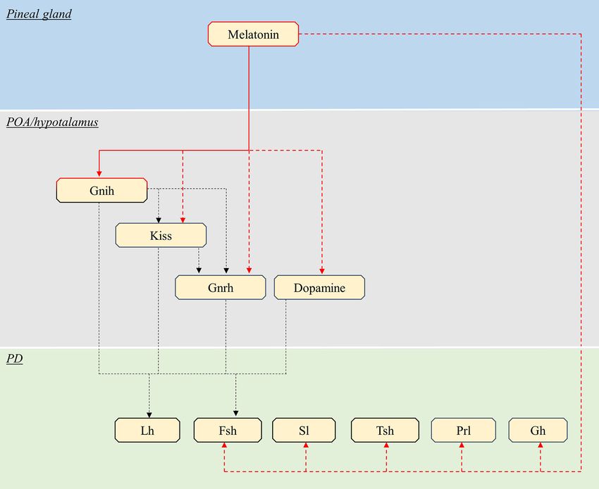
Ciani et al.                                                                                                                          Melatonin Effects on the Pituitary

  FIGURE 1 | Schematic representation of daily and seasonal fluctuation in plasma melatonin levels.

neuroendocrine cells in the preoptic area (POA) and the                               signaling centers in the brain, mainly the POA and hypothalamus,
hypothalamus of the brain, which are considered today as two                          and from peripheral organs, which provide positive and negative
distinct regions (37). The adenohypophysis originates from an up-                     feedback to these centers and to the pituitary (41, 42). In mammals,
growth of the pharyngeal ectoderm and endoderm (38) and can                           POA/hypothalamic neurons project to the median eminence (ME) of
be histologically divided in the pars intermedia (PI), the pars                       the hypothalamus, releasing their hormones into the hypophysial
distalis (PD), and the pars tuberalis (PT), the latter present in                     portal system where they are transported via the blood stream to the
mammals but not in teleosts. The adenohypophysis hosts several                        pituitary endocrine cells (42). Teleosts, on the other hand, lack the
hormone-producing cell types: gonadotropes (producing the                             hypophysial portal system, and instead the POA/hypothalamic
gonadotropins: follicle-stimulating and luteinizing hormones,                         neurons innervate the pituitary, releasing their neurohormones
FSH and LH), lactotropes (prolactin, PRL), somatotropes (growth                       directly at target cells or into pituitary blood vessels (41,
hormone, GH), thyrotropes (thyrotropin, TSH), corticotropes                           43). Pituitary hormonal production is regulated through
(adrenocorticotropin, ACTH), and melanotropes (melanocyte-                            both modulation of the activity of individual cells, and
stimulating hormone, MSH) (39). Teleosts also possess one                             regional reorganizations of the anterior pituitary in terms of
additional cell type, the somatolactotropes responsible for the                       structure or cell composition, as discussed previously for
production of somatolactin (Sl) (40).                                                 gonadotropes (38, 44).
    The activity of pituitary endocrine cells is constantly changing                      While pituitary plasticity is influenced by environmental
over time, adjusting the hormonal production to changing                              factors, the role that melatonin plays in translating fluctuations
physiological needs. It is controlled by factors produced from                        of environmental conditions into pituitary hormonal production

  FIGURE 2 | Schema of the pituitary in mammals and teleosts. The pituitary is composed of two main parts: the neurohypophysis (posterior pituitary) and the
  adenohypophysis (anterior pituitary). The neurohypophysis is mainly composed of neuron terminals from neuroendocrine cells with cell soma located in the preoptic-
  hypothalamic region of the brain. The adenohypophysis contains different hormones producing cell types and can be anatomically divided in pars distalis, pars
  intermedia and, in mammals but not in teleosts, pars tuberalis.

Frontiers in Endocrinology | www.frontiersin.org                                  3                                       January 2021 | Volume 11 | Article 605111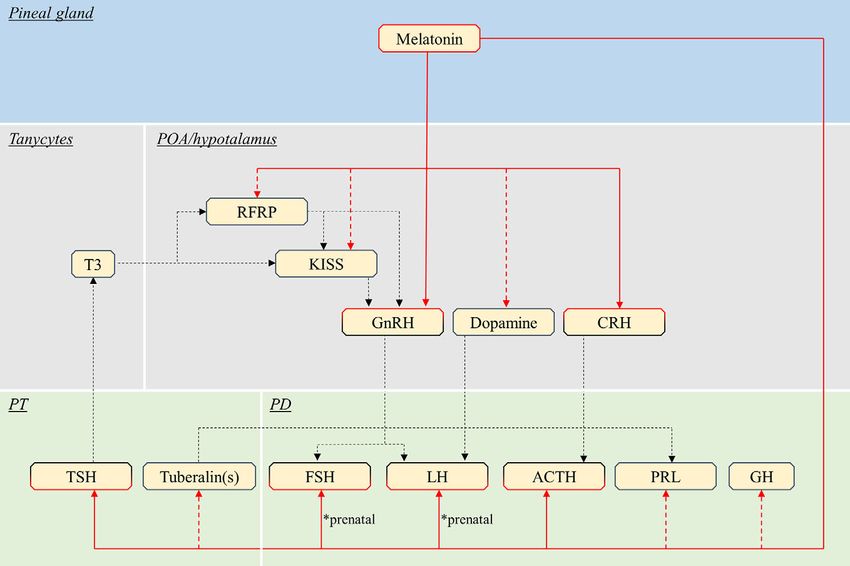
Ciani et al.                                                                                                                             Melatonin Effects on the Pituitary

is not always clear. In addition, the mechanisms of melatonin                           Gonadotropes
action are complex, as both direct effects on pituitary endocrine                       Gonadotropes are the most investigated pituitary cell type in
cells and indirect effects through neuro/hormonal signaling                             relation to melatonin, due to the high scientific and economic
centers combine to regulate pituitary activity. In this review, we                      interest around the seasonal control of reproduction. Indeed,
describe the known pathways through which melatonin modulates                           gonadotropes produce the two gonadotropins (FSH and LH),
anterior pituitary hormonal production, distinguishing between                          key hormones in the control of reproduction, which are
indirect effects mediated by brain centers and direct effects on the                    heterodimeric glycoproteins composed of a common a-subunit
anterior pituitary. We also highlight similar and divergent features                    (GPHa, also shared with TSH) and a hormone-specific b subunit
between teleosts and mammals, and emphasize important                                   (LHb or FSHb) conferring the specific biological activity (42).
unsolved questions for future research.
                                                                                        GnRH
                                                                                        Mammalian gonadotropin-releasing hormone (GnRH1 or
                                                                                        mGnRH-I), a 10-amino acid neuropeptide produced from POA/
BRAIN-MEDIATED EFFECTS OF                                                               hypothalamic neurons, is the main regulator of gonadotropin
MELATONIN ON ANTERIOR PITUITARY                                                         synthesis and secretion (42). Most mammals also possess a
ENDOCRINE CELLS                                                                         second form (GnRH2 or cGnRH-II), expressed in the midbrain
                                                                                        and other organs, which is primarily involved in other functions
Mammals                                                                                 than regulating gonadotropin release. Vertebrates also possess two
Endocrine pituitary cells are primarily controlled by brain                             major types of GnRH receptors (type I with the GnRHR1a and II
signaling centers, mainly the preoptic and hypothalamic area                            with the GnRHR2c) (68), however in many mammalian species,
(42) (Figure 3, Table 1), which integrate nervous and hormonal                          GnRHR2c receptor is not functional (69).
signals of different origins. These brain regions are characterized                        Melatonin influences GnRH production and thus the
by the presence of numerous melatonin binding sites as shown in                         reproductive axis in seasonal breeders. Melatonin administration
rodents and ruminants (60–66). Although the suprachiasmatic                             and short photoperiod (SP) cause testicular regression in the male
nucleus (SCN) of the hypothalamus drives the rhythmic                                   summer breeder jerboa (Jaculus orientalis), a desert hibernating
production of melatonin in mammals (67), the present review                             rodent, by inhibiting GnRH release (47). In contrast, melatonin
will focus on known effects of melatonin on brain centers directly                      administration in the ewe (Ovis aries, a winter breeder) increases the
regulating pituitary endocrine production, as discussed below.                          pulsatile GnRH secretion from hypothalamus, and pituitary LH

  FIGURE 3 | Schematic view of the putative pathways through which melatonin influence pituitary endocrine activity in mammals. Red continuous lines indicate cell
  types directly targeted from melatonin. Dashed red lines indicate cells influenced by melatonin via yet unidentified interneurons, paracrine signals or MTNR. Note that
  melatonin might act only on a few of the illustrated pathway, in different species (see text). Black dashed lines indicate all other interactions between brain and
  pituitary. POA, preoptic area; PT, pars tuberalis; PD, pars distalis; T3, triiodothyronine; RFRP, RFamide-related peptide; KISS, kisspeptin; GnRH, gonadotropin-
  releasing hormone; CRH, corticotropin-releasing hormone; TSH, thyroid-stimulating hormone; FSH, follicle-stimulating hormone; LH, luteinising hormone; ACTH,
  adrenocorticotropic hormone; PRL, prolactin; GH, growth hormone.

Frontiers in Endocrinology | www.frontiersin.org                                    4                                       January 2021 | Volume 11 | Article 605111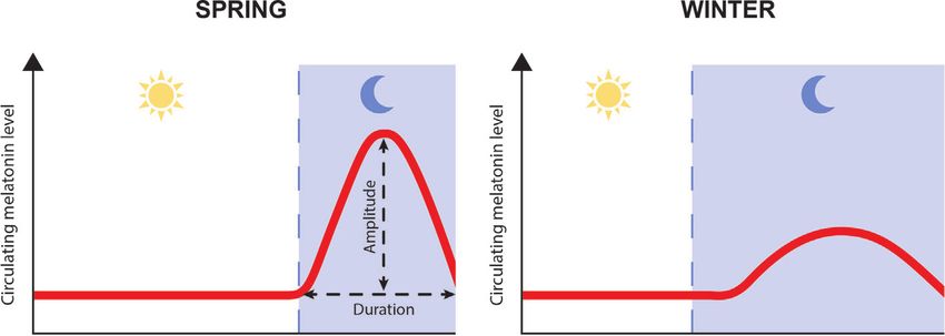
Ciani et al.                                                                                                                          Melatonin Effects on the Pituitary

TABLE 1 | Summary of the known effects of melatonin POA/hypothalamic neurons controlling pituitary hormonal production in mammals.

Target         Effect of Species                   Breeding       Description                                                                      Reference
               melatonin                           season/
                                                   Photoperiod

Mammals
GnRH    Stimulates         Sheep                   Winter/SP      Melatonin administration increases GnRH secretion                                Bittman et al. (45)
                                                                                                                                                   Viguié et al. (46)
               Inhibits    Jerboa                  Summer/LP      Short photoperiod and melatonin administration downregulate GnRH release         El Qandil et al. (47)
               Inhibits    GT1-7 mouse GnRH                       Melatonin reduces GnRH mRNA and protein levels in GT1-7 cell line                Roy et al. (48)
                           cell line
               Modulates   Rat                     non-seasonal   Melatonin augments/reduces GABA-induced currents in GnRH neurons in a            Sato et al. (49)
                                                   breeder        sex dependent way
KISS           Inhibits    Syrian hamster          Summer/LP      Melatonin reduces KISS1 mRNA                                                     Revel et al. (50)
                                                                                                                                                   Ansel et al. (51)
                           Turkish hamster         Summer/LP      Melatonin reduces KISS1 mRNA                                                     Piekarski et al. (52)
                           Striped hamster         Summer/LP      Melatonin reduces KISS1 mRNA                                                     Li et al. (53)
                           Rat                     non-seasonal   Melatonin reduces KISS1 mRNA                                                     Oliveira et al. (54)
                                                   breeder
RFRP           Inhibits    Syrian hamster          Summer/LP      Melatonin (and SP) reduces RFRP-3 mRNA and protein                               Mason et al. (55)
                                                                                                                                                   Revel et al. (56)
                           Siberian hamster        Summer/LP      Melatonin (and SP) reduces RFRP-3 mRNA and protein                               Ubuka et al. (57)
                                                                                                                                                   Revel et al. (56)
Dopamine Stimulates        Syrian hamster          Summer/LP      Melatonin administration stimulates tyrosine hydroxylase activity in males       Alexiuk et al. (58)
         Inhibits          Sheep                   Winter/SP      Melatonin implants inhibit tyrosine hydroxylase activity                         Viguié et al. (59)

secretion (45, 46). While suggesting a connection between melatonin                 stimulates GnRH and gonadotropin secretion in males (73). In male
level and GnRH production, these in vivo experiments do not reveal                  Siberian hamster (Phodopus sungorus), RFRP3 directly injected into
whether melatonin acts directly on GnRH neurons, indirectly via                     the third ventricle inhibits LH release when applied under LP, but
interneurons or through a combination of both. However, in vitro                    has excitatory effects under SP (57), suggesting that melatonin
experiments using the GT1-7 mouse hypothalamic GnRH cell line                       might differentially influence the activity of RFRP3 neurons over
reveal that GnRH neurons express MTNR1A (MT1) and MTNR1B                            the seasons.
(MT2) (48) and demonstrate that melatonin inhibits both GnRH                           In summer breeders, such as Siberian and Syrian hamster, both
mRNA expression and protein secretion (48, 70).                                     SP and melatonin injection reduces RFRP3 protein and mRNA
    Melatonin action might be modulated in a sexually dimorphic                     levels, as well as decrease RFRP3 fibre density and number of
way in rodents as higher mRNA levels of Mtnr1a (MT1) are                            projections to GnRH neurons (55–57). These studies also show that
detected in male than female rat (Rattus norvegicus) GnRH                           pinealectomy abolishes the effects of photoperiod manipulation,
neurons, while Mtnr1b (MT2) is not expressed in either sex (71).                    while subsequent melatonin exposure re-establishes them. While
Sexual dimorphism of the melatonin response in GnRH neurons is                      sex steroids are known to induce positive or negative feedback on
supported by another in vitro study, where melatonin augmented                      hypothalamic signaling centers, the SP-induced reduction in
the membrane current induced by gamma-aminobutyric acid                             RFRP3 protein and mRNA levels observed in male hamsters is
(GABAA) in 70% and attenuated it in 18% of neurons from adult                       not a consequence of reduced circulating steroid levels, since neither
males, while it augmented the current in only 25% and attenuated it                 castration nor testosterone implants alter RFRP3 synthesis. These
in 61% of the neurons from adult females (49). Nevertheless, the                    data therefore strongly suggest that melatonin is responsible for the
physiological relevance of the direct actions of melatonin on GnRH                  inhibition of RFRP3.
neuron activity in vivo remains controversial, as melatonin might                      In winter breeders, such as sheep, SP decreases both RFRP3
additionally act on upstream signals, such as KISS1, RFRP3 and T3,                  mRNA and protein levels, and RFRP3 neuron projections to GnRH
as discussed below.                                                                 neurons (74, 75). Similarly, in brushtail possum (Trichosurus
                                                                                    vulpecula) females, the number of RFRP3 neurons decreases
RFRP3 (GnIH)                                                                        during winter (76). Interestingly, in the laboratory Wistar rat, a
RFamide related peptide3 (RFRP3) is the mammalian orthologue of                     non-photoperiodic breeder, no effect of photoperiod manipulation
avian GnIH, which was originally identified in birds as an inhibitory                was detected on RFRP3 neurons (56). These results suggest that the
factor of gonadotropin synthesis and release, by acting on both                     photoperiodic control of melatonin on RFRP3 is conserved among
GnRH neurons and gonadotropes. RFRP3 neurons are located in                         mammals, with inhibiting effects in both summer and winter
the paraventricular nucleus of the hypothalamus (42). Interestingly,                breeders, while the downstream effects of the RFRP3 system on
the effects of RFRP3 on gonadotropin synthesis are deeply                           gonadotropin secretion might diverge to adapt to long-day or short-
influenced by sex and timing of administration in mammals. For                       day breeding strategies.
instance, in Syrian hamsters (Mesocricetus auratus), RFRP3 inhibits                    Whether melatonin acts directly on RFRP cells in mammals
gonadotropin secretion in ovariectomized females (72) while it                      requires further investigations as there is still a lack of evidence for

Frontiers in Endocrinology | www.frontiersin.org                                5                                        January 2021 | Volume 11 | Article 605111
Ciani et al. Melatonin Effects on the Pituitary
colocalization with MTNR, or studies clearly demonstrating a direct (promoting the release of cortisol from the adrenal gland) and
action of melatonin on RFRP3 neurons, as previously discussed by the control of numerous daily and seasonal physiological
Kriegsfeld and collaborators (77). rhythms (including sleep) (83). Corticotropes are mainly
regulated by corticotropin releasing hormone (CRH) neurons
Kisspeptin
located in the paraventricular nucleus. Melatonin exerts a stress-
KISS neurons produce the neuropeptide kisspeptin (KISS) and
protective effect in mammals (84, 85). Daily melatonin
stimulate GnRH synthesis and secretion, thereby regulating
administration reduces the ACTH secretory response to acute
gonadotrope cell activity (78). Located in two discrete
and chronic stress in rat (86, 87). In humans (Homo sapiens),
hypothalamic nuclei, the arcuate nucleus (ARC) in all mammals
oral melatonin administration in blind individuals normalizes
and the anteroventral periventricular area around the 3rd ventricle in
the temporal pattern of ACTH and cortisol plasma
rodents or the POA in non-rodent mammals, the activity and
concentrations during sleep, suppressing the pituitary-adrenal
number of KISS neurons display a marked photoperiodic/seasonal
activity during early sleep and activating it during late sleep (88).
pattern, as shown below.
Melatonin might modulate ACTH production by acting directly
In the winter breeding sheep, SP upregulates both ARC KISS1
on hypothalamic CRH neurons, which express the MTNR1A in
mRNA and protein, and increases the number of both ARC KISS
humans (89).
neurons and synaptic connections from KISS to GnRH neurons
Lactotrope cells produce PRL, a peptide hormone involved in
(75). In contrast, melatonin inhibits the activity of KISS neurons in
reproduction (lactation), moulting, metabolism, and immune
summer breeders. For instance, using a combination of
responses. PRL secretion is stimulated by releasing factors from
photoperiod manipulation, pinealectomy and melatonin
the PT and inhibited by dopamine secreted by tubero-
administration Revel and colleagues (50) and Ansel and
infindibular dopaminergic neurons located in the dorsomedial
colleagues (51) demonstrated that melatonin clearly reduces ARC
arcuate nucleus (90–92). Exogenous melatonin administration
KISS1 mRNA in Syrian hamsters, an effect further modulated by the
and SP decrease the PRL secretion in ruminants. For instance,
negative steroid feedback. Similar inhibitory effects of melatonin on
oral melatonin administration inhibits PRL secretion in lactating
KISS1 mRNA were detected in Turkish (Mesocricetus brandti) (52)
ewes (93). SP reduces PRL secretion in cows [Bos taurus (94)],
and striped (Cricetulus barabensis) (53) hamsters but also in the rat,
while melatonin oral administration reduces PRL release in both
a non-seasonal breeder (54). Interestingly, in the Siberian hamster,
prepubertal (95) and mature (96) cows. The pathway involved in
ARC KISS1 mRNA levels are lower under LP due to a robust
the melatonin-mediated PRL inhibition seems to be mediated
negative sex steroid feedback overriding the melatonin signal, since
through a dopamine-independent mechanism, since melatonin
castration in LP animals restores high KISS1 mRNA levels (79).
administration inhibits PRL release even in rams where the
Therefore, the role of melatonin among different species, or
hypothalamo-pituitary connection has been surgically
different reproductive stages, might be difficult to identify
disrupted (97, 98) and melatonin implants reduce PRL
considering the impact of steroid feedbacks on ARC KISS neurons.
secretion without altering dopamine content in ewe (59).
Although a direct effect of melatonin on KISS1 mRNA levels
Despite the involvement of both somatotropes and thyrotropes
was detected in a hypothalamic cell line from rat (80), Mtnr
in seasonal physiological activity, there is no clearly established role
expression has not been found in sheep KISS neurons, neither
for melatonin signaling to their POA/hypothalamic regulators.
during the breeding nor during the non-breeding season (81).
Somatotrope cells produce GH, a peptide hormone involved in
These results suggest that the effects of melatonin on KISS
numerous physiological processes including growth, metabolism
neurons could be mediated upstream.
and cellular proliferation. The main hypothalamic regulators of
Dopamine somatotropes are growth hormone releasing hormone (GHRH)
The activity of the dopaminergic neurons located within the and somatostatin, which stimulates and inhibits GH production,
POA/hypothalamic area, which are involved in the inhibition of respectively (99). Thyrotrope cells produce TSH, a heterodimeric
gonadotropin synthesis and release (82), also appears to be glycoprotein hormone, composed of an a- (GPHa) and a b-
regulated by melatonin. For instance, in the ewe, a winter (TSHb) subunit, involved in different seasonal physiological
breeder, melatonin implants inhibit the activity of tyrosine functions including reproduction and growth (100). Two distinct
hydroxylase (the rate-limiting enzyme in the dopamine populations of thyrotropes, with distinct morphology and secretory
synthesis pathway) in the median eminence, while stimulating activity are located in the PT and PD (101–103). Thyrotropin-
LH release (59). In contrast, in male Syrian hamsters, in which SP releasing hormone (TRH) produced by hypothalamic neurons is
elicits testicular regression, melatonin administration stimulates the main regulator of PD TSH synthesis (104). By contrast, TRH has
tyrosine hydroxylase activity in the median eminence, increasing no effect on PT thyrotrope activity (105), which is controlled by
dopamine synthesis and release (58). other signals including melatonin, as discussed in section 3 below on
direct effects of melatonin.
Other Endocrine Cells in the Pars Distalis
Melatonin plays a role in the regulation of other pituitary Teleosts
endocrine cell types in mammals by regulating their main In teleosts, endocrine pituitary cells are also mainly controlled by
hypophysiotropic factors. brain signaling centers in the preoptic and hypothalamic areas
Corticotrope cells produce ACTH, a hormone involved in (41), which are characterized by the presence of numerous
various physiological processes including the stress response melatonin binding sites (106–108). The effects of melatonin on
Frontiers in Endocrinology | www.frontiersin.org 6 January 2021 | Volume 11 | Article 605111Ciani et al. Melatonin Effects on the Pituitary
these brain centers, and thus indirectly on pituitary activity, have inconsistent responses of gnrh highlight the importance of the
been studied mainly in the context of reproduction in teleosts, experimental conditions, and suggest the presence of different
such as in the salmon (109) and eel (110) where melatonin has regulatory mechanisms activated by melatonin exposure and
been shown to play an important role in puberty. Therefore, the photoperiod manipulation, as mentioned in the introduction.
available knowledge and thus the discussion in the present In other teleost species, melatonin inhibits gonadotropin
review, only concern gonadotropes (Figure 4, Table 2). production by downregulating Gnrh expression. In Nile tilapia
(Oreochromis niloticus), whose development and reproduction are
Gonadotropes suppressed under SP (125, 126), intraperitoneal melatonin injections
In contrast to mammals, the teleost gonadotropes mainly produce simultaneously reduce gnrh1 mRNA in the brain and both lhb and
Lh and Fsh in distinct cells, with only a small portion of fshb mRNA in the pituitary (112). Several studies were performed in
gonadotropes producing both hormones in some species (43). male sea bass (Dicentrarchus labrax). Both intraperitoneal melatonin
However, like in mammals, pituitary gonadotropin synthesis and injections (114) and melatonin implants (115) downregulate brain
release are regulated by POA/hypothalamic signaling centers, mRNA levels of the two hypophysiotropic forms of Gnrh, gnrh1 and
including Gnrh, Gnih, Kiss, and dopamine neurons (41, 123, 124). gnrh3. Remarkably, these genes show natural daily variations in
mRNA levels, with lower levels during the mid-dark phase, when
Gnrh plasma melatonin is highest (114). Melatonin implants also decrease
Fish possess up to three genes encoding Gnrh (gnrh1, gnrh2, gnrh3) pituitary mRNA levels of Gnrh receptors (named gnrhr2ba1 and
(16, 41). In some teleost species, melatonin stimulates gonadotropin gnrhr1cb, according to recent phylogeny, (68) but named gnrhr-II-1a
production by upregulating Gnrh expression. For instance, in adult and gnrhr-II-2b, respectively, in the study), as well as fshb (115).
female zebrafish (Danio rerio), melatonin exposure via immersion Interestingly, pituitary Gnrh1 protein content shows daily variation
increases the mRNA levels of brain gnrh3 and pituitary lhb (111). In with minimum levels during night time, under both natural
a second study on adult zebrafish females, brain gnrh3 mRNA levels photoperiod and LP (127). While downregulating the Gnrh
were increased in both constant light (LL) and constant darkness system, melatonin implants also reduce plasma gonadotropins (Lh
(DD) as compared to normal light-dark cycles (117). The and Fsh) and androgens (testosterone, T and 11-keto-testosterone,
FIGURE 4 | Schematic view of the putative pathways through which melatonin influence pituitary endocrine activity in teleosts. Red continuous lines indicate cell
types directly targeted from melatonin. Dashed red lines indicate cells influenced by melatonin via yet unidentified interneurons, paracrine signals or Mtnr Note that
melatonin might act only on a few of the illustrated pathway, in different species (see text). Black dashed lines indicate all other interactions between brain and
pituitary. POA, preoptic area; PD, pars distalis; Kiss, kisspeptin; Gnrh, gonadotropin-releasing hormone; Lh, luteinising hormone; Fsh, follicle-stimulating hormone;
Sl, somatolactin; Tsh, thyroid-stimulating hormone; PRL, prolactin; GH, growth hormone.
Frontiers in Endocrinology | www.frontiersin.org 7 January 2021 | Volume 11 | Article 605111Ciani et al. Melatonin Effects on the Pituitary
TABLE 2 | Summary of the known effects of melatonin POA/hypothalamic neurons controlling pituitary hormonal production in teleosts.
Target Effect of Species Spawning Description Reference
melatonin season/
Photoperiod
Teleosts
Gnrh Stimulates Zebrafish Spring/LP Melatonin exposure via water upregulates brain gnrh3 expression (adult Carnevali et al. (111)
(Daily in females)
captivity)
Inhibits Nile tilapia Spring/LP Melatonin injections downregulate brain gnrh1 expression Kim et al. (112)
Masu salmon Autumn/SP Oral melatonin administration (50 µg/g feed) decreases Gnrh release Amano et al. (113)
Sea bass Spring/LP Melatonin injections downregulate brain gnrh1 and gnrh3 expression Servili et al. (114)
Melatonin implants downregulate brain gnrh1 and gnrh3 and gnrhr-II-1a -2b Alvarado et al. (115)
None European eel Spring/LP Melatonin implants have no effects on gnrh expression Sé bert et al. (116)
Gnih Stimulates Nile tilapia Spring/LP Melatonin injections upregulate brain gnih expression Kim et al. (112)
Inhibits Zebrafish Spring/LP Melatonin downregulates gnih expression in cultured hypothalamus Yumnamcha et al. (117)
(Daily in
captivity)
Kiss Stimulates Zebrafish Spring/LP Melatonin exposure via water upregulates brain kiss1 and kiss2 expression Carnevali et al. (111)
(Daily in (adult females)
captivity)
Inhibits Sea bass Spring/LP Melatonin implants downregulate brain kiss1 and kiss2 expression Alvarado et al. (115)
Dopamine Inhibits European eel Spring/LP Melatonin implants stimulate brain tyrosine hydroxylase activity Sé bert et al. (116)
Carp Spring/LP Melatonin inhibits dopamine release in cultured hypothalamus Popek (118)
Melatonin injections inhibit brain dopamine release Popek et al. (119)
Asian catfish Spring/LP Melatonin inhibits hypothalamic tyrosine hydroxylase activity during preovulatory Chaube and Joy (120)
phase in female
A higher dose of melatonin has no effect on tyrosine hydroxylase activity Senthilkumaran
and Joy (121)
Rainbow trout Autumn/SP Melatonin decreases hypothalamic-pituitary dopamine turnover Herná ndez-Rauda et al.
(122)
11KT) levels, thus impairing sexual maturation (115). Servili and Gnih
collaborators (128) show that in sea bass, the non-hypophysiotropic Melatonin modulates the activity of Gnih neurons by stimulating or
Gnrh2 neurons send their projections to the pineal gland, and directly inhibiting Gnih expression in different species. In adult zebrafish
stimulate melatonin secretion. Taken together, these results suggest females, exogenous melatonin treatment reduces gnih mRNA levels
that melatonin in male sea bass, downregulates the production of the in cultured whole brain, while DD decreases in vivo brain gnih and
hypophysiotropic Gnrh forms (Gnrh1 and Gnrh3) and their release increases both lhb and fshb mRNA in the pituitary (117). In
in the pituitary. This, combined with the reduction of Gnrh receptors contrast, in Nile tilapia (mixed sex), brain gnih mRNA levels
in the pituitary, result in gonadotropin downregulation. The use of increase during the night, in parallel with plasma melatonin levels
different concentrations of exogenous melatonin can modulate its in mature fish (130). Additionally, intraperitoneal melatonin
effects on Gnrh and gonadotropin content. For example, in injections increase gnih and mtnr1c mRNA in the brain and
underyearling masu salmon (Oncorhynchus masou), oral simultaneously decrease lhb and fshb mRNA in the pituitary.
administration of melatonin (50 µg/g feed) under LP increases Fsh Kim and colleagues (130) suggest that, like in birds, melatonin
and T plasma content but has no effect on Lh (129), suggesting that might act on Gnih neuron activity via Mtnr1c (131). Indeed, in the
mimicking SP by melatonin administration stimulates testicular cinnamon clownfish (Amphiprion melanopus), Gnih neurons
development. However, a 10-fold higher dose (500 µg/g feed) express Mtnr1c (named from the authors MT-R1) (132). However,
decreases pituitary Gnrh and Lh content together with plasma T whether this is a species-specific case, or a general condition for all
levels (113). In contrast, the Gnrh system does not respond to teleosts, remains to be investigated.
melatonin in some teleost species such as European eel (Anguilla
anguilla) where melatonin implants had no effects on brain gnrh1 and Kiss
gnrh2 mRNA levels (116). In teleosts, contrary to in mammals, Kiss neurons directly regulate
The specific pathways through which melatonin affects Gnrh pituitary endocrine cells rather than acting through Gnrh neurons
are largely unknown. In sea bass, the effects of melatonin on (133). Teleosts possess two genes encoding kisspeptins (kiss1, kiss2)
Gnrh neuron activity are most likely mediated via interneurons (16, 134). Melatonin is also involved in the control of kiss expression
(114), since the distribution of melatonin receptors does not in teleosts. In adult female zebrafish, melatonin exposure via
match the distribution of gnrh1 and gnrh3 cells (106). In masu immersion increases mRNA transcript levels of both kiss1 and
salmon, melatonin binding sites were localized in the POA (113), kiss2 in the brain, and lhb in the pituitary (111). While kiss1 does not
however no colocalization study was performed to investigate respond to photoperiod manipulations, kiss2 mRNA is induced
their presence in Gnrh neurons. under LL when melatonin plasma levels are at their minimum
Frontiers in Endocrinology | www.frontiersin.org 8 January 2021 | Volume 11 | Article 605111Ciani et al. Melatonin Effects on the Pituitary
(117). In contrast to in zebrafish, prolonged exposure to melatonin gonadotropes play a crucial role in the control of reproduction,
via implants decreases brain mRNA levels of kiss1 and kiss2 in male which often depends on environmental conditions, it is not
sea bass (115). The heterogeneity of kiss response, as seen for gnrh, surprising that especially their response to indirect melatonin
highlight the influence of experimental conditions and suggest the signaling has been studied extensively.
possible involvement of different pathways influenced by the In mammals, melatonin modulates gonadotropin expression
hormonal and nervous photoperiodic signal. by acting on POA/hypothalamic signaling centers. It
It is not known whether melatonin acts directly on kiss downregulates KISS and GnRH production and stimulate
neurons or operates via interneurons in teleosts. In sea bass, dopaminergic activity in summer breeders, while it upregulates
Kiss1 and Kiss2 immunoreactive neurons were identified in the KISS and GnRH production and inhibits dopaminergic activity
lateral tuberal nucleus and parvocellular nucleus, respectively in winter breeders. Interestingly, melatonin reduces GnIH
(135), two locations that also express melatonin receptors (106). neuronal activity in both summer and winter breeders,
However, a clear colocalization of the melatonin receptors in Kiss indicating downstream differences in the signaling cascade.
neurons has not been demonstrated. In teleost fish, melatonin affects these brain signaling hubs in a
more complex manner, both inhibiting and activating the
Dopamine gonadotrope axis, depending on the species. In some species,
In several teleost species, dopaminergic neurons from the POA exert a
melatonin activates the gonadotrope axis by simultaneously
strong negative control on gonadotropes, especially Lh-producing cells
stimulating the production of Gnrh and Kiss, while inhibiting
(82). Melatonin, in turn, influences the activity of hypophysiotropic
Gnih; in others, melatonin exerts a negative action on
dopaminergic neurons. In European eel melatonin implants stimulate
reproduction. Melatonin may downregulate dopamine
the dopaminergic system in the POA, increasing tyrosine hydroxylase
production resulting in increased gonadotropin synthesis, or it
activity, the rate-limiting enzyme of dopamine synthesis (116). As a
can stimulate the dopaminergic system and inhibit gonadotropin
consequence, this treatment downregulates both lhb and fshb mRNA
production. Nevertheless, in both teleosts and mammals, a clear
levels. In contrast to the eel, melatonin inhibits the dopaminergic
picture of the cell types directly targeted by melatonin in the brain is
system in other fish species. For instance, in mature female carp
still scarce. In light of such opposing forces, it is urgent to identify
(Cyprinus carpio), melatonin decreases dopamine release, both in in
the specific cell types targeted by melatonin in both mammals and
vitro cultured hypothalamus (118) and in vivo by direct injection into
teleosts, by determining which ones express MTNR. This is a
the third cerebral ventricle (119). The in vivo inhibition of dopamine
requirement before being able to fully elucidate the mechanisms
release was registered during the spawning period in summer, but not
involved in the integration of environmental signals in the brain
during sexual regression in winter, suggesting that the effect of
neuroendocrine centers.
melatonin on the dopaminergic system might depend of the
maturation stage. Inhibiting effects of melatonin on the
dopaminergic systems were also observed in other species, including
the threespot wrasse (Halichoeres trimaculatus), where intraperitoneal DIRECT EFFECTS OF MELATONIN ON
melatonin injections downregulate brain dopamine content (136), and THE ANTERIOR PITUITARY
rainbow trout (Oncorhynchus mykiss), where melatonin decreases the
hypothalamic-pituitary dopamine turnover (122). In preovulatory Mammals
female catfish (Heteropneustes fossilis), melatonin injections for three In addition to the effects mediated by the brain, melatonin can act
days inhibit tyrosine hydroxylase enzymatic activity in the directly on the pituitary gland in mammals (Figure 3, Table 3).
hypothalamus (120). However, in a previous study using the same
species, reproductive phase and melatonin injection dose, but Melatonin Receptors in Mammalian Pituitary
administrated over a longer period (20 days), failed to affect The main target for melatonin within the mammalian pituitary is
hypothalamic dopamine turnover (121). This indicates that the the PT, as indicated by the important presence of melatonin binding
length of the treatment with melatonin might affect the response of sites in all mammalian species investigated so far. Those include, for
the dopaminergic system. instance, Siberian hamster (154, 155); Syrian hamster, (154–156);
Melatonin binding and mtnr mRNA distribution studies in rat, (154, 155, 157, 158); red deer (Cervus elaphus) (159); ferret
rainbow trout (108) indicate that it is very unlikely that Mtnr is (Mustela putorius furo) (160), rhesus monkeys (Macaca mulatta)
present on hypophysiotropic dopaminergic neurons. Therefore, the (161) and human (89). MTNR1A (MT1) is the main form of
effects of melatonin might be mediated by interneurons in this melatonin receptor present in the PT. Mtnr1a mRNA was
species. Studies in Atlantic salmon (137) goldfish (Carassius detected by in situ hybridization in the PT of sheep, Siberian
auratus) (138) and Japanese catfish (Silurus asotus) (139), identify hamster and rat (154) and in primary PT cell cultures of sheep
melatonin binding sites in the POA, where hypophysiotropic (162). MTNR1A has also been detected in human PT, via immuno-
dopaminergic neurones are located (82); however, a clear staining (89). Identification of the specific cell types expressing
colocalization has not been demonstrated. melatonin receptors is a key requisite to discriminate the direct
effects mediated by melatonin. Double labelling combining in situ
Summary hybridization and immunohistochemistry shows that Mtnr1a is
Melatonin affects pituitary hormonal production in both expressed in most, but not all, thyrotrope cells within the PT, while it
mammals and teleosts by regulating upstream brain factors. As is absent from the other endocrine cells types in the anterior
Frontiers in Endocrinology | www.frontiersin.org 9 January 2021 | Volume 11 | Article 605111Ciani et al. Melatonin Effects on the Pituitary
TABLE 3 | Summary of the known effects of melatonin on pituitary in vitro and ex vivo cultures in mammals.
Species Type of preparation Effects of melatonin Reference
Mammals
Sheep PT cell culture Acute: inhibits forskolin-induced secretion of tuberalin Morgan et al. (140)
PT cell culture Acute: inhibits forskolin-induced cAMP Hazlerigg et al. (141)
PT cell culture Prolonged: increase basal and forskolin-induced cAMP Hazlerigg et al. (141)
PT cell culture Reduces Mtnr1a mRNA Barret et al. (142)
PT explants Reduces Mtnr1a mRNA Fustin et al. (143)
PT explants and cell culture Reduces Egr1 expression Fustin et al. (143)
Rat (neonatal) PD organ cultures Inhibits LH and FSH release Martin and Klein (144)
Martin and Sattler (145)
PD cell culture Inhibits GnRH-induced Ca2+ signal and LH secretion Vaně č ek and Klein (146)
PD cell culture Inhibits GnRH-induced cFOS Sumova et al. (147)
Organ cultures No effect on TRH-induced TSH/PRL or SRIF-induced inhibition of GH Martin and Klein (144)
Martin and Sattler (145)
Rat (maturing) PD cell culture No effect on GnRH-response Rivest et al. (148)
Rat (Adult) TP/ME explants Inhibits Lh release Nakazawa et al. (149)
GH3/GH4 cell line Inhibits secretion of PRL and GH (no effect on cAMP) Griffiths et al. (150)
GH3 cell line Inhibits basal and forskolin-induced PRL secretion and expression Ogura-Ochi et al. (151)
Baboon PD cell culture No effect on LH or FSH secretion Ibá ñez-Costa et al. (152)
Baboon PD cell culture Stimulates GH and PRL expression and release Ibá ñez-Costa et al. (152)
PD cell culture Increases expression of receptors for GhRH and ghrelin, decreases receptors for somatostatin Ibá ñez-Costa et al. (152)
PD cell culture No effect on ACTH or TSH Ibá ñez-Costa et al. (152)
Mouse ATt20 cell line Inhibits cAMP and ACTH release Tsukamoto et al. (153)
pituitary in European hamster (Cricetus cricetus) (163) and rat 172), red deer (159) and ferret (160). A weak staining of
(157). This suggests that thyrotropes of the PT are the main pituitary MTNR1A was detected in human PD by in situ hybridization
target of melatonin action in mammalian species. (89). To date, the identity of the cellular targets of melatonin
Interestingly, the expression of Mtnr within the PT varies within the PD and their contribution to the hormonal regulation
across the year, in response to different neuroendocrine factors of the gland remain largely unknown.
(34), modulating the endocrine response to melatonin between
the different seasons. For instance, Mtnr expression increases Effects of Melatonin on the Pars Tuberalis
during the reproductive season, peaking under LP in summer As mentioned above, melatonin pituitary binding primarily takes
breeders or SP in winter breeders. Simulating winter season in place in the PT. Through these cells, melatonin seems to regulate
the summer breeder Syrian hamster, using artificial SP (164) or the PD activity via at least two different routes, a retrograde route
melatonin injections (156, 165) induces a marked decrease in from the PT to the brain (Figure 5) and an anterograde route
MTNR density in the PT of Syrian hamster. Melatonin injections from the PT to the PD, as discussed below.
decrease MTNR1A density in both Syrian and Siberian hamster
PT (156). Similar effects of SP were shown in European hamster Retrograde Route
(163, 166) and hedgehog, Erinaceus europaeus (167). On the Studies in rodents demonstrate the inhibitory effects of melatonin
contrary, in winter breeders such as the mink (Mustela vison), on PT-TSH synthesis (173) via MTNR1A (174). During the long
the density of melatonin binding sites in the PT plummets in July days in spring/summer, PT-TSH rises independently from TRH
and peaked in October, in concomitance with reactivation of stimulation (175) due to reduced circulating melatonin levels. In the
sexual activity (168). retrograde route (Figure 5), the expression of PT-TSH is rapidly
Melatonin and the pituitary also regulate daily physiological induced, after LP exposure, by the transcription factor EYA3, which
rhythms. As a consequence, variations in the presence of MTNR works with the circadian transcription factor thyrotroph embryonic
within the PT are not limited to annual cycles, but also occur factor (TEF) (176, 177). Melatonin acutely inhibits Eya3 expression,
within the span of a single day. Mtnr1a mRNA levels vary during but at the same time induces a peak of Eya3 12 h later. This leads to a
the day in the PT of Siberian and Syrian hamster, peaking during strong morning peak of Eya3 (and subsequently TSH) during long
daytime and decreasing at night (156). days. Although several other collaborating transcription factors are
Finally, in the PD of foetal rat, Mtnr1a mRNA was also involved in the precise circannual regulation of TSH secretion from
identified by in situ hybridization before activation of the GnRH PT, EYA3 seems to be the one regulated by photoperiod
system, but not in postnatal stages (169). Bae and colleagues via melatonin.
(170) demonstrate that Mtnr1a transcripts are present in the PT-TSH then reaches the brain where it binds to its receptors in
mice aT3-1 gonadotrope cell line, but their expression is tanycytes, specialized ependymal cells within the mediobasal
downregulated after exposure to GnRH. The activation of the hypothalamus, thus regulating the enzymatic activity of local
GnRH system in postnatal stages might therefore be responsible deiodinases (Dio2-Dio3) (173, 178). This controls local thyroid
for the lack of MTNR in the adult PD reported in rat. Melatonin hormone (TH) metabolism by converting thyroxine (T4) to the
binding sites were also detected in the PD of adult sheep (171, bioactive triiodothyroxine (T3), serving as key regulator of
Frontiers in Endocrinology | www.frontiersin.org 10 January 2021 | Volume 11 | Article 605111Ciani et al. Melatonin Effects on the Pituitary
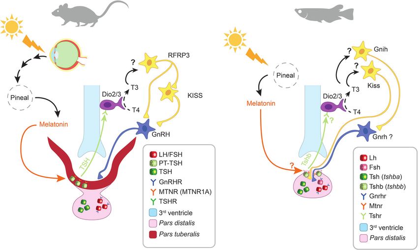
FIGURE 5 | Melatonin-induced retrograde signaling in mammals and teleosts. Thyrotrope and gonadotrope cells are respectively represented as green and red squares
(see legend). Question marks (?) indicate putative pathways not yet demonstrated. In mammals, the photic signal perceived from the retina reaches the pineal gland after
being processed from different brain centers (including the suprachiasmatic nucleus, SCN) thus regulating the rhythmic release of melatonin at night. Circulating melatonin
acts on pars tuberalis (PT) thyrotropes (PT-TSH) via MTRN1A, thus inhibiting PT-TSH release. In spring, when melatonin levels decrease, PT-TSH secretion is stimulated. PT-
TSH, guided by tissue specific glycosylation, binds on its receptors on tanycytes located in the third ventricle of the hypothalamus. Here, PT-TSH regulates local deiodinases
(Dio2/Dio3) influencing thyroid hormone metabolism by promoting the conversion of T4 into the bioactive T3. T3 in turn activates arcuate nucleus (ARC) kisspeptin (KISS)
neurons via a still unknown mechanism. The following increase in gonadotropin releasing hormone (GnRH) release, stimulates gonadotropes activity in the pars distalis (PD).
In teleosts the photic signal is directly perceived from photoreceptive structures within the pineal gland, thus regulating the rhythmic release of melatonin at night. Recent
studies suggest that melatonin might regulate the release of a retrograde signal from the pituitary also in teleosts. A distinct population of thyrotrope cells (expressing a
second Tsh paralogue, tshbb) located near the pituitary stalk, drastically increase tshbb expression under long photoperiod, a similar response to the one occurring in
mammalian PT-TSH. Although melatonin receptors have been described in teleost pituitary and found to display daily and seasonal regulation, their presence in this
thyrotrope population as well as the inhibition of Tsh synthesis and release in response to melatonin, remain to be demonstrated.
seasonality (173). Ikegami and collaborators (175) demonstrate that the enzyme catalysing the conversion of ATP to cyclic AMP
specific post-translational glycosylations allow PT-TSH to (cAMP). The forskolin-induced secretion of tuberalin from PT
exclusively target the hypothalamus, and not the thyroid. While cells was assessed by adding medium from the PT culture to a PD
the cellular and molecular targets remain to be clearly identified, culture and measuring the amount of PRL secreted in response.
recent findings indicate that the increase of T3 in the mediobasal Melatonin acutely inhibited the forskolin-induced secretion of
hypothalamus acts on KISS1 and RFRP3 neurons, which in turn tuberalin but had no effect alone. In support, melatonin inhibited
modulate GnRH secretion (78). The molecular pathway from the forskolin-induced cAMP production in ovine PT cells (141).
melatonin to T3 production appears to be conserved in Furthermore, melatonin downregulated the expression of its own
mammalian species regardless of their reproductive strategy as receptor in PT cells from rat (183) and sheep (142). In ovine PT
summer or winter breeders (67, 179). Therefore, species-specific cells, the downregulation of Mtnr expression involves the cAMP
differences might occur downstream of this common pathway. signaling pathway (143). Together, these results imply that
melatonin works through the MTNR/Gi/cAMP pathway to
Anterograde Route inhibit tuberalin secretion and subsequently regulating PRL
In the anterograde route, melatonin regulates PRL synthesis and production in the PD. Interestingly, incubation with melatonin
secretion in the PD by inhibiting the release from the PT of one for 16 h sensitizes AC, increasing both basal and forskolin-
or more PRL-releasing factors named “tuberalin” (67). To date, induced cAMP production (141). After the prolonged melatonin
the PT-specific factor(s) are still undetermined, as more than 30 exposure, acute application of melatonin still inhibits the
different factors are known to stimulate PRL secretion (92). forskolin-induced cAMP increase.
Several candidates have been proposed including tachykinin-1 For melatonin to inhibit secretion of tuberalin, there must be a
and neurokinin A in sheep (180) or endocannabinoids in stimulating factor that melatonin can oppose. This “tuberalin
hamster (179, 181). Notably, these factors might act through releasing factor” has not been identified but was named StimX by
folliculo-stellate cells to regulate lactotropes (182). Morgan and Williams (184). Dardente and colleagues (67)
Tuberalin secretion can indeed be stimulated in ovine PT cell proposed dopamine as a promising candidate for StimX, arguing
cultures by forskolin (140), an activator of adenylyl cyclase (AC), that it might act through the dopamine receptor D1 expressed in PT
Frontiers in Endocrinology | www.frontiersin.org 11 January 2021 | Volume 11 | Article 605111Ciani et al. Melatonin Effects on the Pituitary
cells, whose activation increases the intracellular cAMP level effect on the pituitary. Rivest and collaborators (148) found that
in neurons. melatonin incubation (5 nM) of pituitary cell cultures from
Downstream of cAMP, melatonin up-regulates or down- sexually maturing rats does not modify the GnRH response.
regulates the expression of a range of genes (67). Several of them Likewise, Ibá ñez-Costa and colleagues (152) found no effect of
are clock genes, including Period1 (Per1) and Cryptochrome-1 melatonin (pM to µM range) on the FSH and LH secretion in
(Cry1) (185). This implies that PT cell activity might be regulated by primary pituitary cultures from adult female baboons.
an internal clock and that the clock itself may be modulated by
melatonin. Interestingly, the same clock genes are not affected
Other Endocrine Cells in the pars distalis
Regarding the effects of melatonin on other PD endocrine cells, the
by melatonin in the suprachiasmatic nucleus (SCN) of the
results are even scarcer. Melatonin (10-8 to 10-6 M) reduces the
hypothalamus, indicating that the SCN clock is working more
production and secretion of both PRL and GH from the rat pituitary
independently than peripheral clocks (186–188). In ovine PT cell
cell line GH4C1, but has no effect on basal or stimulated cAMP
cultures and explants, the expression of the immediate early gene
levels (150). Similarly, Ogura-Ochi and collaborators (151) show
Egr1 is acutely suppressed by melatonin, which otherwise follows a
that melatonin suppresses both basal and forskolin-induced PRL
daily rhythm (143). EGR1 in turn regulates several genes, some
secretion and mRNA abundance in the closely related GH3 cell line.
being upregulated, such as Cry1, others downregulated such as
In contrast, in primary pituitary cell cultures from adult female
Mrnt1a. In contrast to Mtnr1a, the expression of Cry1 was not
baboons, melatonin increases GH and PRL expression and release
affected by changes in cAMP levels.
in a dose-dependent manner, an effect blocked by somatostatin
Other PT endocrine cells beside thyrotropes might also be
(152). Both the common (AC/PKA/Ca-channels) and distinct
regulated by melatonin. Nakazawa and collaborators (149) found
(PLC/Ca-release) pathways seem to be involved. Melatonin (10
that melatonin inhibits LH release from male rat tissue explants
nM) also affects the expression of GHRH receptors, ghrelin and
(consisting of PT and median eminence) in a dose-dependent
somatostatin, but not expression or release of ACTH or TSH. Also
manner. This in turn increases the release of GnRH from the
in pituitary organ cultures from neonatal rats, melatonin has no
median eminence part of the explant.
effect on TRH-induced TSH/PRL release or somatostatin-induced
Effects of Melatonin on the pars distalis inhibition of GH release (144, 145). In the mice corticotrope cell line
Gonadotropes AtT20, melatonin reduces the levels of ACTH, alongside a
In rodents, the effects on PD gonadotropes seem highly age-specific, reduction in cAMP (153).
with clear inhibitory effects in neonatal animals and no effects in
adults. Melatonin (1–10 nM) reduces the GnRH-induced LH and Teleosts
FSH release in pituitary organ cultures from neonatal rats (144, As in mammals, melatonin can also act directly on the pituitary
145). Furthermore, Vanecek and Klein (146) demonstrate that gland in teleosts (Figure 4, Table 4).
melatonin (10 nM) reduces the GnRH-induced Ca2+ signal and
subsequently LH secretion. Similarly, Pelisek and Vanecek (189) Melatonin Receptors in Teleosts Pituitary
demonstrated that melatonin (2 nM) reduces GnRH-induced LH Multiple Mtnr paralogues are expressed in the pituitary of
release, as well as the forskolin-induced cAMP production, in cell teleosts (28). For instance, qPCR analysis detected the mRNA
cultures from neonatal rats. Melatonin (1 nM) directly inhibits of four Mtnr paralogues in the pituitary of medaka (27). Three
GnRH-induced Ca2+ signaling in neonatal gonadotropes, both via were described in Senegalese sole (Solea senegalensis) (200),
plasma membrane Ca2+ channels and endoplasmic reticulum Ca2+ goldfish (201), and Atlantic salmon (25). Two were detected in
release channels (190). The inhibitory effect of melatonin on the chum salmon (Oncorhynchus keta) (202) and pike (Esox lucius)
GnRH-induced Ca2+ oscillations might not be uniform over the (196). One was detected in European sea bass (203), suggesting
gonadotrope cell population as the responses differ between cells, possible multiple effects of melatonin which might also vary
indicating a complex regulatory pathway (191, 192). The role of between species.
MTNR in neonatal PD may not be limited to gonadotrope The exact location of melatonin receptor/binding sites in teleosts
regulation. In light of the previously described role of melatonin is not clear. Despite the aforementioned identification of mtnr
on the regulation of clock genes, Johnston and colleagues (169) mRNA in goldfish pituitary, Martinoli and colleagues (107) observe
suggest that Mtnr1a expression may reflect a developmental no specific binding of melatonin. On the other hand, rainbow
requirement for circadian synchronization between tissues before trout (197) and pike (196) pituitaries have 2-[125I]iodo-melatonin
mature regulatory pathways become established. Additionally, the binding sites. However, the assay used was aimed at characterizing
promoter region of rat Mtnr1a contains response elements for the binding capacity rather than their localization within the
transcription factors involved in pituitary cell differentiation and pituitary, although a regional distribution was reported, with
regulation (169). Melatonin might therefore be involved in the binding sites clustering together in close proximity.
correct development of the embryonic PD. Indeed, melatonin (100 Like in mammals, the abundance of pituitary Mtnr in teleosts
nM) inhibits GnRH-induced increase of cFos (a proto-oncogene varies with the season and the day, suggesting a correlation with
involved in cellular proliferation and differentiation) physiological state. The Senegalese sole (200) shows seasonal
immunoreactivity in neonatal rat pituitary PD culture (147). fluctuations with higher mtnr mRNA levels during the summer
During development and maturation, melatonin binding is spawning period. While a first study in Atlantic salmon carried
reduced, and in adults, melatonin does not have the same direct out in autumn indicated the absence of melatonin binding sites
Frontiers in Endocrinology | www.frontiersin.org 12 January 2021 | Volume 11 | Article 605111You can also read