PROSTATE CANCER METASTASIS AND SOY ISOFLAVONES: A DOGFIGHT OVER A BONE - EXCLI Journal
←
→
Page content transcription
If your browser does not render page correctly, please read the page content below
EXCLI Journal 2019;18:106-126 – ISSN 1611-2156
                               Received: October 25, 2018, accepted: February 12, 2019, published: February 19, 2019

                                                  Review article:

      PROSTATE CANCER METASTASIS AND SOY ISOFLAVONES:
                  A DOGFIGHT OVER A BONE

Vladimir Ajdžanović1,*, Branko Filipović1, Dragana Miljić2, Sanja Mijatović3,
Danijela Maksimović-Ivanić3, Marko Miler1, Jasmina Živanović1, Verica Milošević1
1
    Department of Cytology, Institute for Biological Research “Siniša Stanković”, University
    of Belgrade, Belgrade, Serbia
2
    Clinic for Endocrinology, Diabetes and Diseases of Metabolism, Clinical Center of Serbia,
    Faculty of Medicine, University of Belgrade, Belgrade, Serbia
3
    Department of Immunology, Institute for Biological Research “Siniša Stanković”,
    University of Belgrade, Belgrade, Serbia

* Corresponding author: Vladimir Z. Ajdžanović, PhD, Department of Cytology, Institute
  for Biological Research “Siniša Stanković”, University of Belgrade, Despot Stefan Blvd.
  142, 11060 Belgrade, Serbia, Tel: +381 11 2078 321; Fax: +381 11 2761 433,
  E-mail: avlada@ibiss.bg.ac.rs

http://dx.doi.org/10.17179/excli2018-1836

This is an Open Access article distributed under the terms of the Creative Commons Attribution License
(http://creativecommons.org/licenses/by/4.0/).

                                                      ABSTRACT
Prostate cancer is a complex, progressive, bone-tropic disease, which is usually associated with skeletal issues,
poor mobility and a fatal outcome when it reaches the metastatic phase. Soy isoflavones, steroid-like compounds
from soy-based food/dietary supplements, have been found to decrease the risk of prostate cancer in frequent
consumers. Herein, we present a systematization of the data on soy isoflavone effects at different stages of meta-
static prostate cancer progression, with a particular interest in the context of bone-related molecular events. Spe-
cifically, soy isoflavones have been determined to downregulate the prostate cancer cell androgen receptors, re-
verse the epithelial to mesenchymal transition of these cells, decrease the expressions of prostate-specific anti-
gen, matrix metalloproteinase and serine proteinase, and reduce the superficial membrane fluidity in prostate
cancer cells. In addition, soy isoflavones suppress the angiogenesis that follows prostate cancer growth, obstruct
prostate cancer cells adhesion to the vascular endothelium and their extravasation in the area of future bone le-
sions, improve the general bone morphofunctional status, have a beneficial effect on prostate cancer metastasis-
caused osteolytic/osteoblastic lesions and possibly affect the pre-metastatic niche formation. The observed, mul-
tilevel antimetastatic properties of soy isoflavones imply that they should be considered as promising compo-
nents of combined therapeutic approaches to advanced prostate cancer.

Keywords: prostate cancer, metastasis, bones, soy isoflavones

                 INTRODUCTION                                    dence and mortality (Rebbeck, 2017; Pernar
                                                                 et al., 2018; Kimura and Egawa, 2018). The
    Prostate cancer is the most common can-
                                                                 highest age-adjusted incidence rates were
cer in men worldwide, manifesting consider-
                                                                 observed in developed countries (the Afri-
able racial, ethnic, geographic and socioeco-
                                                                 can-American male population in the United
nomic status-related differences in its inci-

                                                            106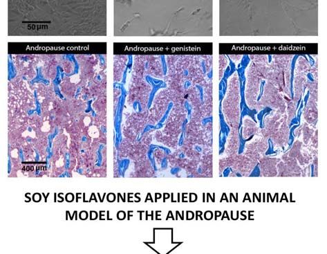
EXCLI Journal 2019;18:106-126 – ISSN 1611-2156
                        Received: October 25, 2018, accepted: February 12, 2019, published: February 19, 2019

States is the most vulnerable in this respect),       tate cancer (with still activated but deviant
while the lowest prostate cancer incidence is         androgen receptor (AR) signaling) may in-
characteristic of Asian men living in their           clude AR-targeted therapy (abiraterone, en-
native countries (Rebbeck, 2017; Pernar et            zalutamide), chemotherapy (docetaxel and
al., 2018). On the other hand, Afro-                  cabazitaxel), immunotherapy (sipuleucel-T),
Caribbean and Sub-Saharan African popula-             bisphosphonates or radionuclides (radium-
tions have the highest prostate cancer mortal-        223) (Grossmann et al., 2001; Nuhn et al.,
ity rates, strongly correlated with limited ac-       2019). Palliative care for patients suffering
cess to medical care, i.e. prostate-specific an-      from metastatic prostate cancer is a challeng-
tigen (PSA)/ultrasound screening and the              ing task that requires a multimodal therapeu-
possibility of early detection of the disease,        tic approach (Das and Banerjee, 2017).
or to adequate therapy (Rebbeck, 2017; Per-                In recent decades, interest in the plant-
nar et al., 2018). Predominance of an andro-          derived compounds relevant for cancer pre-
gen-independent cell phenotype in the pros-           vention and therapy has increased substan-
tate tumor is one of the crucial moments in           tially. Soy isoflavones are steroid-like (the
the malignant disease progression and brings          chemical features of these compounds have
bad news for the patients (Tang and Porter,           been more thoroughly described in our pre-
1997; Arnold and Isaacs, 2002). In parallel,          vious works – Ajdžanović et al., 2012; 2014;
‘bone tropism’ or the preference of prostate          2018), non-nutrient components of soy-
cancer cells for bone invasion and coloniza-          based food and therapeutic dietary supple-
tion, resulting from a sequential series of tar-      ments whose application is inter alia associ-
getable molecular events, underlies the de-           ated with improved bone health in both nor-
creased quality of life, skeletal pain/compli-        mal and osteoporotic male rodents (Chin and
cations and mortality of these cancer patients        Ima-Nirwana, 2013) as well as with low risk
(Rucci and Angelucci, 2014; Ziaee et al.,             of prostate cancer, especially in frequent
2015). The metastasizing of prostate cancer           consumers such as Asian-Pacific men (Mes-
cells to bones is a microenvironment-                 sina, 2010; Ajdžanović et al., 2014;
adjusted process, considering the cancer              Mahmoud et al., 2014; Sak, 2017; Xiao et
cell–bone tissue cross-talk, and involves nu-         al., 2018). There is now a growing body of
merous signaling pathways (Jin et al., 2011;          evidence on the exact mechanisms by which
Ziaee et al., 2015). The poor prognosis of a          these compounds of natural origin may pre-
prostate cancer in its metastatic stage sug-          vent the development or progression of pros-
gests the need for improving the available            tate cancer (Mahmoud et al., 2014). It would
diagnostic methods as well as for finding in-         be too ambitious to compare the specificity
novative approaches to establishing a safe            and therapeutic potential of soy isoflavones
and promising therapeutic strategy. The ex-           with those of the newly developed pharma-
isting treatment protocols and guidelines re-         cotherapeutics; however, since isoflavones
garding prostate cancer highlight a number            have been well recognized as bone-
of factors that should be considered during           modifying agents (Messina, 2010; Messina
therapy, such as: existence of concrete symp-         et al., 2010; Filipović et al., 2010, 2018;
toms, serum androgen and PSA levels, type             Chin and Ima-Nirwana, 2013; Zheng et al.,
of metastasis if present (bone/visceral),             2016), we believe that, in the specific con-
treatment history, performance status, side           text of prostate cancer bone metastasis for-
effects of the therapy, etc. (Crawford et al.,        mation, the effects of their application de-
2015). In line with this, androgen deprivation        serve some attention. Here, we will focus on
therapy (ADT) is the treatment of choice and          the potential role of soy isoflavones in the
has a high response rate in the early stages of       prevention and treatment of bone metastasis
the disease, while the options available for          in prostate cancer, as very important aspects
treating metastatic castration-resistant pros-        of this cancer management.

                                                  107
EXCLI Journal 2019;18:106-126 – ISSN 1611-2156
Received: October 25, 2018, accepted: February 12, 2019, published: February 19, 2019
A BRIEF OVERVIEW OF THE the so-called cancer secretome, which is re-
MECHANISMS OF BONE sponsible for the formation of a pre-
METASTASIS IN PROSTATE CANCER metastatic zone in the particular distant or-
gan (Gartland et al., 2016).
Prostate cancer, like most other solid tu-
A brief summary of the most important
mors, shows an intrinsic tendency toward
mechanisms relevant for prostate cancer me-
metastasizing to distant organs (lungs, liver,
tastasis to bone is given below.
brain), but it has a pronouncedly high prefer-
ence for metastasizing to the bone (verte-
brae, femur, pelvis, ribs; Yang et al., 1999; The role of androgen receptors (ARs)
Androgens, realizing their actions
Jin et al., 2011). This principle of malignant
through ARs, have a crucial role in prostate
disease spreading, with secondary deposits
cancer development and progression, most
formation, is metaphorically presented in the
likely at all stages of the disease (Cunha et
‘seed and soil’ model (Paget, 1889), where
al., 2004; Jin et al., 2011; Ziaee et al., 2015).
cancer cells or ‘seeds’ metastasize to the
The cascade of molecular events implies an-
‘soil’ most appropriate for their growth. Ac-
drogen binding to the AR and translocating
tually, the bone contains chemotactic factors
to the nucleus, where the binding of this
that attract prostate cancer cells and direct
complex to androgen responsive elements
their movement. Stromal-derived factor-1
occurs, which affects the expression of vari-
(SDF-1), epidermal growth factor (EGF), in-
ous genes. As a result, proliferation of pros-
sulin-like growth factor (IGF), hepatocyte
tate cancer cells is favored at the expense of
growth factor (HGF), Type I collagen, oste-
their apoptosis (Jin et al., 2011). ADT
onectin and bone sialoprotein have been
(chemical or surgical castration) is the treat-
shown to act as chemoattractants for prostate
ment of choice for this malignant disease in
cancer cells, predominantly causing them to
gravitate towards the bone (Jacob et al., its early stages. As previously indicated, the
1999; Taichman et al., 2002; Stewart et al., prostate cancer acquiring a castration-
2004; Arya et al., 2006). More broadly, the resistant phenotype is the inevitable next
metastasizing of prostate cancer is a multi- stage of the disease, which relies on the fol-
step process that implies angiogenesis at the lowing events that take place in the cancer
primary site, loss of the cancer cells’ adhe- cells: upregulation and mutation of ARs with
sion, followed by their local migration, in- irregular downstream gene expression, al-
travasation into the vasculature or lymphat- tered expression and function of AR coacti-
ics, transport via circulation, extravasation, vators, ligand independent AR activation, in-
and homing to distant organs, which is again creased autocrine and paracrine production
followed by the angiogenesis step (Arya et of androgens and elevated expression of in-
al., 2006). Interestingly, recent findings have terleukin-6 (Adler et al., 1999; Chen et al.,
suggested the existence of an early pre- 2000; Debes and Tindall, 2004; Linja et al.,
metastatic phase, preceding the homing of 2004; Dutt and Gao, 2009; Bonkoff and
cancer cells to the bone tissue (Gartland et Berges, 2010; Jin et al., 2011; Mahmoud et
al., 2016). This formation of a pre-metastatic al., 2014).
niche could be described as an adaptation of
an apartment for the arrival of a new occu- Insight into the epithelial to mesenchymal
pant. The discovery of pre-metastatic niches transition (EMT) and adhesiveness of
confirms the impressive communication be- prostate cancer cells
tween primary sites and distant tissues, se- The adhesiveness of prostate cells de-
lected for dissemination according to criteria creases if they ‘move along the way’ of ma-
that are still unclear. It has become obvious lignant transformation (Jin et al., 2011;
that this step is navigated by products pre- Jadaan et al., 2015). Namely, in the process
sent in the primary tumor microenvironment, of epithelial to mesenchymal transition
108EXCLI Journal 2019;18:106-126 – ISSN 1611-2156
Received: October 25, 2018, accepted: February 12, 2019, published: February 19, 2019
(EMT), essential for the development of a are the crucial signaling molecules output-
more invasive cancer cell phenotype, the ting focal adhesions, is characteristic of a
static, polarized epithelial cells transform in- migratory metastatic prostate cancer cell
to migratory, spindle-shaped mesenchymal phenotype (Rovin et al., 2002; Kim et al.,
cells (Thiery, 2002; Yang and Weinberg, 2009; Tatarov et al., 2009). Given the fact
2008; Jadaan et al., 2015). The EMT, which that increased membrane fluidity enhances
is specific for higher grades of prostate can- the malignancy of cancer cells in vitro
cer, is accompanied by cadherin protein (Zeisig et al., 2007) and correlates with the
switching, which includes the downregula- decreased cancer cell adhesiveness (Gonda
tion of E-cadherin (characteristic of normal et al., 2010), we will prove that the superfi-
epithelial cells) and upregulation of N- cial membrane fluidity of LNCaP and PC-3
cadherin (abundant in mesenchymal cells) prostate cancer cells (isolated from lymph
(Gravdal et al., 2007). At this stage of pros- node and bone metastasis, respectively) at
tate cancer progression, the expression of β- least partially determines their invasive ac-
catenin also decreases (Jaggi et al., 2005), all tivity (Ajdžanović et al., 2013, 2014), there-
of which contributes to the loosening of con- fore completing the previous observations in
nections between the cells, given the im- this context.
portant role of E-cadherin and β-catenin
complexes in the maintenance of cell-cell Degradation of the extracellular matrix
adhesions (Jin et al., 2011). In parallel, in- (ECM) integrity by prostate cancer cells
trinsic overexpression of miRNA-409 and Prostate cancer invasion and metastasis
downregulation of miRNA-143 and -145 require partial degradation of the ECM integ-
promote the EMT of prostate cancer cells rity. This process is mediated by families of
and support the shaping of their metastatic proteinase enzymes, such as matrix metallo-
phenotype (Peng et al., 2011; Josson et al., proteinases (MMPs), serine proteinases
2014). Of note, somewhat higher expressions (urokinase-type plasminogen activator–uPA
of E-cadherin and β-catenin have been re- and plasmin), and probably by PSA, general-
ported in the metastatic prostate cancer cells ly known as a fibronectin-degrading protein-
that have already reached the bone (Saha et ase (Jin et al., 2011). In human prostate can-
al., 2008), suggesting that reverse, mesen- cer tissues, upregulation of MMPs correlates
chymal to epithelial transition (MET) is a with the loss of tissue inhibitor of MMP-1
prerequisite for the growth of metastatic cells (Brehmer et al., 2003), the metastatic pheno-
at the site of a secondary bone deposit (Jin et type brings high plasma concentrations of
al., 2011). Focal adhesions, or more precise- MMP-2 and MMP-9 (Morgia et al., 2005),
ly the macromolecular complexes mediating while MMP-12 participates in bone-tropic
the extracellular matrix (ECM)–cell cyto- metastasis (Nabha et al., 2008). A biome-
skeleton contacts, convert physical vectors chanical point of view suggests that MMPs
into chemical signaling, thus affecting the activity facilitates the expansion tendency of
cell’s dynamic properties (Bershadsky et al., prostate cancer cells and their navigation
2003; Ajdžanović et al., 2014). Variable ex- through the ‘cracks’ of the degraded matrix
pression of transmembrane integrin proteins, (Ajdžanović et al., 2013, 2014). A similar
regulators of focal adhesions, and the down- pattern of ECM degradation characterizes
stream signaling molecules they affect, is as- the invasive prostate cancer cell-specific ac-
sociated with decreased adhesion of prostate tivity of uPA, the proteinase simultaneously
cancer cells and metastasis (Hao et al., 1996; involved in the activation of latent MMPs
Slack-Davis and Parsons, 2004; Nicolas and and conversion of plasminogen into the func-
Safran, 2006). Overexpression of focal adhe- tional, matrix-degrading enzyme plasmin
sion kinase (FAK) and the Src family of ki- (Sheng, 2001; Arya et al., 2006).
nases, the nonreceptor tyrosine kinases that
109EXCLI Journal 2019;18:106-126 – ISSN 1611-2156
Received: October 25, 2018, accepted: February 12, 2019, published: February 19, 2019
Circulation- and bone-related aspects of 2010). Osteoblastic lesions with irregular,
prostate cancer cell dissemination increased bone formation occur more fre-
Intravasation of metastatic prostate can- quently (Urwin et al., 1985) and represent
cer cells and their entry into the circulation hot spots for bone fractures. Formation of os-
impose the need for survival in a new milieu, teolytic lesions, on the other hand, releases a
and precede the adhesion of malignant cells three-dimensional space for further prostate
to the vascular endothelium and extravasa- cancer metastasis progression and at the
tion into bone. Vascular endothelial growth same time liberates the molecules involved
factor (VEGF) and its receptor (VEGFR), as in further bone and tumor cell proliferation.
potent stimulators of angiogenesis, are high- These findings complete the pool of data
ly expressed in prostate cancer (Pallares et pertinent to dysregulated osteoblast and os-
al., 2006). Recruited and organized endothe- teoclast function in metastatic-related bone
lial cells provide a blood supply that nour- remodeling. The bone aspect, both initially
ishes the prostate cancer mass and facilitate and at the later stages of prostatic cancer dis-
the cancer cell metastasis at distant loci ease, will determine the diagnostic proce-
(Ziaee et al., 2015). The membrane fluidity dures, treatment, complications, quality and
of freely circulating cancer cells is found to duration of life of the patients (Butoescu and
be more than double that of the cells adher- Tombal, 2014).
ing to the inner vascular surface, and around
23 times higher than the membrane fluidity The phenomenon of pre-metastatic niche
of already adhered cancer cells, migrating formation: general and prostate cancer-
over the vascular surface (Gonda et al., specific considerations
2010). A ‘dock and lock’ mechanism has Does the metastatic process start before
been proposed for the instance of cancer detectable cancer cells appear in the bone
cells binding to the vascular endothelium tissue?
(Honn and Tang, 1992). The adhesion mole- As mentioned above, the reposition of
cule P-selectin, expressed by the bone endo- circulating tumor cells (CTCs) into distant
thelial cells, and sialyl-Lewisx carbohydrate, organs is crucial for the development of met-
available on the prostate cancer cell surface, astatic foci. Obviously, this step is critically
play a crucial role in the association (Mar- affected by the local microenvironment at
tensson et al., 1995; Mazo and von Andrian, the place of CTC arrival. Sound data have
1999), while integrin molecules mediate the recently suggested that a primary tumor can
subsequent locking process (Romanov and prearrange its new home before a cancer cell
Goligorsky, 1999). In addition, prostate can- enters the site by inducing a supportive mi-
cer cell- and osteoclast-integrin and cancer croenvironment recognized as a pre-
cell/osteoblast cadherin 11 molecules appear metastatic niche (Kaplan et al., 2005; Liu
to be important in the colonization of these and Cao, 2016). The pre-metastatic niche
malignant cells in the bone (Chu et al., 2008; could be described as an area where fine ac-
Jin et al., 2011). Growth of prostate cancer commodation for the newcomer has been
cells settled in the bone is accompanied by prepared, offering all the conditions for col-
their production of growth factors that stimu- ony formation, in close interaction with the
late proliferation and maturation of osteo- surrounding tissue.
blasts and osteoclasts, and the release of Kaplan et al. (2005) showed that bone
these factors in turn stimulates metastatic marrow-derived hematopoietic progenitor
growth (the ‘vicious cycle’; Jin et al., 2011). cells (BMDC) settle at tumor-specific pre-
The inevitable disbalance between bone metastatic sites and form cellular clusters be-
formation and bone resorption, characteristic fore the arrival of tumor cells. Those cells
of metastatic prostate cancer, results in oste- express vascular endothelial growth factor
oblastic or osteolytic lesions (Ibrahim et al., receptor 1 (VEGFR1), and depletion of
110EXCLI Journal 2019;18:106-126 – ISSN 1611-2156
Received: October 25, 2018, accepted: February 12, 2019, published: February 19, 2019
VEGFR1+ cells from the bone marrow of Osteolytic prostate cancer metastases
wild-type mice disables the formation of pre- Presence of specific bone metastases that
metastatic clusters and prevents metastasis coincide with osteolytic lesions implies a
development, thus confirming the hypothesis pronounced participation of the receptor ac-
of their role in the process of dissemination. tivator of nuclear factor-κB (RANK)/RANK
Since a functional reconstitution of this cell ligand (RANKL)/osteoprotegerin (OPG) axis
population in Id3 (inhibitor of differentiation (Jin et al., 2011). RANKL, regularly pro-
3) knockout mice resulted in the reestab- duced by osteoblasts, can also be the secreto-
lishment of the entire process, from cluster ry product of metastatic prostate cancer cells
formation to the development of metastasis, that directly activates osteoclasts via RANK
it is clear that those cells represent the key (Zhang et al., 2003). The mentioned process
actors in pre-metastatic niche formation of osteoclast activation and bone resorption
(Kaplan et al., 2005). Until today, a long list is inhibited by OPG derived from osteo-
of molecules involved in pre-metastatic blasts; however, this molecule simultaneous-
niche formation has been defined. Among ly protects cancer cells from apoptosis
the niche promoting molecules are TGF-β (Holen et al., 2002; Boyle et al., 2003), thus
(Hiratsuka et al., 2008; Olkhanud et al., expressing its antinomic nature. In this re-
2011), RANK/RANKL (Chu et al., 2014), spect, parathyroid hormone-related protein
Lysil Oxidase enzyme (LOX) (Erler et al., (PTHrP), a homolog of parathyroid hor-
2009), Hypoxia Inducible Factors (HIFs) mone, upregulates RANKL production in os-
(Unwith et al., 2015), etc. LOX enzyme- teoblasts and decreases the OPG expression,
mediated remodeling of ECM in the bones all of which leads to the activation of osteo-
leads to the formation of a pre-metastatic clasts and osteolytic metastasis formation
niche within the bone microenvironment, fa- (Liao et al., 2008). Given that PTHrP also
voring the homing of CTC and subsequent induces differentiation of osteoblasts (Liao et
development of dissemination lesions (Cox al., 2008), its role in osteoblastic lesion for-
et al., 2015). Data about the pivotal role of mation appears certain.
LOX proteins in the development of prostate Osteoclast-derived proteinases and pros-
cancer metastasis has been recently provid- tate cancer cells-secreted PSA and uPA acti-
ed. Crosslinking of collagen, conducted with vate transforming growth factor β (TGF-β),
the LOX derived from stromal cells, was which may also promote osteolytic metasta-
shown to control the movement of prostate ses (Josson et al., 2010; Jin et al., 2011)
cancer cells, while LOX inhibition obstruct- through induction of the proosteolytic gene
ed the same process (Caley et al., 2016). In- expression in cancer cells, with PTHrP in the
terestingly, LOX pro-peptide (LOX-PP), as key position (Yin et al., 1999; Kingsley et
an intermediary product in the formation of al., 2007). PC-3 cells, established from an-
the final form of LOX enzyme, functions as drogen independent prostate cancer bone
a tumor suppressor (Trackman, 2016). De- metastasis, are potent producers of PTHrP
spite its opposite role in comparison with the prometastatic protein (Kingsley et al., 2007).
mature protein counterpart, in the case of in- In addition, TGF-β is one of the important
tramedullary injections of PC-3 LOX-PP ex- factors involved in EMT, whereby the
pressing prostate cancer cell lines, enhanced PI3K/Akt pathway plays a noticeable role in
appearance of osteolytic lesions and subse- this respect (Nakazawa and Kyprianou,
quent bone destruction were discovered in 2017). All together, the bone matrix (con-
vivo (Alsulaiman et al., 2016). It is important taining calcium, TGF-β, insulin-like growth
to underline that the concept of a pre- factors (IGF) I and II, etc.) with its physical
metastatic niche perfectly fits into both the features (low oxygen, local acidity) favors
linear and parallel models of metastasis de- tumor progression and the subsequent for-
velopment.
111EXCLI Journal 2019;18:106-126 – ISSN 1611-2156
Received: October 25, 2018, accepted: February 12, 2019, published: February 19, 2019
mation of new osteolytic lesions, forming an hending the concrete topic, can be found in
amplification loop (Kinsley et al., 2007). our previous review articles (Ajdžanović et
al., 2014, 2015).
Osteoblastic prostate cancer metastases
Osteoblastic lesions are the main type of Soy isoflavone effects on the AR signaling
bone abnormalities in prostate cancer metas- It has been reported that the soy isofla-
tasis (Kingsley et al., 2007). Dysregulated vone genistein, as a component of diet in
osteoblast activities are mediated by numer- concentrations comparable to the human in-
ous ‘osteoblastic’ factors. Endothelin-1 (ET- take (250 or 1000 mg/kg diet, 2 weeks),
1) is a vascular endothelium-produced vaso- downregulates AR mRNA in the rat prostate
constricting peptide of small size that in- (Fritz et al., 2002). In prostate cancer cells,
creases osteoblast proliferation and initiates genistein (at 30 and 50 μM concentrations,
bone matrix formation (Yin et al., 2003; 24 h) was shown to downregulate the AR
Guise et al., 2003). Thus, a diagnosis of os- gene and protein expression in vitro, as well
teoblastic prostate cancer metastasis implies as the receptor transcriptional activity (Davis
elevated plasma levels of ET-1 (Nelson et et al., 2002). A daidzein metabolite equol (50
al., 1995). Proliferation of osteoblasts is also μM, 48 h) was also found to suppress AR
stimulated by Wnt signaling through β- expression in LNCaP prostate cancer cells
catenin-induced gene expression (Behrens et (Itsumi et al., 2016). Furthermore, significant
al., 1996). It should be mentioned that meta- inhibition of some AR pathway-related
static prostate cancer cells are also capable genes in human prostate cancer cells was
of producing Wnt proteins and stimulating identified after genistein application (at 1
osteoblast proliferation (Hall et al., 2006), μM, 5 μM and 25 μM; 48 h) (Takahashi et
and therefore irregular bone formation. Os- al., 2004) (Figure 1). The proposed mecha-
teoblasts produce bone morphogenic protein nism of genistein action in this respect im-
2 (BMP-2) that can activate Akt, ERK and plies its binding to estrogen receptor β (ER-
NFκB signaling, essential for the migration β) and the initiation of a cascade of molecu-
of prostate cancer cells (Lai et al., 2008). Fi- lar events resulting in the AR downregula-
nally, IGF I is upregulated in prostate cancer tion (Bektic et al., 2004). However, some in
bone metastasis and stimulates the cancer vitro studies have demonstrated that
cell proliferation, while elevated levels of genistein, at low doses (2 μM, 24 h) and in
IGFs may coincide with osteoblast prolifera- the presence of a synthetic androgen, may
tion and the following susceptibility to bone have a stimulating effect on the expression
fractures (Rubin et al., 2004; Jin et al., of AR pathway-related genes (PSA, KLK4,
2011). NKX3.1, STAMP2) in metastatic prostate
cancer cells (Lazarevic et al., 2008) (Figure
PROSTATE CANCER METASTASES 1). This phenomenon could at least partly be
AND SOY ISOFLAVONES APPLICA- explained by the AR mutations associated
TION – THE BONE ENDPOINTS with prostate cell malignant transformation
In the next section of this analytical arti- and the ARs acquiring the capability to in-
cle, we will focus on prostate cancer metas- teract with a wide range of steroid-like com-
tasis formation in connection with the appli- pounds (Mahmoud et al., 2014).
cation of soy isoflavones. This up-to-date re-
port attempts to elucidate the effects of these EMT and adhesiveness of prostate cancer
steroid-like compounds along the sequence cells upon soy isoflavones application
of events relevant to the metastatic process, The EMT of prostate cancer cells can be
which culminates in the bones. A detailed reversed with the active participation of soy
mechanistic overview of the soy isoflavone isoflavones (Mahmoud et al., 2014). Name-
actions, which may be useful for compre- ly, culturing of IA8-ARCaP cells with low
112EXCLI Journal 2019;18:106-126 – ISSN 1611-2156
Received: October 25, 2018, accepted: February 12, 2019, published: February 19, 2019
dosed genistein (15 μM/L for 24 h) morpho- matrix elements (Skogseth et al., 2006) (Fig-
logically changed these human prostate can- ure 1), which would be the desired effect in
cer cells, from a fibroblast-like shape to an target bones, but not at the site of initial dis-
epithelial-like shape (Zhang et al., 2008). In- semination. Such findings call for caution
cubation with genistein that lasted 48 h led to and highlight the importance of correctly
enhanced cell-cell contacts in this context timing soy isoflavones application during
(Zhang et al., 2008) (Figure 1). The authors prostatic cancer disease, so they shouldn't be
observed that genistein markedly increased overlooked in a serious evaluation of their
the expression of E-cadherin and significant- antimetastatic properties.
ly decreased the expression of the mesen-
chymal marker vimentin, when applied in Soy isoflavone effects on the ECM-
low doses to IA8-ARCaP as well as degrading enzymes
LNCaP/HIF-1a prostate cancer cells (Zhang Genistein was shown to exert a concen-
et al., 2008). By establishing a balance be- tration-dependent inhibitory effect on PSA
tween the expressions of epithelial and mes- (fibronectin-degrading proteinase) secretion
enchymal protein markers which is actually in androgen-dependent LNCaP metastatic
characteristic of MET, genistein decreases prostate cancer cells. After 5 days of treat-
the invasiveness of prostate cancer cells ment, genistein in a concentration of 100 nM
(Figure 1). Although the soy isoflavones, decreased PSA secretion by 25 %; 5 μM of
genistein and daidzein (at concentrations of genistein caused a 50 % decrease in the same
40 μM and 110 μM, 48 h), may affect the parameter, while a 90 % reduction of secret-
expression of certain miRNAs in prostate ed PSA was detected with 50 μM genistein
cancer cell clones (Rabiau et al., 2011), to (Davis et al., 2000). In androgen-independ-
the best of our knowledge, their concrete ef- ent VeCaP metastatic prostate cancer cells,
fects on miRNA-409, -143 and -145 (respon- the same duration of genistein treatment in-
sible for the EMT and affirmation of a meta- duced an inhibitory effect on PSA secretion
static phenotype in prostate cancer cells) still only at higher (nutritionally irrelevant) con-
remain unknown. Morphological flattening centrations. Namely, the PSA secretion was
of highly metastatic prostate cancer PC-3-M decreased by 25 % after 10 μM and by 50 %
cells upon genistein (50 μM, 2 h – 3 days) upon administration of 50 μM of genistein,
treatment was found to be accompanied by while lower, nutritionally relevant concentra-
an increase in cell adhesion (Bergan et al., tions (0.1–5 μM) were ineffective in this re-
1996). Genistein caused FAK accumulation spect (Davis et al., 2000). The basis for the
in the areas of focal cell attachment, and observed decrease in the PSA secretion from
simultaneous complexing between β-1- these metastatic prostate cancer cell lines
integrin and FAK was shown to occur with- represents a genistein-induced multirange
out the requirement for FAK activation inhibition of the PSA gene and protein ex-
(Bergan et al., 1996; Liu et al., 2000) (Figure pression (Davis et al., 2000) (Figure 1). Sim-
1). Tumor expression of FAK increased, but ilarly, culturing of LNCaP cells with the
the levels of activated FAK and the cancer presence of soy milk digestion extract (0.79
invasion decreased in mice implanted with mg/ml; containing a mixture of genistein,
PC-3-M cells and administered genistein daidzein, glycitein and other isoflavones,
(250 mg/kg of food, 4 weeks) (Lakshman et whereby ~26 mg/100 g was the concentra-
al., 2008). On the other hand, some results tion of aglycones) significantly reduced the
have suggested that genistein application (30 gene expression levels of PSA (Kang et al.,
μg/ml, 48 h) decreased the expression of β-1- 2016). Interestingly, some clinical studies
integrins by 40 % in PC-3 and by 22 % in have suggested that prolonged consumption
DU-145 metastatic prostate cancer cells, of a soy isoflavone mixture (450 mg
suppressing the cell adhesion to extracellular genistein + 300 mg daidzein + other isofla-
113EXCLI Journal 2019;18:106-126 – ISSN 1611-2156
Received: October 25, 2018, accepted: February 12, 2019, published: February 19, 2019
vones, daily for 6 months) may not affect genistein action at the level of the LNCaP
PSA levels in prostate cancer patients cell surface immobilized the membrane an-
(deVere White et al., 2010). The crucial im- drogen receptor containing lipid rafts, down-
pact of soy isoflavones in preventing the regulated these specific androgen receptors
ECM degradation and prostate cancer cells involved in fast signaling from the cell sur-
expansion is realized through an unambigu- face and silenced the related downstream
ously inhibitory effect on the MMP and uPA pathways (Oh et al., 2010; Ajdžanović et al.,
proteinases (Figure 1). Increased expression 2015) (Figures 1, 2). The effects of genistein
of MMP-2 at least partly underlies prostate on invasive activity and the resulting dynam-
cancer aggressiveness, while the metastatic ic phenotype were more prominent in the
potential of PC-3 and LNCaP cells coincides PC-3 metastatic cell clone, while daidzein,
with an increased expression of MMP-9 even in higher doses (25 μg/ml), was ineffec-
(which is twofold higher in more invasive tive when it comes to membrane fluidity and
PC-3 cells) (Upadhyay et al., 1999; invasiveness of the tested metastatic prostate
Aalinkeel et al., 2004). Genistein has shown cancer cells (Ajdžanović et al., 2013; 2014)
a dose- and time-dependent inhibitory effect (Figure 2). Considering the fact that the
on the MMP-2 protein expression levels in membrane fluidity value of cancer cells is
both LNCaP and PC-3 cells, being the most reciprocal to their adhesiveness (Gonda et
effective at 50 μg/ml during 48 h (Kumi- al., 2010), the effects of genistein observed
Diaka et al., 2006). In line with this observa- in this context (adhesiveness promotion) are
tion, MMP-2 activity and gene expression desirable at the site of initial dissemination,
were decreased after genistein application but appear unwanted in distant secondary
(50 μM, 24 h) in several normal and malig- bone deposits.
nant prostate cell lines (Huang et al., 2005;
Xu et al., 2009). In PC-3 cells, genistein (50 Angiogenesis following prostate cancer
μM, 24–72 h) was shown to downregulate growth and soy isoflavones
MMP-9 activity, gene and protein expression Along with the soy isoflavone effects
(Li et al., 2006). Equol, applied at concentra- that suppress the evolution of metastatic
tions of 10 μM and 50 μM for 24 h, was prostate cancer cell malignancy, which is
found to decrease uPA mRNA expression in aimed at enabling the cells entry into the
prostate cancer DU-145 cells, which possess bloodstream or lymphatics, their influence
moderate metastatic potential (Zheng et al., on the process of angiogenesis, following the
2012) (Figure 1). expansion of cancer growth, deserves some
attention. Genistein (10-50 μM, 72 h) was
Membrane fluidity and invasiveness of shown to significantly inhibit basal and hy-
prostate cancer cells after soy isoflavones poxia-stimulated VEGF gene expression in
application PC-3 cells (Guo et al., 2007) (Figure 1).
As previously indicated, the increased Subcutaneous inoculation of metastatic pros-
membrane fluidity of metastatic prostate tate cancer LNCaP cells to immuno-deficient
cancer cells represents one of the intriguing mice resulted in reduced cancer mass growth
definers of their invasiveness (Ajdžanović et and diminished density of cancer-pervading
al., 2013, 2014). We have demonstrated that vessels if a soy phytochemicals-rich diet
short-term exposure to genistein (12.5 μg/ml, (containing 341 mg or 1705 mg of isofla-
10 min) significantly decreased superficial vone equivalents/kg) was applied (Zhou et
membrane fluidity in LNCaP and PC-3 cells, al., 1999). In line with this, a soy isoflavone
which corresponded with the genistein- concentrate (49 % of isoflavones; 200 mg/L,
induced poor invasive trends of these cells in 48 h) reduced the mRNA expression and
2.5 D extracellular matrix – Matrigel (Aj- protein level of the pro-angiogenic cytokine
džanović et al., 2013, 2014). More precisely,
114EXCLI Journal 2019;18:106-126 – ISSN 1611-2156
Received: October 25, 2018, accepted: February 12, 2019, published: February 19, 2019
interleukin-8 in PC-3 cells (Handayani et al., comparison with orchidectomized controls
2006) (Figure 1). (Filipović et al., 2010, 2018) (Figure 2).
Elaboration of the andropausal rat pituitary–
Soy isoflavones and the bone aspects of adrenocortical axis, under the same condi-
prostate cancer tions, suggested that genistein and daidzein
Metastatic prostate cancer cells adhesion decreased the capacity for production and
to the vascular endothelium and their extrav- secretion of corticosterone (Ajdžanović et
asation in the areas of future bone lesions al., 2009a, b, 2011) (Figure 2), which is im-
may also be obstructed by the use of soy iso- portant in light of the fact that glucocorti-
flavones. Genistein (10 nM, 24 h) was re- coids are well known to be osteoporosis-
ported to decrease the gene expression of the promoting factors (Ringe, 1989). Our results
endothelial cell adhesion molecule P-selectin confirm numerous other experimental studies
(Sandoval et al., 2010), important for the reporting beneficial effects of soy isofla-
process of association with the prostate can- vones on the male skeleton in androgen defi-
cer cell surface (Figure 1). While the charac- ciency situations (frequent during prostate
ter of the inhibitory effects of genistein on cancer therapy; Ishimi et al., 2002; Khalil et
the expression of prostate cancer cells- al., 2005; Soung et al., 2006), while the re-
derived, adhesion-locking integrins as well lated clinical data are still expected.
as on the adhesion-defining cancer cell
membrane fluidity has already been de- Soy isoflavones versus osteolytic prostate
scribed above, it should be mentioned here cancer metastases
that, in cancer cells, genistein slightly inhib- Prostate cancer metastasis-caused osteo-
its the mRNA and protein levels of cadherin lytic lesions, which release the space for fur-
11 (Moiseeva et al., 2007), another bone me- ther malignant growth in the targeted bone,
tastasis-promoting marker (Chu et al., 2008). are characterized by significant RANK/
The bone morphofunctional status in prostate RANKL/OPG signaling (Jin et al., 2011).
cancer patients appears to be important both Genistein (1 g/kg of diet) was found to sig-
initially and during treatment, but also inde- nificantly inhibit the protein expression of
pendently from the process of metastatic RANKL in PC-3-induced tumors in severe
cells invasion (Miñana et al., 2014). Our ex- combined immunodeficiency (SCID) mice
perimental experience suggests some multi- (Li et al., 2006). Genistein and glycitein (10
dimensional, bone health-related benefits of nM, 14 days) significantly decreased the os-
soy isoflavones application in an animal teoblast RANKL gene expression in vitro
model of the andropause (orchidectomized, (Winzer et al., 2010) (Figure 1). Also,
15–16 months old Wistar male rats). genistein (50 μmol/L, 24 h) inhibited the se-
Genistein and daidzein, s.c. administrated in cretion of RANKL protein into the medium
a dose of 30 mg/kg b.m., for three weeks, in RANKL-transfected PC-3 cells (Li et al.,
significantly increased the cancellous bone 2006). In line with this, differentiation of
area, trabecular thickness and trabecular RANKL-induced RAW264.7 cells (osteo-
number, but decreased the trabecular separa- clast precursor macrophages) to osteoclasts
tion, in the proximal tibial metaphysis of this was inhibited by genistein (10 μmol/L)
animal model (Filipović et al., 2010, 2018). treatment (Li et al., 2006). The complex gene
The molecular mechanisms through which and protein expression as well as the micro-
soy isoflavones reinforce the bone microar- array data analysis revealed OPG upregula-
chitecture in andropausal rats primarily in- tion in genistein-treated (50 μM, 2–3 days)
volve ER-dependent pathways (Filipović et PC-3 cells (Li et al., 2006). Two times high-
al., 2010, 2018). In parallel, significant re- er levels of OPG mRNA were found in PC-3
ductions in serum osteocalcin levels and uri- bone tumors in SCID mice preventively
nary Ca2+ concentrations were observed, in treated with genistein (1 g/kg of diet, 58
115EXCLI Journal 2019;18:106-126 – ISSN 1611-2156
Received: October 25, 2018, accepted: February 12, 2019, published: February 19, 2019
days; Li et al., 2004) (Figure 1). In human pool of factors that actively mediate osteolyt-
osteoblastic MG-63 cells, daidzein (0.01, 0.1 ic metastases when isoflavones are applied.
and 1 μM, 3 days) increased OPG, but de-
creased RANKL gene and protein levels, all Soy isoflavones versus osteoblastic prostate
via ER-α and ER-β (Sun et al., 2016). Soy cancer metastases
extract (0.001 mg/ml, 6 days) was shown to Metastasis-related osteoblastic lesions
increase the OPG secretion levels and to de- i.e. foci of irregular bone formation, imply
crease those of RANKL in a conditioned osteoblast proliferation, stimulated by Wnt
medium of MC3T3-E1 osteoblasts (Park et signaling, through β-catenin-induced gene
al., 2014). An increased OPG/RANKL ratio expression (Behrens et al., 1996). In PC-3
was observed in LNCaP and PC-3 cells upon cells, genistein downregulated Wnt-4 protein
genistein or daidzein (10–50 μM, 72 h) ap- expression, while the soy isolate mildly de-
plication (Alonso et al., 2009) (Figure 1). All creased β-catenin protein levels (Liss et al.,
these data suggest specific effects of soy iso- 2010) (Figure 1). Genistein-mediated inhibi-
flavones on concrete constituents of the tion of IGF-1 (usually upregulated in pros-
RANK/RANKL/OPG triad that synergisti- tate cancer bone metastasis) also silences the
cally strive to deactivate osteoclasts and pre- β-catenin pathway (Rubin et al., 2004;
vent resorption of the affected bone. A Mahmoud et al., 2014). Finally, considering
somewhat reserved attitude towards the OPG the property of E-cadherin to bind to β-
increase still remains, given its role in the catenin and immobilize it, the increased ex-
prevention of apoptosis of prostate cancer pression of E-cadherin upon genistein appli-
cells (Holen et al., 2002). In addition, the cation (15 μM/L, 24 h or 48 h) to prostate
PTHrP molecule, generally known to elevate cancer cells may suggest some indirect bene-
the RANKL production in osteoblasts and to fits in impeding metastasis (Zhang et al.,
decrease the OPG expression and activate 2008; Mahmoud et al., 2014).
osteoclasts in the formation of metastatic le-
sions (Liao et al., 2008), is susceptible to soy Soy isoflavones against the pre-metastatic
isoflavones actions. Genistein and daidzein niche formation: a new horizon?
(0.01–50 μM, 48 h) were shown to induce Until now, the potential of soy isofla-
the PTHrP gene expression in LNCaP cells, vones to influence pre-metastatic niche for-
while both isoflavones increased the PTHrP mation has not been evaluated directly.
protein expression in these cells even at nM However, the capacity of these naturally oc-
doses during 72 h (Alonso et al., 2009) (Fig- curring molecules to influence the estab-
ure 1). Actually, these results reflect certain lishment of pre-metastatic fields in bones,
pro-survival effects of soy isoflavones in re- prior to cancer cell arrival, can be discussed
spect to the metastatic prostate cancer cells in light of the numerous findings confirming
(Alonso et al., 2009). Soy isoflavones may their modulatory effect on the mediators in-
directly or indirectly affect TGF-β, the cyto- volved in this important phase of the cancer
kine that also promotes osteolytic metastases dissemination route. One of the factors pre-
(Jin et al., 2011). Gene and protein expres- sumably important in the pre-metastatic
sion of TGF-β2 in PC-3 cells is reduced up- ECM bone rearrangement is Hypoxia Induc-
on genistein (50 μM, 6 h, 36 h and 72 h) ap- ible Factor 1α (HIF-1α) (Semenza, 2016).
plication (Li and Sarkar, 2002). Isoflavones Generally, this molecule is the main regula-
downregulate the levels of PSA and uPA, tor of the expression of genes critical to cell
TGF-β-activating molecules, in metastatic survival under hypoxic conditions. One of
prostate cancer cells (Davis et al., 2000; the proteins regulated by HIF-1α is LOX en-
Josson et al., 2010; Zheng et al., 2012), zyme, a key factor in matrix remodeling in
which most likely excludes TGF-β from the pre-metastatic fields (Joo et al., 2014).
Singh-Gupta et al. (2009) found that pre-
116EXCLI Journal 2019;18:106-126 – ISSN 1611-2156
Received: October 25, 2018, accepted: February 12, 2019, published: February 19, 2019
treatment of prostate cancer cells with soy pre-metastatic bone lesions and its appear-
isoflavones downregulated the Stc/STAT3/ ance is remarkably connected with bone-
HIF-1α pathway and prevented the transloca- tropism and relapses (Cox et al., 2015). In
tion of HIF-1α into the nucleus. This finding view of these circumstances, soy isoflavones
was important from the perspective of soy may be of use in the prevention of bone me-
isoflavones usage in the sensitization of tastases, starting from the earliest phase of
prostate cancer cells to radiotherapy. Yet, the this process. Also, the soy isoflavone-
discovery of pre-metastatic fields and the induced inhibition of RANKL protein secre-
role of HIF-1α at this important stage of the tion into the medium in RANKL-transfected
metastatic process have given another im- PC-3 cells (Li et al., 2006) and inhibition of
portant context to the ability of genistein/ RANKL protein expression in a PC-3 xeno-
daidzein to inhibit HIF-1α nuclear action. graft model (Li et al., 2006) have been dis-
Namely, it is known that hypoxic conditions cussed above. Furthermore, there are numer-
are typical for the bone microenvironment, ous sources highlighting the potential of soy
and together with the hypoxia related high isoflavones to directly or indirectly affect the
LOX activity, this is one of the main charac- expression and function of TGF-β and, con-
teristics of the bone tissue matrix (Kingsley sequently, the pathways regulated by this
et al., 2007). A global quantitative analysis molecule (Davis et al., 2000; Li and Sarkar,
has confirmed that the dominant molecule of 2002; Josson et al., 2010; Zheng et al.,
the hypoxic cancer secretome, LOX, induces 2012). While RANK/RANKL and TGF-β
Figure 1: Highlights of the effects of soy isoflavones application along the sequence of events rele-
vant to metastasis formation in prostate cancer (sorted in a clockwise direction; references are provid-
ed in the appropriate section of the article). ARs – androgen receptors, EMT – epithelial to mesen-
chymal transition, FAK – focal adhesion kinase, MET – mesenchymal to epithelial transition, MMPs –
matrix metalloproteinases, OPG – osteoprotegerin, PSA – prostate-specific antigen, PTHrP – parathy-
roid hormone-related protein, RANKL – receptor activator of nuclear factor-κB ligand, uPA – uroki-
nase-type plasminogen activator, VEGF – vascular endothelial growth factor
117EXCLI Journal 2019;18:106-126 – ISSN 1611-2156
Received: October 25, 2018, accepted: February 12, 2019, published: February 19, 2019
are on the list of mediators crucial to the es- CONCLUSIONS AND EVIDENCE-
tablishment of pre-metastatic niches, it is BASED PERSPECTIVES
important to note that soy isoflavones can be The impression is that the phrase ‘better
effective in this (possibly) critical stage for safe than sorry’ is more than adequate in the
prostate cancer dissemination. context of prostate cancer and soy isofla-
vones, i.e., the prevention of concrete, multi-
variable malignant disease by using soy iso-
flavones appears to be more promising than
the treatment, especially when the cancer is
in a metastatic phase. On the other hand, the
applied soy isoflavones have demonstrated
beneficial effects at different stages of meta-
static prostate cancer progression, including
its culmination in the bones. The experience
so far indicates that the therapeutic potential
of plant-derived compounds is generally ex-
hausted after they have been formulated as
dietary supplements/nutraceuticals, and they
certainly haven't been positioned as first-line
therapeutics for metastatic cancer. Despite
certain limitations regarding soy isoflavone
actions in prostate cancer cells (prevention of
their apoptosis, coupling with mutated ARs,
general prevalence of in vitro studies that
should be extended with the use of isofla-
vone metabolites) as well as the dose- and
timing-related specificity of isoflavones ac-
tion, the solid evidence of these compounds-
induced metastatic sequence disruption (pos-
sibly including the aspect of pre-metastatic
niche formation) presented herein suggest it
might be useful to re-evaluate their therapeu-
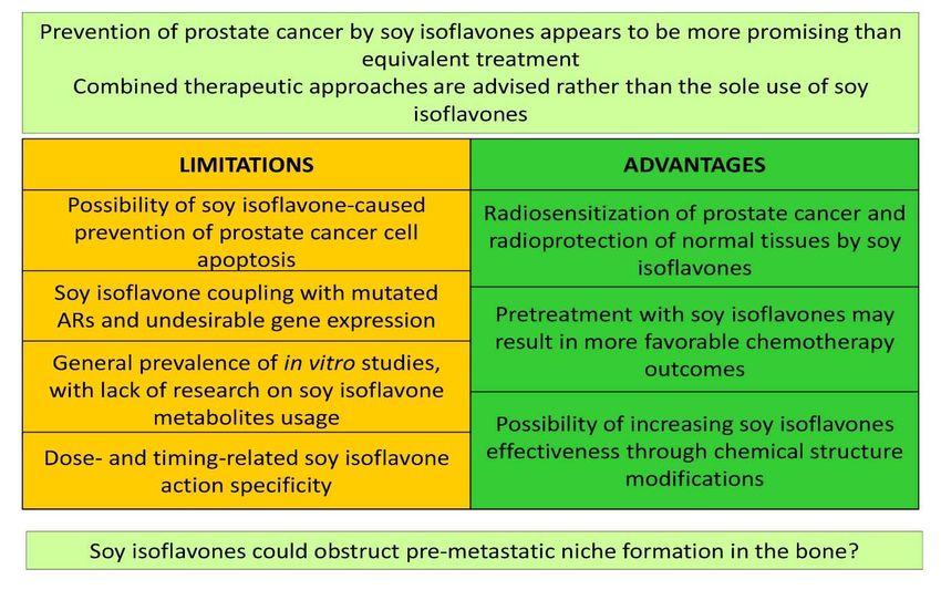
tic ranking (Figure 3). Thus, soy isoflavones
could participate more widely in the com-
bined therapeutic approaches, following the
already demonstrated radiosensitization of
prostate cancer and radioprotection of nor-
mal tissues and organs in the field of radia-
tion, all achieved with the use of soy isofla-
vones (Raffoul et al., 2007; Ahmad et al.,
2010; Hillman, 2019), or given the observed,
more effective docetaxel-induced apoptosis
of prostate cancer cells pretreated with
genistein (Li et al., 2005) (Figure 3). How-
Figure 2: Dynamic phenotype of metastatic
prostate cancer cells (LNCaP and PC-3) and the
ever, studies reporting a lack of combination
general bone morphofunctional status in an an- effects of soy isoflavones and taxane chemo-
dropausal subject, after treatments with soy iso- therapy on castration-resistant prostate can-
flavones (Ajdžanović et al., 2009a, b, 2011, cer should also be considered (Eskra et al.,
2013, 2014, 2015; Filipović et al., 2010, 2018). 2019). The possibility of increasing isofla-
118EXCLI Journal 2019;18:106-126 – ISSN 1611-2156
Received: October 25, 2018, accepted: February 12, 2019, published: February 19, 2019
vone lipophilicity through complexation with the Figure 2 legend while the full reference
transient metal cations and derived inputs on is available in the section “References”. The
the cell signaling machinery during modified authors, Vladimir Ajdžanović, Jasmina
compounds application (Tarahovsky et al., Živanović and Verica Milošević, participate
2014; Ajdžanović et al., 2015) additionally in the COST Action FA 1403 POSITIVe (In-
open the gate for therapy fine tuning (Figure terindividual variation in response to con-
3). sumption of plant food bioactives and deter-
minants involved), supported by the COST
Acknowledgements (European Cooperation in Science and
This work was supported by the Ministry Technology). We are grateful to Mrs. Maja
of Science, Education and Technological Vojvodić, an English language professional,
Development of the Republic of Serbia, for her help in proofreading the manuscript.
Grant number 173009. Part of the Figure 2
was adopted from our previous publication Conflict of interest
and reprinted by permission of the Licensor- The authors declare that they have no
publisher Springer (Ajdžanović et al., The conflict of interest.
Journal of Membrane Biology 246: 307–314,
2013). An appropriate citation is provided in
Figure 3: Evaluation of soy isoflavones significance for prostate cancer therapy. ARs – androgen re-
ceptors
119EXCLI Journal 2019;18:106-126 – ISSN 1611-2156
Received: October 25, 2018, accepted: February 12, 2019, published: February 19, 2019
REFERENCES Ajdžanović V, Trifunović S, Miljić D, Šošić-Jurjević
B, Filipović B, Miler M, et al. Somatopause, weak-
Aalinkeel R, Nair MP, Sufrin G, Mahajan SD, nesses of the therapeutic approaches and the cautious
Chadha KC, Chawda RP, et al. Gene expression of optimism based on experimental ageing studies with
angiogenic factors correlates with metastatic potential soy isoflavones. EXCLI J. 2018;17:279-301.
of prostate cancer cells. Cancer Res. 2004;64:5311-
21. Alonso V, Pérez-Martínez FC, Calahorra FJ, Esbrit P.
Phytoestrogen modulation of bone-related cytokines
Adler HL, McCurdy MA, Kattan MW, Timme TL, and its impact on cell viability in human prostate can-
Scardino PT, Thompson TC. Elevated levels of circu- cer cells. Life Sci. 2009;85:421-30.
lating interleukin-6 and transforming growth factor-
beta 1 in patients with metastatic prostatic carcinoma. Alsulaiman M, Bais MV, Trackman PC. Lysyl oxi-
J Urol. 1999;161:182-7. dase propeptide stimulates osteoblast and osteoclast
differentiation and enhances PC3 and DU145 prostate
Ahmad IU, Forman JD, Sarkar FH, Hillman GG, cancer cell effects on bone in vivo. J Cell Commun
Heath E, Vaishampayan U, et al. Soy isoflavones in Signal. 2016;10:17-31.
conjunction with radiation therapy in patients with
prostate cancer. Nutr Cancer. 2010;62:996-1000. Arnold JT, Isaacs JT. Mechanisms involved in the
progression of androgen-independent prostate can-
Ajdžanović VZ, Šošić-Jurjević BT, Filipović BR, Tri- cers: it is not only the cancer cell's fault. Endocr Relat
funović SL, Brkić DD, Sekulić MI, et al. Genistein af- Cancer. 2002;9:61-73.
fects the morphology of pituitary ACTH cells and de-
creases circulating levels of ACTH and corticosterone Arya M, Bott SR, Shergill IS, Ahmed HU, William-
in middle-aged male rats. Biol Res. 2009a;42:13-23. son M, Patel HR. The metastatic cascade in prostate
cancer. Surg Oncol. 2006;15:117-28.
Ajdžanović V, Šošić-Jurjević B, Filipović B, Tri-
funović S, Manojlović-Stojanoski M, Sekulić M, et al. Behrens J, von Kries JP, Kuhl M, Bruhn L, Wedlich
Genistein-induced histomorphometric and hormone D, Grosschedl R, et al. Functional interaction of beta-
secreting changes in the adrenal cortex in middle-aged catenin with the transcription factor LEF-1. Nature.
rats. Exp Biol Med (Maywood). 2009b;234:148-56. 1996;382:638-42.
Ajdžanović VZ, Šošić-Jurjević BT, Filipović BR, Tri- Bektic J, Berger AP, Pfeil K, Dobler G, Bartsch G,
funović SL, Milošević VLj. Daidzein effects on Klocker H. Androgen receptor regulation by physio-
ACTH cells: immunohistomorphometric and hormo- logical concentrations of the isoflavonoid genistein in
nal study in an animal model of the andropause. His- androgen-dependent LNCaP cells is mediated by es-
tol Histopathol. 2011;26:1257-64. trogen receptor beta. Eur Urol. 2004;45:245-51.
Ajdžanović V, Milošević V, Spasojević I. Glucocorti- Bergan R, Kyle E, Nguyen P, Trepel J, Ingui C,
coid excess and disturbed hemodynamics in advanced Neckers L. Genistein-stimulated adherence of prostate
age: the extent to which soy isoflavones may be bene- cancer cells is associated with the binding of focal ad-
ficial. Gen Physiol Biophys. 2012;31:367-74. hesion kinase to beta-1-integrin. Clin Exp Metastasis.
1996;14:389-98.
Ajdžanović V, Mojić M, Maksimović-Ivanić D, Bula-
tović M, Mijatović S, Milošević V, et al. Membrane Bershadsky AD, Balaban NQ, Geiger B. Adhesion-
fluidity, invasiveness and dynamic phenotype of met- dependent cell mechanosensitivity. Annu Rev Cell
astatic prostate cancer cells after treatment with soy Dev Biol. 2003;19:677-95.
isoflavones. J Membr Biol. 2013;246:307-14.
Bonkhoff H, Berges R. From pathogenesis to preven-
Ajdžanović VZ, Medigović IM, Pantelić JB, Mi- tion of castration resistant prostate cancer. Prostate.
lošević VLj. Soy isoflavones and cellular mechanics. 2010;70:100-12.
J Bioenerg Biomembr. 2014;46:99-107.
Boyle WJ, Simonet WS, Lacey DL. Osteoclast differ-
Ajdžanović V, Medigović I, Živanović J, Mojić M, entiation and activation. Nature. 2003;423:337-42.
Milošević V. Membrane steroid receptor-mediated ac-
tion of soy isoflavones: tip of the iceberg. J Mem- Brehmer B, Biesterfeld S, Jakse G. Expression of ma-
brane Biol. 2015;248:1-6. trix metalloproteinases (MMP-2 and -9) and their in-
hibitors (TIMP-1 and -2) in prostate cancer tissue.
Prostate Cancer Prostatic Dis. 2003;6:217-22.
120You can also read