Splice-correcting oligonucleotides restore BTK function in X-linked agammaglobulinemia model
←
→
Page content transcription
If your browser does not render page correctly, please read the page content below
The Journal of Clinical Investigation                                                                                                                                 Research article

Splice-correcting oligonucleotides restore BTK function
in X-linked agammaglobulinemia model
Burcu Bestas,1 Pedro M.D. Moreno,1,2 K. Emelie M. Blomberg,1 Dara K. Mohammad,1 Amer F. Saleh,3 Tolga Sutlu,4 Joel Z. Nordin,1
Peter Guterstam,5 Manuela O. Gustafsson,1 Shabnam Kharazi,4 Barbara Piątosa,6 Thomas C. Roberts,7,8 Mark A. Behlke,9
Matthew J.A. Wood,7 Michael J. Gait,3 Karin E. Lundin,1 Samir El Andaloussi,1,7 Robert Månsson,4 Anna Berglöf,1
Jesper Wengel,10 and C.I. Edvard Smith1
Department of Laboratory Medicine, Clinical Research Center, Karolinska Institutet, Karolinska University Hospital Huddinge, Stockholm, Sweden. 2Instituto de Engenharia Biomédica, Universidade
1

do Porto, Porto, Portugal. 3Medical Research Council, Laboratory of Molecular Biology, Cambridge, United Kingdom. 4Department of Laboratory Medicine, Center for Hematology and Regenerative Medicine,
Karolinska Institutet, Karolinska University Hospital Huddinge, Stockholm, Sweden. 5Department of Neurochemistry, Stockholm University, Stockholm, Sweden. 6Histocompatibility Laboratory,
Children’s Memorial Health Institute, Warsaw, Poland. 7Department of Physiology, Anatomy and Genetics, University of Oxford, Oxford, United Kingdom. 8Department of Molecular and
Experimental Medicine, The Scripps Research Institute, La Jolla, California, USA. 9Integrated DNA Technologies (IDT), Coralville, Iowa, USA. 10Nucleic Acid Centre, Department of Physics,
Chemistry and Pharmacy, University of Southern Denmark, Odense, Denmark.

             X-linked agammaglobulinemia (XLA) is an inherited immunodeficiency that results from mutations within the gene encoding
             Bruton’s tyrosine kinase (BTK). Many XLA-associated mutations affect splicing of BTK pre-mRNA and severely impair B
             cell development. Here, we assessed the potential of antisense, splice-correcting oligonucleotides (SCOs) targeting mutated
             BTK transcripts for treating XLA. Both the SCO structural design and chemical properties were optimized using 2′-O-methyl,
             locked nucleic acid, or phosphorodiamidate morpholino backbones. In order to have access to an animal model of XLA,
             we engineered a transgenic mouse that harbors a BAC with an authentic, mutated, splice-defective human BTK gene. BTK
             transgenic mice were bred onto a Btk knockout background to avoid interference of the orthologous mouse protein. Using this
             model, we determined that BTK-specific SCOs are able to correct aberrantly spliced BTK in B lymphocytes, including pro–B
             cells. Correction of BTK mRNA restored expression of functional protein, as shown both by enhanced lymphocyte survival and
             reestablished BTK activation upon B cell receptor stimulation. Furthermore, SCO treatment corrected splicing and restored
             BTK expression in primary cells from patients with XLA. Together, our data demonstrate that SCOs can restore BTK function
             and that BTK-targeting SCOs have potential as personalized medicine in patients with XLA.

Introduction                                                                                               signaling, which is a determinant for proliferation, differentiation,
X-linked agammaglobulinemia (XLA) is the most common form of                                               and survival of the early B cell stages (3, 11, 12).
inherited agammaglobulinemia, accounting for 85% of all cases (1,                                               Patients with XLA are prone to recurrent infections by pyo-
2). In this disease, a B cell lineage developmental block, manifested                                      genic bacteria, such as pneumococci and streptococci. Affected
at the transition between the pro–B and pre–B cell stages (3), leads to                                    subjects are also unduly susceptible to enteroviruses, which cause
the essential lack of mature B and plasma cells, with a concomitant                                        dermatomyositis and fatal chronic encephalomyelitis (13–16).
pronounced reduction of immunoglobulin levels. XLA is caused by                                            The current treatment for XLA consists of prophylactic, regular
defects in the gene encoding Bruton’s tyrosine kinase (BTK) (4–6).                                         intravenous, or subcutaneous gammaglobulin replacement ther-
BTK is a cytoplasmic, nonreceptor tyrosine kinase belonging to the                                         apy and generous administration of antibiotics. This consider-
TEC family of tyrosine kinases (7) and is expressed in all stages of                                       ably improves quality of life, but patients still suffer from chronic
B cell development, with the exception of long-lived, antibody-pro-                                        infections (17, 18). Furthermore, life expectancy is reduced despite
ducing plasma cells (8, 9). Besides B lymphocytes, many other                                              appropriate care (16, 19–21).
hematopoietic cells also express BTK (5, 8–10), but clinically rele-                                            Splicing defects have been identified as an important cause
vant functional impairment seems to be confined to this lineage.                                           of genetic disease. Many mutations affecting splicing disrupt the
Thus, the severe clinical outcome of BTK mutations demonstrates                                            regular 5′ splice site (5′ss) or 3′ss at exon-intron junctions. Further,
the essential role of BTK already occurring in pre–B cell receptor                                         mutations can create new splice sites, which may result in the inclu-
                                                                                                           sion of intronic sequences or loss of part of the exons in the tran-
                                                                                                           script. When such a new splice site occurs in an intron in the vicin-
Authorship note: Burcu Bestas and Pedro M.D. Moreno contributed equally to this work.                      ity of a suitable pseudo splice site, the intervening intronic region
Conflict of interest: Patent applications have been filed relating to the technologies
                                                                                                           can be included in the mRNA as a cryptic exon. This mechanism
described herein, which are assigned to IDT. Mark A. Behlke is employed by IDT but does
not personally own any shares or equity in IDT. IDT is not a publicly traded company.
                                                                                                           has been observed in multiple diseases, including cystic fibrosis
Submitted: March 21, 2014; Accepted: July 3, 2014.                                                         (22) and ataxia telangiectasia (23, 24) among others. In XLA, we
Reference information: J Clin Invest. 2014;124(9):4067–4081. doi:10.1172/JCI76175.                         have previously identified and described 2 such families (25, 26).

                                                                                                           jci.org   Volume 124   Number 9   September 2014                                  4067
Research article The Journal of Clinical Investigation
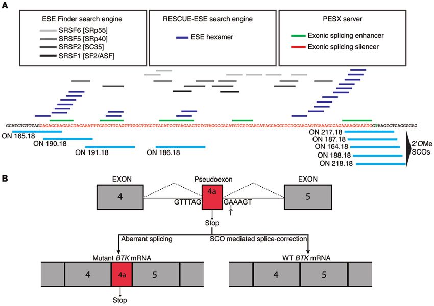
Figure 1. Design of SCOs using bioinformatic tools to search for ESE sites in the disease-causing pseudoexon. (A) Three different algorithms from the
corresponding web-based servers, ESE-Finder, RESCUE-ESE, and PESX, were used, and the hits were aligned to the pseudoexon sequence to find locations
with the highest correlation between algorithms. SCOs targeting the probable ESE regions were subsequently designed. SCOs are schematically presented
with their binding-positions along the pseudoexon. (B) Predicted outcome of splicing, with or without SCOs, indicated schematically.
The XLA defect studied in this work arises from one of these exon-skipping SCOs has been used successfully to generate a
families, which has an A-to-T transition in intron 4 of the BTK gene truncated but partially functional protein (31, 32).
generating a novel 5′ss. This, together with a preexisting cryptic Here, we describe the feasibility of splice correction by pre-
3′ss upstream in the same intron, results in the inclusion of a cryp- venting cryptic exon inclusion as a personalized therapy for XLA.
tic exon (exon 4a) of 109 nucleotides between exons 4 and 5 in the In order to achieve this, we designed SCOs targeting various sites
mRNA (25). This changes the reading frame and completely abol- in the pseudoexon region of BTK pre-mRNA. Different oligonucle-
ishes BTK protein expression. The erroneous inclusion of exon 4a otide (ON) chemistries have been developed over recent years in
prompted us to investigate the possibility of using splice-correct- order to improve resistance to degradation, enhance target affin-
ing antisense oligonucleotides (SCOs), which bind to and restore ity, and promote cellular uptake. In this study, we investigated
the splicing of the pre-mRNA, a concept also exploited in other SCOs with 2′-O-methyl phosphorothioate (2′OMePS) or phos-
diseases, as reviewed recently (27, 28). phorodiamidate morpholino (PMO) backbone chemistries. For
Apart from the splice sites themselves, splicing is also con- simplicity, the splice-correcting PMO-based oligomers are also
trolled by short regulatory sequence motifs in both exons and referred to as SCOs.
introns. These motifs are designated exonic or intronic splic- The PMO was also tested in a cell-penetrating peptide–conju-
ing enhancers (ESEs or ISEs, respectively) or exonic or intronic gated (CPP-conjugated) version. CPPs are a class of peptides with
splicing silencers (29). These are also of interest since SCOs the ability to translocate rapidly into cells and to transport various
targeting exonic splicing silencers have been shown to induce macromolecular cargos. PMO-based SCOs conjugated with CPPs
the inclusion of exon 7 in the transcript of the SMN2 gene in have been shown to be very efficient as therapeutics against splice
spinal muscular atrophy (30). Similarly, exonic splicing silencer abnormalities, especially in the mdx mouse model of DMD (33).
regions have been targeted in the case of Duchenne muscular Furthermore, we investigated nucleic acid modifications, such as
dystrophy (DMD), which is caused by mutations in the DMD locked nucleic acid (LNA), since they have been used successfully
gene. In this case, restoration of a disrupted reading frame by in many different settings, such as antisense gapmers, siRNAs,
4068 jci.org Volume 124 Number 9 September 2014The Journal of Clinical Investigation Research article
has been shown recently to have potentiating effect for antago-
Table 1. SCOs used in the study
mirs, it was also tested (37).
ON (SCO) Sequence (5′→3′) We targeted primary patient cells as well as lymphocytes from a
2′-O-methyl RNA – genetically altered mouse created for this purpose. This transgenic
164.18 ugagacuuaccaccuccu mouse has several unique features; it carries the entire human
165.18 uugcucuccuaaacagau mutant BTK locus and is devoid of endogenously expressed BTK
186.18 cagaguucucaggaugua protein. Notably, in contrast to the treatment concept for DMD,
187.18 gagacuuaccacuuccuu in which a truncated protein with reduced function is formed, the
188.18 cugagacuuaggacuucc strategy in XLA permits the restoration of the authentic reading
190.18 auuuguaguucuugcucu frame generating a fully functional protein. Importantly, because of
191.18 caaacugaagaccaaauu the developmentally regulated expression of BTK, it may become
217.18 agacuuaccacuuccuuu feasible to achieve long-term correction of the disease phenotype.
218.18 ccugagacuuaccacuuc Moreover, besides proving the viability of splice-modifying therapy
705.18 (unrelated control) ccucuuaccucaguuaca for a novel target disease both ex vivo and in vivo, we demonstrate
LNA/2′-O-methyl RNA and DNA – for the first time that both primary B cells and primary monocytes
190.15-5LNA uTugTagTucTugCu are amenable to splice correction strategies.
187.15-5LNA aGacTuaCcaCuuCc
187.18-6LNA gAgaCuuAccAcuTccT Results
187.12-8LNA AcTTaCCaCTTc
Design of SCOs using bioinformatics tools. Different SCOs directed
187.12-8LNA-4LNAgly AcXXaCCaCXXc
against the BTK pseudoexon region were designed, taking into
187.10-9LNA TTACCaCTTC
consideration not only the 3′ss and 5′ss of exon 4a, but also puta-
186.15-3LNA gAguucuCaggauGu
186-15-5LNA gAguTcuCagGauGu tive ESE sites. For this purpose, we used 3 different algorithms
186.15-6LNA-2UNA gAgTucuCaGgaTGu from the web-based servers ESE Finder, PESX, and RESCUE-ESE.
186.15-3LNA-2UNA gAguucuCaggauGu The sequence hits were aligned to exon 4a, as depicted in Figure 1,
186.12-8LNA-3LNAgly TXcXCAggAXgT which also shows the studied mutation and the expected out-
186.12-8LNA-4LNAgly TXcXCAggAXgX comes with or without splice correction. This analysis prompted
186.12-8LNA-5LNAgly XXcXCAggAXgX us to generate a set of SCOs, as schematically indicated in Figure 1.
186.12-8LNA TTcTCAggATgT They were designed to cover the 3′ss and 5′ss and the ESEs sites
5′186.12-7LNA AgTTcTCagGaT that demonstrated the most concordance between the algorithms
GAA.12-6LNA cTaTaTTCaCga used. The first set of SCOs was composed of the commonly used
GAA.19-3LNA cuTccuuuTcuggcuuTca 2′OMePS modification, taking into consideration the GC con-
GAA.19-6LNA cuTccTuuTcuGgcTuuCa tent, self-dimerization, and nucleic acid melting temperature
Scrambled cAcuCuaTauAacTg (Tm) values (38). Additional sets were then designed with vari-
ZEN LNA/2′-O-methyl RNA – ous modifications and lengths. A schematic representation of the
186.15-5LNA-ZEN g/ZEN/AguTcuCagGauG/ZEN/u SCOs is shown in more detail in Supplemental Figure 1 (supple-
186.18-ZEN c/ZEN/agaguucucaggaugu/ZEN/a mental material available online with this article; doi:10.1172/
PMO & B-PMO – JCI76175DS1), and the modifications are indicated in Table 1.
B-PMO 186.25 B-peptide-CTACAGAGTTCTCAGGATGTAAGCA SCOs restore luciferase mRNA in a reporter cell line model. We first
Scrambled B-PMO (DMD) B-peptide-GGCCAAACCTCGGCTTACCTGAAATT designed a reporter construct containing the expression cassette
B-peptide N-RXRRBRRXRRBRXB-C
for a fusion of EGFP and firefly luciferase but interrupted by the
B-peptide was conjugated to PMO 186.25 to generate B-PMO 186.25. mutated patient-derived BTK intron 4. When uncorrected, this mini-
2′-O-methyl RNA SCOs (including those modified by LNA and ZEN) gene produces a corrupt EGFP-luciferase mRNA. However, intro-
have phosphorothioate backbone. LNA bases are in uppercase letters;
duction of an SCO capable of rescuing the splicing of the mutated
2′-O-methyl RNA bases are in lowercase letters; UNA bases are underlined;
intron would promote restoration of EGFP-luciferase fusion protein
DNA bases are in bold letters; “X” represents amino-glycyl-LNA-T.
expression. The model was constructed as a first functional read-
out for SCO activity and used to generate a stable U2OS reporter
cell line expressing the above-mentioned corrupt transcript. Sub-
anti-microRNAs (antagomirs), and anti-gene approaches (34, 35). sequently, the fully 2′OMePS-modified SCOs (sequences depicted
An LNA-containing ON with its 3′-endo conformation is consid- in Table 1) were transfected, and luciferase enzymatic activity was
ered to be an RNA mimic, making it effective toward structured determined (Figure 2A). Several of the tested SCOs showed prom-
RNA regions, which can be beneficial when targeting pre-mRNAs. ising results when compared with the unrelated, control SCO 705
This is due to the fact that position-dependent substitution with directed against a mutated β-globin intron (39). SCO 187, covering
LNA bases changes the thermodynamic property of the duplexes the 5′ss, performed the best in these experiments, whereas SCO
(34, 36). Additionally, we also investigated LNA in combination 165, covering the 3′ss, demonstrated very low efficacy. SCOs 186,
with other derivatives, such as unlocked nucleic acid (UNA) or 187, and 190 were chosen for further experiments. SCOs 186 and
amino-glycyl-LNA (34). Moreover, since the nonbase modifier 190 were selected because of their activity and the fact that they
N,N-diethyl-4-(4-nitronaphthalen-1-ylazo)-phenylamine (ZEN) were not targeted to splice sites but instead to the predicted ESEs.
jci.org Volume 124 Number 9 September 2014 4069Research article The Journal of Clinical Investigation
Figure 2. Splice correction–induced upregulation of reporter minigene activity. The U2OS cell line stably carrying a minigene interrupted by the
mutated BTK intron 4 was transfected with different SCOs, and efficacy was measured as luciferase activity or mRNA restoration. (A) The relative
luciferase activity in comparison to nontreated cells (mock) 24 hours after SCO transfection. “705” indicates an unrelated control SCO, targeting position
705 of a mutated β-globin intron, not complementary to the BTK pre-mRNA. All tested SCOs are 2′OMePS based (Table 1). Data represent mean + SD of
3 independent experiments, each with 2 replicates. **P ≤ 0. 01, ***P ≤ 0.001, versus 705. (B) Increase in luciferase activity by 2′OMePS-modified SCOs
versus LNA SCOs is presented as fold increase over nontreated cells (mock). Graph represents mean + SD of 3 independent experiments, each with 2 rep-
licates. **P ≤ 0. 01, ****P ≤ 0.0001, 186.18 versus 186.15-5LNA. (C) Total RNA RT-PCR showing the 389-bp and 271-bp bands corresponding to aberrant
and corrected (with BTK intron 4 excised) mRNA bands, respectively, with a lower band of ribosomal RNA (18S) serving as an RNA quality control. The
lane on the right represents RNA input from nontreated (NT) cells. “Scr” represents the control LNA/2′OMePS SCO not complementary to the reporter
gene sequence (Table 1). A representative gel from 2 independent experiments is shown. Statistical significance was determined using 1-way ANOVA,
followed by Bonferroni’s multiple-comparison test.
SCO chemistry affects splice correction activity in reporter cell of the corrected form at low SCO concentrations. The 190 series
line. We modified the selected SCOs with LNA bases, which are of SCOs was not further assessed, since at higher concentrations
known to significantly enhance Tm and increase the efficiency of there were signs of toxicity in the form of cell death 24 hours after
ONs both in vitro and in vivo (40–44). More specifically, we chose transfection (data not shown).
a 2′OMe RNA backbone with LNA modifications, as this combi- Based on these results we further modified the 186 and 187
nation has previously demonstrated high potency (45–48). Since series, including a reduction of the ON length further to 10 to 12
size-reduced SCOs are generally considered to be more efficient nucleotides. We also synthesized 3 additional SCOs, referred to as
(34, 42, 49), with lowered risk of off-target effects and improved the GAA series. These were designed to target GAA-triplets, known
uptake, we designed LNA-modified 15-mer versions of the 186, to be present in certain splicing-enhancers (26, 50), as shown in
187, and 190 series (Supplemental Figure 1 and Table 1). Figure 2 Supplemental Figure 1 and Table 1. Supplemental Figure 2 depicts
shows the activity of LNA-modified SCOs in the U2OS reporter the effect of SCOs derived from the 186, 187, and GAA series, as
cell line, as measured by luciferase expression and as also con- analyzed by RT-PCR. The doses were lowered, as compared with
firmed by the correction of mRNA determined by RT-PCR. It is previous experiments (Figure 2), in order to identify the most
clearly shown that the LNA-modified SCOs mediate enhanced potent SCOs. As can be seen in Supplemental Figure 2, LNA-mod-
luciferase activity at low concentrations. The same tendency ified SCOs displayed higher potency as compared with their corre-
was confirmed at the RNA level, at which we observed a reduc- sponding 2′OMePS versions. 186.15-5LNA and 186.15-3LNA (with
tion of the aberrant mRNA band and a concomitant appearance 5 and 3 LNA substitutions, respectively) showed the best efficiency
4070 jci.org Volume 124 Number 9 September 2014The Journal of Clinical Investigation Research article
Figure 3. Splice correction–induced upregulation of reporter minigene
activity following naked uptake of SCOs. SCOs from the 186 series,
including PMO 186.25, with and without the CPP moiety (B-PMO 186.25),
were tested, and activity was measured as restoration of mRNA. A concen-
tration of 6 μM was used for the scrambled control SCO (Scr) (Table 1). A
representative gel from 2 independent experiments is shown.
in the reporter cell assay. The 12-mer versions of both the 186 and that ZEN modification is more useful when used in the context of
187 series did not exhibit any enhanced activity under these in vitro a pure 2′OMe backbone and not when combined with shorter LNA
conditions, and the 10-mer of SCO 187 was inactive (Supplemen- ONs. Taken together, these additional modifications did not show
tal Figures 2 and 3). Moreover, the newly designed GAA series did enhanced activity and were not studied further.
not show better efficiency compared with 186.15-5LNA or 186.15- LNA-based SCOs and PMOs conjugated to a CPP are efficiently
3LNA and was therefore not studied further. taken up (gymnosis) in the reporter cell line. We next investigated
In addition, we investigated 3 more chemical modifications the naked uptake efficiency in the reporter cell line under “gym-
(Table 1): amino-glycyl-LNA, UNA, and the nonbase modifier nosis” conditions. The term gymnosis was coined by Stein et al.
ZEN. The amino-glycyl modification adds a glycyl residue to the to describe uptake of LNA gapmers by cells in the absence of any
N2′ position of 2′-amino-LNA, thereby providing a positive charge transfection reagent (53). The authors suggested that this mode
(51). UNA is known to decrease the Tm and destabilize the duplex, of in vitro delivery better correlated to in vivo uptake, making
which potentially could increase target specificity if appropriately this method of general interest. Compared with lipofection, con-
designed (52). ZEN is a naphthyl-azo modifier; when placed at siderably higher concentrations of ONs are needed for gymnosis.
or near both ends of a 2′OMe ON, it
increases Tm and enhances the sta-
bility against endo- and exonucleases
(37). These new modifications were
also tested in the reporter cell line
model. As can be seen in Supple-
mental Figure 3, the glycyl-modified
12-mer versions of 186 and 187 series
showed reduced activity with increas-
ing numbers of amino-glycyls cor-
relating to further reduction in activ-
ity. This was somewhat unexpected,
since the reduced net negative charge
potentially could have enhanced their
splice-correcting capacity by allow-
ing better hybridization to the cog-
nate pre-mRNA. On the other hand,
decreased net negative charge might
also have reduced the complexation
with the transfection reagent Lipofect-
amine 2000, which potentially could
have altered both the complex con-
formation and the amount transfected
into cells. Conversely, UNA incorpora-
tion into the 186 series of SCOs caused
reduced activity at low LNA content, Figure 4. Full-length human mutated BTK pre-mRNA corrected by SCOs yielding normal-sized transcripts
whereas with higher LNA content and detectable BTK protein. (A) B cells from human BAC transgenic mice were treated with 1.6 μM or
3.2 μM 186.15-3LNA SCO and analyzed by RT-PCR. To identify human RNA in cells from BAC transgenic and
the activity was somewhat restored. KO mice, a primer pair only detecting human BTK was used. To measure endogenous mouse Btk mRNA
Moreover, ZEN-modified SCO levels in splenocytes derived from WT mice, a different primer set selective for mouse Btk was used. A rep-
(186.15-5LNA-Zen), as compared with resentative gel from 3 independent experiments is shown. Ribosomal RNA (18S) was used as an RNA qual-
186.15-5LNA (Supplemental Figure 2), ity control. (B) FACS analysis of cells 48 hours after treatment with 186.15-3LNA. B cells were stained with
did not show much difference in activ- antibodies directed against CD19 or BTK protein (recognizing both mouse and human protein). Histograms
for BTK expression in each sample from a representative experiment as well as the percentage of corrected
ity. The slightly longer 186.18-ZEN had cells from 3 independent experiments are shown (mean + SD). A concentration of 3.2 μM was used for the
almost the same effect as 186.15-5LNA scrambled control SCO (Table 1). Mean fluorescence intensity, WT NT: 2,314; BAC SCO treated: 1,210. Per-
and 186.15-3LNA. This might suggest centage of WT BTK RNA was calculated as WT RNA fraction × 100/(misspliced + WT RNA fractions).
jci.org Volume 124 Number 9 September 2014 4071Research article The Journal of Clinical Investigation
Figure 5. Enhanced survival and anti-IgM–induced BTK tyrosine phos-
phorylation in SCO-treated BAC transgenic B cells. (A) Spleen B cells
from BAC transgenic mice were treated with 3.2 μM SCO (186.15-3LNA or
scrambled control) and stimulated with 20 μg/ml anti-IgM 48 hours after
electroporation (time 0). Cells were counted, and viability was determined
by trypan blue exclusion 24 and 48 hours after stimulation. The initial
number of cells from both WT and BAC transgenic mice was 2 × 10⁶.
Data represent 2 independent experiments with duplicates. Note that
nontreated and scrambled ON–treated cell numbers overlap. Statistical
significance was analyzed by using 1-way ANOVA followed by Bonferroni’s
multiple-comparison test. ****P ≤ 0.0001 for BAC SCO treated versus BAC
Scr and NT at 24 hours. (B) Analysis of BTK tyrosine phosphorylation after
3.2 μM SCO (186.15-3LNA) treatment and anti-IgM stimulation (20 μg/ml).
Blots show pY551 phosphorylation of BTK and total BTK. Whole cell
lysate (WCL) analysis shows BTK protein and actin. Each duplicate (–/+)
corresponds to starved or anti-IgM–activated conditions. A representa-
tive blot is shown from 2 independent experiments. Note that the higher
molecular weight band, located immediately above the pY551-BTK band
in activated samples (IP pY551 gel), is of unknown origin and unrelated
to BTK, since it also exists in B cells obtained from Btk KO mice follow-
ing stimulation. The bar graph shows the quantitative analysis of Y551
phosphorylation as a percentage of relative intensity signal from the blots
according to ImageJ Software.
We selected the most efficient SCOs based on the results from A-to-T change as seen in the XLA-family described previously. We
the lipofection experiments and assessed their gymnotic activ- confirmed that the BAC transgenic mice carry the mutated human
ity. Although 186.15-5LNA exhibited good activity, we continued BTK gene by sequencing of chromosomal DNA (data not shown).
with the similarly efficient 186.15-3LNA, owing to the fact that The founder animals were then backcrossed onto Btk knockout
reduced LNA content potentially could reduce off-target effects (KO) mice in order to generate transgenic animals with a disrupted,
(45). Although ZEN-modified SCOs had similar activity compared nonfunctional endogenous Btk locus, which were therefore only
with the LNA-containing 186.15 versions, they could not be stud- capable of expressing the mutated human BTK gene (Supplemen-
ied, since their availability was limited. tal Figure 4). Throughout this report we refer to these mice as BAC
In addition, we also investigated the PMO backbone (54, 55), transgenic mice. We also verified that the insertion of the transgene
with the 186 sequence in the gymnosis setting. Owing to the rules did not influence hematopoietic development; Supplemental Fig-
of PMO design, this SCO was further extended to be 25 nucleo- ure 5 shows that the common lymphoid progenitors and B cell com-
tides (Supplemental Figure 1 and Table 1). The rationale for length- partment are similar in Btk KO and BAC transgenic mice.
ening PMOs is the lower target affinity (56). We further conjugated SCOs correct mRNA and restore BTK protein in primary B cells ex
the PMO with a CPP moiety named B-peptide (B-PMO), since it is vivo. To test the activity of the SCOs in the context of a full-length
known to be very active for exon skipping in the mdx mouse model pre-mRNA transcribed from the mutant human BTK gene, we
(57). Thus, the PMOs tested were either unmodified or covalently isolated splenic B cells from mice expressing the human mutant
conjugated to B-peptide. BTK transcript. As control, we used B cells from WT and Btk KO
As can be seen from Figure 3, in gymnosis setting, 186.15- mice. For transfections of SCOs, we optimized a protocol for
3LNA and B-peptide–conjugated PMO 186.25 (B-PMO) showed electroporation of mouse primary B cells. At 48 hours after elec-
high potency in the reporter cells when analyzed by RT-PCR. troporation, cells were collected, and human BTK RNA was ana-
We also tested higher (up to 10 μM) concentrations of PMO lyzed by semiquantitative RT-PCR, using primers detecting only
186.25, which were devoid of any CPP; however, the exon-skip- human BTK. Another primer set was used to specifically amplify
ping effect was very low. mouse Btk RNA in order to verify that this transcript is unaffected
Generation of a transgenic mouse carrying the mutated human by the SCOs (Figure 4A). The results clearly show that, by using
BTK locus. In order to have access to an unlimited supply of mutated SCO 186.15-3LNA, splicing of the full-length human mutant BTK
B-lineage cells as well as a relevant in vivo model, we generated a pre-mRNA was corrected, as demonstrated by the appearance of
mouse carrying the corresponding human BTK locus. A suitable a lower molecular weight amplicon corresponding to the size of
BAC clone named RP11-125I5 was chosen for the development unmutated, native BTK mRNA. The correction was also verified by
of BAC transgenic mice. This clone encompasses 175 kb and con- sequencing the cDNA (data not shown).
tains an extended human genomic BTK locus. The BAC was sub- The RT-PCR results were also confirmed by flow cytometry
sequently subjected to site-specific mutagenesis, generating a sin- analysis on cells collected 48 hours after transfection (Figure 4B).
gle-point mutation at position 602 of BTK intron 4, the very same As can be seen from Figure 4B, treatment with 186.15-3LNA gen-
4072 jci.org Volume 124 Number 9 September 2014The Journal of Clinical Investigation Research article
Figure 6. B-PMO–mediated restoration of BTK mRNA and protein in pri-
mary spleen B cells from human BAC transgenic mice. Cells were treated
with PMO 186.25 or B-PMO 186.25 in different concentrations. “Scr”
indicates a B-PMO SCO for an unrelated target (DMD). (A) RT-PCR prod-
ucts from SCO-treated B cells at 1 and 3 μM concentrations. Ribosomal
RNA (18S) serves as an RNA quality control. Of note, for the splenocytes
derived from WT mice, a different primer set selective for mouse Btk
was used. The percentage of WT BTK RNA was calculated as WT RNA
fraction × 100/(misspliced + WT RNA fraction). (B) Western blot from the
same treatment. Experiments were repeated 3 times, and representative
gels are depicted. Of note, for this experiment, anti-BTK from BD (see
Methods) was used, yielding different unspecific bands. The bottom band
originates from BTK, while the top band is unspecific. The lanes separated
by a black line were run on the same gel, but since some lanes were
considered uninformative, they were omitted as indicated by the black
line. The bar graph shows the quantitative analysis of BTK protein as a
percentage of relative intensity signal according to ImageJ Software. Scr,
treated with scrambled ON.
receptor–induced signaling leads to tyrosine Y551 phosphorylation
of BTK. The corresponding tyrosine is conserved in all cytoplasmic
protein-tyrosine kinases and upon phosphorylation significantly
enhances their catalytic activity owing to a conformational change
(3, 13). SCO-treated B cells from BAC transgenic mice were com-
pared with B cells from WT mice. 48 hours after electroporation,
cells were serum starved for 6 hours and subsequently incubated
with 20 μg/ml anti-IgM before harvest. BAC transgenic B cells
treated with 186.15-3LNA SCO showed pronounced phosphoryla-
erated a mean of over 40% of cells expressing substantial levels tion when activated with anti-IgM (Figure 5B). The level was almost
of BTK protein. These experiments demonstrate that not only as high as in WT cells, suggesting functional restoration of BTK.
the reporter minigene-derived transcript carrying the mutated B-PMOs efficiently promote splice correction in primary B cells.
BTK intron, but also the full-length human BTK pre-mRNA is We next evaluated the effects of naked delivery into primary cells.
amenable to SCO therapy. The non-PMO SCOs, initially evaluated in the reporter gene assay
SCOs restore functional BTK in primary B cells from the BAC (Supplemental Figure 2), were again selected based on potency
transgenic mouse. Since we could conclusively show the presence of and assessed in primary B cells. In contrast to the gymnotic effect
BTK protein by flow cytometry analysis, we investigated whether seen in the reporter cell line, we were unable to detect any signifi-
the synthesized protein is also functionally active. For this pur- cant reproducible correction of the BTK mRNA in primary B cells
pose, 2 functional assays were carried out, cell viability and phos- under these conditions (data not shown). We subsequently evalu-
phorylation of the BTK regulatory loop tyrosine (Y551). ated the B-PMO 186.25 as well as the corresponding control, PMO
The rationale for the cell viability test is that impaired matura- 186.25. Notably, treatment with 3 μM B-PMO 186.25 produced
tion and survival of B cells are hallmarks of XLA (1, 2, 13). Moreover, corrected BTK mRNA (Figure 6A) as well as restored BTK protein
it is known that BTK-deficient mouse B cells do not proliferate in expression (Figure 6B) in B cells from the BAC transgenic mice.
response to anti-IgM stimulation, while WT cells
do (58, 59). Therefore, we stimulated BAC trans-
genic mouse B cells 48 hours after SCO treatment
with anti-IgM and incubated them for another
24 or 48 hours in the absence of the survival-pro-
moting CpG stimulus (see Methods), conditions
rather harsh for primary B cells. Figure 5A shows
the viability of primary B cells after 24 or 48 hours
of incubation with anti-IgM. As seen, under these
experimental conditions SCO-treated BAC trans-
genic B cells showed enhanced survival compared
with nontreated or scrambled, SCO-treated cells.
As a sign of the harsh environment, WT mouse B Figure 7. Restoration of BTK protein expression after splice correction in pro–B cells from BAC
cells were also reduced in numbers. As a second transgenic mice. Cells were electroporated with the 186.15-3LNA SCO. After 48 hours, cells were
stained with anti-CD19 and anti-BTK (recognizing both mouse and human protein) for FACS
proof of BTK functionality, we analyzed the BTK analysis. Numbers in the 3 right upper quadrants indicate the percentage of CD19-positive and
phosphorylation after B cell receptor stimulation. BTK-positive cells. The experiment was repeated twice with a similar outcome, and a represen-
In normal B cells, upon anti-IgM treatment, B cell tative result is presented.
jci.org Volume 124 Number 9 September 2014 4073Research article The Journal of Clinical Investigation
IL-7, the expansion of pro–B cells was induced in vitro
(see Methods). After 9 days, an essentially pure pro–B
cell population was obtained (Supplemental Figure
6). We subsequently transfected these cells with the
186.15-3LNA SCOs and assessed BTK protein expres-
sion at 48 hours. Encouragingly, as many as 33.8% of
the pro–B cells were BTK positive (Figure 7).
In vivo administration of SCOs restores BTK expres-
sion in BAC transgenic mice. In order to investigate
whether the SCOs also had the capacity to correct the
defect in vivo, 4 animals were treated with a multiple-
dose regimen using B-PMO 186.25. They received
B-PMO 186.25 (30 mg/kg) 4 times: 3 doses intrave-
nously every second day during 1 week, followed by a
single subcutaneous dose; animals were assayed after
another 3 or 4 days, respectively. As is depicted in
Figure 8, A and B, and Supplemental Figure 7, a robust
therapeutic effect was observed both in the bone mar-
row and in the spleen. In contrast to control mice, all
treated mice expressed corrected BTK mRNA as well
as BTK protein. Moreover, we isolated B cells from 2
treated and 2 untreated animals in order to be able to
analyze BTK restoration specifically in this cell type.
As shown in Figure 8C, we found that B-PMO was
taken up by B cells both in bone marrow and spleen, as
evidenced by the restored BTK expression.
Collectively, this suggests that the splice correction
concept could become a viable treatment option for
patients with XLA, although standardization of multi-
ple parameters is needed prior to any clinical approach.
SCOs correct splicing and restore BTK protein in
monocytes from patients with XLA ex vivo. As a final
proof of concept in a human setting, we applied our
strategy to primary patient cells. Similar to B cells,
monocytes express the BTK gene but, in contrast, do
Figure 8. Restoration of BTK expression upon splice correction after in vivo treatment not need BTK protein for normal development or sur-
of BAC transgenic mice. Four mice were treated with B-PMOs, as described in Methods,
vival (9, 13). Since patients with XLA essentially lack
and assayed for BTK restoration. (A) RT-PCR analysis of total spleen and total bone
marrow cells from B-PMO–treated transgenic mice. A representative gel from 2 animals, circulating B cells but do have normal numbers of cir-
both from the treated and the untreated group, is shown. “WT-2” represents RNA from culating monocytes, we obtained monocytes from a
normal mice, and the slightly smaller amplicon in this lane results from a differential patient with the BTK intron 4 cryptic splice site muta-
primer set discriminating human and mouse RNA. To ascertain the detection of corrected tion. Since splicing factors in mice and humans are
transcripts for samples from treated and untreated BAC transgenic animals, we decided
similar but not identical, it was important to ascertain
to use 3 times more cDNA and 5 more amplification cycles. (B) Western blot analysis of
BTK restoration in 2 of 4 treated animals; total cells from bone marrow and spleen. (C) that splice correction of full-length pre-mRNA also
Western blot analysis of the isolated B cells both from spleens and bone marrow, show- could take place in primary patient cells.
ing BTK expression from 2 representative animals. The percentage of WT BTK RNA was Electroporation conditions for transfection of
calculated as WT RNA fraction × 100/(misspliced + WT RNA fraction). Bar graphs show SCOs into human monocytes were optimized, and
the quantitative analysis of BTK protein as a percentage of relative intensity according to
their therapeutic effect was assessed. Using 186.15-
ImageJ Software. T, treated.
3LNA, splice correction was observed for BTK both
at the RNA (Figure 9A) and protein level (Figure 9B).
SCOs restore BTK protein expression in pro–B cells from the BAC Moreover, we also evaluated naked uptake of B-PMO 186.25 in
transgenic mouse. Patients with XLA are devoid of mature B cells but patient monocytes, and SCOs readily restored BTK mRNA in these
harbor differentiation-arrested pro–B cells in their bone marrow cells (Figure 9C) as well as yielded the corresponding protein dose
(13, 60). Because of this fact, it was important to establish whether dependently (Figure 9D). Even at a very high concentration (10 μM),
these cells are also amenable to SCO treatment as a proof of con- PMO 186.25, devoid of the CPP moiety, did not restore BTK pro-
cept. For this reason, we first isolated and differentiated c-KIT+ pro- tein synthesis, suggesting the crucial importance of the CPP.
genitor cells from the bone marrow of BAC transgenic mice. With Thus, the results obtained in splice-corrected monocytes
support of OP9 stromal cells, Flk-2/Flt3 ligand, KIT ligand, and derived from a patient with XLA are in concordance with the
4074 jci.org Volume 124 Number 9 September 2014The Journal of Clinical Investigation Research article
Figure 9. SCO-induced BTK restoration in patient monocytes. (A) Results from RT-PCR 48 hours after electroporation of monocytes from a patient with
XLA with the intron 4 mutation. Monocytes were transfected with 1 μM 186.15-3LNA. Human control monocytes were also included. Ribosomal RNA (18S)
serves as an RNA quality control. (B) Western blot showing BTK protein 48 hours after electroporation with or without treatment with 1 μM 186.15-3LNA.
The right lane with a mouse B cell sample serves as a positive control for BTK (mouse and human BTK protein are both composed of 659 amino acids and
recognized by the same antiserum). (C) RT-PCR results from patient monocytes treated with B-PMO 186.25 (3 μM and 6 μM). (D) Western blot showing
BTK protein from B-PMO 186.25– and PMO 186.25–treated patient monocytes. Note that the anti-BTK antibody used for the monocyte lysates generates
a strong, nonspecific band appearing just below the BTK-specific band, while the specific band is completely absent from the nontreated patient control
cells (lane 1). Actin served as a loading control. Representative gels from 3 independent experiments are shown. The lanes separated by a black line were
run on a different gel. The percentage of WT BTK RNA was calculated as WT RNA fraction × 100/(misspliced + WT RNA fraction). The bar graphs show the
quantitative analysis of BTK protein as a percentage of relative intensity according to ImageJ Software. MΦ, monocytes.
findings in B lymphocytes from BAC transgenic mice both in Mutations interfering with splicing events are estimated
vitro and in vivo. Collectively this demonstrates that splicing of to account for a substantial amount of all alterations leading
full-length mutant human BTK pre-mRNA can be efficiently cor- to disease (62), and XLA is no exception, with 14% of all muta-
rected by the SCOs, thereby suggesting that our strategy is viable tions being caused by defects related to splicing (63, 64). In this
in a human setting. study, we investigated a case of XLA caused by a single nucleotide
replacement (A to T) in intron 4 of the BTK gene, which leads to
Discussion aberrant splicing resulting in the inclusion of a cryptic exon con-
In this report, we describe a novel BAC transgenic mouse model taining an in-frame termination codon. The aim was to promote
for human XLA and demonstrate its feasibility for the study of the exclusion of the erroneous exon and restore normal splicing
splice correction therapy. To the best of our knowledge, there is in both patient with XLA and mouse model cells. Here, we have
only a single existing human transgenic animal model for hemato- tested a number of ON chemistries and demonstrated their differ-
poietic disease caused by aberrant splicing (61). The unique fea- ent properties and utility as therapeutics in leukocytes.
tures of our transgenic mouse are that the entire affected locus Design of efficient SCOs requires careful examination of both
is of human origin and that the corresponding endogenous gene the Tm and the uniqueness of the target sequence and, when pos-
has been deleted. This is also the first human transgene model for sible, analysis of the secondary structure of the target site. Fur-
treatment strategies caused by defective splicing in the lymphoid thermore, the in vivo activity of any SCO is not only dependent on
compartment. Furthermore, owing to the fact that the BAC also hybridization properties, but also on resistance to degradation and
encompasses the HNRNPH2 splice-factor encoding gene, these clearance as well as on uptake and intracellular release in the target
mice express an additional component of human origin, mak- cell. We initially tested a battery of 2′OMePS RNA ONs in a model
ing this model even more similar to the situation in XLA. Taken cell line expressing an EGFP-luciferase fusion reporter gene inter-
together, this suggests that our human BAC transgenic mouse rupted by the BTK intron 4 with the same mutation as in the studied
could become of broad interest. family with XLA. We scanned the whole exon 4a by designing differ-
jci.org Volume 124 Number 9 September 2014 4075Research article The Journal of Clinical Investigation
ent SCOs primarily targeting ESE sites. Using the reporter cell line successfully used by Gatti and collaborators to treat lymphoblas-
readout, we could observe robust splice correction, as evidenced toid cell lines derived from patients with ataxia telangiectasia (24).
by restored mRNA, thus confirming the potential of this approach, In the latter report, it was also demonstrated that fluorescently
and the best-performing SCO candidates were selected for the next labeled CPP-PMOs could cross the blood-brain barrier.
round of experiments. Of note here, SCOs complementary to the While almost full restoration was obtained with B-PMO
3′ss of the cryptic exon showed lower efficiency compared with the 186.25 in patient monocytes, the correction under gymnosis con-
other targeted sites. This might be due to the presence of secondary ditions was weaker in primary B cells as compared with that under
structures in the 3′ss, making the SCOs less efficient. electroporation. It should be noted that the restored mRNA, as
Studies in the cell line model allowed us to screen different expected, was more stable than the aberrantly spliced transcript.
LNA-modified versions of the SCOs to achieve further enhanced Here, it is likely that optimization of primary cell culture condi-
splice correction. Since LNA ONs are normally more potent, we tions enabling long-term survival would allow uptake over several
investigated size-reduced versions. By decreasing the length days, which is necessary for optimal gymnosis (53). Besides, the
from the initial 18-mers to 15-mers, the idea was that smaller phagocytic nature of monocytes might enable easier translocation
SCOs could provide an advantage in terms of cellular uptake and of CPPs in a shorter time. Since there is frequently a high capacity
release and hybridization specificity (49, 65). Thus, the efficiency in endogenous signal transduction pathways, with only a fraction
of 2′OMePS SCOs was compared with the corresponding shorter of proteins being, for example, phosphorylated, it is possible that
versions. Hence, in comparison to an 18-mer 2′OMePS RNA, the correction of every defective BTK pre-mRNA is not needed for a
various 15-mers composed of LNA/2′OMePS RNA had similar, or therapeutic effect. Our findings with CPP-conjugated SCOs are
even higher, activity. The size factor is of relevance, since shorter reminiscent of the results obtained in DMD mice (57, 69, 70), in
ONs have reduced risk of accidentally targeting irrelevant RNA which B-PMOs were extraordinarily active.
sequences if appropriately designed. The resulting enhanced This is also the first time that such modified ONs have been
target specificity can potentially allow lower doses to be adminis- proven to work ex vivo in primary lymphoid cells, suggesting that
tered while activity in vivo is maintained (34, 49, 53). However, we the B-PMO design could become a versatile combination chem-
also observed that further shortening of the SCOs, as measured in istry not restricted to muscle cell delivery. Of note is the fact that
this in vitro model, reduced the splice-correcting activity of both we observed a decrease in cell viability with increasing B-PMO
the 186 and the 187 series. This may be secondary to the fact that concentrations in B cells, whereas monocytes were more resis-
the avidity of very short ONs simply is too low or to the fact that tant. This may be due to the fragile nature of BTK-defective pri-
short ONs are less likely to interfere with splicing. mary B cells compared with monocytes, which are not functionally
Next, the activity of the SCOs was assessed in the context affected by lack of this protein. Moreover, the fact that lympho-
of full-length, mutated human BTK pre-mRNA using either cells blastoid cell lines treated with B-PMO SCOs showed restoration of
derived from the human BAC transgenic mouse or from primary the ataxia telangiectasia protein for a period of 21 days following a
patient monocytes carrying the intron 4 mutation. Although not single treatment (24) demonstrates that at least certain B-lineage
all SCOs with different chemistries and lengths were compared cells are highly suitable targets.
between these 2 readouts, those tested behaved similarly. Thus, Comparing delivery by electroporation in human monocytes
there was no indication of any unique secondary pre-mRNA struc- and primary B cells, we observed that lower concentrations of
tures imposing an altered sensitivity to the SCO-induced correc- LNA-containing SCOs (1 μM) were sufficient for the correction
tion. This suggests that the identified SCOs, at least from this per- in monocytes. This could be attributed to the electroporation
spective, could be applied in a clinical setting. method itself, which monocytes are able to resist better, whereas
To the best of our knowledge, this is the first time that B cells get more damaged. It is possible that the electroporation
exon-skipping SCOs have been successfully attempted as a treat- conditions per se reduced the actual number of lymphocytes that
ment in primary B lymphocytes, adding this lineage to the list of are both successfully transfected and surviving, which could result
potential targets for such a therapeutic intervention (66). Of cru- in an underestimation of the potency of the SCO after electropo-
cial importance was the fact that we had the chance to obtain pri- ration in B cells. On the other hand, there may be differences in
mary monocytes from a patient with XLA and successfully treat the internalization of the SCOs and their subsequent intracellular
them with SCOs. This further substantiates the therapeutic poten- routing. Moreover, the fact that primary human monocytes were
tial of our SCO-based approach. readily transfectable also suggests that SCOs could represent a
To this end, we also wanted to study naked delivery to both treatment option for monocyte disease.
primary B cells and patient monocytes. Recently, PMO-based Demonstrating not only protein restoration, but also the cor-
SCOs have proven to be very efficient in DMD animal models, responding biological activity, was crucial for the splice-switch
especially when conjugated to a short arginine-rich peptide (i.e., concept as a potential treatment modality. The fact that 2 different
B-PMO) belonging to the CPP group (67). Moreover, a d–amino functional tests, cell viability and Y551 phosphorylation of BTK,
acid–containing CPP-PMO was used in a thalassemia transgenic yielded confirmatory results strongly suggests that the BTK pro-
mouse model (68). Thus, in the context of splice switching, sev- tein generated is fully active.
eral CPPs, including the B-PMO, have been used to enhance PMO Moreover, we also demonstrate that B-PMO 186.25 could be
delivery, allowing the dose to be reduced by 1 order of magnitude systemically administered in vivo, correcting the defect in mice
or more in vivo (33). Moreover, antisense PMOs (23) and, even lacking the endogenous protein but expressing the mutated human
more potently, when conjugated to the B-peptide, such SCOs were BTK gene. We could show that treated animals had substantial lev-
4076 jci.org Volume 124 Number 9 September 2014The Journal of Clinical Investigation Research article
els of BTK expression both in the bone marrow and spleen, organs to patients with XLA, vaccination against them could potentially
considered challenging for SCO therapy. Moreover, we could also yield an adequate humoral immune response by SCO-corrected B
demonstrate that isolated B cells, both from the bone marrow cells. The primary B lymphocytes responding to the vaccine would
and spleen, expressed BTK, further proving that B-PMO reached subsequently maturate into BTK-independent long-lived plasma
the target cells and induced a therapeutic effect. At this stage, we cells exerting long-term protection. Due to the fact that the B cell
have not made measurements allowing the detailed comparison receptor is not expressed in plasma cells, this strategy is valid for
between transcript and protein levels, for example, but rather, the all primary immunodeficiencies caused by splice site mutations
analysis should be regarded as semiquantitative. The observation affecting this receptor complex (1).
that upon treatment the relative amount of BTK in bone marrow From a therapeutic point of view, it might be feasible to
cells seems to be lower is not unexpected, since it is likely that obtain pro–B cells, carry out the treatment ex vivo, and return the
the splenocyte exposure to SCOs will be greater, due to the fact splice-corrected cells to the patient or, alternatively, to administer
that more of the cardiac output reaches the spleen. One can also the SCOs systemically in vivo. Ex vivo treatment has the advantage
speculate that a fraction of BTK-expressing B-lineage cells in the that any potential side effect resulting from the uptake of SCOs in
bone marrow might constitute progenitor cells, adding a further non–B-lineage cells could be avoided. On the other hand, such a
therapeutic value. In this study, only a single multidose regimen treatment would be more cumbersome. Thus, in vivo administra-
was investigated, so it remains to titrate the dose and frequency of tion might seem more straightforward, and even if the dosage has
treatments to identify an optimal dosage. Importantly, treatment not been studied in detail, it is likely that rather large doses will be
for just 1 week was sufficient for obtaining significant BTK restora- required. If such a medicine would become available, there would
tion. Thus, it is quite possible that treatment periods longer than a be additional aspects to take into consideration, e.g., the cost of syn-
week will further increase the number of corrected cells. The opti- thesis, which is likely to be higher for in vivo delivery. Whether splice
mal mode of administration also remains to be tested. Since the correction will ever become a treatment option for B cell disorders,
treated mice were subject to combined intravenous and subcuta- or other hematopoietic diseases, remains to be seen. Here, we have
neous delivery, their respective contribution to the final treatment developed a unique animal model allowing both ex vivo and in vivo
result cannot be estimated but deserves further studies. research along this line to be conducted in a preclinical setting.
Splenic B cells are easily accessible and constitute the preferred
experimental source of mouse B cells. However, for obvious rea- Methods
sons, splenocytes are not the candidate target cells for SCO therapy Design of SCOs. The bioinformatic algorithms of 3 different web-based
in XLA and, instead, bone marrow–resident pro–B cells are. There- splice servers, namely ESE Finder (74), PESX (75), and RESCUE-ESE
fore, we differentiated primary bone marrow progenitor cells into (76), were used to aid in the design of the SCOs with the highest prob-
pro–B cells and subsequently transfected them with SCOs. Already ability of success.
after 48 hours, substantial amounts of BTK protein were detected ONs. A list of the SCOs used is presented in Table 1. 2′OMePS RNA
in a high proportion of these cells, approximately to the same extent ONs were synthesized at GE Healthcare. LNA-containing ONs were syn-
as that observed in mature B lymphocytes. This is an important thesized at the University of Southern Denmark. PMO-based ONs were
piece of information, which suggests that the SCO treatment con- purchased from Gene Tools and conjugation to B-PMO made at the MRC
cept could become applicable in a patient setting. Furthermore, it is Laboratory of Molecular Biology. ZEN-modified ONs were made by IDT.
known that pro–B cells from human bone marrow can be expanded Reporter plasmids. The pEGFPLuc plasmid (Clontech) was used to
ex vivo as described before (71). It was gratifying to observe that in construct a reporter plasmid in which the luciferase cDNA was inter-
vivo administration of B-PMOs yielded BTK restoration not only rupted by the introduction of a mutated BTK intron 4 containing an
in the spleen, but also in the bone marrow. This bodes well for the A-to-T change known to result in aberrant splicing resulting in the
concept of correcting human pro–B cells. inclusion of the cryptic exon (25). The introduction of this exon in the
We also investigated the possibility of using a dual approach, EGFP-luciferase fusion mRNA abolishes most of the luciferase activ-
namely to complement the SCO therapy with treatment with ity. The final plasmid was designated pEGFPLuc/Btkint4mut. Cloning
antagomirs targeting miR-185, which has been reported to nega- of the mutant BTK intron 4 into the EGFPLuc plasmid was done first
tively regulate BTK expression in primary mouse cells (72). How- by PCR amplification of a WT BTK gene intron 4, followed by site-spe-
ever, in preliminary experiments, the antagomir strategy was not cific mutagenesis to recreate the specific A-to-T change found in the
sufficiently robust (unpublished observations). A single antagomir patient. The cloning work was performed by Mutagenex Inc.
design was used in these experiments, and it may well be that fur- Cell culture and SCO transfections. The U2OS cell line stably
ther optimization could improve the outcome. Thus, combining expressing pEGFPLuc/Btkint4mut was generated by transfection of
different ON strategies remains an attractive option with recent the plasmid with Fugene 6 (Roche) and selection of stable integrants
success in experimental DMD (73). for 3 weeks with 1 mg/ml Geneticin (Life Technologies). The stable cell
A key feature of XLA is that BTK is expressed in all B-lineage line was designated U2OS/EGFPLucBTKint4mut. Cells were normally
stages, except for the mature plasma cell (8, 9). This opens the cultured in DMEM with 10% FBS and maintained at 37°C, 5% CO2
possibility that even a transient correction of B-lineage cells could atmosphere. For SCO transfections, cells were plated the day before at
result in a long-term treatment effect. Thus, if BTK expression a density of 50,000 cells per well in a 24-well plate. Transfections were
could be transiently restored at the pro–B cell stage, yielding a poly- performed using Lipofectamine 2000 (Life Technologies) according
clonal native B cell repertoire, such cells are expected to respond to the manufacturer’s protocol, and cells were further incubated for 24
to immunization. Since infectious agents represent a major threat hours. For the gymnosis/naked uptake experiments, cells were plated
jci.org Volume 124 Number 9 September 2014 4077You can also read