Biomarker Discovery in Attention Decit Hyperactivity Disorder: RNA sequencing of Whole Blood in Discordant Twin and Case-Controlled Cohorts
←
→
Page content transcription
If your browser does not render page correctly, please read the page content below
Biomarker Discovery in Attention De cit
Hyperactivity Disorder: RNA sequencing of Whole
Blood in Discordant Twin and Case-Controlled
Cohorts
Timothy McCaffrey ( mcc@gwu.edu )
George Washington University Medical Center https://orcid.org/0000-0002-4648-7833
Georges St. Laurent III
College Ahuntsic Bibliotheque Laurent-Michel-Vacher
Dmitry Shtokalo
Institut sistem informatiki imeni A P Ersova SO RAN
Denis Antonets
Laboratoire d'Informatique de Modelisation et d'optimisation des Systemes
Yuri Vyatkin
Janssen Research and Development LLC
Daniel Jones
Amgen Inc
Eleanor Battison
Oregon Health & Science University - West Campus
Joel Nigg
Oregon Health & Science University
Research article
Keywords: attention-de cit/hyperactivity disorder (ADHD), RNA sequencing, transcriptome, GIT1,
galactose, twins
DOI: https://doi.org/10.21203/rs.3.rs-20756/v2
License: This work is licensed under a Creative Commons Attribution 4.0 International License.
Read Full License
Page 1/33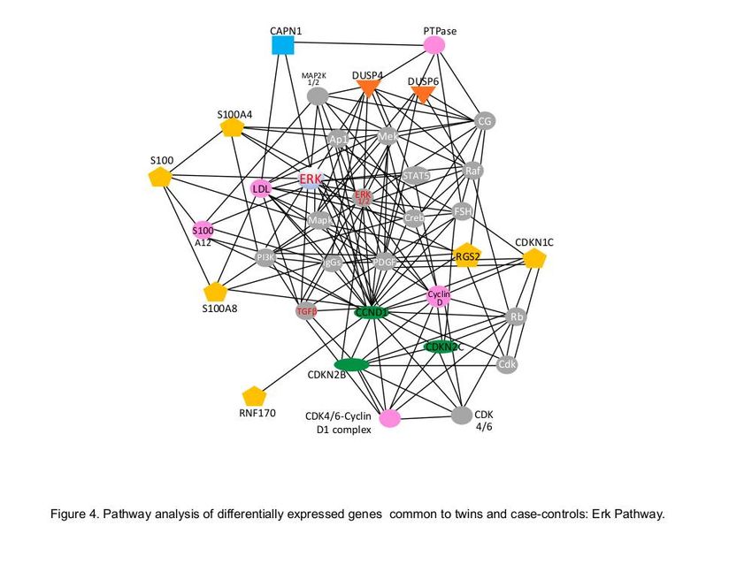
Abstract
Background: A variety of DNA-based methods have been applied to identify genetic markers of attention
de cit hyperactivity disorder (ADHD), but the connection to RNA-based gene expression has not been fully
exploited. Using well de ned cohorts of discordant, monozygotic twins from the Michigan State
University Twin Registry, and case-controlled ADHD cases in adolescents, the present studies utilized
advanced single molecule RNA sequencing to identify expressed changes in whole blood RNA in
ADHD. Multiple analytical strategies were employed to narrow differentially expressed RNA targets to a
small set of potential biomarkers of ADHD.

Results: RNA markers common to both the discordant twin study and case-controlled subjects further
narrowed the putative targets, some of which had been previously associated with ADHD at the DNA
level. The potential role of several differentially expressed genes, including ABCB5, RGS2, GAK, GIT1 and
3 members of the galactose metabolism pathway (GALE, GALT, GALK1) are substantiated by prior
associations to ADHD and by established mechanistic connections to molecular pathways relevant to
ADHD and behavioral control.

Conclusions: The convergence of DNA, RNA, and metabolic data suggests these may be promising
targets for diagnostics and therapeutics in ADHD.

Background
Attention de cit hyperactivity disorder (ADHD) is the most common psychiatric disorder in childhood and
adolescence, affecting roughly 5% of youth worldwide [1]. Although heritability is substantial [2],
environmental modulation is well known and environmental in uences are of keen interest. Further, ADHD
is a complex disease, with no single gene showing an overwhelming effect, except in very rare cases [2].
Further, ADHD is likely to be etiologically complex and to re ect multiple underlying etiology types [3].
Thus, ADHD is potentially a collection of disorders, which could involve inherited DNA variations, somatic
epigenetic changes during neural differentiation, and environmental modi ers. A variety of genetic
technologies have been applied to help identify either biomarkers or clues to causative mechanism,
including GWAS, exome sequencing, and others [4].

Physiologically, numerous avenues have been examined for a potential role in ADHD. One theory holds
that ADHD involves changes in the glutaminergic neurotransmitter systems, a major excitatory pathway
in the brain, however genome-wide SNP analysis has so far failed to identify glutamate receptor DNA
variants as a signi cant association with ADHD [5]. Several lines of evidence, including the utility of
drugs such as methyphenidate, amphetamines, and atomoxetine has suggested that de cits in the
dopaminergic and adrenergic systems could underlie the neurochemical basis of ADHD [6].

While a major focus has been on neurodevelopmental pathways, there are reasons to be cognizant of
potentially important changes in non-neural endocrine systems. The strong linkage of ADHD with male

                                                  Page 2/33
gender has raised potential connections to steroid hormones, but it is di cult to exclude sex-linked
genetic elements, and/or cultural perceptions related to the diagnosis of ADHD [7].

It has recently been suggested that RNA sequencing may be informative for identifying ADHD-related
biomarkers, based on positive ndings in a small paired analysis in 3 multiplex cases [8]. Additionally,
targeted and candidate gene studies have stimulated interest in miRNAs in ADHD [9]. The present studies
employed a broad and relatively unbiased strategy to obtain exploratory data for hypothesis generation
using two types of ADHD cohorts analyzed by high-precision, genome-wide single molecule RNA
sequencing.

The present studies employed cutting-edge RNA sequencing technology to very accurately quantify
transcript levels across the entire transcriptome, for both coding, and non-coding transcripts. The RNA
pro ling approach was applied to stabilized whole blood samples drawn from two well-characterized
cohorts: a) children matched on ADHD and non-ADHD status and, b) identical twins that were discordant
for ADHD traits. The results provide a deep and broad map of RNA changes potentially related to ADHD,
and can be combined with other ‘omic data, such as genome-wide association studies (GWAS), DNA
methylation, and proteomic analysis to help identify new biomarkers, and secondarily, new clues to
biological processes related to ADHD.

Methods
Participants.

Non-twin case-controls. Case-controls were selected from the Oregon ADHD Longitudinal Study Cohort
(for illustrative prior papers see [10, 11]. For that cohort, families were recruited for a case-control study of
ADHD and non-ADHD, by soliciting community volunteers with public advertisements and mass mailings
using commercial mailing lists. The local Institutional Review Board approved the studies. Parents
provided written informed consent; children provided written informed assent. All families completed a
full multi-informant, multi-method screening process to establish eligibility and diagnostic group
assignment for ADHD, non-ADHD, as well as comorbid disorders.

After screening, the research team conducted a diagnostic evaluation using standardized, well-normed
rating scales from parents and teachers, parent semi-structured clinical interview, child intellectual testing,
and clinical observation. Best-estimate research diagnoses and nal eligibility were established by a team
of two experienced clinicians (a child psychiatrist and a child psychologist), who independently assigned
  nal diagnoses and comorbid disorders including ADHD, oppositional de ant disorder (ODD), and any
lifetime mood disorder (major depression, dysthymia, or other), as reported herein. History of seizures or
head injury, parent–teacher rating discrepancy making diagnosis uncertain, psychosis, mania, current
major depressive episode, Tourette’s syndrome, autism, and estimated IQ
From 2144 volunteers, 850 eligible children were identi ed. The group used in the current study were a
subset selected because they (a) clearly met Diagnostic and Statistical Manual of Mental Disorders-5
(DSM-5) criteria for ADHD or non-ADHD comparison group (rather than subthreshold), (b) had no prior
history of psychiatric medication, (c) were Tanner stage 1 or 2 by parent report on the Pubertal
Development Questionnaire, (d) were not a sibling of another child in the cohort; and, (e) were willing to
give a blood sample. This process provided a set of non-twin case-controls (n=48: 24 ADHD, 24 controls)
for whom Paxgene-stabilized, frozen blood samples were submitted for RNA sequencing. After RNA
sequencing, 23 ADHD and 21 case controls were available, as described in Table 1.

Twins. Twins were recruited from the Michigan State University Twin Registry (MSUTR; [12, 13]. The
registry has over 30,000 twin pairs between the ages of 3 and 55. For the current study, recruitment was
carried out via anonymous mailings to all MSUTR, identical (monozygotic, MZ) twin families with twins
age 7-17 years old. Volunteering families were then screened for eligibility. They were restricted for the
current study to (a) never-medicated youth, (b) age 7-17 (it proved impractical to restrict to pre-pubertal
due to the relative rarity of discordant pairs), (c) no major medical illness, autism, or neurological
condition in the screen record (later con rmed by clinical interview), (d) believed to be MZ (later con rmed
by genotyping), (e) eligible for outreach (limited to accommodate multiple studies accessing the registry).

Those who responded then completed a phone screen for eligibility and, if eligible, were scheduled for a
home visit. At the home visit, a trained staff member completed a clinical interview, drew blood into an
RNA Paxgene stabilizer tube, and collected saliva into an Oragene tube for DNA isolation. Self- and
parent-report rating scales were also collected, as described below.

The number of discordant pairs was limited by two major factors (a) about 1/3 of twin births are
monozygotic, (b) ADHD has high heritability, so discordant pairs are rare. The nal samples available for
statistical analysis after clinical screening, RNA sequencing, and data quality checks is shown in Table 1,
with additional recruitment details provided in Supplementary Data 1. After RNA sequencing, there were
16 discordant pairs meeting quality control metrics for analysis. In both cohorts, the size of the nal
groups was determined by the number of available samples meeting inclusion/exclusion criteria, and
then by successful RNAseq analysis.

MEASURES

ADHD Evaluation. ADHD was evaluated by the following measures: parent completed the Conners' Rating
Scales-3rd Edition short form [14]; Strengths and Di culties Questionnaire long form including the
impairment module (SDQ) [15]; the ADHD Rating Scale (ADHD-RS) [16], and a semi-structured clinical
interview (Kiddie Schedule for Affective Disorders and Schizophrenia, KSADS) administered by a
Master’s-degree level clinician trained to reliability with an outside trainer (EB) [17]. For the twin data,
these data were used to evaluate consensus ADHD status by two of the authors (JN,EB) blind to the RNA
data. For the case-control data, teacher ratings were also available and consensus diagnosis was arrived
at by an independent clinical team as described in the online appendix.

                                                  Page 4/33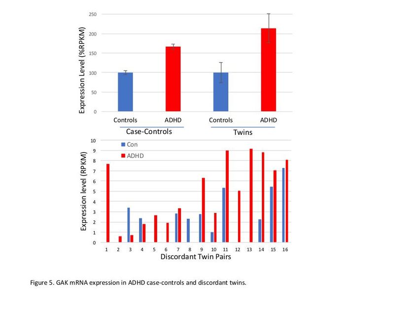
De nition of Discordance. Twins were considered discordant if they met the following criteria:

  1. At least a 3-symptom separation on the ADHD-RS or KSADS, with one twin below diagnostic
     threshold and never identi ed with ADHD; or
  2. At least a 10 point T score difference on the ADHD-RS, or
  3. One was previously diagnosed and treated and the other was not (and the untreated twin did not
     meet ADHD criteria by our measures), or
  4. Any combination of these criteria.

ADHD Discrepancy score. To create a quantitative estimate of degree of discordance, the following four
variables were created:

  1. Absolute difference between higher and lower scoring twin on an ADHD–RS (by parent) raw score
    (range 0-27) obtained at the same time as the RNA blood tube was collected, and rated over the past
    6 months for (i) inattention, (ii) hyperactivity-impulsivity, and (iii) total. The total is reported here.
  2. For the fourth variable, each twin pair was ranked on “degree of discordance” based on considering
     all available data including multiple time points rating scales (Conners, ADHD-RS, SDQ), the KSADs,
    and comorbid and substance use conditions. This was achieved by consensus rankings by JN and
    EB blind to the RNA data.

Whole blood RNA isolation. For complete methods, see Supplementary Data 1. Whole blood RNA was
isolated from the Paxgene blood collection tube using the Paxgene Blood Isolation kit and the Qiagen
QIACube Automation System. The RNA was quanti ed by optical absorbance at 260 and 280 nm using
the NanoDrop 1000. The resulting nucleic acids were treated with TurboDNAse to remove residual DNA
and then depleted of ribosomal RNA (rRNA) using the Illumina Ribo-Zero rRNA Removal Kit (H/M/R). RNA
concentration was measured using the absorbance at 280 and 260 nm (Nanodrop).

Single molecule sequencing. First strand cDNA synthesis was carried out with 50-100 ng RNA at 95°C for
5 minutes using 50 ng/µL random hexamers (Invitrogen #51709) and 1 µL of 10 mM dNTP mix
(Invitrogen #Y02256). 3’ Poly A tailing was achieved with terminal transferase (NEB #M0315). Cleaned
samples were denatured and hybridized to the poly dT surface of SeqLL ow cells. Samples were
sequenced using SeqLL’s True Single Molecule Sequencing (tSMS) technology that allows for RNA
sequencing without requiring PCR ampli cation or library preparation ligation steps.

Read alignment and quanti cation. Data output from the sequencer is in raw short read format (SRF)
 les. SRF les were processed using the HeliSphere Bioinformatics package, rst converting to SMS
format for alignment. SMS reads were trimmed for leading T homopolymers and were ltered for reads
with a minimal length of 25 bases after trimming. Trimmed reads were aligned to the HG38 human
genome supplemented with the complete ribosomal repeat unit (GenBank Accession U13369.1) using the
HeliSphere BASIC analysis pipeline.

                                                  Page 5/33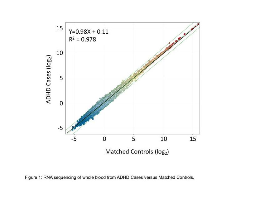
Statistical analysis of differentially expressed genes. From the aligned reads, a variety of analytical
approaches were employed in order to identify differentially expressed genes (DEGs) which passed one or
more ltering strategies. To minimize the impact of any one statistical method, the sample set was
analyzed by 9 methods, deriving from 5 types of analysis: using the EdgeR Bioconductor package [18, 19]
using either dispersion analysis (comEdgeR), or generalized regression analysis (glmEdgeR); with TCC
package [20] using either edgeR (tccEdgeR), DESeq [21] (tccDESeq) or DESeq2 [22] (tccDESeq2) methods
for differential expression analysis; with voom [23] from limma [24]; and also with baySeq [25] and
ALDEx2 [26] packages. To nd the most commonly identi ed DEGs, the results of each analysis were
ranked by the resulting p-value likelihood of a difference between groups adjusted for multiple testing
using the Benjamini-Hochberg method. To constrain the size of the DEG list, it was predetermined to
select the top 100 from each method and then combine them to achieve a single ranked list across
methods. These were then ranked by the number of times a given DEG appeared in each of the 9 lists.
Jaccard similarity analysis reveals the concordance between methods edgeR, DESeq and DESeq2. The
most different results were obtained with BaySeq.

A second general strategy that has proven useful, due its simplicity and absence of assumptions about
the distribution of RNAseq data, is to adjust the transcript counts only by correction for the transcript size
and total informative reads. The RPKM calculation compensates for the size of the transcript, and for the
total number of reads acquired. The number of reads aligned to each transcript are divided by the size of
transcript in thousands of base pairs (per K), and then divided by the total number of informative reads
obtained for that subject (in Millions), yielding RPKM. To further constrain the DEG list to transcripts with
detectable expression, the total aligned read counts for the HG38 genome (GRCh38, n=195,187) were
 ltered to include transcripts present at >0.01 RPKM in 70% of the samples of at least one diagnostic
group, leaving ~95K working transcripts for analysis (see Figure 1 for scatter plot of RPKM expression per
transcript).

Results
Clinical parameters of the study subjects.

Case Controlled Cohort. A total of 100 never-medicated children on whom Paxgene stabilized blood was
available were used to compose 24 pairs of ADHD affected or unaffected case controls. They were
selected so that each ADHD case was matched by age and gender to an unaffected control. From these
24 pairs, 23 ADHD and 21 controls were successfully RNA sequenced to pre-speci ed criteria of read
depth. The characteristics of the groups are shown in Table 1.

Discordant Twins Cohort. An initial cohort of 50 never-medicated, potentially discordant identical twin
pairs was identi ed and then narrowed for clinical criteria focusing on a high degree of discordance in the
severity of ADHD symptoms. A set of 24 pairs of twins with strong to moderate discordance were
identi ed and 16 pairs had su cient RNA quality and yield, and successful RNAseq data for further
analysis (Table 1).

                                                   Page 6/33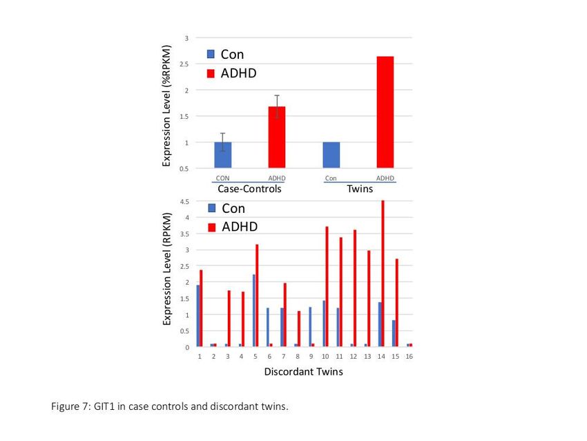
Analysis of differentially expressed genes

Case Controlled Cohort.

The average yield of nucleic acids was 5.4 ug/tube with an average 260/280 ratio of 2.3 and a
BioAnalyzer RIN RNA quality index of 8.97. The tSMS method produced a very broad pro ling of
ribosome-depleted RNA transcripts in stabilized whole blood. By averaging all subjects in each group,
  ltering out low expressing transcripts of
The fth transcript, RP11-35015.2 (↓1.6X) is a poorly annotated transcript that lies within intron 1 of the
IGF1 receptor (IGF1R), and thus, di cult to more clearly understand.

The complete list of 391 selected (Supplemental Table 1) includes other interesting transcripts, such as
BACE2 (↓1.6X) and MED6 (↑1.5X). However, we proceeded to further narrow this case-control list by virtue
of analyzing the cohort of discordant twins, and then determining whether any systematic patterns of
similarity emerged.

Discordant Twins Cohort

tSMS of 16 discordant twin pairs produced transcript pro ling of similar breadth and linearity as
observed in the case-control study (Suppl. Figure 1). Using an essentially identical analytical approach to
the case controls (Figure 2), the results of RNAseq from monozygotic ADHD-discordant twins were
subjected to 9 analytical approaches and then the top 100 transcripts from each were ranked by their
presence on the 9 lists. The resulting list of 385 transcripts can be found in Supplementary Table 2.

Discordant twins DEGs: A total of 10 transcripts passed 8 of 9 lters and contains transcripts with close
similarity to several of the case-control DEGs (Figure 2). These high-ranking transcripts present potential
hypotheses for further study with regard to ADHD, as follows:

ARL6IP5 (↓1.4X) is an ADP ribosylation factor-like GTPase 6-interacting protein, but is also known as
JWA, a homologous gene of the glutamate-transporter-associated protein 3-18 (GTRAP3-18), and
addicsin. ARL6IP5/JWA is expressed at high levels in the hippocampus and ARL6IP5/JWA knockout
mice showed spatial cognitive potentiation and enhanced neurite growth in newborns [36]. Conditional
astrocytic ARL6IP5/JWA null mice demonstrates a role as a neuroprotective factor against dopaminergic
neuronal degeneration [37]. ARL6IP5/JWA has been associated with increased expression in the
amygdala after chronic morphine treatment [38], and with morphine dependence via the delta opioid
receptor [39].

CCDC107 (↓1.9X) is closely related to CCDC132 and CCDC84 found in the case control study. While
relatively little is known about these coiled coil family members, coiled coil helix proteins (e.g. Chchd2)
have been implicated in ADHD-like mouse models [40].

CCND1, cyclin D1 (↓2.9X), is related to CCNC and CCNL2 from the case control studies. While the cyclins
are largely studied in relationship to cell cycle control, they can serve a variety of regulatory functions in
cells.

DBF4B, DBF4 Zinc Finger B (↑2.5X), has been extensively studied as an activator of the Mcm2-7 helicase,
a partner to Cdc7 kinase, and thus important for the initiation of DNA replication. Potentially of interest, it
has also been associated with autism spectrum disorders via a semaphorin 5A (SEMA5A) eQTL network
[41].

                                                   Page 8/33
Dual speci city phosphatase 4 (DUSP4, ↓3.2X) is a family member to DUSP6 from the case-control study,
and has recently been described as a control element in the suprachiasmatic clock network via
modulation of vasoactive intestinal peptide signaling to ERK1/2 [42].
FAM159A (↑1.7X) has counterparts FAM104A, FAM134B, FAM157C, FAM162A, and FAM213B as
differentially expressed in the case-control study. Little is known about FAM159A, but FAM134B, aka
RETREG1 reticulophagy regulator 1, has a substantial literature connecting it with various functions
including autophagy, and sensory neuropathy in humans [43] and Border Collies [44]. Inhibition of
FAM134A causes impaired proteostasis in the endoplasmic reticulum due to the accumulation of
misfolded proteins, which has been implicated in vascular dementia [45]. FAM162A is associated by
GWAS to a gene-by-alcohol dependence interaction study of risky sexual behaviors and so it could be
related to behavioral control [46]. Coincidentally, ADHD is associated with increased sexual risk taking
[47].
RARS2 (↓1.6X) is the arginyl-tRNA synthetase gene that has been associated with a spectrum of
neurological disorders including myoclonic epilepsy, mental retardation, spasticity, and extrapyramidal
features [48]. Patients with RARS2 mutations exhibit early onset hypotonia, epileptic seizures,
encephalopathy, and feeding di culties in a syndrome termed pontocerebellar hypoplasia type 6 (PCH6)
[49].
RN7SL454P (↓1.95X), has counterparts RN7SL423P and RN7SL687P as DEG in the case control cohort.
It appears to be a small non-coding transcript, intronic to the dynein axonemal heavy chain 17 gene on
chromosome 17 (DNAH17), but with no known function.
SIK3.IT1 (↑2.1X) is salt-inducible kinase 3, with known relations to sleep and circadian rhythm, and to
glucose and lipid homeostasis, steroidogenesis, and adipogenesis [50].
SPICE1(↑1.4X) is a spindle and centriole-associated protein, which might relate to DBF4B and CCND1 in
regards to cell cycle control. Computational screening identi es it as an aurora kinase substrate and it is
known to cooperate with CEP120 in centriole elongation. Interestingly, SIK3 also interacts with aurora A,
aurora B, and polo-like kinases, and SIK3 repression enhances the antimitotic effect of aurora inhibition
[50]. Likewise, CCND1 has known interactions with aurora kinases [51]. The coiled coil proteins,
potentially including CCDC107, are commonly associated with the centrosome maturation and aurora
kinases [42], suggesting several possible coregulatory scenarios for SPICE1, CCND1, DBF4B, and SIK3 in
ADHD, potentially in a non-mitotic, but centriolar/aurora kinase-mediated control of gene expression.
DEGs common to both cohorts. From the 385 DEG list compiled from 9 analyses of the discordant twins
(Suppl. Table 2), 6 transcripts are identical to the 391 DEG case-control results obtained by similar
methods (Suppl. Table 1). While 6 identical matches between 2 different cohorts of ADHD subjects could
occur by random chance (Fisher’s exact text p=0.318), it does suggest that these transcripts may merit
further analysis as hypotheses in future studies.
Page 9/33HLA.DQB1.AS1, as the name implies, is an antisense transcript to the HLA-DQB1 locus on chromosome 6,
which is elevated about 2-fold in ADHD cases. As noted previously, particularly in the case-control study, a
substantial group of transcripts were HLA or IgG related, implying that some type of immune defect is at
work. Because whole blood is being pro led, one must be cautious about an over-representation of
immune-related transcripts (which are very plentiful in whole blood), but conversely, one cannot dismiss
immune involvement as noted earlier. The potential role of in ammatory factors in ADHD has been raised
over the years and is supported by various circumstantial data as recently reviewed [32], and recent
analysis suggests the potential role of HLA loci in neurodevelopmental disorders such as ASD, and to a
lesser degree ADHD [52].
In the same vein, IGHV3-74 is the variable region heavy chain transcript involved in antigen recognition by
encoding IgM antibodies. While speculative, increased levels in the 2 cohorts could suggest some type of
immune or autoimmune activity in ADHD.
The regulator of G protein signaling RGS2 is increased in both cohorts. RGS2 has diverse actions
including promoting the translation of stress-associated proteins ATF4 and CHOP via an eIF-2B inhibitory
domain [53]. Of potential importance, RGS2 variants have been associated with childhood adversities as
predictors of anxious and depressive responses [54], as well as the regulation of nicotine-induced
anxiolytic activity in mice, and cocaine-induced rewarding effects [55, 56]. Likewise, RGS2 is thought to
mediate the anxiolytic effects of oxytocin [57], and affects T cell activation, anxiety, and male aggressive
behavior [58]. RGS2 knockout mice exhibit increased fear learning, spatial learning, and neophobia [59].
Further, RGS2 modulates the activity and internalization of the dopamine D2 receptor in neuroblastoma
cells [60], and has been implicated in dopamine receptor signaling during amphetamine self-
administration [61].
Of potential interest is Park7 RNA (DJ-1), which is extensively investigated as related to early onset
Parkinson’s Disease [62]. Based on some symptomatic similarities between Parkinson’s and ADHD,
especially impulsivity [63], it was suggested there may be shared underlying causative factors. However,
the circulating plasma protein levels of Park7 were not associated with ADHD in 125 ADHD patients
versus 66 healthy controls [64], although whole blood RNA levels were not assessed.
Changes in RNU1-14P, which is a small nuclear pseudogene, is quite di cult to interpret, as is the RP11-
661A12 transcript, though the latter is potentially an upstream ORF or alternate 5’ exon for the zinc nger
CCCH-type containing 3 (ZC3H3) transcript, which is involved in nuclear adenylation and export of
mRNAs [65].
Pathway Analysis. An additional set of transcripts had very similar isoforms reported in the case control
results, for example, MED7 vs MED6, CLIC2 vs CLIC1, JRK vs JRKL. While there is no assurance that
these close family members perform similar functions, it is worth considering whether a similar pattern is
re ected. A list of 66 of these overlapping transcripts was submitted for an unbiased analysis using pre-
curated relationships between the gene products (Ingenuity Pathway Analysis). Several plausible
relationships are identi ed in a manner that could identify latent variables that might account for a
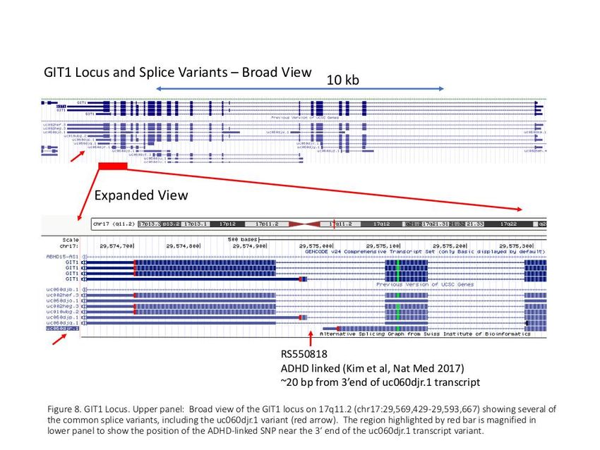
Page 10/33substantial subset of the transcript variations. Statistically, the top pathway identi ed centered around the well-characterized Akt/Insulin/PI3kinase/NfKB axis, as shown in Figure 3. Underlying changes in glucose to insulin signaling could drive broader changes into the MED6/MED7/CCNC pathway as well as VAMP8/VAMP3/MMP/NDUF pathway. A second, and related, high scoring pathway is the Erk pathway, which would explain the CCND1/CDKN2B/CDKN2C/CDKN1C/RGS2 changes, and also the S100A12/S100A4/S100A8/CAPN1/DUSP4/DUSP6 transcript alterations (Figure 4). RPKM analysis of the case-control and discordant twin datasets. In the context of a hypothesis- generating, exploratory study such as this, the prior analysis using 9 DEG methods may risk missing biologically important pathways in favor of statistical rigor. The datasets were re-analyzed using an RPKM threshold of 0.01, and combined fold-change (>1.5) and p-value (
methylmalate in human physiology is incompletely studied, but methyphenidate treatment in rats causes
signi cant changes in the citrate, malate, and isocitrate synthetic enzyme levels in the brain [76].
GAK (↑2.1X twins), cyclin G associated kinase, is potentially interesting in relation to ADHD. GAK (auxilin-
2) has known involvement in synaptic function and neurological diseases [77], and is associated by
GWAS with overlapping properties of Parkinson’s Disease and autoimmune diseases [78]. GAK was
elevated in both cohorts (Figure 5A) and in 14/16 of the discordant twin pairs, often in fairly striking
fashion (Figure 5B). GAK mRNA expression across a range of human tissues shows relatively high
expression in the cerebellum, about twice the level observed in whole blood (Suppl. Figure 2, GTEX).
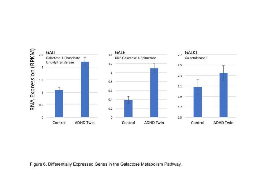
GALE (↑2.9X twins), UDP-galactose-4-epimerase, is one of 3-4 key enzymes in the synthesis and
utilization of galactose, and changes in the other members of this family, especially GALT and GALK,
were noticeably affected in the ADHD cases, with all 3 of these enzymes in the galactose processing
pathway being elevated in the ADHD-affected twins (Figure 6).
GIT1 (↑2.6X twins) was elevated >2-fold in the ADHD subjects in both the discordant twin and case-
control cohorts (Figure 7, upper panel). Several striking coincidences draw attention to GIT1 as potential
target. First, of the 15 known GIT1 isoforms, the changes in both cohorts seems largely restricted to a
single isoform (uc060djr.1), which was elevated in 12 of 16 discordant twin pairs (Figure 7, lower panel).
GIT1 SNPs were previously associated with ADHD by genome-wide association studies (GWAS) studies
that employ a relatively unbiased view of known genetic variation [79], although other cohorts did not
support this association [80]. Fine mapping identi es an intronic SNP in GIT1 which causes reduced
expression of GIT1 RNA and protein [81]. Strikingly, GIT1 is extensively spliced (Figure 8), and the intronic
SNP localizes to within 20 bp of 3’ terminus of the uc060djr.1 isoform identi ed in the present RNAseq
analysis (Figure 7). GIT1 knockout mice have ADHD-like traits including a shift in the neuronal
excitation/inhibition balance associated with a decreased glial GABA intensity [4], and behavioral
correction with methyphenidate and amphetamine [79]. Mechanistically, GIT1 is thought to play an
important role in neurite outgrowth [82], synapse formation [83], and the turnover of ß2-adrenergic and
other G-protein coupled receptors [84]. GIT1 is expressed at relatively high levels (10-fold above blood) in
most brain regions, tibial nerve, and the testes (Suppl. Figure 3, GTEX). While we cannot rule out a type I
error, the present data suggests GIT1 merits further consideration as a factor in ADHD.
STAM2 (↑1.5X twins), signal transducing adaptor molecule 2, is a member of the endosome-associated
ESCRT-0 complex that is highly expressed in neurons, especially in the cerebral and cerebellar cortex,
hippocampus, and medial habenula [85]. STAM2 regulates signaling via Jak2 and Jak3, which are
directly involved in c-myc induction of IL-2 [86].
The remaining targets identi ed in both cohorts are more di cult to interpret. ERCC6L2 (↑2.9X twins) is
excision-repair like 2, which has known relevance in cancer, but is di cult to connect with ADHD. HDLBP
(↑1.3X twins) is highly relevant to high density lipoprotein metabolism, but has only a tenuous connection
to ASD by virtue of a 2q27 deletion that causes reduced expression of HDLBP and 2 other genes [87]. IDS
(↑1.4X twins), iduronate 2-sulfatase, is highly studied in Hunter syndrome mucopolysaccharidosis [88],
Page 12/33but has no known relation to ADHD. UBE2J2 (↑2.5X twins) directs the ubiquitination of hydroxylated
amino acids in the ER, but has no reported connection to ADHD or other developmental disorders [89].
Correlation between ADHD Discordance and Gene Expression Discordance.
To further narrow candidate gene expression to potentially important correlates of ADHD, we moved from
a categorical to a dimensional analysis of ADHD severity, building on evidence that ADHD functions like a
trait in the population [90-92]. As explained earlier, ADHD severity scores (based on parent ADHD-RS raw
scores) between the identical twins were compared to create a ‘discrepancy score’ for the twins. These
scores were then ranked, with highest discrepancy (most different) being ranked 1, and then correlated to
the difference in gene expression (fold change) between the paired twins, for the 505 RPKM list of
transcripts (Suppl. Table 4). In an ideal scenario, the fold change would inversely correlate to the rank
discrepancy (high fold change in gene expression, i.e. 10, associates with lowest numerical rank, ie 1,
most discrepant). Negative correlations of r > -0.4 were observed for several transcripts of interest (boxed
yellow, Suppl. Table 5), and closer inspection suggests they might have potential relevance to ADHD.
Among the highly correlated DEGs, RN7SKP194 (r=-0.60) bears some general similarity to the
RN7SL454P target identi ed by the 9 lter approach, and discussed above. Both of these small nuclear
pseudogenes are likely to have as yet unknown regulatory functions [93]. SRP14, signal recognition
particle 14, is potentially interesting because it is 5-7 fold lower in the ADHD twins, and it is part of a
larger riboprotein complex thought to regulate translational arrest during protein synthesis in dendrites
[94]. GMFG, glia maturation factor gamma, is almost 16-fold lower in the ADHD twins and affects a
diverse range of cell types. MICU1, elevated 3-fold in ADHD twins, encodes a Ca+2-sensing, regulatory
subunit of the mitochondrial uniporter, and mutations in MICU1 cause a range of symptoms that include
progressive extrapyramidal signs, learning disabilities, and fatigue [95]. Among positively correlated
transcripts, whereby the most discordant pairs showed the least fold-change in expression was GIT1 with
r=0.585 and an average increase of 2.4 fold in the ADHD twins. Unfortunately, the changes in RNA levels
are generally not perfectly correlated with changes in protein expression (r=~0.6) [96], and so this
unexpected relationship may not be a signi cant impediment to GIT1’s relevance to ADHD.
Comparison to prior genetic studies
Prior exome sequencing of sporadic ADHD cases compared to sibling/parent triads identi ed ~8
interesting targets [4]. Of those, exome mutations in TBC1D9 are a relatively close match to RNAseq
expression changes in TBC1D17 observed in the present discordant twin pairs. This suggest a closer look
at this family of proteins, likely to be important in vesicle transport, may be warranted. A second possible
match is between exome mutations in WDR83, and expression changes in WDR45B in discordant twins,
and WDR74 in case control subjects. Also, we observed some similarity to transcripts identi ed in ADHD
by Liao et al [97], whereby transcripts MED8 and ARTN had suggestive p-values in our analysis.
Discussion
Page 13/33Among the strengths of the present approach are the unique and well-characterized ADHD cohorts. In
particular, monozygotic, but discordant twins present a powerful genetic model for comparison, and here
demonstrated some intriguing similarity in expression pattern with a case-control cohort. They open the
possibility of understanding environmental in uences while largely controlling for genotype. An important
methodological detail is that the RNAseq analysis examines the expression pattern on a relatively high
resolution scale to the level of transcript isoforms. Other common RNAseq analytic platforms tend to
aggregate expression to ‘gene level’ expression as a single transcript, which has the effect of masking
changes in alternatively spliced transcripts. The case of GIT1 is an excellent example of where very
speci c changes in one splice variant might have high relevance to the disorder in question.
Limitations to the present study principally derive from the observational nature of the studies, a
necessarily small samples for MZ discordant twins, and the necessity to use peripheral blood RNA, as
opposed to a tissue more proximal to the presumed neural in uences on ADHD. Further, the mRNA
pro ling gives us a very comprehensive view of the transcriptome, albeit at a speci c point in time, and
without strict control of the mental or physical state of the participants. Of course, causality is
indeterminate: we cannot evaluate, for instance, whether changes in the galactose pathway ‘cause’ ADHD,
or somehow result from the increased activity or altered diet or other behaviors of the children. A third
option, which must be considered, is that both ADHD and galactose changes could result from changes
in a different pathway, or from coincidental differences in ADHD teens, such as diet, drugs, or activity.
While we can exclude ADHD medications as a source of variations herein, it is di cult to exclude other
type of nutritional or nutraceutical differences.
Technically speaking, the RNAseq approach is intrinsically limited by the known genome and
transcriptomes that are used to align and interpret the reads. Every RNA pro ling method has unique
‘gaps’ and biases that can in uence the outcomes, and thus it should not be surprising if a different
RNAseq method identi ed other different differentially expressed transcripts. Further, the RNAseq of
whole blood allows for the possibility that the types of cells present in blood at the time of sampling
differ from patient to patient, or group to group. Because blood cell counts were not available on the
subjects at the time of the blood draw, we were unable to identify such differences or adjust for them.
Additionally, a valuable approach to understanding this large data set would be to conduct co-expression
analysis, which is a logical next step that might reveal systematic changes not apparent from the current
analysis [98-100].
While the present results therefore should be seen as preliminary, the nature of this work is largely
unprecedented and therefore it is valuable to note that several patterns were identi ed that are suggestive
as hypotheses for further investigation. Collectively, the results a rm some prior targets, such as GIT1,
that were identi ed by DNA-based technologies, as also relevant to ADHD at the RNA level. Several new
pathways are brought to light as potentially productive ground for further exploration. Based on a variety
of lines of evidence, it would be quite unlikely if there is a single etiologic cause for ADHD, and the present
results demonstrate that none of the RNA transcript changes were observed to occur in all of the youth. It
is interesting to speculate that changes in GAK or GIT1, which showed quite strong changes in some, but
Page 14/33not all subjects, could indicate particular genetic/epigenetic subtypes of ADHD. The cohort sizes
obviously were not powered for a detailed subtype analysis.
An intriguing future direction would explore the possible role of the galactose pathway in ADHD, either as
a modulator of core energy sensing via the insulin/AKT/NFkB/mediator pathway (Figure 3), or as a
regulator of galactosylation of key factors in the neurotransmitter pathway, as highlighted by consistent
changes in GALE/GALK/GALT. Galactose metabolism could be related to energy sensing and
in ammation via the well-established glucose/lactose/galactose connection to the
immune/in ammatory pathways that is key to the obesity/insulin resistance/in ammation connection
(reviewed in [101]). Additionally, galactose modi cation to proteins alters their in ammatory potential and
is thought to be a key component of ‘in ammaging’ [102]. Potentially the most obvious effect of altered
galactose metabolism would be the direct effect on the transport of dopamine to the brain, via galactose
modi cation of dopamine [103]. Dopamine itself is poorly absorbed in the brain, but galactosylated
dopamine has increased transit across the blood-brain barrier, and in mouse models increases attention
without reducing activity [104]. Given a well-documented relationship between dopaminergic dysfunction
and ADHD [105], it is quite plausible that perturbation of the galactose pathway in humans could produce
an ADHD-like syndrome.
Conclusions
These results are the most extensive discordant MZ study of RNAseq expression in ADHD. The results,
while preliminary, suggest several interesting hypotheses for further study.
Declarations
Ethics approval and consent to participate: All human subjects gave informed consent under protocols
approved by their respective study sites at either the Oregon Health and Science University (approval #
IRB10543) or the Michigan State University Twin Registry. For subjects under 18 years old, written
informed consent was obtained from the parents or legal guardians. Additionally, for subjects under age
16, written informed assent of the subject was obtained, and for subjects over age 16, written informed
consent was obtained. All research complied with applicable ethical and human participant guidelines.
Consent for publication: Not applicable, no individually identi able information is provided.
Availability of data and material: Expression-level de-identi ed data will be freely shared by online access
at Gene Expression Omnibus (GEO Accession xxxx) for any investigators seeking to reproduce the results,
or conduct new analysis within the bounds of the IRB-approved work. For the purpose of respecting the
privacy of the participants, and their permission to use their samples only for speci c research projects,
sequence-level data or studies outside of the scope of the existing IRB-approved work would be made
available upon reasonable request of the corresponding author.
Page 15/33Competing Interests: TM holds an equity interest in True Bearing Diagnostics, Inc., which is developing
blood RNA-based biomarkers for several disorders, but not speci cally in the area of ADHD at this time.
DJ is employed by and holds an equity interest in SeqLL, Inc. All other authors do not report competing
interests.
Author Contributions: JN, GSL, and TM were responsible for the overall conception, design, and execution
of the studies. GSL, DS, and DJ conducted the RNA sequencing, alignment, and DEG analysis. JN and EB
conducted clinical assessments of ADHD. TM and JN analyzed DEGs for pathways and patterns relevant
to ADHD. The manuscript was principally drafted by TM and JN, with input from all the authors. All the
authors have read and approved the manuscript.
Funding: The authors are grateful to The Abracadabra Foundation, and the St. Laurent Family, through
The St. Laurent Institute, for their generous philanthropic support of this work. The funding organizations
were apprised of, but had no role in the design, analysis, or interpretation of the data, nor were they
involved in the writing of the manuscript.
Acknowledgements: The authors are grateful to The Michigan State University Twins Registry for
providing blood samples and clinical data for these studies, and to the OHSU Genomics Core, especially
Beth Wilmot, for RNA sample processing. The authors are also very grateful to Debra Dederich of SeqLL
for her dedicated work in sample processing for RNAseq data generation. The authors are grateful to
Richard Wargowsky for preparation of color gures.
Abbreviations
ADHD, attention-de cit hyperactivity disorder; Diagnostic and Statistical Manual of Mental Disorders-5
(DSM-5); DEG, differentially expressed genes; GWAS, genome-wide association studies; KSADS, Kiddie
Schedule for Affective Disorders and Schizophrenia; MSUTR, Michigan State University Twin Registry;
MZ, monozygotic; ODD, oppositional de ant disorder; RNAseq, RNA sequencing; RPKM, reads per
thousand bases of transcript per million transcripts sequenced; SDQ, Strengths and Di culties
Questionnaire; SRF, short read format; tSMS, True Single Molecule Sequencing;
References
1. Polanczyk G, de Lima MS, Horta BL, Biederman J, Rohde LA: The worldwide prevalence of ADHD: a
systematic review and metaregression analysis. Am J Psychiatry 2007, 164(6):942-948.
2. Faraone SV, Larsson H: Genetics of attention de cit hyperactivity disorder. Mol Psychiatry 2019,
24(4):562-575.
3. Karalunas SL, Fair D, Musser ED, Aykes K, Iyer SP, Nigg JT: Subtyping attention-de cit/hyperactivity
disorder using temperament dimensions: toward biologically based nosologic criteria. JAMA
Psychiatry 2014, 71(9):1015-1024.
Page 16/334. Kim YS, Woo J, Lee CJ, Yoon BE: Decreased Glial GABA and Tonic Inhibition in Cerebellum of Mouse
Model for Attention-De cit/Hyperactivity Disorder (ADHD). Exp Neurobiol 2017, 26(4):206-212.
5. Huang X, Wang M, Zhang Q, Chen X, Wu J: The role of glutamate receptors in attention-
de cit/hyperactivity disorder: From physiology to disease. Am J Med Genet B Neuropsychiatr Genet
2019, 180(4):272-286.
6. Sharma A, Couture J: A review of the pathophysiology, etiology, and treatment of attention-de cit
hyperactivity disorder (ADHD). Ann Pharmacother 2014, 48(2):209-225.
7. Pinares-Garcia P, Stratikopoulos M, Zagato A, Loke H, Lee J: Sex: A Signi cant Risk Factor for
Neurodevelopmental and Neurodegenerative Disorders. Brain Sci 2018, 8(8).
8. Lorenzo G, Braun J, Munoz G, Casarejos MJ, Bazan E, Jimenez-Escrig A: RNA-Seq blood
transcriptome pro ling in familial attention de cit and hyperactivity disorder (ADHD). Psychiatry Res
2018, 270:544-546.
9. Srivastav S, Walitza S, Grunblatt E: Emerging role of miRNA in attention de cit hyperactivity disorder:
a systematic review. Atten De c Hyperact Disord 2018, 10(1):49-63.
10. Miller LL, Gustafsson HC, Tipsord J, Song M, Nousen E, Dieckmann N, Nigg JT: Is the Association of
ADHD with Socio-Economic Disadvantage Explained by Child Comorbid Externalizing Problems or
Parent ADHD?J Abnorm Child Psychol 2018, 46(5):951-963.
11. Nigg JT, Gustafsson HC, Karalunas SL, Ryabinin P, McWeeney SK, Faraone SV, Mooney MA, Fair DA,
Wilmot B: Working Memory and Vigilance as Multivariate Endophenotypes Related to Common
Genetic Risk for Attention-De cit/Hyperactivity Disorder. J Am Acad Child Adolesc Psychiatry 2018,
57(3):175-182.
12. Burt SA, Klump KL: The Michigan State University Twin Registry (MSUTR): an update. Twin Res Hum
Genet 2013, 16(1):344-350.
13. Klump KL, Burt SA: The Michigan State University Twin Registry (MSUTR): genetic, environmental
and neurobiological in uences on behavior across development. Twin Res Hum Genet 2006,
9(6):971-977.
14. Conners CK: Conners’ rating scales: Revised technical manual. In. New York, NY: Multi-Health
Systems; 2003.
15. Goodman R: Psychometric properties of the strengths and di culties questionnaire. J Am Acad Child
Adolesc Psychiatry 2001, 40(11):1337-1345.
16. DuPaul G, Power T, Anastopoulos A, Reid R: ADHD Rating Scale—IV: Checklists, Norms, and Clinical
Interpretation. New York, NY: Guilford Press; 1998.
17. Puig-Antich J, Ryan N: Kiddie schedule for affective disorders and schizophrenia. Pittsburgh, PA:
Western Psychiatric Institute; 1986.
18. Robinson MD, McCarthy DJ, Smyth GK: edgeR: a Bioconductor package for differential expression
analysis of digital gene expression data. Bioinformatics 2010, 26(1):139-140.
Page 17/3319. McCarthy DJ, Chen Y, Smyth GK: Differential expression analysis of multifactor RNA-Seq
experiments with respect to biological variation. Nucleic Acids Res 2012, 40(10):4288-4297.
20. Sun J, Nishiyama T, Shimizu K, Kadota K: TCC: an R package for comparing tag count data with
robust normalization strategies. BMC Bioinformatics 2013, 14:219.
21. Anders S, Huber W: Differential expression analysis for sequence count data. Genome Biol 2010,
11(10):R106.
22. Love MI, Huber W, Anders S: Moderated estimation of fold change and dispersion for RNA-seq data
with DESeq2. Genome Biol 2014, 15(12):550.
23. Law CW, Chen Y, Shi W, Smyth GK: voom: Precision weights unlock linear model analysis tools for
RNA-seq read counts. Genome Biol 2014, 15(2):R29.
24. Ritchie ME, Phipson B, Wu D, Hu Y, Law CW, Shi W, Smyth GK: limma powers differential expression
analyses for RNA-sequencing and microarray studies. Nucleic Acids Res 2015, 43(7):e47.
25. Hardcastle TJ, Kelly KA: Empirical Bayesian analysis of paired high-throughput sequencing data with
a beta-binomial distribution. BMC Bioinformatics 2013, 14:135.
26. Fernandes AD, Macklaim JM, Linn TG, Reid G, Gloor GB: ANOVA-like differential expression (ALDEx)
analysis for mixed population RNA-Seq. PLoS One 2013, 8(7):e67019.
27. Lutz NW, Banerjee P, Wilson BJ, Ma J, Cozzone PJ, Frank MH: Expression of Cell-Surface Marker
ABCB5 Causes Characteristic Modi cations of Glucose, Amino Acid and Phospholipid Metabolism in
the G3361 Melanoma-Initiating Cell Line. PLoS One 2016, 11(8):e0161803.
28. Glessner JT, Brad eld JP, Wang K, Takahashi N, Zhang H, Sleiman PM, Mentch FD, Kim CE, Hou C,
Thomas KA et al: A genome-wide study reveals copy number variants exclusive to childhood obesity
cases. Am J Hum Genet 2010, 87(5):661-666.
29. Nigg JT, Johnstone JM, Musser ED, Long HG, Willoughby MT, Shannon J: Attention-
de cit/hyperactivity disorder (ADHD) and being overweight/obesity: New data and meta-analysis.
Clin Psychol Rev 2016, 43:67-79.
30. Xu M, Xie YA, Abouzeid H, Gordon CT, Fiorentino A, Sun Z, Lehman A, Osman IS, Dharmat R, Riveiro-
Alvarez R et al: Mutations in the Spliceosome Component CWC27 Cause Retinal Degeneration with
or without Additional Developmental Anomalies. Am J Hum Genet 2017, 100(4):592-604.
31. Xiahou Z, Wang X, Shen J, Zhu X, Xu F, Hu R, Guo D, Li H, Tian Y, Liu Y et al: NMI and IFP35 serve as
proin ammatory DAMPs during cellular infection and injury. Nat Commun 2017, 8(1):950.
32. Dunn GA, Nigg JT, Sullivan EL: Neuroin ammation as a risk factor for attention de cit hyperactivity
disorder. Pharmacol Biochem Behav 2019, 182:22-34.
33. Han MR, Han KM, Kim A, Kang W, Kang Y, Kang J, Won E, Tae WS, Cho Y, Ham BJ: Whole-exome
sequencing identi es variants associated with structural MRI markers in patients with bipolar
disorders. J Affect Disord 2019, 249:159-168.
34. Jin J, Bhatti DL, Lee KW, Medrihan L, Cheng J, Wei J, Zhong P, Yan Z, Kooiker C, Song C et al: Ahnak
scaffolds p11/Anxa2 complex and L-type voltage-gated calcium channel and modulates depressive
Page 18/33behavior. Mol Psychiatry 2019.
35. Haase H, Alvarez J, Petzhold D, Doller A, Behlke J, Erdmann J, Hetzer R, Regitz-Zagrosek V, Vassort G,
Morano I: Ahnak is critical for cardiac Ca(V)1.2 calcium channel function and its beta-adrenergic
regulation. FASEB J 2005, 19(14):1969-1977.
36. Sha S, Xu J, Lu ZH, Hong J, Qu WJ, Zhou JW, Chen L: Lack of JWA Enhances Neurogenesis and
Long-Term Potentiation in Hippocampal Dentate Gyrus Leading to Spatial Cognitive Potentiation.
Mol Neurobiol 2016, 53(1):355-368.
37. Miao SH, Sun HB, Ye Y, Yang JJ, Shi YW, Lu M, Hu G, Zhou JW: Astrocytic JWA expression is
essential to dopaminergic neuron survival in the pathogenesis of Parkinson's disease. CNS Neurosci
Ther 2014, 20(8):754-762.
38. Ikemoto MJ, Inoue K, Akiduki S, Osugi T, Imamura T, Ishida N, Ohtomi M: Identi cation of
addicsin/GTRAP3-18 as a chronic morphine-augmented gene in amygdala. Neuroreport 2002,
13(16):2079-2084.
39. Wu Y, Chen R, Zhao X, Li A, Li G, Zhou J: JWA regulates chronic morphine dependence via the delta
opioid receptor. Biochem Biophys Res Commun 2011, 409(3):520-525.
40. Goncalves TM, Southey BR, Rodriguez-Zas SL: Interplay Between Amphetamine and Activity Level in
Gene Networks of the Mouse Striatum. Bioinform Biol Insights 2018, 12:1177932218815152.
41. Cheng Y, Quinn JF, Weiss LA: An eQTL mapping approach reveals that rare variants in the SEMA5A
regulatory network impact autism risk. Hum Mol Genet 2013, 22(14):2960-2972.
42. Hamnett R, Crosby P, Chesham JE, Hastings MH: Vasoactive intestinal peptide controls the
suprachiasmatic circadian clock network via ERK1/2 and DUSP4 signalling. Nat Commun 2019,
10(1):542.
43. Park GY, Jang DH, Lee DW, Jang JH, Joo J: Hereditary Sensory and Autonomic Neuropathy 2B
Caused by a Novel RETREG1 Mutation (c.765dupT) and Paternal Uniparental Isodisomy of
Chromosome 5. Front Genet 2019, 10:1085.
44. Forman OP, Hitti RJ, Pettitt L, Jenkins CA, O'Brien DP, Shelton GD, De Risio L, Quintana RG, Beltran E,
Mellersh C: An Inversion Disrupting FAM134B Is Associated with Sensory Neuropathy in the Border
Collie Dog Breed. G3 (Bethesda) 2016, 6(9):2687-2692.
45. Islam F, Gopalan V, Lam AK: RETREG1 (FAM134B): A new player in human diseases: 15 years after
the discovery in cancer. J Cell Physiol 2018, 233(6):4479-4489.
46. Polimanti R, Zhao H, Farrer LA, Kranzler HR, Gelernter J: Ancestry-speci c and sex-speci c risk alleles
identi ed in a genome-wide gene-by-alcohol dependence interaction study of risky sexual behaviors.
Am J Med Genet B Neuropsychiatr Genet 2017, 174(8):846-853.
47. Hechtman L, Swanson JM, Sibley MH, Stehli A, Owens EB, Mitchell JT, Arnold LE, Molina BS,
Hinshaw SP, Jensen PS et al: Functional Adult Outcomes 16 Years After Childhood Diagnosis of
Attention-De cit/Hyperactivity Disorder: MTA Results. J Am Acad Child Adolesc Psychiatry 2016,
55(11):945-952 e942.
Page 19/3348. Mathew T, Avati A, D'Souza D, Therambil M: Expanding spectrum of RARS2 gene disorders:
Myoclonic epilepsy, mental retardation, spasticity, and extrapyramidal features. Epilepsia Open 2018,
3(2):270-275.
49. Zhang J, Zhang Z, Zhang Y, Wu Y: Distinct magnetic resonance imaging features in a patient with
novel RARS2 mutations: A case report and review of the literature. Exp Ther Med 2018, 15(1):1099-
1104.
50. Chen H, Huang S, Han X, Zhang J, Shan C, Tsang YH, Ma HT, Poon RY: Salt-inducible kinase 3 is a
novel mitotic regulator and a target for enhancing antimitotic therapeutic-mediated cell death. Cell
Death Dis 2014, 5:e1177.
51. Jantscher F, Pirker C, Mayer CE, Berger W, Sutterluety H: Overexpression of Aurora-A in primary cells
interferes with S-phase entry by diminishing Cyclin D1 dependent activities. Mol Cancer 2011, 10:28.
52. Nudel R, Benros ME, Krebs MD, Allesoe RL, Lemvigh CK, Bybjerg-Grauholm J, Borglum AD, Daly MJ,
Nordentoft M, Mors O et al: Immunity and mental illness: ndings from a Danish population-based
immunogenetic study of seven psychiatric and neurodevelopmental disorders. Eur J Hum Genet
2019.
53. Wang CJ, Chidiac P: RGS2 promotes the translation of stress-associated proteins ATF4 and CHOP
via its eIF2B-inhibitory domain. Cell Signal 2019, 59:163-170.
54. Asselmann E, Hertel J, Schmidt CO, Homuth G, Nauck M, Beesdo-Baum K, Grabe HJ, Pane-Farre CA:
Interplay between RGS2 and childhood adversities in predicting anxiety and depressive disorders:
Findings from a general population sample. Depress Anxiety 2018, 35(11):1104-1113.
55. Rorabaugh BR, Chakravarti B, Mabe NW, Seeley SL, Bui AD, Yang J, Watts SW, Neubig RR, Fisher RA:
Regulator of G Protein Signaling 6 Protects the Heart from Ischemic Injury. J Pharmacol Exp Ther
2017, 360(3):409-416.
56. Rorabaugh BR, Sprague L, Norman H, Seeley SL, D'Souza MS: Regulator of G protein signaling 2
differentially regulates nicotine-induced anxiolytic- and antidepressant-like effects in mice. Eur J
Neurosci 2018, 48(5):2110-2117.
57. Okimoto N, Bosch OJ, Slattery DA, P aum K, Matsushita H, Wei FY, Ohmori M, Nishiki T, Ohmori I,
Hiramatsu Y et al: RGS2 mediates the anxiolytic effect of oxytocin. Brain Res 2012, 1453:26-33.
58. Oliveira-Dos-Santos AJ, Matsumoto G, Snow BE, Bai D, Houston FP, Whishaw IQ, Mariathasan S,
Sasaki T, Wakeham A, Ohashi PS et al: Regulation of T cell activation, anxiety, and male aggression
by RGS2. Proc Natl Acad Sci U S A 2000, 97(22):12272-12277.
59. Raab A, Popp S, Lesch KP, Lohse MJ, Fischer M, Deckert J, Hommers L: Increased fear learning,
spatial learning as well as neophobia in Rgs2(-/-) mice. Genes Brain Behav 2018, 17(4):e12420.
60. Luessen DJ, Hinshaw TP, Sun H, Howlett AC, Marrs G, McCool BA, Chen R: RGS2 modulates the
activity and internalization of dopamine D2 receptors in neuroblastoma N2A cells.
Neuropharmacology 2016, 110(Pt A):297-307.
61. Calipari ES, Sun H, Eldeeb K, Luessen DJ, Feng X, Howlett AC, Jones SR, Chen R: Amphetamine self-
administration attenuates dopamine D2 autoreceptor function. Neuropsychopharmacology 2014,
Page 20/3339(8):1833-1842.
62. van Duijn CM, Dekker MC, Bonifati V, Galjaard RJ, Houwing-Duistermaat JJ, Snijders PJ, Testers L,
Breedveld GJ, Horstink M, Sandkuijl LA et al: Park7, a novel locus for autosomal recessive early-onset
parkinsonism, on chromosome 1p36. Am J Hum Genet 2001, 69(3):629-634.
63. Kehagia AA, Housden CR, Regenthal R, Barker RA, Muller U, Rowe J, Sahakian BJ, Robbins TW:
Targeting impulsivity in Parkinson's disease using atomoxetine. Brain 2014, 137(Pt 7):1986-1997.
64. Lee CJ, Wu CC, Chou WJ, Lee MJ, Chou MC, Lee SY, Wang LJ: Mitochondrial-associated protein
biomarkers in patients with attention-de cit/hyperactivity disorder. Mitochondrion 2019, 49:83-88.
65. Hurt JA, Obar RA, Zhai B, Farny NG, Gygi SP, Silver PA: A conserved CCCH-type zinc nger protein
regulates mRNA nuclear adenylation and export. J Cell Biol 2009, 185(2):265-277.
66. Fallahi P, Katz R, Toma I, Li R, Reiner J, VanHouten K, Carpio L, Marshall L, Lian Y, Bupp S et al:
Aspirin Insensitive Thrombophilia: Transcript pro ling of blood identi es platelet abnormalities and
HLA restriction.Gene 2013, 520(2):131-138.
67. Chawla LS, Toma I, Davison D, Vaziri K, Lee J, Lucas R, Seneff MG, Nyhan A, McCaffrey TA: Acute
appendicitis: transcript pro ling of blood identi es promising biomarkers and potential underlying
processes. BMC Med Genomics 2016, 9(1):40.
68. Ashtari N, Jiao X, Rahimi-Balaei M, Amiri S, Mehr SE, Yeganeh B, Marzban H: Lysosomal Acid
Phosphatase Biosynthesis and Dysfunction: A Mini Review Focused on Lysosomal Enzyme
Dysfunction in Brain. Curr Mol Med 2016, 16(5):439-446.
69. Sittig LJ, Carbonetto P, Engel KA, Krauss KS, Palmer AA: Integration of genome-wide association and
extant brain expression QTL identi es candidate genes in uencing prepulse inhibition in inbred F1
mice. Genes Brain Behav 2016, 15(2):260-270.
70. Rubio RM, Depledge DP, Bianco C, Thompson L, Mohr I: RNA m(6) A modi cation enzymes shape
innate responses to DNA by regulating interferon beta. Genes Dev 2018, 32(23-24):1472-1484.
71. Du K, Zhang L, Lee T, Sun T: m(6)A RNA Methylation Controls Neural Development and Is Involved in
Human Diseases. Mol Neurobiol 2019, 56(3):1596-1606.
72. Stojkovic V, Fujimori DG: Mutations in RNA methylating enzymes in disease. Curr Opin Chem Biol
2017, 41:20-27.
73. Habtemichael EN, Alcazar-Roman A, Rubin BR, Grossi LR, Belman JP, Julca O, Lo er MG, Li H, Chi
NW, Samuel VT et al: Coordinated Regulation of Vasopressin Inactivation and Glucose Uptake by
Action of TUG Protein in Muscle. J Biol Chem 2015, 290(23):14454-14461.
74. Sharma SR, Gonda X, Tarazi FI: Autism Spectrum Disorder: Classi cation, diagnosis and therapy.
Pharmacol Ther 2018, 190:91-104.
75. Strittmatter L, Li Y, Nakatsuka NJ, Calvo SE, Grabarek Z, Mootha VK: CLYBL is a polymorphic human
enzyme with malate synthase and beta-methylmalate synthase activity. Hum Mol Genet 2014,
23(9):2313-2323.
Page 21/33You can also read