Genotype-phenotype correlations and novel molecular insights into the DHX30- associated neurodevelopmental disorders - Genome Medicine
←
→
Page content transcription
If your browser does not render page correctly, please read the page content below
Mannucci et al. Genome Medicine       (2021) 13:90
https://doi.org/10.1186/s13073-021-00900-3

 RESEARCH                                                                                                                                          Open Access

Genotype–phenotype correlations and
novel molecular insights into the DHX30-
associated neurodevelopmental disorders
Ilaria Mannucci1, Nghi D. P. Dang2, Hannes Huber3, Jaclyn B. Murry4,5, Jeff Abramson6, Thorsten Althoff6,
Siddharth Banka7,8, Gareth Baynam9,10,11, David Bearden12, Ana Beleza-Meireles13, Paul J. Benke14, Siren Berland15,
Tatjana Bierhals1, Frederic Bilan16,17, Laurence A. Bindoff18,19, Geir Julius Braathen20, Øyvind L. Busk20,
Jirat Chenbhanich21, Jonas Denecke22, Luis F. Escobar23, Caroline Estes23, Julie Fleischer24, Daniel Groepper24,
Charlotte A. Haaxma25, Maja Hempel1, Yolanda Holler-Managan26, Gunnar Houge15, Adam Jackson7,8,
Laura Kellogg27, Boris Keren28, Catherine Kiraly-Borri29, Cornelia Kraus30, Christian Kubisch1,
Gwenael Le Guyader16,17, Ulf W. Ljungblad31, Leslie Manace Brenman32, Julian A. Martinez-Agosto5,33,34,35,
Matthew Might36, David T. Miller37, Kelly Q. Minks12, Billur Moghaddam27, Caroline Nava28, Stanley F. Nelson5,35,38,
John M. Parant2, Trine Prescott20, Farrah Rajabi37, Hanitra Randrianaivo39, Simone F. Reiter15,
Janneke Schuurs-Hoeijmakers40, Perry B. Shieh41, Anne Slavotinek21, Sarah Smithson13,
Alexander P. A. Stegmann40,42, Kinga Tomczak43, Kristian Tveten20, Jun Wang2, Jordan H. Whitlock36,
Christiane Zweier30,44, Kirsty McWalter45, Jane Juusola45, Fabiola Quintero-Rivera4,5,46, Utz Fischer3, Nan Cher Yeo2*,
Hans-Jürgen Kreienkamp1* and Davor Lessel1*

  Abstract
  Background: We aimed to define the clinical and variant spectrum and to provide novel molecular insights into
  the DHX30-associated neurodevelopmental disorder.
  Methods: Clinical and genetic data from affected individuals were collected through Facebook-based family
  support group, GeneMatcher, and our network of collaborators. We investigated the impact of novel missense
  variants with respect to ATPase and helicase activity, stress granule (SG) formation, global translation, and their
  effect on embryonic development in zebrafish. SG formation was additionally analyzed in CRISPR/Cas9-mediated
  DHX30-deficient HEK293T and zebrafish models, along with in vivo behavioral assays.
  (Continued on next page)

* Correspondence: nyeo@uab.edu; kreienkamp@uke.de; d.lessel@uke.de
2
 Department of Pharmacology and Toxicology, University of Alabama,
Birmingham, USA
1
 Institute of Human Genetics, University Medical Center Hamburg-Eppendorf,
20246 Hamburg, Germany
Full list of author information is available at the end of the article

                                       © The Author(s). 2021 Open Access This article is licensed under a Creative Commons Attribution 4.0 International License,
                                       which permits use, sharing, adaptation, distribution and reproduction in any medium or format, as long as you give
                                       appropriate credit to the original author(s) and the source, provide a link to the Creative Commons licence, and indicate if
                                       changes were made. The images or other third party material in this article are included in the article's Creative Commons
                                       licence, unless indicated otherwise in a credit line to the material. If material is not included in the article's Creative Commons
                                       licence and your intended use is not permitted by statutory regulation or exceeds the permitted use, you will need to obtain
                                       permission directly from the copyright holder. To view a copy of this licence, visit http://creativecommons.org/licenses/by/4.0/.
                                       The Creative Commons Public Domain Dedication waiver (http://creativecommons.org/publicdomain/zero/1.0/) applies to the
                                       data made available in this article, unless otherwise stated in a credit line to the data.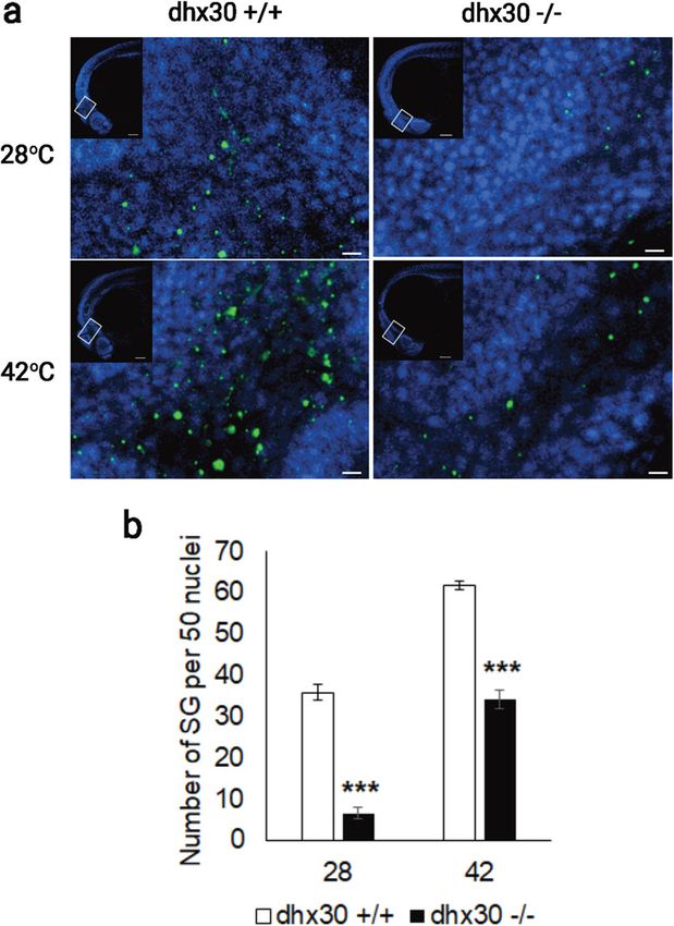
Mannucci et al. Genome Medicine   (2021) 13:90                                                                    Page 2 of 19

 (Continued from previous page)
 Results: We identified 25 previously unreported individuals, ten of whom carry novel variants, two of which are
 recurrent, and provide evidence of gonadal mosaicism in one family. All 19 individuals harboring heterozygous
 missense variants within helicase core motifs (HCMs) have global developmental delay, intellectual disability, severe
 speech impairment, and gait abnormalities. These variants impair the ATPase and helicase activity of DHX30, trigger
 SG formation, interfere with global translation, and cause developmental defects in a zebrafish model. Notably, 4
 individuals harboring heterozygous variants resulting either in haploinsufficiency or truncated proteins presented
 with a milder clinical course, similar to an individual harboring a de novo mosaic HCM missense variant.
 Functionally, we established DHX30 as an ATP-dependent RNA helicase and as an evolutionary conserved factor in
 SG assembly. Based on the clinical course, the variant location, and type we establish two distinct clinical subtypes.
 DHX30 loss-of-function variants cause a milder phenotype whereas a severe phenotype is caused by HCM missense
 variants that, in addition to the loss of ATPase and helicase activity, lead to a detrimental gain-of-function with
 respect to SG formation. Behavioral characterization of dhx30-deficient zebrafish revealed altered sleep-wake activity
 and social interaction, partially resembling the human phenotype.
 Conclusions: Our study highlights the usefulness of social media to define novel Mendelian disorders and exemplifies
 how functional analyses accompanied by clinical and genetic findings can define clinically distinct subtypes for ultra-
 rare disorders. Such approaches require close interdisciplinary collaboration between families/legal representatives of
 the affected individuals, clinicians, molecular genetics diagnostic laboratories, and research laboratories.

Background                                                         Previously, we reported 12 unrelated individuals with
RNA helicases (RH) are highly specialized proteins which         global developmental delay (GDD), intellectual disability
use ATP hydrolysis for the unwinding of RNA secondary            (ID) accompanied by severe speech impairment and gait
structures and the remodeling of ribonucleoprotein particles     abnormalities, harboring one of six different de novo mis-
(RNPs) [1, 2]. RHs are classified into six known superfamilies   sense variants located within highly conserved HCMs of
based on their sequence and structure [1]. Among these, the      DHX30 [8]. Moreover, a recent study reported gonadal
large helicase superfamily 2 (SF2) contains more than 50         mosaicism in two brothers carrying a de novo missense
members in humans [3]. These are designated DDX and              variant, p.(Ser737Phe), which resides within a HCM [11].
DHX proteins based on the consensus amino acid sequence          Here, we performed clinical, genetic, and functional ana-
DExD or DExH signature in their ATP-binding motif II             lyses to provide further understanding of DHX30-related
(Walker B motif) [3]. All SF2 RNA helicases are built around     neurodevelopmental disorders through the identification
a highly conserved helicase core region consisting of two do-    of 25 previously unreported individuals. This systematic
mains that resemble the bacterial recombination protein re-      clinical and research approach, partially facilitated through
combinase A (referred to as RecA-1 and RecA-2). Within           social media, establishes novel genotype-phenotype corre-
these two core helicase domains, eight highly conserved se-      lations based on in-depth functional analyses accompanied
quence elements, helicase core motifs (HCMs) play a role in      by clinical and genetic findings.
either RNA binding, or ATP binding and hydrolysis. The
roles of SF2 RNA helicases include regulation of splicing, nu-
clear mRNA export, translation, transcription, facilitation of   Methods
mRNA decay, microRNA processing, and cytoplasmic trans-          Human subjects and genetic analyses
port and storage of RNAs [1]. So far, many of the RHs have       Written informed consent for all 25 subjects was ob-
been studied in various cancers revealing the role of transla-   tained from the parents or legal guardians in accordance
tion in carcinogenesis [4], and serve as potential biomarkers    with protocols approved by the respective ethics com-
for diagnosis and prognosis, and novel drug targets [5]. The     mittees of the institutions involved in this study. Next-
importance and functional relevance of certain SF2 RHs in        generation sequencing-based analyses were performed in
human neurodevelopment is demonstrated by the identifica-        various independent research or diagnostic laboratories
tion of pathogenic germline variants in DDX3X [6], DDX6          worldwide, using previously described procedures [8,
[7], DHX30 [8], and DDX5 [9] in individuals with neurodeve-      12–16]. Trio-whole exome sequencing (WES) was per-
lopmental disorders. Additionally, a paralog-based study im-     formed in families of subjects 1, 4, 5, 6, 10, 11, 13, 14,
plicated a role for DHX16, DHX34, DHX37, and DDX54, in           15, 17, 18, 19, 20, 21, 22, and 23. Single WES was per-
human neurodevelopmental disorders and suggested that            formed in subjects 2, 3, 8, 16, and 25. Targeted Sanger
DHX8, DDX47, and DHX58 may also be neurodevelopmen-              sequencing was performed in subject 9, half-sister of
tal genes [10].                                                  subject 8. For subject 7, WES was performed as duo with
                                                                 DNA sample of his mother. Trio-whole genome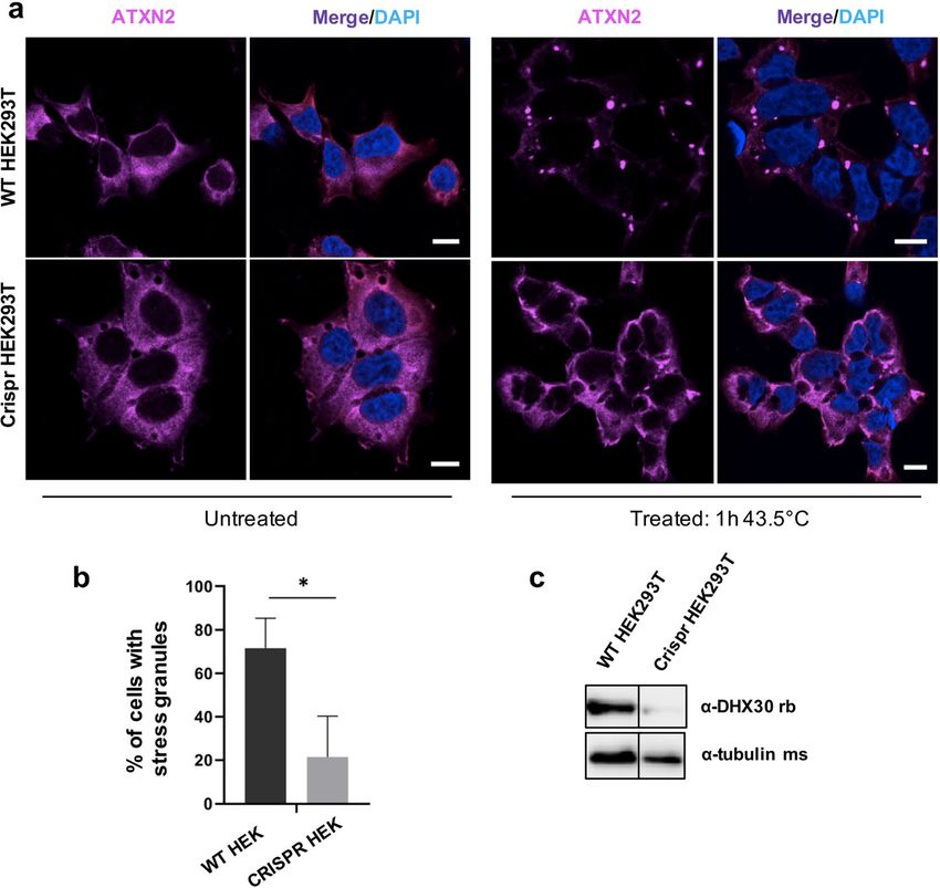
Mannucci et al. Genome Medicine    (2021) 13:90                                                              Page 3 of 19

sequencing (WGS) in family of the subject 12 was done         min at 4 °C. GFP-containing proteins were purified from
on an Illumina system using Nextera DNA Flex Library          the supernatant by immunoprecipitation using 20 μl of
Prep. Reads were aligned to human genome build                GFP-Trap_A matrix (Chromotek, Munich, Germany).
GRCh38 and analyzed for sequence variants using Cpipe         Precipitates were washed twice in RIPA buffer, and twice
analysis tool [17]. Classification the identified variants    in phosphate-free ATPase assay buffer (40 mM KCl; 35
was based on the American College of Medical Genetics         mM HEPES pH 7.5; 5 mM MgCl2; prepared in plastic
and Genomics (ACMG) guidelines [18]. Clinical                 ware to avoid phosphate contamination). Precipitates were
Chromosomal Microarray analysis in family 24 was per-         then incubated in 50 μl phosphate-free buffer supple-
formed using standardized platforms [19]. Interpretation      mented with 2 mM ATP and 2 mM DTT at 30 °C for 30
of identified copy number variants followed ACMG              min (for assaying ATPase activity in the absence of ex-
guidelines [20]. Most individuals were enrolled in the        ogenous RNA). After brief centrifugation (1 min, 1000×g),
present study through the “DHX30 family support               the supernatant was removed and precipitated samples
group” on Facebook: https://www.facebook.com/                 were incubated in phosphate-free buffer containing 2 mM
groups/1808373282809332. In such a case the families/         ATP; 2 mM DTT, and 100 μg/ml yeast RNA for 30 min at
legal representatives were asked to provide the contact de-   30 °C (for assaying ATPase activity in the presence of ex-
tails of attending physicians in order to obtain objective    ogenous RNA). The amount of free phosphate released by
and accurate clinical and genetic data. Others presented in   ATP hydrolysis was determined photometrically using
the University Medical Center Hamburg-Eppendorf,              Biomol Green reagent (Enzo Life Sciences, Lörrach,
Hamburg, Germany, or were recruited through Gene-             Germany). Subsequently, bead-attached proteins were de-
Matcher [21] and our network of collaborators. For all 25     natured in SDS-sample buffer, and the amount of DHX30
individuals, clinical data and information on genetic test-   protein was determined by western blotting using anti-
ing were uniformly obtained from attending physicians         GFP (Covance). In each case, ATPase activity was normal-
using a structured clinical summary (Additional file 1) and   ized to the amount of GFP-tagged DHX30 protein at-
clinical table (Additional file 2: Table S1).                 tached to the GFP-trap matrix.

Cell culture and in-vitro assays                              Helicase assay
Human embryonic kidney 293 T (HEK 293 T) cells and            6xHis-SUMO-DHX30 wild-type and mutant proteins
human bone osteosarcoma epithelial (U2OS) cells were          were expressed in the E. coli BL21 (DE3) pLysSpRARE
grown in Dulbecco’s modified Eagle’s medium (DMEM)            cells (Novagen, Germany). Proteins were purified from ly-
supplemented with 10% fetal bovine serum (FBS) as de-         sates using Ni-NTA beads (Qiagen, Germany) as previ-
scribed previously [8]. DXH30 expression vectors based        ously described [22]. To test the RNA unwinding activity
on pEGFP-C3 (leading to an N-terminal GFP-tag) and            of DHX30, a [32P]-labeled RNA duplex was synthesized
pEGFP-N2 (for expression of the mitochondrial form of         using the T7 RNA polymerase from a linearized DNA
DHX30 with a C-terminal GFP-tag) have been described          template designed by Tseng-Rogenski and Chang [23].
previously [8]. Newly identified missense variants were in-   Helicase activity was measured in 20 μl of reaction mix-
troduced into both vectors using Quick-Change II site-        ture containing 0.13 pmol of purified protein (=20 ng of
directed mutagenesis kit (Agilent, Waldbronn, Germany).       full-length protein), 25 fmol [32P]-labeled RNA duplex, 17
HEK293T and U2OS cells were transfected with Turbo-           mM HEPES-KOH pH 7.5, 150 mM NaCl, 1 mM MgCl2,
Fect or Lipofectamine 2000, respectively, transfection        2 mM DTT, 1 mM spermidine, 0.3% PEG8000, 5% gly-
reagent (ThermoFisher Scientific) according to the manu-      cerol, 150 mM KCl, 20 units of RNasin™ Plus (Promega,
facturer’s recommendations. Immunocytochemistry and           USA), 1 mM ATP. The mixture was incubated for 1 h at
puromycin incorporation assay in U2OS cells were per-         37 °C, mixed with 2X non-denaturing loading dye and
formed utilizing the following antibodies at manufacturers'   subjected to gel electrophoresis through non-denaturing
recommended dilutions: anti-Puromycin mouse monoclo-          8% PAGE (19:1) in 0.5X TBE at 4 °C. Reaction products
nal (Millipore, #MABE343); goat anti-mouse coupled to         were visualized by autoradiography. For more information
Alexa Fluor 555 (ThermoFisher Scientific). A custom           see Additional file 1: Supplementary methods.
made anti-ATXN2 (#8G3, kindly provided by Dr. S. Kind-
ler, Human Genetics, UKE; Hamburg) rat monoclonal             Generation of a HEK293T DHX30 stable knockout line
antibody was used at a 1:10 dilution as previously de-        HEK293T DHX30-deficient cells were generated by
scribed [8]. ATPase assay was performed as previously de-     transfecting a plasmid (pLentiCRISPR v2, GenScript,
scribed [8]. Briefly, after transfection of HEK293T cells     #52961) encoding a single guide RNA (CGAGTGCTAG
with DHX30 expression vectors, followed by lysis in 1 ml      CTGATCGCTT) targeting exon 7, the Cas9 endonucle-
of radioimmunoprecipitation assay buffer (RIPA), the ly-      ase and a puromycin resistance gene under the control
sates were clarified by centrifugation at 20,000×g for 20     of the EFS promoter. Cells were transfected with

Mannucci et al. Genome Medicine   (2021) 13:90                                                               Page 4 of 19

TurboFect transfection reagent (Thermo Scientific) and        Tol2 mRNA and 25 ng/μl equal mixture of pTol2pA2-
treated with puromycin for 3 days. Surviving cells were       cmlc2:EGFP;tuba1a:DHX30 with the respective variant
then subjected to single cell sorting using BD FACS           DNA were injected into 1-cell stage zebrafish embryos.
Aria™ IIIu Cell Sorter (BD Biosciences). Single-cell          The embryos were raised and scored for abnormal devel-
clones were grown in 96-well plates for two weeks and         opment 1–7 days post fertilization. Zebrafish were
then expanded into 6 well dishes. DHX30 knockout effi-        maintained in the Zebrafish Research Facility at the
ciency was assessed by Western blotting using an anti-        University of Alabama at Birmingham using standard
DHX30 rabbit polyclonal antibody (Bethyl, #A302-218A)         protocols. All fish were maintained at 28 °C and kept at
(1:500).                                                      14-h light and 10-h dark cycle under standard laboratory
                                                              conditions.
Stress treatment
HEK293T WT and DHX30-deficient cells were plated              Generation of zebrafish dhx30 stable knockout line
on glass coverslips coated with poly-L-Lysine. After 24 h,    The zebrafish dhx30 stable knockout line was generated
cells were heat stressed at 43.5 °C for 1 h, fixed in 4%      using CRISPR/Cas9 with sgRNA target sequence 5′-
paraformaldehyde and permeabilized with 0.1% Triton           TCAAGTTCAGCTGCACGGAT-3′ made by Integrated
X-100 (Sigma). Blocking was performed using 10% horse         DNA Technologies (IDT) according to the manufac-
serum (HS). Rat monoclonal anti-ATXN2 was used as a           turer’s protocol. The mutant contains an 8-bp deletion
primary antibody (1:10 in 2 % HS in PBS), followed by         that shifts the translational reading frame after amino
goat anti-rat IgG coupled to Alexa Fluor 647 (Thermo          acid 90 and results in a premature stop codon at amino
Fisher Scientific). Coverslips were mounted on glass          acid 107, compared to 1173 amino acids for the wild-
microscope slides with ProLong Diamond Antifade               type (WT) protein. Mutant animals were genotyped and
Mountant with DAPI (Thermo Fisher Scientific). Im-            sequenced using primers 5′-ATCTTCACGCCAAAAA
munofluorescence images were acquired using a con-            CCTG-3′ and 5′-GACCACGGTTCAGCTCTCTC-3′.
focal microscope (Leica TCS SP5, 63x/1.25 objective)          The dhx30 heterozygous mutants were outcrossed to the
and processed with ImageJ software.                           parental AB strain for at least two generations before
                                                              use in experiments to eliminate potential off-target vari-
Construction of Tol2 plasmids                                 ants. After each assay described below, test animals were
DHX30 cDNA plasmids were assembled using the Tol2             individually genotyped using PCR with primers 5′-
MultiSite Gateway® kit (Invitrogen, USA). Briefly, the        ATCTTCACGCCAAAAACCTG-3′                 and     5′-GACC
cDNA of the wild-type DHX30 and DHX30 containing re-          ACGGTTCAGCTCTCTC-3′ and high-resolution melt-
spective missense variants were amplified from the            ing (HRM) analysis as previously described [24].
pEGFP-C3-DHX30 plasmids, using primers containing
the appropriate att site sequences for BP recombination       Stress treatment and zebrafish whole-mount
reactions. PCR products were purified and cloned into a       immunostaining
pDONR221 donor vector using BP Clonase II enzyme              The dhx30 +/− animals were in-crossed to generate
mix following the manufacturer’s manual. The resulting        dhx30 +/+, +/−, and −/− sibling progeny for heat shock
middle entry clones pME-DHX30 were purified and veri-         and immunostaining analyses. Twenty-four-hour post-
fied by direct sequencing. To assemble the final expression   fertilization embryos were dechorionated and incubated
plasmids, p5E-tuba1a promoter and pME-DHX30 were              at 28 °C or 42 °C for 1 h. After treatment, embryos were
cloned into a Tol2-based destination vector, pDest-           fixed overnight in cold 4% paraformaldehyde (PFA). Em-
Tol2CG2 containing cmlc2:EGFP transgenesis marker,            bryos were then dehydrated with acetone at − 20 °C for
using LR Clonase II Plus enzyme mix following the manu-       7 min, washed in PBST [PBS+ 0.1% Tween 20], and
facturer’s instructions. The resultant pTol2pA2-cmlc2:        blocked with 10% goat serum for at least 1 h at room
EGFP;tuba1a:DHX30 vectors were purified and verified by       temperature. Thereafter, embryos were incubated with
direct sequencing.                                            rabbit anti-TIAL-1 (Novus Biologicals, NBP1-79932; 1:
                                                              200) overnight at 4 °C, washed with PBST, and incubated
Zebrafish maintenance and manipulation                        with secondary antibody Alexa Fluor 488-conjugated
Tol2 transposase mRNA were synthesized using mMES-            goat anti-rabbit IgG (Invitrogen, A11034; 1:200) for 2 h
SAGE mMACHINE™ T7 Transcription Kit (Ambion)                  at room temperature. Embryos were washed with PBST,
per the manufacturer’s instructions. Twenty-five nano-        incubated with 100 uM DAPI (1:500) to counterstain
grams/μl Tol2 mRNA and 25 ng/μl of pTol2pA2-cmlc2:            nuclei for 10 min, and stored in PBS at 4 °C. For im-
EGFP;tuba1a:DHX30 DNA were injected into 1-cell               aging, stained embryos were mounted in 1% low melting
stage zebrafish embryos (Danio rerio AB strain). To in-       agarose and imaged using a Nikon A1 inverted confocal
vestigate a potential dominant-negative effect, 25 ng/μl      microscope at approximately 50-μm Z-stacks at 5.6 μm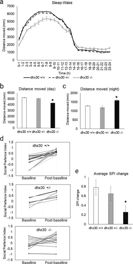
Mannucci et al. Genome Medicine   (2021) 13:90                                                                 Page 5 of 19

intervals. The number of TIAL-1-labeled stress granules       Statistical analyses
per 50 nuclei was quantified using Nikon NIS Element.         All cell line data (U2OS and HEK293T) are presented as
After imaging, test animals were individually genotyped       mean ± SD and analyzed by One-Way ANOVA followed
by PCR and HRM analysis to delineate the dhx30                by Dunnett’s multiple comparisons test or unpaired Stu-
genotype.                                                     dent’s t test as indicated in figure descriptions. All
                                                              zebrafish-related data are presented as mean ± SEM and
                                                              analyzed by unpaired Student’s t test. The percentage of
Behavioral assays                                             developmental defects observed upon overexpression of
For each behavioral experiment, dhx30 +/− animals were        dhx30 was analyzed by the χ2 test.
in-crossed to generate dhx30 +/+, +/−, and −/− sibling
progeny.                                                      Results
                                                              Identification of likely causative variants in DHX30
                                                              We identified 25 individuals carrying likely causative
Twenty-four-hour sleep-wake activity                          variants in DHX30 (Fig. 1). Of these, 12 individuals carry
For each sleep-wake study, zebrafish larvae at 5-day          a previously reported heterozygous missense variant lo-
post-fertilization (dpf) were chosen randomly and placed      calizing within highly conserved helicase core motifs
individually into each well of a flat-bottom 24-well plate.   (HCMs): p.(Arg493His), p.(His562Arg), p.(Arg782Trp)
The activity of each larva was tracked for 24 h consisting    (5 individuals including two half-sisters indicative of go-
of 14-h light and 10-h dark using the DanioVision sys-        nadal mosaicism), p.(Arg785Cys) (4 individuals), and
tem (Noldus Information Technology). The average              p.(Arg785His). Further, 7 individuals have a novel het-
swimming distance was measured for 24 h per 1-h time-         erozygous missense variant classified as either “likely
bins using EthoVision XT software (Noldus).                   pathogenic” or “pathogenic” according to The American
                                                              College of Medical Genetics and Genomics (ACMG)
                                                              guidelines (Additional file 2: Table S1) [18]. Indeed, each
Social preference assay (SPA)                                 of these variants alters a highly conserved amino acid
We adopted and modified a previously described social         within a HCM predicted to be responsible for ATP bind-
preference assay (SPA) [25]. Briefly, SPA was performed       ing and/or hydrolysis (Fig. 1 and Additional file 3: Figure
using a flat-bottom 12-well plate and custom-built re-        S1). p.(Gly462Glu) identified in a single individual affects
movable opaque dividers. The individual “test” animals,       motif I, also referred to as Walker A motif, that binds γ
whose behaviors were analyzed, were placed in each of         phosphate and coordinates, together with motifs II and
the 4 middle wells of the plate, and a WT conspecific of      VI, ATP binding and hydrolysis in other DExH family
similar age and size was placed in a well either above or     members [26, 27]. p.(Ala734Asp) identified in two unre-
below each middle well. The activity of each test larva       lated individuals, one of which (individual 6) appears to
was tracked using the DanioVision (Noldus Information         have mosaicism for the variant (Additional file 4: Figure
Technology) system and data analyzed using EthoVision         S2), and p.(Thr739Ala) identified in a single individual,
XT software (Noldus). Before data acquisition, animals        both affect motif V which regulates both ATP binding
were given 5-min habituation period. The “baseline” ac-       and/or hydrolysis and RNA binding [2, 27]. Three indi-
tivity of the test fish was then recorded while the opaque    viduals carry p.(Arg782Gln), located within motif VI af-
dividers were inserted between each well to prevent the       fecting the identical arginine residue (Arg782), that we
animals from seeing each other. The dividers were then        previously reported p.(Arg782Trp) [8], which was identi-
removed, allowing each test animal to view one well           fied here in five additional individuals.
containing a conspecific animal and one empty well. The          Moreover, a homozygous variant p.(Arg725His) lo-
fish were given another 5-minute habituation period,          cated within the helicase core region albeit between mo-
followed by a 10-min “post-baseline” recording. For data      tifs IV and V, unlike all the missense variants mentioned
analyses, wells containing test fish were divided into two    above, was identified in individual 4 and classified as
0.5 cm × 2.2 cm zones, one closest to the well containing     “variant of uncertain significance”. Additionally, a het-
a conspecific animal and one closest to the empty well.       erozygous de novo variant p.(Arg908Gln) was identified
The amount of time spent by a test fish in each zone          in individual 21. This was the only variant not located
during the baseline and post-baseline periods was ana-        within the helicase core region and was classified as
lyzed. The social preference of each test fish was quanti-    “likely pathogenic”. Predictions based on homology to
fied by calculating the social preference index (SPI) =       other SF2 helicases [26, 28] and published structures of
(time spent in zone near the conspecific fish – time          the Prp43 [29] and Mle [30] revealed three novel highly
spent in zone near the empty well)/time spent in both         conserved C-terminal regulatory domains (CTD). These
zones as previously described [25].                           include a winged helix (WH), a ratchet-like (RL) and an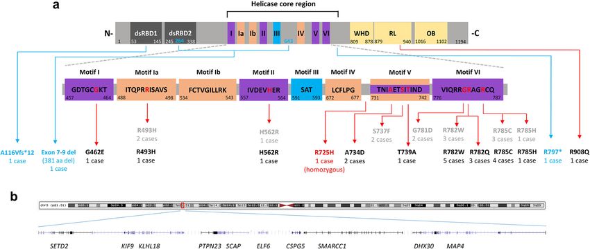
Mannucci et al. Genome Medicine         (2021) 13:90                                                                                      Page 6 of 19

 Fig. 1 Location of identified DHX30 germline variants. a Highly conserved sequence motifs within the helicase core region are shown with color
 coding that corresponds to the primary function of the motif (as previously described by Lessel et al., 2017). Double-stranded RNA-binding domains
 (dsRBD) 1 and 2 at the N-terminus (N-) are shown in gray. A winged helix domain (WHD), a ratchet-like (RL) domain, and an oligosaccharide binding
 (OB) domain are shown in yellow at the C-terminus (-C). The position of the first and last amino acid within each motif/domain is indicated below.
 Previously reported heterozygous missense variants and newly identified DHX30 variants are denoted in gray and black, respectively. Frameshift and
 nonsense variants are denoted in blue. Mutated amino acid residues within the helicase core region are marked in red. The position of previously and
 newly identified variants are indicated with red arrows. b Genomic region, chr3.hg19:g.(47098509-48109065)del, of the ~ 1 Mb deletion identified in
 case 24

oligosaccharide binding (OB) fold domain (Fig. 1) with a                     DHX30 is, with a probability of being loss-of-function
potential role in coupling ATP hydrolysis to RNA un-                         intolerant (pLI) score of 1 and a loss-of-function ob-
winding [31]. Notably, the p.(Arg908Gln) affects a highly                    served/expected upper bound fraction (LOEUF) score of
conserved residue within the RL domain (Figure S1).                          0.04, extremely loss-of-function intolerant [35]. Add-
   Furthermore, we identified four individuals bearing                       itionally, the degree of intolerance to deleterious variants
likely pathogenic loss-of-function variants. A heterozygous                  of DHX30 according to the Residual Variation Intoler-
de novo frameshift variant, p.(Ala116Valfs*12) in individ-                   ance (RVI) score, which quantifies gene intolerance to
ual 22, a heterozygous nonsense variant, p.(Arg797*) in in-                  functional variants, is − 1.51 (3.54th percentile) and thus
dividual 23 inherited from a mosaic mother, and a de novo                    even lower than the average RVI score for genes in-
in-frame deletion encompassing exons 7-9 of DHX30,                           volved in developmental disorders (0.56; 19.54th per-
leading to deletion of 381 amino acids, in individual 25.                    centile) [36, 37].
Individual 24 has a large heterozygous de novo deletion
(arr[GRCh37] 3p21.31 (47098509_48109065)del) encom-                          Clinical spectrum of the DHX30-associated
passing ten genes including two disease genes previously                     neurodevelopmental disorders
associated with an autosomal dominant inheritance,                           All 19 individuals harboring a heterozygous missense
SETD2 [32–34] and DHX30 (Fig. 1b and Additional file 5:                      variant within a highly conserved motif in the helicase
Figure S3), possibly pointing to a dual diagnosis. The                       core domain have global developmental delay (GDD), in-
whole gene deletion results in haploinsufficiency, whereas                   tellectual disability (ID), severe speech impairment, and
the in-frame deletion, frameshift, and nonsense variant, if                  gait abnormalities, similar to our initial findings [8]. In
they were to result in stable proteins, are predicted to lead                more detail, all individuals had an intellectual disability,
to loss of functionally important domains (Fig. 1a).                         only nine (47%) learned to walk, all with an ataxic gait.
   Notably, none of these DHX30 alterations was present                      The majority had no speech (74%), four individuals
in the gnomAD dataset v2.1.1 (Additional file 2: Table                       spoke only single words, and only individual 6, who is
S1) [35], indicating that they are extremely rare in the                     mosaic for the de novo p.(Ala734Asp) variant, spoke
population and unlikely to be variants unrelated to dis-                     simple sentences. It is worth noting that the individuals
ease. As previously noted, DHX30 is one of the most                          highly benefit from communication devices (tablets,
missense-intolerant genes in the human genome [8].                           smartphones, and eye-driven tablet communication sys-
Furthermore, according to the gnomAD v2.1.1 dataset                          tems) which significantly reduced frustration-related
Mannucci et al. Genome Medicine            (2021) 13:90                                                                               Page 7 of 19

behavior (D.L. personal communication with legal                                 observe a recognizable facial gestalt, similar to our previ-
guardians and family members). Additional phenotypic                             ous findings [8].
features included muscular hypotonia in eighteen (95%),                            Two individuals (#4 and #21) clearly stand out pheno-
feeding difficulties in sixteen (84%), microcephaly in                           typically. Individual 4 was homozygous for p.(Arg725His),
thirteen (81%, 13/16), joint hypermobility in fourteen                           developed early-onset infantile epileptic encephalopathy,
(74%), structural brain anomalies in eleven (65%, 11/17),                        and died at 11 months. In contrast, individual 21, who
sleep disturbances in nine (47%), strabismus in eight                            harbors the de novo p.(Arg908Gln) variant, had unre-
(42%), autistic features in five (33%, 5/15), and seizures                       markable psychomotor development until the age of 8
in four (21%) individuals (Table 1, Additional file 2:                           years when she presented with progressive balance impair-
Table S1 and Additional file 6). Noteworthy, individual 6                        ment with truncal ataxia. Subsequently, she experienced a
had a relatively milder clinical course, with a moderate                         decline in motor skills and developed cognitive problems
intellectual disability, independent walking at 2 years and                      with reduced concentration (Table 1, Additional file 2:
8 months, and the ability to speak in simple sentences at                        Table S1 and Additional file 6).
the age of 15 years. This individual´s presentation is
similar to that of the four individuals (#22, #23, #24, and                      Effect of novel DHX30 missense variants on ATPase
#25), who carry either a frameshift or nonsense variant,                         activity
whole-gene deletion or in-frame deletion, respectively,                          To corroborate the pathogenicity of the novel missense vari-
who all learned to walk in the second year of life, had a                        ants identified in this study, along with the recently reported
mild muscular hypotonia and spoke at least 20 words by                           p.(Ser737Phe) [11], we performed several previously estab-
the age of 3 years. Although some individuals displayed                          lished functional assays [8]. First, we analyzed the ATPase ac-
some dysmorphic features (Additional file 6) we did not                          tivity of wild-type (WT) and mutant forms of DHX30. As
Table 1 Clinical features in 25 individuals bearing pathogenic DHX30 variants and frequency of these features in previously reported
individuals
DHX30            Heterozygous                p.(Ala734Asp)    Haploinsufficiency/       Homozygous Heterozygous       Heterozygous missense
variant          missense variants           mosaic           protein truncating        p.(Arg725His) p.(Arg908Gln)   variants within a HCM
                 within a HCM                (this study)     variants                  (this study)  (this study)    (previous studies: Lessel
                 (this study)                                 (this study)                                            et al. 2017 and Cross et al.
                                                                                                                      2020)
Sex              11 females/7 males          Female           1 female/3 males          Male          Female          8 females/6 males
Intellectual     18/18                       +                4/4                       ?             −               13/13
disability
Speech ability 14/18 non-verbal              Simple           20 words to normal        ?             Normal speech 11/13 non-verbal
               4/18 single words             sentences        speech ability                          ability       2/13 single words
Motor            18/18                       +                4/4 mild                  +             −               14/14
development
delay
Muscular         17/18                       +                3/4                       +             −               14/14
hypotonia
Gait             10/18 no independent        Ataxic gait      0/4 no independent        ?             Ataxic gait     7/13 no independent walking
abnormalities    walking                                      walking                                                 6/13 ataxic gait
                 8/18 ataxic                                  3/4 ataxic gait
Feeding          15/18                       +                1/4                       +             −               11/14
difficulties
Microcephaly     13/15                       +                0/4                       −             −               7/10
Joint         13/18                          +                1/3                       −             −               6/14
hypermobility
Brain MRI        11/17                       −                2/3                       +             +               10/14
anomalies
Sleep            8/18                        +                2/3                       +             −               7/12
disturbance
Strabismus       8/18                        −                2/4                       −             +               6/14
Autistic         4/14                        +                0/3                       ?             −               7/12
features
Seizures         3/18                        +                2/3                       Severe        −               3/14
+, present; −, absent; ?, too young to evaluate; NA, unkown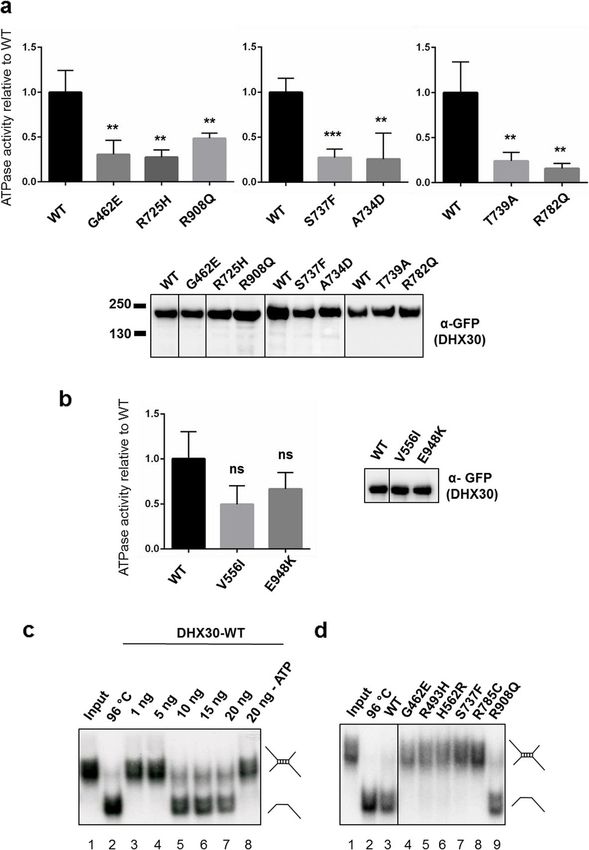
Mannucci et al. Genome Medicine    (2021) 13:90                                                                     Page 8 of 19

previously shown, DHX30-WT acts as an RNA-dependent               inclusion bodies or its direct degradation [38]. Thus, we
ATPase, and its ATPase activity is strongly stimulated by the     could not analyze its impact on the helicase activity.
addition of RNA [8]. In contrast, and similar to the previ-
ously analyzed mutants [8] all missense variants (p.(Gly462-      Subcellular localization and effect on global translation of
Glu),     p.(Arg725His),     p.(Ala734Asp),      p.(Ser737Phe),   novel DHX30 missense variants
p.(Thr739Ala), p.(Arg782Gln), and p.(Arg908Gln)) show a           We have previously shown that the expression of mutant
significant reduction in ATPase activity in the presence of ex-   forms of DHX30 induces the formation of stress gran-
ogenous RNA (Fig. 2a). For control experiments, we included       ules, concomitant with a global down-regulation of
two common non-synonymous DHX30 variants found in                 translation [8]. Therefore, we repeated this analyses for
public repositories [35]. Namely, p.(Val556Ile) is located        selected novel missense variants. In keeping with the
within the helicase core region albeit not within a HCM,          previous results [8], we observed that mutants within a
similar to p.(Arg725His), and p.(Glu948Lys) in the vicinity of    HCM, p.(Gly462Glu), p.(Ala734Asp), p.(Ser737Phe), and
p.(Arg908Gln). Notably, in comparison to the missense vari-       p.(Thr739Ala), also strongly accumulated in cytoplasmic
ants identified in affected individuals, the ATPase activity      foci shown to be stress granules upon co-staining with
was not significantly reduced neither for p.(Val556Ile) nor       Ataxin-2 (ATXN2). Expression of the p.(Arg908Gln)
for p.(Glu948Lys) (Fig. 2b).                                      mutant, however, resulted in localization to cytoplasmic
                                                                  aggregates that co-stained with Ataxin-2 in only 50% of
                                                                  the transfected cells. In contrast, p.(Arg725His) was
RNA helicase activity of DHX30 is disrupted by missense           mostly diffusely localized throughout the cytoplasm
variants within the helicase core motifs                          similar to the DHX30-WT (Fig. 3 and Additional file 8:
DHX30 has been classified as an RH due to the presence            Figure S5). Global translation was measured by incorp-
of the highly conserved motifs in its helicase core region        oration of puromycin into nascent peptide chains, which
and sequence similarity to other RHs. To confirm that it          were visualized with a puromycin-specific antibody.
indeed possesses RNA helicase activity we established an          Interestingly, expression of both the HCM mutants and
RNA unwinding assay for recombinant full-length                   the p.(Arg908Gln) mutant resulted in dramatically de-
DHX30 purified from bacteria as a His6-SUMO-tagged                creased puromycin incorporation, suggestive of a global
protein. As a substrate, we used a synthetic [32P]-labeled        decrease in protein synthesis (Fig. 3). Analogous to the
RNA molecule which carries a sequence with a strong               results obtained in the ATPase assay, the two common
propensity to self-anneal and form a double helix. Ana-           variants p.(Val556Ile) and p.(Glu948Lys) were diffusely
lysis of this RNA substrate by non-denaturing PAGE re-            localized throughout the cytoplasm, resembling the
sulted in a single band of low electrophoretic mobility,          DHX30-WT (Additional file 8: Figure S5).
corresponding to the dimer linked by the double helical
segment. This dimer could be resolved into a band of              In vivo analyses of selected DHX30 missense variants
higher mobility, the monomer, by pre-incubation at                Given the somewhat conflicting results of functional analyses
96 °C (Fig. 2c). To identify the amount of the DHX30-             of the p.(Arg725His) and p.(Arg908Gln) variants, and in
WT necessary to resolve the dimeric form we performed             order to gain a better understanding of the impact of DHX30
a titration analysis from 1 to 160 ng. In the presence of         missense variants in vivo, we utilized a zebrafish model. Pre-
ATP, 10 ng of DHX30-WT was sufficient to resolve the              vious studies showed that overexpression of pathogenic al-
dimer into the monomeric form, confirming that                    leles in zebrafish results in defective embryonic development
DHX30 indeed possesses the ATP-dependent RNA heli-                [39, 40]. Thus, we overexpressed human wild-type DHX30
case activity (Fig. 2c and Additional file 7: Figure S4).         cDNA or DHX30 cDNA harboring selected missense vari-
We next analyzed the impact of selected missense vari-            ants, p.(Arg493His), p.(Arg725His), p.(Arg785Cys), and
ants on the helicase activity, each affecting a different         p.(Arg908Gln) as well as p.(Val556Ile) and p.(Glu948Lys) in
helicase core motif (p.(Gly462Glu) in motif I,                    zebrafish using Tol2 transposition. Tol2 mRNA and
p.(Arg493His) in motif Ia, p.(His562Arg) in motif II,             pTol2pA2-cmlc2:EGFP;tuba1a:DHX30 were co-injected into
p.(Ser737Phe) in motif V, and p.(Arg785Cys) in motif              1-cell stage zebrafish embryos. For analyses, we selected em-
VI) along with p.(Arg908Gln) located in the RL domain.            bryos with strong cmlc2:EGFP expression which indicates a
All missense variants within a HCM failed to unwind               high level of transgene integration in somatic cells. Overex-
the RNA substrates in this assay, whereas the                     pression of DHX30-WT, p.(Val556Ile) or p.(Glu948Lys) had
p.(Arg908Gln) mutant behaved similarly to DHX30-WT                little or no impact on zebrafish embryonic development: over
(Fig. 2d). It is worth noting that we subsequently failed         88% of embryos displayed normal development and morph-
to purify the p.(Arg725His) mutant protein product.               ology. However, expression of DHX30 harboring one of the
This finding suggests misfolding of this mutant protein,          missense variants resulted in developmental defects in 75–
followed by either deposition of the insoluble protein in         90% of embryos (Fig. 4 and Additional file 9: Figure S6),
Mannucci et al. Genome Medicine (2021) 13:90 Page 9 of 19
Fig. 2 Protein variants of DHX30 affect ATPase and helicase activity. a, b ATPase assays were performed for DHX30-WT, novel DHX30 missense variants
(a), and two common polypmorphisms, p.(Val556Ile) and p.(Glu948Lys) (b) in the presence of exogenous RNA. ATPase activity was calculated by
subtracting phosphate values obtained with GFP alone from those obtained with GFP-tagged DHX30-WT and mutants. These figures were then
normalized on precipitated protein amounts using the intensities of the GFP signal in the western blot. Means ± standard deviation values are based
on 3 replications. **,***: significantly different from DHX30-WT, ns: not significantly different from DHX30-WT (**p< 0.01;***p< 0.001; n=3; One-Way
ANOVA, followed by Dunnett’s multiple comparisons test). Values were normalized on DHX30-WT ATPase activity obtained in the presence of RNA. c
Increasing amounts of His6-SUMO-tagged DHX30 WT protein were incubated with a 32P-labeled RNA substrate in the presence (lane 3–7) or absence
(lane 8) of ATP and analyzed by native PAGE. The position of the RNA duplex and the single-stranded RNA are indicated in the first and second lanes,
respectively. Their schematic representation is shown at the right side. d Helicase assay was repeated for selected DHX30 missense variants affecting
either conserved motifs within the helicase core region (lane 4–8) or the auxiliary RL domain (lane 9)
suggesting that these mutant alleles interfere with normal HCMs, we further investigated the nature of the latter.
embryonic development and supporting the pathogenicity of First, we analyzed the localization of the RFP-tagged
p.(Arg725His) and p.(Arg908Gln). DHX30-WT co-expressed with respective GFP-tagged
missense variants. Notably, their equimolar expression re-
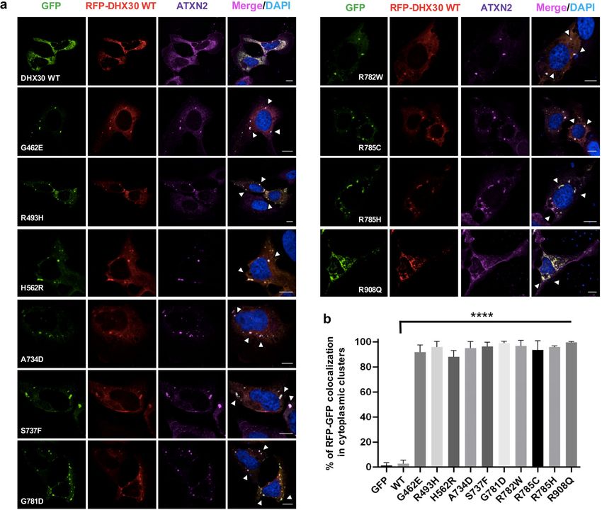
Analyses of the nature of the DHX30 missense variants sulted in each case in DHX30-WT being localized in
Given the somewhat milder clinical presentation of indi- Ataxin-2 positive cytoplasmic clusters (Fig. 5). These data
viduals carrying a whole gene deletion, in-frame deletion, suggest that these missense variants either exert a domin-
frameshift, or nonsense variant as compared to the indi- ant negative effect on the wild-type or lead to a gain-of-
viduals harboring a de novo missense variant in one of the function since both overexpressed DHX30-WT andMannucci et al. Genome Medicine (2021) 13:90 Page 10 of 19
120.0
100.0 **** **** **** ****
80.0
Percentage
60.0
40.0
20.0
0.0
WT V556I E948K R493H R725H R785C R908Q
Normal/Survived Dead at 4dpf Dead at 5dpf
Dead at 6dpf Dead at 7dpf
Fig. 4 Protein variants of DHX30 lead to embryonal developmental
defects in zebrafish. In vivo analyses of selected DHX30 missense
variants. Assessment of embryonic development after injection of
DHX30 WT and mutant cDNAs in a zebrafish model. Bar graph
indicating the percentage of cmcl2-GFP positive zebrafish embryos
assessed 4–7 days post fertilization (dpf). The presented data are
derived from three independent studies. The total number of
embryos assessed are 45, 23, 21, 30, 34, 33, and 29 for WT, V556I,
E948K, R493H, R725H, R785C, and R908Q, respectively. ****:
significantly different from WT (****p< 0.0001; χ2 test)
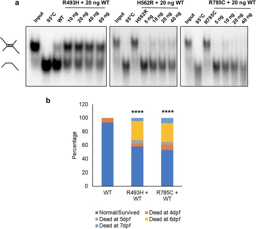
rescued the abnormal phenotypes associated with both
p.(Arg493His) and p.(Arg785Cys) (Fig. 6b), a finding that
could potentially support both loss-of-function and a
dominant negative effect as a mechanism underlying
disease.
Fig. 3 Missense variants in DHX30 initiate the formation of cytoplasmic DHX30 deficiency impairs stress granules formation in
aggregates and impair global translation. Puromycin incorporation assay in HEK293T cells and zebrafish
U2OS cells expressing DHX30-GFP fusion proteins (green). Translation was To further characterize the role of DHX30 we estab-
monitored by staining against puromycin (red), SGs were detected by
lished HEK293T DHX30 stable knockout lines. CRISPR/
ATXN2 (magenta) and nuclei via DAPI staining (blue). Arrows indicate
transfected cells. Note the correlation between formation of clusters and Cas9 based knockout (KO) of DHX30 in HEK293T cells
lack of puromycin staining. Scale bars indicate 10 μm yielded several cell lines with a residual DHX30 immu-
noreactivity of less than 10 % (Fig. 7). Given that
DHX30 is recruited to SGs, we wondered whether
endogenous DHX30 are recruited to cytoplasmic clusters DHX30 additionally plays a role in SG formation. There-
only after stress [8]. fore, we assessed the ability of KO cells to induce SGs or
Next, we analyzed if the DHX30-WT can rescue the cytoplasmic clusters following heat stress treatment. By
inability of p.(Arg493His), p.(His562Arg), and incubating cells at 43.5 °C, a condition after which en-
p.(Arg785Cys) to unwind RNA. The addition of DHX30- dogenous DHX30 accumulates in SGs [8], we observed
WT to p.(His562Arg) and p.(Arg785Cys) efficiently re- that KO cells had a significantly reduced number of SG-
solved the dimer into the monomeric form even in the positive cells as compared to HEK293T WT cells (Fig.
presence of increased amounts of the respective mu- 7). These data suggest a previously unknown role of
tants. However, we observed only a partial rescue when DHX30 in SG assembly. Combined with our previous
DHX30-WT was added to the p.(Arg493His) variant findings (Fig. 3 and 5) these data actually suggest that
(Fig. 6a). Our data suggest that the mutants cause a loss the HCM missense variants exhibit a gain of function by
of helicase function rather than having a dominant nega- triggering SG formation which results in global transla-
tive effect. Given these somewhat contradictory results, tion inhibition.
we turned again to the zebrafish model. We co-injected Next, we generated a predicted null allele in the single
pTol2pA2-cmlc2:EGFP;tuba1a:DHX30 p.(Arg493His) or zebrafish dhx30 ortholog using CRISPR/Cas9. At day
p.(Arg785Cys) with wild-type DHX30 cDNA and five post fertilization, transcript levels of dhx30 were
assessed embryonic development. Interestingly, co- barely detectable in homozygous mutant animals com-
injection of DHX30-WT, at a similar level, partially pared to wild-type, whereas heterozygous siblingsMannucci et al. Genome Medicine (2021) 13:90 Page 11 of 19 Fig. 5 Recombinant protein variants of DHX30 induce translocation of the DHX30-WT in the cytoplasmic clusters. Immunocytochemical detection of RFP-DHX30 WT (red) and ATXN2 (magenta) after co-expression of DHX30-GFP mutants (green) in U2OS cells. Bar graph indicating the percentage of cells where RFP-DHX30 WT co-localizes with DHX30-GFP mutants within cytoplasmic clusters identified as SGs via co-staining with ATXN2 (****: significantly different form DHX30-WT: **** p < 0.0001; n > 100 from 3 independent transfections; one-way ANOVA followed by Dunnett’s multiple comparisons test). Scale bars indicate 10 μm displayed ~ 30% lower dhx30 expression as compared to staining for TIAL-1, an established stress granule marker wild type, potentially due to nonsense mediated decay of (Fig. 8). Although an increase in SG formation occurred the mutated alleles (Additional file 10: Figure S7). The upon heat shock, the number of TIAL-1-labeled SGs homozygous mutant animals are viable, fertile, and mor- remained significantly lower in the homozygous mutants phologically indistinguishable from their wild-type and compared to sibling controls (Fig. 8). Thus, these data heterozygous siblings (data not shown). Previous studies show that SG formation is compromised in the homozy- have demonstrated that during early embryonic develop- gous mutants and suggests an evolutionarily conserved ment, zebrafish exhibit robust SG formation in response role for DHX30 in SG assembly. to stress, such as heat shock [41]. Therefore, based on our in vitro findings, we first asked whether dhx30 mu- Dhx30-deficient zebrafish display altered behavioral tant zebrafish also exhibit impaired SG formation activity in vivo. At 24-h post-fertilization and normal condition, We next examined whether dhx30-deficient zebrafish compared to dhx30-WT the homozygous mutant exhib- exhibit abnormal sleep-wake activity and social behav- ited significantly lower number of SGs, determined by iors, similar to those recently observed in a zebrafish
Mannucci et al. Genome Medicine        (2021) 13:90                                                                                   Page 12 of 19

 Fig. 6 Analyses of the nature of missense variants within the helicase core motifs (HCM). a RNA unwinding activity of purified DHX30 R493H, H562R,
 and R785C mutants was analyzed upon addition of DHX30 WT protein. Increasing amounts of mutant proteins were incubated with 20 ng of WT
 protein and assayed for their ability to unwind a radiolabeled RNA duplex in the presence of ATP. b Assessment of embryonic development after co-
 injection of DHX30 R493H and R785C with DHX30 WT cDNA in a zebrafish model. Bar graph indicating the percentage of cmcl2-GFP positive zebrafish
 embryos 4–7 days postfertilization (dpf). The presented data are derived from three independent studies. The total number of embryos assessed are
 58, 43, and 51 for WT, R493H+WT, and R785C+WT, respectively. ****: significantly different from WT (****p< 0.0001; χ2 test)

model of the NR3C2-related neurodevelopmental dis-                          consistent with findings in DHX30-related neurodeve-
order [25]. We first analyzed sleep-wake behaviors in 5-                    lopmental disorders.
day-old dhx30 KO mutants. Compared to wild-type and
heterozygous siblings, the homozygous mutants dis-                          Discussion
played significantly less activity during the day and more                  Our study has allowed further delineation of the clinical
nocturnal activity (Fig. 9a–c), mimicking somewhat the                      spectrum of DHX30-related neurodevelopmental disor-
sleep disturbances in individuals affected by a DHX30-                      ders through analysis of 25 novel affected individuals,
related neurodevelopmental disorder. Additionally, using                    partially facilitated by the use of a social media-based
an established social preference assay, we observed that                    family support group. Individuals harboring heterozy-
the wild-type and heterozygous animals showed the pre-                      gous missense variants affecting highly conserved resi-
viously described social behavior of preferring to stay                     dues within a HCM present with global developmental
close to conspecific fish of similar age and size, whereas                  delay, intellectual disability, muscular hypotonia, severe
the homozygous animals did not show this preference                         gait abnormalities (if walking is acquired), and remain
(Fig. 9d–e). There were no obvious dysmorphic pheno-                        non-verbal or speak only single words. We also identi-
types in the homozygous mutant animals compared to                          fied microcephaly as an additional common feature. In-
their wild-type and heterozygous siblings. We propose,                      dividuals with either a mosaic missense variant within a
therefore, that the mutant phenotype was not simply                         HCM, or with variants resulting in haploinsufficiency or
due to developmental delay but influenced by abnormal-                      with protein-truncating variants all learned to walk in
ities in complex neural circuitry. Taken together, our                      the second year of life, had a mild muscular hypotonia,
data indicate that dhx30 KO zebrafish have a social be-                     and spoke at least 20 words by the age of 3 years. There-
havioral deficit with altered sleep-wake activity, which is                 fore, based on the clinical and molecular findings we
Mannucci et al. Genome Medicine         (2021) 13:90                                                                                    Page 13 of 19

 Fig. 7 DHX30 deficiency in HEK293T cells leads to reduced formation of stress granules. a Immunocytochemical detection of endogenous ATXN2
 (magenta) in WT HEK293T cells and DHX30-deficient HEK293T cells before (left panel) and after (right panel) heat shock at 43.5 °C for 1 h. Note
 that, upon heat stress and depletion of DHX30 (right hand, lower panel), ATXN2 does not alter its diffuse cytoplasmic distribution to accumulate
 in cytoplasmic foci, as observed in WT HEK293T cells (right hand, upper panel). Nuclei are identified via DAPI staining (blue). Scale bars indicate
 10 μm. b Bar graph indicating the percentage of cells containing stress granules. (*: significantly different from WT HEK293T cells: *p< 0.05; n >
 200 from 3 independent experiments; unpaired t test ). c Western blotting detection of DHX30 knock-out efficiency in HEK293T cells. Expression
 of DHX30 was reduced by 90% as detected by a DHX30 specific antibody. Tubulin was used as loading control

suggest a classification in two DHX30-associated neuro-                      words, thus did not have a completely absent language.
developmental disorder subtypes.                                             Therefore, we suggest referring to these conditions as
  It is worth noting that the identified heterozygous                        DHX30-associated neurodevelopmental disorders in the
deletion in individual 24 also encompasses the first                         future.
15 SETD2 exons, suggestive of a dual diagnosis. How-                           To provide further evidence for the pathogenicity of
ever, given the phenotypic differences in the seven in-                      the novel DHX30 variants and gain better insight into
dividuals reported to date, some of whom inherited                           the genotype-phenotype correlation we performed sev-
their SETD2 variant from an apparently unaffected                            eral in vitro and in vivo analyses. For this, we have now
parent [32–34], we are unsure to what extent loss of                         formally confirmed that DHX30 possesses ATP-
SETD2 contributed to the phenotype observed in this                          dependent RNA helicase activity. In line with their ab-
individual.                                                                  sence from public databases and high evolutionary con-
  Identification of affected individuals with milder                         servation of affected amino acid residues, all novel
phenotype challenges naming of this disorder “Neurode-                       missense variants within a HCM resulted in impaired
velopmental disorder with severe motor impairment and                        ATPase activity (all were within an ATP binding and hy-
absent language” (NEDMIAL; OMIM # 617804). Not-                              drolysis motif), impaired helicase activity, and showed an
ably, only 9 of the 25 individuals (36%) presented here                      increased propensity to trigger stress granule (SG) for-
had a severe motor impairment (never learned to walk)                        mation resulting in inhibition of global translation, as
and 9 out of 25 individuals (36%) spoke at least single                      expected from the previous study [8]. In addition,
Mannucci et al. Genome Medicine            (2021) 13:90                                                                                  Page 14 of 19

 Fig. 8 DHX30 deficiency in zebrafish cells leads to reduced formation of
 stress granules. a Representative confocal images of TIAL-1-labeled stress
 granules (green) in dhx30 wild-type (+/+) and homozygous mutants (−/−).
 Zebrafish underwent normal conditions or heat shock treatment at 42 °C.
 Nuclei were counterstained with DAPI (blue). b Analyses of TIAL-1-labeled
 stress granules per 50 nuclei. The total number of embryos assessed are 8,
 9, 8, and 8 for dhx30 +/+ (28 °C), dhx30 −/− (28 °C), dhx30 +/+ (42 °C), and
 dhx30 −/− (42 °C), respectively. Data are presented as means ± standard
 error of mean based on the indicated number of embryos. ***: significantly
 different from DHX30+/+ (***p< 0.001; unpaired Student’s t test)

selected HCM missense variants interfere with normal
                                                                                Fig. 9 Behavioral analyses of dhx30 mutant zebrafish. a Distance
zebrafish embryonic development.                                                moved of dhx30 mutants and wild-type sibling controls measured at
  We have previously suggested that the missense vari-                          5 days post fertilization. b Average of distance moved during 14-h
ants within HCM might have a more severe effect than a                          daytime. c Average of distance moved during 10-h nighttime. N =
loss of one gene copy [8]. This hypothesis is now sup-                          15, 18, and 25 for +/+, +/−, and −/− animals, respectively. d Social
                                                                                preference index (SPI) calculated during 10-min baseline and post-
ported by identification of four affected individuals car-
                                                                                baseline period. SPI = 1 indicates a fish that spends 100% of its time
rying variants that result in either haploinsufficiency or a                    near a conspecific, SPI = − 1 indicates a fish that spends 100% of its
truncated protein, all of whom presented with a milder                          time near the empty well, and SPI = 0 indicates a fish that spends
phenotype as compared to the individuals harboring                              equal amounts of time near the conspecific and near the empty
missense variants within HCM. To gain further insight                           well. e The change in SPI between baseline and post-baseline,
                                                                                indicating the preference of zebrafish to stay close to conspecific
into the nature of these variants we determined that
                                                                                fish. N = 13, 6, and 17 for +/+, +/−, and −/− animals, respectively.
DHX30-WT can rescue the inability of selected HCM                               Data are presented as means ± standard error of mean based on
missense variants to unwind an RNA duplex, and that                             the indicated number of embryos. *: significantly different from
co-injection of DHX30-WT with selected HCM mis-                                 dhx30+/+ (*p< 0.05; unpaired Student’s t test)
sense variants can partially ameliorate the observed zeb-
rafish phenotypes. These data point to loss-of-function
effects of the HCM mutants on a molecular level. How-
ever, co-expression of HCM missense variants together
Mannucci et al. Genome Medicine (2021) 13:90 Page 15 of 19
with the DHX30-WT resulted in recruitment of Beyond providing the molecular explanation for the
DHX30-WT into SG´s, a finding that might possibly genotype-phenotype correlation of these two subtypes
suggest a dominant negative effect. It is worth noting we additionally performed in vivo behavioral modeling
that DHX30-WT, as well as the endogenous protein, are of zebrafish dhx30 KO’s. Zebrafish exhibit all the hall-
recruited to the SG´s after stress induction [8]. Thus, marks of mammalian sleep by utilizing neurotransmit-
HCM missense variants might actually result in a detri- ters known to coordinate sleep and wake states in
mental gain-of-function by inducing SG formation with humans [44]. Analysis of dhx30-deficient animals re-
concomitant global translation impairment even without vealed a compromised sleep/wake behavior, as they were
endogenous or exogenous stressors. less active during the day but more active and slept less
To gain further clarity we focused on the relation of at night than dhx30-WT animals. This is partially remin-
DHX30 to SG formation. Using CRISPR/Cas9 based iscent of the sleep disturbances observed in almost half
technology, we established two DHX30 knockout of DHX30-affected individuals. Additionally, homozy-
models. Analyses of both, DHX30-deficient HEK293T gous dhx30 KO animals displayed altered social behavior
cells and zebrafish, revealed an impairment of SG forma- as manifested by their performance in the social prefer-
tion upon heat stress, pointing to an essential and evolu- ence assay, e.g., showing reduced preference for conspe-
tionary conserved role of DHX30 in SG assembly. These cifics as compared to dhx30-WT zebrafish. The
findings provide a molecular explanation for the above- observed social behavioral deficits and altered sleep-
mentioned phenotypic differences, as they strongly sug- wake activity are similar to the findings in zebrafish
gest that pathogenic missense HCM variants, in addition models of other neurodevelopmental disorders [25, 45],
to the loss of ATPase or RNA-binding activity and with and to some extent recapitulate the clinical findings in
impaired helicase function, exert a selective gain-of- individuals affected by the DHX30-related neurodevelop-
function by triggering SG formation. This is in line with mental disorder.
our hypothesis that due to SG hyper-assembly these Furthermore, we present here two individuals who
pathogenic variants generate a chronic condition of im- clearly stand out both in terms of their clinical presenta-
paired translation [8]. Noteworthy, impaired translation tion and their identified DHX30 variant. Individual 4
due to aberrant SG formation is associated with a broad with an early-lethal infantile epileptic encephalopathy
variety of neurodegenerative and neurodevelopmental carries a homozygous missense variant, p.(Arg725His),
diseases [8, 42]. Furthermore, repeat expansion under- and individual 21 with a de novo p.(Arg908Gln) variant
lying C9orf72-associated neurodegenerative disorders shows late-onset progressive ataxia. Trio-WES analysis
has recently been suggested to result in chronic cellular performed in both individuals identified these DHX30
stress due to aberrant SG formation [43]. variants as the only candidates (Supplementary Data). As
Table 2 Summary of functional analyses of missense variants
DHX30 p.(Gly462Glu), p.(His562Arg), p.(Ala734Asp), p.(Arg493His) p.(Arg725His) p.(Arg908Gln) p.(Val556Ile) p.(Glu948Lys)
variant p.(Ser737Phe), p.(Thr739Ala), p.(Gly781Asp)
p.(Arg782Gln) p.(Arg782Trp), p.(Arg785Cys),
p.(Arg785His)
Location in Helicase core motifs I, II, V, or VI (nucleotide- Helicase core Helicase core Ratchet-like Helicase core C-terminal
DHX30 interacting motifs) motif Ia region, domain region, region
(nucleic acid- between between
binding) motifs IV and V motifs Ib and II
gnomAD Not identified Not identified Not identified Not identified 0/39/282352 1/49/282090
v2.1.1
ATPase Reduced Similar to wt* Reduced Reduced Similar to wt Similar to wt
activity
RNA binding n.d. Reduced* n.d. n.d. n.d. n.d.
capacity
Helicase Reduced** Reduced n.d.*** Similar to wt n.d. n.d.
activity
Cellular Stress granules Stress granules Cytoplasmic, Cytoplasmic Cytoplasmic, Cytoplasmic,
localization similar to wt aggregates similar to wt similar to wt
Puromycin Impaired Impaired* Similar to wt Impaired n.d. n.d.
incorporation
Zebrafish Impaired** Impaired Impaired Impaired Similar to wt Similar to wt
development
n.d., not determined; *, Lessel et al. 2017; **, only selected variants analyzed; ***, unable to purify the proteinYou can also read