DRUG DESIGN AND REPURPOSING WITH DOCKTHOR VS WEB SERVER FOCUSING ON SARS COV 2 THERAPEUTIC TARGETS AND THEIR NON SYNONYM VARIANTS
←
→
Page content transcription
If your browser does not render page correctly, please read the page content below

www.nature.com/scientificreports
OPEN Drug design and repurposing
with DockThor‑VS web server
focusing on SARS‑CoV‑2
therapeutic targets and their
non‑synonym variants
Isabella A. Guedes1, Leon S. C. Costa1, Karina B. dos Santos1, Ana L. M. Karl1,
Gregório K. Rocha2, Iury M. Teixeira1, Marcelo M. Galheigo1, Vivian Medeiros1,
Eduardo Krempser3, Fábio L. Custódio1, Helio J. C. Barbosa1, Marisa F. Nicolás4* &
Laurent E. Dardenne1*
The COVID-19 caused by the SARS-CoV-2 virus was declared a pandemic disease in March 2020 by
the World Health Organization (WHO). Structure-Based Drug Design strategies based on docking
methodologies have been widely used for both new drug development and drug repurposing to find
effective treatments against this disease. In this work, we present the developments implemented
in the DockThor-VS web server to provide a virtual screening (VS) platform with curated structures
of potential therapeutic targets from SARS-CoV-2 incorporating genetic information regarding
relevant non-synonymous variations. The web server facilitates repurposing VS experiments providing
curated libraries of currently available drugs on the market. At present, DockThor-VS provides
ready-for-docking 3D structures for wild type and selected mutations for Nsp3 (papain-like, PLpro
domain), Nsp5 (Mpro, 3CLpro), Nsp12 (RdRp), Nsp15 (NendoU), N protein, and Spike. We performed
VS experiments of FDA-approved drugs considering the therapeutic targets available at the web
server to assess the impact of considering different structures and mutations to identify possible new
treatments of SARS-CoV-2 infections. The DockThor-VS is freely available at www.dockthor.lncc.br.
Pandemic COVID-19 caused by the infection of severe acute respiratory syndrome (SARS) coronavirus 2 (SARS-
CoV-2), initially described near the end of 20191, left the world in lockdown and reached almost 35.6 million
confirmed cases including 1,042,798 deaths by September 2020 (WHO Coronavirus Disease (COVID-19) Dash-
board, 06 September 2020 https://covid19.who.int). SARS-CoV-2 belongs to the genus Betacoronavirus of the
subfamily Coronavirinae, which includes other coronaviruses (CoVs), such as those responsible for the outbreaks
of the severe acute respiratory syndrome (SARS-CoV) in 2002/2003 and the Middle East respiratory syndrome
(MERS-CoV) in 2 0121,2. The emergence of these CoVs, including SARS-CoV-2, demonstrated versatile host
ranges and tissue tropism of these pathogens that can infect the respiratory, gastrointestinal, hepatic, and central
nervous systems of humans, birds, bats, pangolins, and many other wild animals3.
The CoVs genome is a single-stranded positive-sense RNA (+ ssRNA) of approximately 30 Kb with 5′-cap
and 3′-poly-A tail4. The SARS-CoV-2 genomic sequence is more similar (96.2%) to bat SARS (SARSr-CoV;
RaTG13)5 than to human SARS-CoV (about 79%) or MERS-CoV (about 50%)6. However, SARS-CoV-2 uses
the same receptor on the eukaryotic membrane surface as SARS-CoV, the angiotensin II-converting enzyme
(ACE2)7. Although a new Spike-mediated CD147 receptor viral invasion route has recently been demonstrated
for SARS-CoV-2, mediating viral invasion and disseminating virus among other c ells8,9.
Structurally, the SARS-CoV-2 genome is organised in the order of the 5′-replicase polyprotein (ORF1/ab)-
structural proteins [Spike (S)-Envelope (E)-Membrane (M)-Nucleocapsid (N)]-3′. There is a frameshift of -1
1
Grupo de Modelagem Molecular em Sistemas Biológicos (GMMSB), National Laboratory for Scientific Computing
- LNCC, Petrópolis, RJ, Brazil. 2Instituto Federal Fluminense - IFF, Macaé, RJ, Brazil. 3Fundação Oswaldo Cruz
- Fiocruz, Rio de Janeiro, RJ, Brazil. 4Laboratório de Bioinformática (Labinfo), National Laboratory for Scientific
Computing - LNCC, Petrópolis, RJ, Brazil. *email: marisa@lncc.br; dardenne@lncc.br
Scientific Reports | (2021) 11:5543 | https://doi.org/10.1038/s41598-021-84700-0 1
Vol.:(0123456789)
www.nature.com/scientificreports/
between ORF1a and ORF1b, leading to the production of two polypeptides: pp1a and pp1ab. Also, these poly-
peptides are processed into 16 non-structural proteins (Nsps, Nsp1‐16), whose cleavage is mediated by the Mpro
(Nsp5, 3CLpro, or main protease) and a papain-like protease (Nsp3, PLpro)10. Other ORFs in a third of the
genome near the 3′ terminal encode four main structural proteins: Spike, membrane, envelope, and nucleocapsid
proteins. The new CoV encodes accessory structural proteins, namely ORF3a, 6, 7a/b, and ORF8, altogether
totalling 29 proteins. All structural proteins are translated from viral subgenomic messenger RNAs (sgRNAs)
produced by the replication and transcription complex (RTC), which includes both the RNA-dependent RNA
polymerase (Nsp12, RdRp)4,10 and the 3′-5 ’exoribonuclease with a functional proofreading-repair activity (Nsp14,
ExoN)4.
Albeit their high copying fidelity, mutations in CoVs are observed as consequences of three known processes,
namely (i) as lesions during the error-prone repair process; (ii) as a mechanism of RNA recombination and seg-
ment reassortment; (iii) by host-dependent RNA editing systems, such APOBECs and A DARs11. These genetic
processes give rise to clouds of intra-host variants, according to the viral quasispecies dynamics12. Consequently,
during the pandemic outbreak, the generations of polymorphic viral quasispecies can promptly arise interacting
within the host, allowing viral immune evasion, resistance to antiviral drugs, as well as affecting the sensitivity of
molecular diagnostic tests. Therefore, the monitoring of genomic changes in SARS-CoV-2 for identifying regions
associated with drug resistance and vaccine evasion is essential in designing antiviral therapies.
There is a global effort from academic and non-academic groups to evaluate and develop an effective treat-
ment for COVID-19. Many drug repositioning studies and compound evaluation to develop new antiviral drugs
against SARS-CoV-2 have been developed using e xperimental13–15 and theoretical/computational16–20 approaches.
More specifically, there is a vast interest in using high-throughput virtual screening approaches using different
molecular modelling methodologies to investigate drug repositioning libraries and a plethora of compounds
databases focusing on distinct SARS-CoV-2 molecular targets. Such computational studies often serve as a basis
for further in vitro and in vivo studies. However, the success of virtual screening experiments depends on factors
that are often not trivial to be addressed by non-specialist researchers21,22. Among the main factors are: (i) the
correct choice and preparation of the molecular target structure (e.g., protonation states of ionizable residues,
incorrect side-chain conformations); (ii) proper preparation of the compound libraries (e.g., pH-dependent
protonation states, tautomerism, isomerism); (iii) receptor flexibility and, especially, induced-fit effects due to
ligand binding; (iv) ligand flexibility (especially for peptides and macrocycle containing molecules); (v) perfor-
mance of particular docking programs and scoring functions; and (vi) availability of computational resources
for high-throughput virtual screening experiments. Moreover, possible genomic variations in the active/binding
site region of molecular targets can drastically affect the binding mode and affinity of ligands and, consequently,
change the identification of promising compounds.
Nowadays, the fast-increasing amount of available structural and genomic data of SARS-CoV-2 protein targets
enhances the success of virtual screening and molecular modelling experiments. Herein, we adhere to research
groups worldwide to enhance both new antiviral and drug repurposing research to develop an effective treatment
for COVID-19. Our focus is on improving the DockThor-VS web server as a virtual screening platform to moni-
tor the emergence of relevant non-synonymous mutations on SARS-CoV-2 target proteins. The DockThor-VS
web server is freely available for the scientific community since 2013 as a docking platform for drug discovery
containing the main steps of protein, cofactor, and ligand preparation, being able to deal efficiently with a wide
diversity of protein–ligand systems for both binding mode and affinity prediction.
Herein, we present the developments implemented in the DockThor-VS web server as an effort to provide for
the scientific community the possibility to perform COVID-19-related virtual screening experiments with: (1)
curated structures of potential therapeutic targets from SARS-CoV-2 Nsp3 (papain-like, PLpro domain), Nsp5
(Mpro, 3CLpro), Nsp12 (RdRp), Nsp15 (NendoU), Nucleocapsid phosphoprotein (N) and Spike considering
wild types and selected variants, and (2) curated libraries of the currently available drugs on the market.
To the best of our knowledge, the DockThor-VS platform is the unique virtual screening server that provides
the users with curated datasets of both wild type and relevant mutants from SARS-CoV-2 therapeutic targets
and prepared datasets for drug repurposing.
Besides considering relevant protein target mutations, our approach differs from other platforms mentioned
in the literature23,24 by accounting for the following relevant aspects (cited above) when performing a virtual
screening experiment. (i) For each protein target, we chose a set of structures representing different relevant
conformations complexed with varying types of ligand (e.g., ligands covalently bound or not, side chains con-
formations closing or opening the binding site), thus providing an ensemble of representative structures of the
flexibility of the receptor. (ii) For each protein target, the protonation states of ionisable residues were carefully
investigated in the available literature. (iii) The repurposing libraries open to the users were carefully prepared
considering the different protonation states and tautomerism for each compound. (iv) The DockThor docking
program25 was specially developed to deal with highly flexible ligands, and it is very suitable to dock highly flex-
ible peptides26. (v) Macrocycle drugs were subjected to special treatment to generate distinct conformations.
(vi) The availability of the dataset of SARS-CoV-2 therapeutic targets allows the users to perform target fishing
projects to search for the most promising target for the investigated compounds.
The DockThor-VS platform (freely available at https://www.dockthor.lncc.br) is coupled to the SDumont
Brazilian supercomputer platform, supporting virtual screening experiments with up to 200 compounds for
guest users and 5,000 compounds for registered projects.
In the next sections, we will show the technical details of the target selection and generation of the com-
pound libraries and some general features of the DockThor-VS platform. We reveal and discuss the results of
repurposing virtual screening experiments, considering the six therapeutic targets. We also assess the impact of
considering different protein structures and mutations to identify possible new lead compounds to treat SARS-
CoV-2 infections.
Scientific Reports | (2021) 11:5543 | https://doi.org/10.1038/s41598-021-84700-0 2
Vol:.(1234567890)
www.nature.com/scientificreports/
Figure 1. Conformations of the Nsp3-PLpro domain provided at the DockThor-VS web server as representative
structures of the protein flexibility. The apo structure is coloured blue (PDB code 6W9C) and the structure
initially complexed with a covalently bound peptide inhibitor is coloured brown (PDB code 6 WX429).
Results and discussion
Dataset of potential therapeutic targets. Non‑structural protein 3 (Nsp3, papain‑like protease—PL‑
pro). The multi-domain non-structural protein 3 (Nsp3) is the largest protein produced by the coronavirus,
comprising 16 different domains and regions that regulate viral infection, with the papain-like protease domain
(PLpro) being the most widely targeted domain from Nsp3. Since the outbreaks of SARS-CoV in 2003 and
MERS-CoV in 2012, the three-dimensional structure of Nsp3 has been solved by X-ray crystallography and
nuclear magnetic resonance (NMR) spectroscopy. Currently, we provide in the DockThor-VS web server mono-
meric structures and genomic variations of the PLpro domain. Structural information and selected mutations
regarding the Macrodomain I/II/III (MacI/II/III) or active ADP-ribose-100-phosphatase domain (ADRP, app-
1″-pase) will be available soon.
PLpro is a cysteine protease that processes, through self-catalyzed cleavage reaction, the amino-terminal
end of the replicase polyprotein (pp1a) generating mature Nsp1, Nsp2 and Nsp3 proteins27. This protein is also
responsible for aiding the coronavirus in its invasion by counteracting host innate immunity. The PLpro is a
multifunctional enzyme capable of cleaving the viral polyprotein, and also functions as a deubiquitinase (DUB)
and deISGylating (deconjugating interferon-stimulated gene 15 [ISG15] molecule from modified substrates),
using identical catalytic r esidues28. Thus, the therapeutic inhibition of PLpro would have two antiviral effects:
restoration of the antiviral effect of deubiquitinylation/ISGylation and inhibition of viral replication by blocking
polyprotein cleavage29.
The SARS-CoV-2 PLpro catalytic site is composed of a classic triad Cys111-His272-Asp286. Cys111 performs
the nucleophilic attack on the peptidic substrate, while His272 and Asp286 act by stabilizing the intermediate of
the reaction27. Trp106 forms the oxyanion site participating in the stabilization of the negatively charged inter-
mediate. Many non-covalent inhibitors interact at an allosteric site near the catalytic site. This allosteric site is
mainly composed of the Asn267-Tyr268-Gln269 residues, forming a β-turn secondary s tructure27. Its flexibility
is well described in the literature and is mainly characterized by distinct conformations of the residue Tyr268,
which usually makes stacking interactions with aromatic groups of some inhibitors. The protonation state of the
catalytic triad residues was defined based on the mechanism of reaction proposed in the literature for SARS-
CoV27: neutral Cys111, His272 neutral at NE2 and Asp286 negatively charged.
To date, there are 16 PLpro crystal structures in the PDB. Given the flexibility observed for Tyr268 from the
allosteric site and Leu162, located at the entrance of the catalytic site, we provide to the users two prepared struc-
tures related with the PDB codes 6W9C (apo structure) and 6 WX429 (solved in complex with a covalently bound
peptide inhibitor) (Fig. 1). In both structures, the Tyr268 is presented on an open conformation, allowing the
binding of ligands with different sizes. The recently solved structure of PLpro complexed with a non-covalently
bound compound (PDB code 7JIW) will be provided soon in the DockThor-VS.
Non‑structural protein 5 (Nsp5, Mpro, 3CLpro). As is well-known in coronaviruses, the two overlapping poly-
proteins pp1a and pp1ab, firstly produced after infection, are further proteolytically processed into 16 non-struc-
tural proteins (Nsp1–16). This proteolytic process is carried out in a coordinated manner by the PLpro and the
Mpro30. Mpro is also known as 3-chymotrypsin-like cysteine protease (CCP or 3CLpro), that first is auto-cleaved
from polyprotein pp1a to yield the mature enzyme and then digests the remaining pp1a (at least by 11 conserved
sites) to produce the downstream non-structural proteins (Nsps 6 to 16)31. Given the pivotal role of Mpro in the
viral life cycle, it becomes an attractive target for the design of anti-SARS drugs.
Scientific Reports | (2021) 11:5543 | https://doi.org/10.1038/s41598-021-84700-0 3
Vol.:(0123456789)
www.nature.com/scientificreports/
Figure 2. Conformations of the Mpro provided at the DockThor-VS web server as representative structures
of the protein flexibility. The protein complexed structure with a covalent inhibitor is coloured blue (PDB code
6LU7, the ligand is not shown) and the structure initially complexed with a non-covalent inhibitor is coloured
green (PDB code 6W63). The protein–ligand hydrogen bonds are represented as yellow dashed lines.
The Mpro consists of a homodimer with each polypeptide composed of three domains: I (residues 8–101),
II (residues 102–184) and III (residues 201–303). The substrate-binding site is located in a cleft between the
domains I and II. It has the Cys145-His164 catalytic dyad as the reaction center, following a mechanism similar
to other coronaviruses. There are currently 191 structures of the Mpro deposited in the PDB, with many of
them complexed with covalent or non-covalent inhibitors. At this moment, we provide to the users the dimeric
structure of the Mpro in two distinct conformations (PDB codes 6LU730 and 6W63), which are complexed with
covalent and non-covalent inhibitors, respectively. The structures’ superposition highlights some significant
conformational changes within the ligand-binding site, mainly the residues Met49, Asn142 and Gln189. That
reinforces the importance of considering multiple protein conformations in virtual screening experiments to
accommodate distinct compounds (Fig. 2). While the 6LU7 conformation was the first structure experimentally
solved for the SARS-CoV-2 Mpro with an inhibitor, the 6W63 contains a drug-like reversible inhibitor at the
binding site. In the preparation process, the protonation states and flips of key residues were manually adjusted
to provide the Mpro structures with neutral His41 at ND1, the catalytic Cys145 protonated (i.e., neutral), neutral
His163 at NE2 and neutral His164 at NE2.
Non‑structural protein 12 (Nsp12, RdRp). To replicate and transcript positive ssRNA, an RNA-dependent RNA
polymerase (RdRp, also known as Nsp12) of coronaviruses has evolved to perform this process forming an
intricate complex with several non-structural proteins (Nsps) produced as cleavage products of the ORF1a and
ORF1ab viral polyproteins. Nsp12 catalyzes the synthesis of viral RNA and possibly with the assistance of Nsp7
and Nsp8 that function as cofactors32. In SARS-CoV-2 the overall architecture of the Nsp12-Nsp7-Nsp8 complex
is similar to that of SARS-CoV with a root mean square deviation (RMSD) value of 0.82 for 1078 Ca a toms33.
RdRp is considered an interesting target for therapeutic solutions against COVID-19, for which the inhibitor
remdesivir (RDV, GS-5734), a nucleoside analogue prodrug of the ebola virus (EBOV) RdRP has been already
approved34. Since the nucleoside analogues have a high structural similarity, other similar drugs such as favip-
iravir, which was effective in clinical trials, can be used as an i nhibitor35.
The conserved architecture of the Nsp12 core consists of a right-hand RdRp domain (residues Ser367 to
Phe920) and a nidovirus-specific N-terminal extension domain (residues Asp60 to Arg249) that adopts a nidovi-
rus RdRp-associated nucleotidyltransferase (NiRAN) a rchitecture33. The average length of the core RdRp domain
is less than 500 amino acids and is folded into three subdomains, namely thumb, palm, and fingers resembling
a right-handed c up36. The NIRAN and RdRp domains are connected by an interface domain (residues Ala250
to Arg365). Besides, COVID-19 virus Nsp12 possesses a newly identified β-hairpin domain at its N terminus33.
The active site of the SARS-CoV-2 RdRp domain is formed by the conserved polymerase motifs A to G
in the palm subdomain and configured like other RNA p olymerases33. Remarkably, the motifs A and C have
conserved residues characteristic of viruses + ssRNA, such as the catalytic aspartates in motifs A (Asp618) and
C (Asp760)37. Motif B has highly conserved Ser682 that is crucial for recognizing the 2′-OH group of the NTP
ribose and Gly683, which is conserved in all RdRps37. Motif D and motif A, both guide the structural change of
the active site during c atalysis38. Regarding the nucleotide (NTP) selection by RdRp, motif D has a prime role
in NTP addition’s efficiency and fidelity. Indeed, NMR studies have indicated the inability of motif D to achieve
its optimal conformation for catalysis when an incorrect nucleotide is incorporated, thereby demonstrating its
role in selecting NTPs39. Motif E together with motif C interact (in the upstream position) with the newly syn-
thesized backbone of the second and third nucleotides, motif F establishes the upper limit for the entry path of
NTPs37. In the motif F of SARS-CoV-2, the residue Ala547 of the N-Terminal region is equivalent to the highly
conserved glutamate in almost all + ssRNA viruses37. This amino acid change leads to structural and possibly
dynamic differences in this region, which can interfere with the RNA synthesis40. The motif G uses the residues
S96 and A97 that interact with residues + 1 and + 2 of the template ribbon’s backbone to move it vertically37,41.
Scientific Reports | (2021) 11:5543 | https://doi.org/10.1038/s41598-021-84700-0 4
Vol:.(1234567890)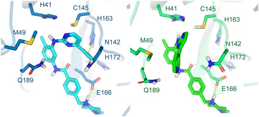
www.nature.com/scientificreports/
Figure 3. Structure of the Nsp12 complexed with remdesivir and an RNA primer (PDB code 7BV2, ligand
coloured greed and RNA represented as an orange cartoon). Mg2+ ions are represented as green spheres. The
protein–ligand hydrogen bonds are represented as yellow dashed lines.
To date, eight RdRp structures have been deposited at the PDB. We provided at the DockThor-VS platform
the RdRp conformation found in the RdRp-RNA-remdesivir complex (PDB code 7BV242, Fig. 3) without the
RNA primer and the inhibitor remdesivir to allow the virtual screening experiments with the free binding site.
Non‑structural protein 15 (Nsp15, endoribonuclease, NendoU). The Nsp15 of SARS-CoV-2 is a nidoviral RNA
uridylate-specific endoribonuclease (NendoU) that displays its RNA endonuclease activity (specific for uridine)
acting on both, single-stranded RNA and double-stranded R NA43. Recently, Susan Baker’s Lab revealed for the
first time, the molecular mechanism of Nsp15, in which the NendoU activity limits the generation of 5′-polyu-
ridines from negative-sense viral RNA, termed PUN. The PUN can act as a CoVs MDA5-dependent pathogen-
associated molecular pattern (PAMP), which in turn can activate the type I interferon (IFN) response in mac-
rophages. The authors found that NendoU cleaves the polyU sequence on the PUN RNA, limiting the length and
abundance of the polyU extension. These studies revealed that the function of NendoU during replication is to
reduce the length of polyU sequences, thus limiting the potential to generate PAMPs and activate the host sensor
MDA5. Consequently, the NendoU activity delays recognition by the host innate immune sensors, and there-
fore, Nsp15 is a highly conserved virulence factor and a potential target for antiviral and vaccine s trategies43.
The Nsp15 endoribonuclease from SARS-CoV-2 comprises 347 amino acid residues (sequence from Met1
to Gln347)44. The SARS-CoV-2 Nsp15 monomers group into a functional hexamer, composed by a dimer of
trimers44. The hexameric form is pivotal for the enzymatic activity. Each monomer presents three domains: (i)
the N-terminal (Nsp15-NTD, residues 1–62), formed by an antiparallel β-sheet wrapped around two α-helices;
(ii) the central middle (residues 63–191), composed by β-strands and short helices; and (iii) the C-terminal
catalytic NendoU domain (NendoU, residues 192–347), formed by two antiparallel β-sheets. There are currently
seven high-resolution crystal structures of Nsp15 endoribonuclease from SARS‐CoV‐2 available at the PDB
containing the three domains.
The active site is located at the CTD, flanked by five α-helices in its concave surface, in a shallow groove
between two β sheets, and contains six highly conserved residues: His235, His250, Lys290, Thr341, Tyr343 and
Ser294. Based on the similar arrangement of its active site with that of Ribonuclease A, the residues His235,
endoU44. The prepared Nsp15 structure is based
His250 and Lys290 are suggested to be the catalytic triad of the N
on the conformation of the protein complexed with tipiracil (PDB code 6WXC) prepared considering the pH
of 6.2 and consists of His235 and His250 neutral at NE2 and ND1, respectively, and Lys290 positively charged
(Fig. 4). New Nsp15 conformations will be available soon at the DockThor-VS platform.
Nucleocapsid phosphoprotein (N protein). The nucleocapsid protein has an essential structural function in
CoVs. This target is a multifunctional phosphoprotein that establishes an arrangement with genomic RNA form-
ing the ribonucleoprotein (RNP) complex and plays a critical role during transcription, virus assembly and
antagonism of host’s innate immunity. The N protein can form a helical filament structure that is assembled into
virions by interactions with the viral membrane (M) p rotein45. Despite its location within the virion rather than
on its surface, N protein is highly immunogenic and abundantly expressed during viral infection46. Interest-
ingly, it has been demonstrated that the antibody to the SARS-CoV-2 N protein is more sensitive than the Spike
protein antibody during the early infection47. Regarding the context of viral infection, the nucleocapsid protein
acts as a viral suppressor of RNAi (VSRs), and thereby antagonizes one of the cell-intrinsic antiviral immune
defence mechanisms of the host48. Mainly, during RNA viral infection, virus-derivated dsRNA (vi-dsRNA) are
generated, which could be recognised and cleaved by the host endonuclease Dicer into virus-derived siRNAs
(vsiRNAs). These vsiRNAs ultimates are integrated into de Argonaute protein within the RNA-induced silencing
Scientific Reports | (2021) 11:5543 | https://doi.org/10.1038/s41598-021-84700-0 5
Vol.:(0123456789)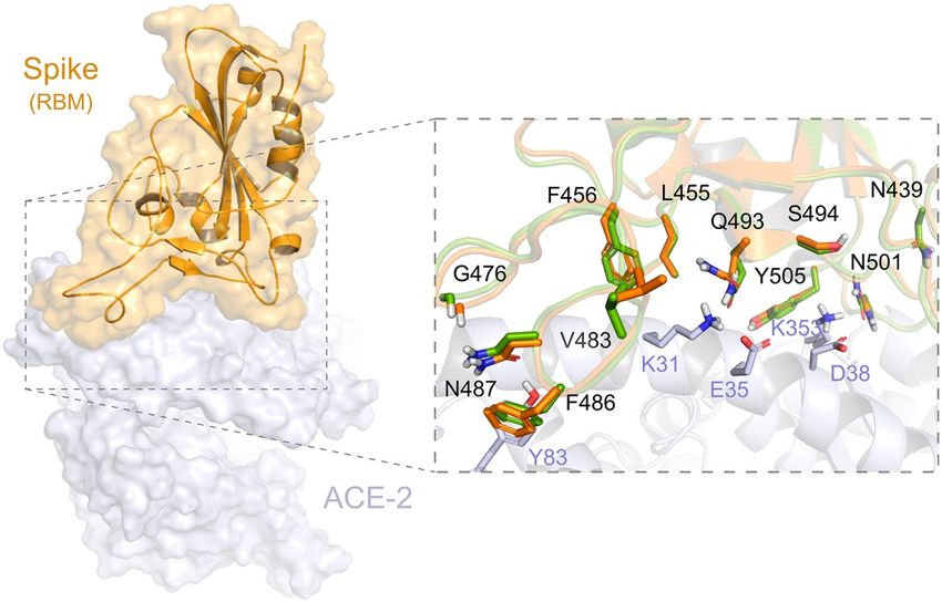
www.nature.com/scientificreports/
Figure 4. Structure of the Nsp15 trimer complexed with Tipiracil (PDB code 6WXC). The protein–ligand
hydrogen bonds are represented as yellow dashed lines.
complex (RISC) directing the destruction of cognate viral RNAs in infected c ells49. Jingfang Mu and collabora-
tors (2020) showed that nucleocapsid of SARS-CoV-2 associates with dsRNA and suppresses RNAi by seques-
trating viral dsRNA in cells, which probably prevents its recognition and cleavage by the host endonuclease
Dicer48. Therefore, the N protein also represents a prime immune evasion factor of SARS-CoV-2, contributing
to the pathogenicity of this new coronavirus. Consequently, the nucleocapsid phosphoprotein can be an attrac-
tive target, for example, to inhibit the viral life cycle stages, or else to recover the host’s immunity mediated by
an antiviral RNAi system.
The structure of N protein from coronavirus is composed of three domains: N-terminal RNA binding
(N-NTD), C-terminal dimerisation (N-CTD) and central Ser/Arg (SR)-rich linker50. At the time of writing of
this manuscript, there are 12 experimentally solved structures for the N protein, where three of them are related
to the N-NTD. We provide to the users experimentally solved structures of the N-NTD already prepared for
docking. The preparation of the N-CTD will be available soon. Currently, there are no drugs or potential com-
pounds experimentally validated as SARS-CoV-2 N protein inhibitors.
Herein, we provide five monomeric structures of N-NTD obtained by NMR experiments (PDB code 6YI3) to
account for the protein flexibility. Specifically, we depicted the basic finger moiety, which is commonly locked in
one conformation in the X-ray solved structures available due to crystal lattice c ontacts51. According to studies
with the N protein from the Influenza virus, antiviral drugs targeting N proteins should stabilise the monomeric
form or induce abnormal oligomerization or interfere with the RNA b inding52. Also, they suggested that the
monomeric form binds to the replicating viral RNA in infected cells.
Recently, surface plasmon resonance (SPR) analysis experiments of SARS-CoV-2 N-NTD show low binding
affinities for different ribonucleotide (AMP/UMP/CMP), except GMP, suggesting potential distinct ribonucle-
otide-binding mechanism between SARS-CoV-2 and HCoV-OC43 N p rotein50. Some important characteristics
observed in the experimentally solved structures of SARS-CoV-2 N-NTD protein that may explain these findings
are: (i) the N-terminal tail is highly flexible and adopts more opened conformations than in HCoV, probably
allowing the interaction with viral RNA genome of high order structure, (ii) replacement of Tyr102 in HCoV to
Arg89 located near to the nitrogenous base recognition site, (iii) phosphate-binding site containing Thr54 and
Ala55 in SARS-CoV-2 instead of Ser67 and Gly68 in HCoV.
According to NMR-based titration experiments of N-NTD with a short double-stranded RNA (5′-CAC
UGAC-3′ and 5′-GUCAGUG-3′), the amino acid residues Ala50, Thr57, His59, Arg92, Ile94, Ser105, Arg107,
Arg149, Tyr172 were proposed to form the molecular interface of the N-NTD: RNA c omplex51. Curiously, some
critical residues involved in RNA recognition on other CoV N proteins such as HCoV, Tyr109 and Tyr111, were
not affected by the RNA binding in the NMR titration experiments. However, they are well conserved among
the coronavirus and remain to occupy the same spatial region in the SARS-CoV-2 structures compared to the
HCoV-OC43 structure (PDB code 4LI453).
Thus, we provide to the users five distinct conformations of the N-NTD solved with NMR after clustering
the 31 conformations containing the Glu174 on an opened conformation and selecting representative structures
according to the flexibility of the residues Arg102 and Tyr109 (Fig. 5). The suggested binding site for docking
experiments is centred on the hotspot located at the surface of the N-NTD between the finger and palm subdo-
mains, which have been claimed as essential for RNA binding and a target site for small molecules51.
Scientific Reports | (2021) 11:5543 | https://doi.org/10.1038/s41598-021-84700-0 6
Vol:.(1234567890)
www.nature.com/scientificreports/
Figure 5. States from the NMR spectroscopy selected as the five representative structures of the N-NTD
flexibility after clustering based on the conformations of Arg92 and Tyr109.
Figure 6. Conformations of Spike (RBM domain) available at the DockThor-VS platform. The human receptor
ACE2 is coloured grey. The Spike-ACE2 interface is illustrated with zoom. The two Spike conformations (PDB
codes 6M0J and 7BZ5) are coloured orange and green, respectively.
Spike glycoprotein. Spike protein (S) is a class I virus fusion protein54, and it is the limiting factor for the virus
to enter the host cell55 using the Angiotensin-Converting Enzyme 2 (ACE2) as the main receptor56.
Spike is a homotrimer in which each monomer is about 180 kDa and contains approximately 1,273 residues57.
It consists of the N-Terminal-Domain (NTD) S1 subunit covering residues 1—667 and will direct the link
with the receptor, and the C-Terminal-Domain (CTD) S2 subunit that covers residues 668—1,273 and will
be responsible for the merger between the virus and host membranes57. The S1 subunit is the main target for
the development of new drugs because it has a region responsible for the interface of interaction with the host
receptor called Receptor Binding Domain (RBD). The RBD is between residues 319 and 541 and its region that
performs direct contact with the host’s receptor is called Receptor Binding Motif (RBM) and is located between
residues 437 and 508 (Uniprot ID P0DTC2)58.
By describing the conformation states "up" and "down" of Spike’s S1 structure, it is possible to illustrate the
states of interaction with the r eceptor59. In the “down” configuration, the receptor is in an inaccessible state, while
in the “up” configuration the receptor is in an accessible state. Since the ACE2 receptor only interacts with the
RBD when it is in the “up” conformation, the down conformation would leave the RBD inaccessible to ACE2 or
even to any possible inhibitor on this i nterface60. For this reason, all the structures that we are providing to the
users are in the “up” conformation.
Until now, there are 69 structures available in the PDB related to the SARS-CoV-2 Spike protein, of which
nine are complexed with ACE2, and four with neutralising antibodies. We currently provide at the DockThor-
VS platform three Spike structures: the Spike-ACE2 complex and without ACE2 (PDB code 6 M0J58). The Spike
conformation found in the PDB code 7BZ561 without the neutralizing antibody (Fig. 6).
Scientific Reports | (2021) 11:5543 | https://doi.org/10.1038/s41598-021-84700-0 7
Vol.:(0123456789)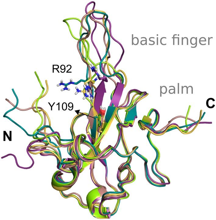
www.nature.com/scientificreports/
Figure 7. Position of Spike amino acid residues corresponding to the mutations highlighted in this study.
Amino acid residues in the RBD (green) and outside of RBD (gray and light brown) mapped onto a structure of
SARS-CoV-2 Spike determined by electron microscopy (PDB code 7DF4). For this analysis, only the mutations
with those residues falling into RBD were considered. Critical residues outside of RBD related to substitutions:
D614G, P681H, T716I, S982A, and D1118H, found in the variant VOC-202012/01, are shown.
In the preparation process, we kept the Asn and Gln flips predicted by the PrepWizard/PROPKA tool since
some of them are part of the Spike protein–protein interaction interface and may be influenced by the interact-
ing partner. For example, Gln493 and Asn501 were predicted with different flips when Spike is complexed with
ACE2 (PDB code 6M0J) or the neutralizing antibody (PDB code 7BZ5).
Non‑synonymous variations in the selected targets. The ongoing pandemic spread of SARS-COV-2
resulted in the increasing generation of thousands of genome sequences (available in the GISAID repository,
https://www.epicov.org, 376,417 sequences on 15/01/2021). Massive sequencing of SARS-CoV-2 genomes
allows performing innumerable comparative, evolutionary and epidemiological analyses, as well as to identify
the circulating genomic variants containing diverse genetic mutations, such as synonymous or non-synonymous
variations (NSVs), deletions and nucleotide insertions. Notably, for the rational drug design, more attention is
given to the study of NSVs in the coding regions since the substitution of amino acids can affect fold, binding
affinity, post-translational modification, protein–protein interaction (PPI) and other protein c haracteristics62.
Even so, some NSVs may not produce visible changes in the structure of the protein; in that case, the muta-
tion may not have a biological impact (neutral). Alternatively, with the intra- and inter-host viral evolution
in infected humans (quasispecies dynamics), the purifying selection can eliminate deleterious mutations over
time, which are more detrimental to the pathogen’s fitness, or else the positive selection promotes the spread of
beneficial ones63.
The estimated mutation rate underlying the global diversity of SARS-CoV-2 is approximately 6 × 10⁻⁴ nucleo-
tides/genome/year64, which is considered moderate for coronaviruses with the Nsp14 proofreading correction
mechanism. Currently, the genomic analysis of almost 243 thousand circulating genomes from patient samples
showed that there are more than 32 thousand replacements among 26 out of 29 proteins encoded on the SARS-
CoV-2′s genomes in comparison with the reference genome sequence of isolate Wuhan-Hu-1 (NC_045512.2)
(CoV-GLUE65, accessed on January 15, 2021). The distribution of this genomic diversity shows high allele fre-
quency for five replacements in just four proteins, namely Spike (D614G, 91.28%, Fig. 7), Nsp12 (P323L, 91.04%),
N (R203K, 36.81%, G204R, 36.55%) and ORF 3a (Q57H, 21.25%). The remainder corresponds to numerous NSVs
with low alleles frequency (~ 11% to 0.002%). Ultimately, this can be explained by the positive selection that
acts at a higher rate after the zoonotic transfer, suggesting an increasing mutant load in the circulating strains of
SARS-CoV-2 in the epidemiological s cenario66.
Considering 242,865 SARS-CoV-2 genomes, the total number of replacements per target selected for this
study varied from 875 (Nsp5), 1,322 (Nsp15), 1,560 (N), 2,491 (Nsp12), 4,360 (Spike) up to 6,869 (Nsp3) (CoV-
GLUE65, accessed on January 15, 2021). Here we selected a total of 19 NSVs covering the six selected targets
(Table S1) with their variant structures already available to the users through the DockThor-VS web server. We
assessed some parameters for the mutation’s choice, such as the impact of the residue’s occurrence in the catalytic
region and its possible interaction with a ligand and the amino acid properties (hydropathy, charge and side
chain). Alternatively, we also have considered the effect on the biological function (neutral vs deleterious) and
Scientific Reports | (2021) 11:5543 | https://doi.org/10.1038/s41598-021-84700-0 8
Vol:.(1234567890)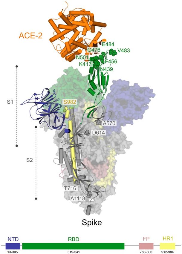
www.nature.com/scientificreports/
searched in the literature any mutagenesis experiments with evidence for alteration in the protein’s molecular
function or viral fitness in CoVs involving the focused residue.
For PLpro, we chose three amino acid substitutions with neutral functional effect, whose corresponding
residues fall into the Peptidase C16 domain (Table S1). So far, we have selected only the replacement M165I
on the Mpro, highlighting that the residue falls on a beta-sheet and is directly part of the ligand-binding site,
with the side chains oriented towards the ligand (Table S1). Here, we describe one selected non-synonymous
variation on the RdRp, namely G683V with deleterious functional effect (Table S1). Replacement G683V on the
RdRp increases the volume of the side chain of a highly conserved g lycine37, and it has already been described
in vitro as a deleterious NSV33. We selected the four non-synonymous variations on the Nsp15, S293A, S293T
(Ser294 in PDB 6VWW), Y342C and Y342H (Tyr343 in PDB 6VWW), whose residues are falling directly on the
ligand interaction binding site. Particularly NSVs S293A and S293T are interesting since the Ser293 accounts as
the critical residue for enzyme discrimination between uracil to cytosine or adenine to guanine b ases44. For the
nucleocapsid phosphoprotein, we selected the NSVs A50V, R92S and R149L, whose residues fall on the RNA-
binding surface of its cognate domain67. These substitutions have a neutral (A50V and R92S) or deleterious
(R149L) predicted functional effect (Table S1).
Finally, for the Spike glycoprotein of the SARS-CoV-2, we selected seven amino-acid substitutions (K417N,
N439K, F456L, G476S, V483A, E484K and N501Y) (Table S1 and Fig. 7), whose residues are within the RBD
(from residues 319 to 541) or RBM (from residues 437 to 508)58. The residues Phe456 and Asn439 are both critical
for the interaction interface with the human receptor ACE2. Regarding Phe456, it is interesting to mention that a
single amino acid substitution on the equivalent residue in SARS-CoV Spike glycoprotein (Leu443) affected both
the antibody binding and neutralization58. Similarly, a mutagenesis assay in SARS-CoV on the equivalent residue
of Asn439 (Arg426) demonstrated that at least two amino acid substitutions significantly reduced the binding to
ACE268. Concerning the residue Gly476, deletion mutagenesis of the equivalent positively charged region in the
RBD of the SARS-CoV Spike (SΔ, 422–463) abolished the ability to induce potent neutralising antibodies in vivo
as well as mediate viral entry69. On the other hand, studies with the equivalent position of the residue Val483
in MERS-CoV (Ile529) showed that single amino acid substitution reduced the host’s receptor affinity, with the
consequent increase in resistance to antibody-mediated n eutralization70. We further included three lineage-
defining mutations in the spike protein with critical residues in the RBD: K417N, E484K and N501Y. The three
amino acid substitutions are characteristic (but not unique) to the South Africa lineage B.1.351 (also known as
501Y.V2 variant), which emerged after the first epidemic wave in the worst affected metropolitan area within
the Eastern Cape (EC) P rovince71. The N501Y mutation has also been identified in a new lineage in the United
Kingdom: B.1.1.7 of Variant of Concern (VOC) designated VUI-202012/01 on the detection and re-designated
as VOC-202012/01 on 18/12/2072. Asn501 forms a hydrogen-bonding interaction with Tyr41 in the binding loop
region of ACE2, contributing to higher ACE2-binding affinity of SARS-CoV-2 than SARS-CoV-173,74. A deep
mutational scanning analysis showed that the N501Y replacement enhances binding affinity to ACE2, which was
also demonstrated through in a mouse model75,76. While N501Y mutation may have a role in escaping class 1
neutralizing antibodies, Xie et al.77 found that antibodies generated by 20 participants with viruses carrying the
N501Y mutation were roughly as potent as antibodies produced to combat Asn501-carrying viruses71,77. Lys417
forms a salt bridge with Asp30 of ACE2 across the binding groove, which contributes to enhanced binding affinity
with the human receptor that is characteristic of SARS-CoV-2 compared to SARS-CoV-174. Despite that, deep
mutational scanning suggests that the K417N replacement negatively impacts binding affinity to A CE275. Also,
Tegally et al.71 disclosed that the K417N mutation would abolish critical interactions with class 1 neutralizing
antibodies and contribute to immune evasion at this site71. Glu484 is a critical residue within the RBM of RBD
and interacts with the Lys31 residue of ACE2 and considered the site of most concern for viral mutations that
impact binding and neutralization by polyclonal serum antibodies targeting the RBD74,78. Recently, the E484K
replacement has been associated with escape from neutralizing antibodies.
Virtual screening for drug repurposing. We performed virtual screening experiments with DockThor-
VS for the e-Drug3D dataset at the reference pH (6.6 to 7.4) for all SARS-CoV-2 targets available at the platform
so far (e.g., PLpro, Mpro, RdRp, NendoU, Spike and N protein) using the wild type genomic variant. When the
protein target has more than one conformation, we adopted an ensemble docking strategy to select the top-
scored binding pose according to each drug’s predicted affinity (see Sect. 3.6 for details).
The virtual screening results of some drugs currently under clinical trials against COVID-19 are presented
in Table 1. The compounds were selected apart of their main proposed mechanism of action and or the expected
biological effect of the ongoing clinical trials.
The screening experiments primarily purpose in this work is to identify some drugs that could interact
with one or more SARS-CoV-2 targets using a reverse docking protocol. However, it is important to state that
a virtual screening campaign serves as a filter to identify the most probable molecules to affect the protein
function. These molecules should be further validated/evaluated in experimental assays and clinical trials to
confirm their biological/therapeutic efficacy. Moreover, the top-scored molecules’ binding mode could be visu-
ally inspected and subjected to more sophisticated simulation techniques before going to the more expensive
and time-consuming experimental validation step. Thus, the docking results present in this work must not be
used for guiding clinical practice.
Another relevant point is that some drugs currently ongoing clinical trials listed on Table 1 do not have
evidence related to antiviral properties, but are claimed to be important in treating, for example, the cytokine
storm such as dexamethasone. However, it is also possible that some drugs with known mechanisms of action,
related to the interaction with host proteins or even with the virus entry pathway, can also inhibit one or more
targets of the pathogen, thus exhibiting multiple mechanisms for the antiviral effect.
Scientific Reports | (2021) 11:5543 | https://doi.org/10.1038/s41598-021-84700-0 9
Vol.:(0123456789)www.nature.com/scientificreports/
Name # Studies1 PLpro Mpro RdRp NendoU N protein Spike
Acetylcysteine 5 − 6.13 − 6.28 − 6.48 − 6.32 − 6.28 − 6.74
Amodiaquine 1 − 7.57 − 8.23 − 7.23 − 7.64 − 7.96 − 7.79
Atorvastatin 4 − 7.65 − 8.73 − 7.37 − 6.92 − 7.97 − 8.71
Atovaquone 2 − 7.68 − 8.28 − 7.91 − 8.50 − 8.33 − 8.12
Azithromycin 71 − 7.90 − 7.73 − 7.73 − 7.88 − 7.75 − 8.40
Baricitinib 12 − 7.53 − 7.96 − 6.43 − 8.26 − 7.98 − 7.61
Chloroquine 29 − 7.92 − 8.06 − 7.49 − 7.88 − 8.10 − 7.65
Chlorpromazine 2 − 7.68 − 8.41 − 6.74 − 7.71 − 7.45 − 7.61
Ciclesonide 4 − 8.04 − 8.68 − 7.42 − 8.45 − 8.92 − 8.00
Cobicistat 2 − 8.36 − 9.46 − 8.58 − 8.82 − 9.16 − 9.31
Daclatasvir 6 − 8.83 − 9.43 − 8.63 − 9.02 − 9.50 − 9.02
Darunavir 3 − 8.07 − 8.45 − 7.84 − 8.27 − 8.32 − 7.87
Deferoxamine 3 − 7.56 − 7.86 − 7.43 − 7.87 − 8.20 − 7.81
Dexamethasone 18 − 7.28 − 7.83 − 6.90 − 8.01 − 7.74 − 7.60
Disulfiram 1 − 7.23 − 7.80 − 6.74 − 7.75 − 7.52 − 7.22
Eltrombopag 1 − 7.96 − 8.48 − 7.78 − 7.60 − 8.56 − 8.40
Emtricitabine 3 − 6.47 − 7.03 − 6.51 − 6.55 − 6.64 − 6.60
Fingolimod 1 − 7.96 − 7.46 − 6.83 − 8.10 − 7.64 − 7.94
Hydroxychloroquine 185 − 7.76 − 7.83 − 7.42 − 7.66 − 8.10 − 7.70
Ibuprofen 2 − 6.86 − 7.43 − 6.70 − 7.26 − 7.19 − 6.89
Icatibant 1 − 6.95 − 7.58 − 8.97 − 7.50 − 8.34 − 9.04
Imatinib 4 − 8.79 − 10.10 − 7.69 − 8.78 − 8.66 − 8.61
Isotretinoin 5 − 7.04 − 7.20 − 6.93 − 7.03 − 8.03 − 7.72
Ivermectin 36 − 8.37 − 9.26 − 8.62 − 8.94 − 9.44 − 8.78
Ledipasvir 3 − 8.47 − 10.59 − 9.16 − 9.25 − 9.36 − 9.66
Leflunomide 2 − 7.15 − 7.83 − 7.13 − 7.79 − 7.42 − 7.17
Lopinavir 43 − 8.80 − 8.47 − 7.77 − 10.12 − 9.06 − 8.76
Losartan 12 − 7.56 − 8.42 − 7.51 − 8.51 − 8.39 − 8.03
Mefloquine 1 − 7.77 − 7.46 − 7.03 − 7.49 − 7.17 − 7.47
Methylprednisolone 17 − 7.76 − 7.99 − 6.81 − 8.05 − 7.74 − 7.68
Montelukast 1 − 8.38 − 9.08 − 8.65 − 8.52 − 9.02 − 9.28
Niclosamide 3 − 7.44 − 8.04 − 7.28 − 7.29 − 7.47 − 7.68
Nitazoxanide 19 − 7.41 − 7.77 − 6.87 − 7.29 − 7.57 − 7.18
Oseltamivir 9 − 7.12 − 7.03 − 6.83 − 7.33 − 7.25 − 7.64
Prazosin 2 − 8.84 − 8.57 − 7.56 − 8.41 − 8.53 − 7.38
Ribavirin 7 − 6.91 − 6.45 − 6.10 − 6.80 − 6.84 − 6.54
Ritonavir 48 − 8.69 − 8.70 − 7.63 − 9.00 − 8.85 − 8.61
Ruxolitinib 19 − 8.16 − 8.41 − 7.06 − 7.83 − 7.95 − 7.89
Sildenafil 2 − 8.26 − 8.64 − 7.79 − 8.08 − 8.62 − 8.07
Sofosbuvir 7 − 7.92 − 8.49 − 7.29 − 8.10 − 7.40 − 7.48
Telmisartan 9 − 8.55 − 9.38 − 8.12 − 8.42 − 9.09 − 8.60
Tenofovir 1 − 6.86 − 6.89 − 6.86 − 6.65 − 6.73 − 6.90
Thalidomide 2 − 7.15 − 7.40 − 6.66 − 7.17 − 6.71 − 7.21
Tranexamic acid 4 − 6.34 − 6.59 − 6.15 − 6.18 − 6.54 − 7.12
Table 1. Virtual screening results of drugs ongoing clinical trials (accessed on 2020-09-03) for the drug targets
available at DockThor-VS. Affinity predictions (kcal/mol) are given for the top-energy pose according to the
ensemble docking strategy. The top-scored drug for each target is underlined and the most promising target
for each drug is highlighted in bold. The molecular weight (MW) and the number of rotatable bonds (RotB)
of each compound are listed in Table S2. 1 Number of clinical trials reported related to COVID-19 by Mapped
Drug Intervention at the ClinicalTrials.gov (accessed on 2020-09-03, https://clinicaltrials.gov/ct2/covid_view/
drugs).
Considering all targets and all drugs evaluated, we found that the majority of the predicted binding affini-
ties are moderate in the range of high to low micromolar affinity units (scores higher than -6.8 kcal/mol cor-
respond to binding affinity values higher than ~ 10 µM, whereas scores lower than -8.2 kcal/mol corresponds to
Scientific Reports | (2021) 11:5543 | https://doi.org/10.1038/s41598-021-84700-0 10
Vol:.(1234567890)www.nature.com/scientificreports/
submicromolar affinities). This result is expected since many drugs under clinical trials exhibited no activity or
only modest inhibitory effects against SARS-CoV-2 in some experimental studies reported in the l iterature79–81.
However, we found some interesting results that deserve to be highlighted. We identified ledipasvir, imatinib,
lopinavir and daclatasvir as the most promising drugs under clinical trials. They all show a good multi-target
profile and exhibit some predicted binding affinities at a low nanomolar concentration (Table 1). Ivermectin,
montelukast, posaconazole, ritonavir and telmisartan also show an interesting multi-target profile having pre-
dicted binding affinities below sub-micromolar concentration for at least five targets.
Ledipasvir is an antiviral to treat chronic Hepatitis C targeting the non-structural protein 5A from the HCV
(NS5A) and was predicted as the most potent Mpro inhibitor (score = -10.59 kcal/mol) among the drugs cur-
rently ongoing clinical trials against COVID-19. Also, ledipasvir was predicted as the most potent drug under
clinical trials against Spike and RdRp (scores of -9.66 kcal/mol and -9.16 kcal/mol, respectively). Daclatasvir
was the top-ranked drug against the N protein (score = -9.50 kcal/mol). Daclatasvir is also an inhibitor of NS5A
from HCV and might also be a promising Nsp5 inhibitor, with a predicted binding score of -9.43 kcal/mol.
Lopinavir is another antiviral drug inhibitor of the HIV-1 protease administered in combination with other
antiretrovirals in the treatment of AIDS. Herein, it was predicted as the most potent drug against Nsp15 in the
virtual screening experiments.
The anticancer imatinib is an Abl kinase inhibitor with an immune-modulatory effect that was reported to
exhibit anti-SARS-CoV action by blocking the viral envelope fusion with the cell m embrane82, being an inter-
esting drug that might have a dual-purpose effect. According to the virtual screening results, it also potentially
inhibits the SARS-CoV-2 Mpro target with a docking score of -10.10 kcal/mol, interacting at the binding site
with key residues such as His163 and Met49. Imatinib was previously reported to inhibit SARS-CoV and MERS
infections in cell culture assays83. However, a recent study reported that imatinib had no effects on SARS-CoV-2
infection and replication in a standard viral replication a ssay84.
Other examples of drugs proposed to be involved in the symptomatic treatment but obtained good scores
against at least one SARS-CoV-2 target are atorvastatin and prazosin.
Atorvastatin is an oral antilipemic agent that inhibits the HMG-CoA reductase and also possess anti-inflam-
matory and immune-modulatory effects, being the statins suggested as effective against viral infections85–87 and
associated with reduced disease severity of COVID-19 i nfection88. According to our screening protocol, atorv-
astatin achieved moderate affinities against both Mpro and Spike proteins (predicted affinities of − 8.73 kcal/mol
and − 8.71 kcal/mol, respectively). Together with the other reported beneficial effects, if its anti-SARS-CoV-2
action is confirmed by experimental methods, atorvastatin could be a very promising drug to be repurposed
against COVID-19 due to its pleiotropic effect and low cost.
Prazosin is an alpha-1 antagonist used to treat hypertension that is currently under clinical trials to evaluate
its efficacy and safety in preventing the COVID-19 cytokine storm. In our virtual screening experiments, it was
predicted as the best Nsp3 drug among those currently under clinical trials with a moderate docking score of
− 8.84 kcal/mol.
Ivermectin is an antiparasitic drug with reported in vitro antiviral effects against SARS-CoV-2, with a reduc-
tion of 99.98% in viral load in Vero/hSLAM cells after 48 h of treatment89, with the antiviral effect hypothesized
to be due to the inhibition of nuclear import of viral proteins targeting the host importin α/β1 h eterodimer90. In
our screening experiments, Ivermectin achieved good scores against all SARS-CoV-2 targets evaluated in our
screening experiments, mainly N protein (− 9.44 kcal/mol) and Mpro (− 9.26 kcal/mol), suggesting that it could
exhibit an on-target antiviral effect inhibiting SARS-CoV-2 proteins. Indeed, other mechanisms of action for
ivermectin are reported in the literature, including the interaction with the NS3 helicase from dengue virus91.
The moderate (> − 8.2 kcal/mol) to low (> − 6.8 kcal/mol) binding affinities predicted for some drugs with
antiviral activities found experimentally suggest that their primary mechanism of action might not be due to
the interaction with SARS-CoV-2 targets, at least for the proteins evaluated in our experiments. Examples of off-
target mechanisms to achieve antiviral effects include the inhibition of clathrin-mediated endocytosis (e.g., the
antipsychotic drug chlorpromazine92–95) and the lysosomotropic effect (e.g., the antimalarial drugs chloroquine
and hydroxychloroquine96) and on-target mechanisms interfering in the function of host proteins such as ACE-2
and TMPRSS2 (e.g., the serine protease inhibitor camostat-mesylate97). Until now, there is no evidence that such
exemplified drugs inhibit SARS-CoV-2 protein targets.
We also evaluated the top-20 drug candidates for repurposing that are not ongoing clinical trials for each
SARS-CoV-2 target (Table S3). One of the most interesting results is associated with the antiviral drug elbasvir,
which was predicted to interact with both Mpro and N protein at docking scores lower than − 10 kcal/mol, sug-
gesting a multi-target effect of this drug. In addition to Mpro and N protein, elbasvir also was predicted to be
within the top-20 drugs for other SARS-CoV-2 targets, i.e., Spike (− 9.79 kcal/mol), NendoU (score = − 9.61 kcal/
mol) and RdRp (score = − 9.21 kcal/mol).
None of the top-ranked drugs to target Nsp3 was predicted with binding affinities lower than − 10 kcal/
mol. However, we can highlight bazedoxifene (score = − 9.65 kcal/mol) and menaquinone (score = − 9.50 kcal/
mol) as interesting findings. Bazedoxifene is a selective estrogen receptor modulator used to prevent postmeno-
pausal osteoporosis, and strong antiviral effects have been reported for SARS-CoV-279,98. Due to its inhibitory
effect on IL-6 signalling, bazedoxifene has been proposed as a promising drug to prevent the cytokine storm,
ARDS and mortality in severe COVID-19 p atients99–101. Menaquinone (Vitamin K2) is one of the three types
of Vitamin K, and a recent preliminary study suggested that patients with Vitamin K deficiency were related to
poor prognostic102. However, there is no yet evidence if menaquinone administration helps to treat or prevent
COVID-19 infection.
The Mpro screening predicted six antiviral drugs within the top-20 best-affinity compounds, where two
of them (i.e., ledipasvir and velpatasvir) are currently ongoing clinical trials. The anti-HCV drugs elbasvir,
pibrentasvir, velpatasvir and ombitasvir are still not being evaluated in clinical trials but could be promising
Scientific Reports | (2021) 11:5543 | https://doi.org/10.1038/s41598-021-84700-0 11
Vol.:(0123456789)You can also read