Hepatitis C Virus Translation Regulation - MDPI
←
→
Page content transcription
If your browser does not render page correctly, please read the page content below
International Journal of
            Molecular Sciences

Review
Hepatitis C Virus Translation Regulation
Michael Niepmann *              and Gesche K. Gerresheim
 Institute of Biochemistry, Medical Faculty, Justus-Liebig-University, Friedrichstrasse 24, 35392 Giessen, Germany;
 Gesche.Gerresheim@gmx.de
 * Correspondence: michael.niepmann@biochemie.med.uni-giessen.de

 Received: 6 March 2020; Accepted: 25 March 2020; Published: 27 March 2020

 Abstract: Translation of the hepatitis C virus (HCV) RNA genome is regulated by the internal
 ribosome entry site (IRES), located in the 5’-untranslated region (50 UTR) and part of the core protein
 coding sequence, and by the 30 UTR. The 50 UTR has some highly conserved structural regions, while
 others can assume different conformations. The IRES can bind to the ribosomal 40S subunit with
 high affinity without any other factors. Nevertheless, IRES activity is modulated by additional cis
 sequences in the viral genome, including the 30 UTR and the cis-acting replication element (CRE).
 Canonical translation initiation factors (eIFs) are involved in HCV translation initiation, including eIF3,
 eIF2, eIF1A, eIF5, and eIF5B. Alternatively, under stress conditions and limited eIF2-Met-tRNAi Met
 availability, alternative initiation factors such as eIF2D, eIF2A, and eIF5B can substitute for eIF2 to
 allow HCV translation even when cellular mRNA translation is downregulated. In addition, several
 IRES trans-acting factors (ITAFs) modulate IRES activity by building large networks of RNA-protein
 and protein–protein interactions, also connecting 50 - and 30 -ends of the viral RNA. Moreover, some
 ITAFs can act as RNA chaperones that help to position the viral AUG start codon in the ribosomal
 40S subunit entry channel. Finally, the liver-specific microRNA-122 (miR-122) stimulates HCV
 IRES-dependent translation, most likely by stabilizing a certain structure of the IRES that is required
 for initiation.

 Keywords: HCV; Hepacivirus; internal ribosome entry site; IRES; initiation; ribosome; 40S; eIF3; ITAF;
 stress response

1. Introduction
      Hepatitis C virus (HCV) is an enveloped positive strand RNA virus that preferentially replicates
in the liver [1], and it is classified in the genus Hepacivirus in the family Flaviviridae. Worldwide, about
71 million people are infected with HCV [2]. The infection is usually noticed only when coincidentally
diagnosed by routine testing, for example, during hospitalization, or when the liver disease becomes
acute. In the latter case, liver damage by virus replication and the resulting immune responses can
lead to impaired bilirubin conjugation in the liver, and unconjugated bilirubin deposits can then be
noticed as a yellowish color (called jaundice), often first in the sclera in the eyes and when more severe
also in the skin. An acute infection can result in severe liver damage, in rare cases even resulting in
death [3,4]. However, most HCV infections remain inapparent [5,6], and the virus infection can become
chronic in about 60% to 70 % of all infections [7], often without being noticed. Chronic infection
can, in the long run, result in liver cirrhosis and liver cancer (hepatocellular carcinoma, HCC) [8–10],
while a metabolic reprogramming of the infected cells according to the “Warburg effect” like in cancer
cells can be observed only a few days after the onset of HCV replication [11]. Moreover, inapparent
replication of the virus usually results in unnoticed spread of the virus to other individuals, a fact
that is a major challenge for surveillance, health care, and treatment [12]. Meanwhile, very effective
treatment regimens using direct acting antivirals (DAAs) are available, although they are still very

Int. J. Mol. Sci. 2020, 21, 2328; doi:10.3390/ijms21072328                              www.mdpi.com/journal/ijms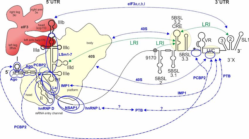
Int. J. Mol. Sci. 2020, 21, 2328 2 of 33
expensive [13,14]. Although the error rate of the viral replicase is high and can, in principle, easily give
Int. J. Mol. Sci. 2020, 21, x FOR PEER REVIEW 2 of 33
rise to resistance mutations, the conserved nature of the replicase active center and the often occurring
reducedreplicase
fitness isofhigh mutants
and can,result in the rare
in principle, easilyappearance of resistance
give rise to resistance mutations
mutations, against
the conserved nucleoside
nature
inhibitors such
of the as sofosbuvir
replicase [15,16].
active center and An effective
the often vaccination
occurring reducedisfitness
not yet available,
of mutants alsoinpartially
result the rare due to
the highappearance
variability of of
resistance
the viral mutations against nucleoside
RNA genome. inhibitors
Thus, further such as
research onsofosbuvir [15,16]. An
HCV is urgently required
effective vaccination is not yet available, also partially due to the high variability of the viral RNA
to combat HCV infections, and despite much progress in the understanding of HCV replication the
genome. Thus, further research on HCV is urgently required to combat HCV infections, and despite
molecular mechanisms of HCV replication are still far from being completely understood [12].
much progress in the understanding of HCV replication the molecular mechanisms of HCV
HCV molecular
replication biology
are still far fromresearch essentially
being completely started[12].
understood with the first cloning of the HCV genome
(“non-A non-B HCVhepatitis”) [17] (Figure
molecular biology research1). Furtherstarted
essentially important landmarks
with the were
first cloning theHCV
of the development
genome of a
(“non-A
subgenomic non-B system
replicon hepatitis”) [17]
[18] (Figure
that 1). Further intracellular
first analyzed important landmarks were the development
virus replication, of a
and the development
subgenomic
of infectious replicon
full-length system
clones that[18]
werethat first analyzed
capable of goingintracellular virus replication,
through a complete and the cycle
viral replication
development of infectious full-length clones that were capable of going through a complete viral
including infection and new virus production [19–21].
replication cycle including infection and new virus production [19–21].
Figure 1. cis-Elements
Figure in the
1. cis-Elements hepatitis
in the hepatitis CCvirus
virus (HCV)
(HCV) RNARNAgenomegenome thatthat are involved
are involved in translation
in translation
regulation. The HCV plus strand RNA genome. The internal ribosome entry site (IRES) HCV
regulation. The HCV plus strand RNA genome. The internal ribosome entry site (IRES) in the in the HCV
5´-untranslated
50 -untranslated regionregion (5´UTR),
(50 UTR), thethe entire33´UTR
entire 0 UTR andand the
thecis-acting
cis-actingreplication element
replication (CRE) in
element the in the
(CRE)
NS5B coding region are involved in translation regulation [22–24]. Those regions of the 5´UTR, 3´UTR,
NS5B coding region are involved in translation regulation [22–24]. Those regions of the 5 UTR, 30 UTR, 0
and CRE that bind to the ribosomal 40S subunit are underlayed in light yellow. Stem-loops (SLs) in
and CRE that bind to the ribosomal 40S subunit are underlayed in light yellow. Stem-loops (SLs) in
the 5´UTR are numbered by roman numerals. The region of the IRES is surrounded by a dotted line.
the 50 UTR
The are
IRESnumbered
includes SLsby roman
II–IV of thenumerals. The region
5´UTR but spans of the
into the core IREScoding
protein is surrounded
region. The by a dotted line.
canonical
The IRES includes SLs in
II–IV ofofthe 0
AUG start codon SL IV the55´UTR
UTR(black
but spans
circle)into
givesthe
risecore protein coding
to translation region. The
of the polyprotein whichcanonical
AUG startis cleaved
codontoinyieldSL IVstructural 0 UTR (black
of the 5proteins and non-structural
circle) gives (NS-)
riseproteins, including
to translation ofthe
theviral replicase which
polyprotein
NS5B.
is cleaved The 3´UTR
to yield contains
structural the variable
proteins andregion, a poly(U/C)
non-structural tract proteins,
(NS-) (U/C), and including
the so-calledthe3´Xviral
region replicase
including SLs 1, 2, and 3. The stem-loop 5BSL3.2 in the 3´-region of the NS5B coding region is the
NS5B. The 3 UTR contains the variable region, a poly(U/C) tract (U/C), and the so-called 30 X region
0
CRE, flanked by upstream stem-loop 5BSL3.1 and downstream 5BSL3.3. The polyprotein stop codon
including SLs 1, 2, and 3. The stem-loop 5BSL3.2 in the 30 -region of the NS5B coding region is the
is located in the stem-loop 5BSL3.4 (asterisk). Some other start codons which give rise to the
CRE, flanked by upstream
alternative reading framestem-loop
(ARF) in5BSL3.1
the core+1and downstream
reading frame [25–28] 5BSL3.3.
are shownTheinpolyprotein
grey. Positivelystopandcodon is
located negatively
in the stem-loop 5BSL3.4 RNA–RNA
acting long-range (asterisk). interactions
Some other startare
(LRIs) codons
shownwhich
in greengive riserespectively,
or red, to the alternative
readingwith
frame the(ARF)
sequencein “9170”
the core+1
shown reading
as greenframe
circle. [25–28]
Selected are shown
binding sitesin
forgrey. Positively(miR-122)
microRNA-122 and negatively
are shown in RNA–RNA
acting long-range blue. interactions (LRIs) are shown in green or red, respectively, with the
sequence “9170” shown as green circle. Selected binding sites for microRNA-122 (miR-122) are shown
The HCV RNA genome is about 9600 nucleotides (nts) long and codes for one long polyprotein
in blue.
that is co- and post-translationally processed into the mature gene products [29–31]. The viral
structural proteins include the core protein, which is contained in the virus particle, as well as the
The HCV RNA genome is about 9600 nucleotides (nts) long and codes for one long polyprotein
envelope glycoproteins E1 and E2. The non-structural (NS) proteins are involved in HCV RNA
that is co- and post-translationally
replication processed
and particle assembly. intoNS
The first theprotein
mature is gene
p7, a products
viroporin,[29–31].
which isThe viral structural
involved in
proteinsassembly.
includeNS2theiscore protein,
a protease which
and acts is contained
as a cofactor ininthe
involved virus particle,
assembly. as well
NS3 has protease ashelicase
and the envelope
glycoproteins E1and
functions, andthe
E2. The non-structural
protease activity of NS3 is(NS) proteins
responsible forare
the involved
cleavage ofin HCV RNA
downstream NSreplication
protein and
particle assembly. The first NS protein is p7, a viroporin, which is involved in assembly. NS2 is a
protease and acts as a cofactor involved in assembly. NS3 has protease and helicase functions, and the
protease activity of NS3 is responsible for the cleavage of downstream NS protein precursor cleavage
sites. NS4A is a NS3 cofactor, whereas NS4B is involved in the membrane reorganization of replication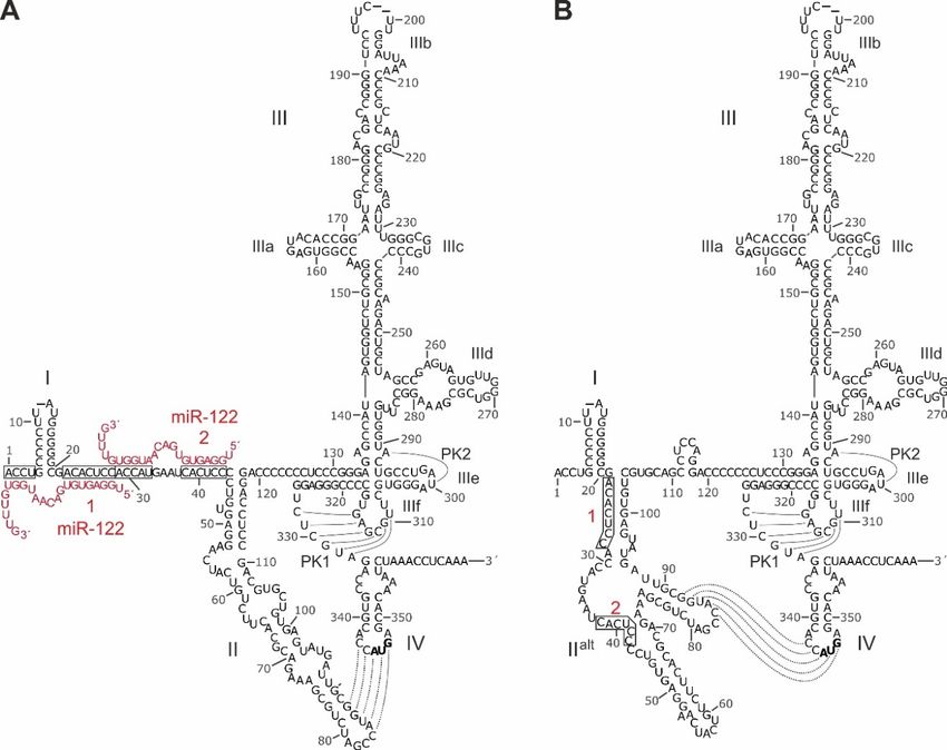
Int. J. Mol. Sci. 2020, 21, 2328                                                                    3 of 33

complex formation. NS5A is involved in RNA replication and assembly, and NS5B actually is the viral
replicase (RNA-dependent RNA polymerase, RdRp) [29–31].
      The HCV virus particle comes as a lipo-viro-particle [32,33]. After binding to a variety of surface
receptors including the low-density lipoprotein (LDL) receptor [33–35] and entry into the cytoplasm,
the viral NS proteins produced in the first (“pilot”) round of translation recruit cellular membranes that
originate from the endoplasmic reticulum (ER) and form a so-called membranous web in which the
viral proteins and genomes form spatially coordinated replication complexes [31,36,37]. Each incoming
plus strand which survived cellular immune responses [5,38,39] and degradation is replicated by the
NS5B replicase and gives rise to one antigenome minus strand copy, which in turn generates about
10 progeny plus strands [30]. HCV RNA synthesis is regulated by a variety of RNA signals that reside
close to the very 30 - and 50 -ends of the genome, but also sequence and RNA secondary structure
elements in the coding region (largely in the 30 -terminal NS5B coding sequence) contribute to the
regulation of RNA synthesis [23,30,40–45]. Bulk translation from the progeny plus strand genomes,
then, yields a vast excess of viral proteins over the number of genomes [30,46]. Then, progeny plus
strand RNA genomes together with viral proteins are packaged into newly assembled virions [32,47],
together with some cellular proteins such as apolipoproteins (Apo) A-I, B, C-II, and E, which contribute
to liver tropism of the virus by binding to the next infected hepatocyte´s surface receptors [48].
      Unlike most cellular mRNAs, the 50 -end of the HCV genomic RNA has no cap nucleotide attached
which would govern efficient cap-dependent translation initiation [49–51]. Instead, HCV translation
is mediated by virtue of an internal ribosome entry site (IRES) [22,52–55], which is located largely
in the 50 UTR but also slightly spans into the coding region (Figure 1). While the resulting low
efficient translation coincides with the “undercover” strategy of HCV replication that often leads
to chronic infection and further unnoticed spread of the virus to uninfected individuals, the use of
such IRES elements has two more big advantages. The first advantage is that in particular the very
ends of the RNA genome do not need to serve functions in translation control such as in capped
and polyadenylated cellular mRNAs. Instead, RNA signals that are involved in genome replication
can be directly placed at the very genome ends [23,30,45]. The second benefit of cap-independent
translation is that the virus escapes antiviral countermeasures of the cell in terms of the downregulation
of cap-dependent translation, which is largely conferred by phosphorylation of eIF2 and the resulting
inhibition of cap-dependent translation initiation [56].
      In addition to the hepatocyte surface receptors mentioned above, another determinant of the liver
tropism of HCV is the microRNA-122 (miR-122). microRNAs are small single-stranded guide RNAs
that direct effector complexes involving Argonaute (Ago) proteins to cellular mRNAs and usually
negatively influence the mRNA´s translation efficiency and induce degradation of the mRNA [57,58].
miR-122 is expressed almost exclusively in the liver and constitutes about 70% of all microRNAs in
hepatocytes [59], whereas it is nearly not expressed in other tissues [60]. In contrast to the negative
influence of microRNAs on cellular mRNAs, HCV utilizes miR-122 to promote its own replication [61],
thereby making the liver-specific miR-122 another determinant for the liver tropism of HCV. There are
five to six target sequences conserved in the HCV genome, two in the 50 UTR [62], one in the 30 UTR,
and two to three in the NS5B coding region (depending on genotype) [63]. Cooperative binding of
two miR-122 molecules to the two adjacent target sites in the HCV 50 UTR contributes to RNA stability
by protecting against cellular nucleases [64,65] and has a positive effect on the efficiency of HCV
translation [66–72]. Although some studies investigated the possible roles of the conserved potential
miR-122 binding sites in the NS5B coding region and in the 30 UTR, it is not yet clear if physical binding
of miR-122 to these sites results in effector functions [73–76], leaving some doubts why these sequences
are conserved among HCV isolates.
      In this review, we focus on the sequences, cellular factors, and molecular mechanisms involved in
the regulation of translation by the HCV IRES. Thereby, we touch the functions of miR-122 specifically
only with regard to translation regulation, while another review by Joyce Wilson in this review series
thoroughly covers miR-122 action in all aspects of HCV replication.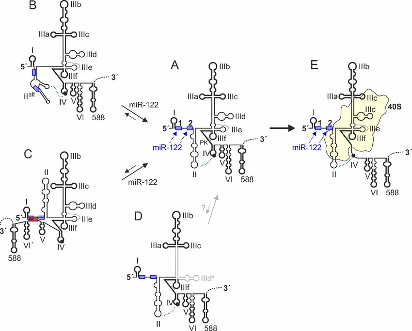
Int. J. Mol. Sci. 2020, 21, 2328                                                                    4 of 33

2. An Overview over HCV Genome Regions Involved in Translation Regulation
      The RNA cis-elements that control HCV RNA genome translation largely reside in the 50 and 30
regions of the genome (Figure 1). The IRES element is located in the 50 UTR plus some 30 nts of the core
coding region (dotted line in Figure 1) [52,53,77,78]. It contains the large branched domain (Dom) III
(or stem-loop (SL) III, respectively) with several small subdomain stem-loops, the SL IV and a double
pseudoknot, and the upstream SL II. In contrast, the SL I located at the very 5-end of the genome is not
involved in translation regulation but in replication control. The sequence between the SLs I and II,
which contains the two miR-122 binding sites located directly upstream of the IRES, is formally not
counted as belonging to the IRES, but this sequence can influence translation activity upon binding of
miR-122 [66]. The IRES binds the small ribosomal 40S subunit with high affinity of about 2 nM [79]
(underlayed in yellow in Figure 1).
      At the 3-end of the HCV genome, the 30 UTR [80], as well as the cis-acting replication element (CRE)
with its stem-loops 5BSL3.2 and 5BSL3.3 in the 30 -terminal region of the NS5B coding region [81,82] are
also involved in translation regulation (Figure 1). The function of the 30 UTR and the possible function
of the CRE in translation regulation are related to two types of long-range interactions (LRIs), first
RNA–RNA LRIs, and second those long-range interactions that are mediated by RNA-binding proteins
or by the ribosomal 40S subunit. This 30 - to 50 -end communication is reminiscent of that occurring with
cellular mRNAs. There, the polyA tract at the 30 -end stimulates translation at the 50 -end. In principle,
this interaction in cis can only indicate that the RNA to be translated is not degraded but intact, and
only then it makes sense to translate this RNA efficiently.
      Reports about a contribution of the HCV 30 UTR to the regulation of translation at the IRES have
been quite different in the past. Some previous reports have shown a positive influence of the 30 UTR
on IRES-directed translation [80,83–87]. One report showed a negative influence of the 30 UTR [88], and
other studies reported no influence [89–92]. These discrepancies could have been caused by the use of
different reporter systems and artificial extensions at the reporter RNA´s 30 -end (discussed in [86]).
Currently, it is rather clear that the 30 UTR actually stimulates translation, and it is recognized that
suitable reporter assays should use RNA transfection (not DNA) and a precise authentic 30 -end of the
HCV 30 UTR [86].
      The CRE (see Figure 1, also called SL5B3.2) was first described to be involved in control of overall
HCV genome replication [93] and binds physically to the NS5B replicase [94–96]. Moreover, the CRE
5BSL3.2 plus the flanking downstream 5BSL3.3 [82], as well as the poly(U/C)-tract of the 30 UTR [82,97]
were shown to bind to the small 40S ribosomal subunit. Thereby, the CRE binds with KD of about 9 nM
to the 40S subunit, while the poly(U/C) tract of the 30 UTR binds even stronger (KD = 1 nM); together,
the CRE and 30 UTR bind 40S subunits with a comparable affinity as the IRES [82,97].
      Long-range RNA–RNA interactions are very important in HCV replication. Several such putative
interactions have been described [24,45,63,82,98–108]. By far the most important interactions (see
Figure 1) that have been demonstrated to be functionally relevant by several studies are the following.
First, the interaction between the apical loop of the CRE 5BSL3.2 and the apical loop of the SL 2 in the
30 UTR (also named “kissing loop” interaction) [98,99] is important for HCV replication. Second, the
interaction between the bulge of the CRE 5BSL3.2 (GCCCG) with a sequence about 200 nts upstream
(CGGGC) (“9170” in Figure 1 and in [23]) was also shown to be important for replication [100,103]
and third, the internal CRE 5BSL3.2 bulge can alternatively interact with the apical loop of the SL IIId
(UGGGU) in the IRES [101].
      Regarding translation regulation, conflicting results have been reported with respect to a possible
role of the CRE. One study showed that deletion of the CRE 5BSL3.2 conferred an increase of translation
efficiency from HCV-luciferase translation reporter constructs 18 h after transfection (but not after
6 h) [102], suggesting that the CRE 5BSL3.2 is involved in inhibiting HCV translation and, together with
the flanking upstream 5BSL3.1 and the downstream 5BSL3.3, the CRE 5BSL3.2 inhibits translation [102].
This implicates that this LRI could have something to do with translation regulation or with a possible
switch between translation and replication. In contrast, another study showed that the inhibition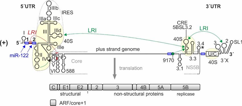
Int. J. Mol. Sci. 2020, 21, 2328 5 of 33
Int. J. Mol. Sci. 2020, 21, x FOR PEER REVIEW 5 of 33
of the CRE by hybridization with locked nucleic acid (LNA) oligonucleotides impairs translation of
luciferase reporter RNAs
6 h after transfection [81],orleading
replication-incompetent
to the conclusionreplicons 6 h after
that the CRE transfection
rather stimulates[81], leading toThis
translation. the
conclusion
discrepancythat the be
could CRE rather
due stimulates in
to differences translation. Thisassay
the reporter discrepancy
systemscould be due
and yet needsto differences in
to be further
the reporter
elucidated. assay systems and yet needs to be further elucidated.
In addition, aa hybridization
In addition, hybridization between
between sequences
sequences in the core-coding
in the core-coding region
region and
and the
the region
region between
between
0
55´UTR
UTR SLs
SLs II and
and IIII (red
(red in
in Figure
Figure 1)1) had
had been
been described,
described, which
which has
has aa negative
negative effect
effect onon translation
translation
efficiency. This interaction is important in terms of IRES structure, function,
efficiency. This interaction is important in terms of IRES structure, function, and miR-122and miR-122 action and
action andis
discussed
is discussedin in
thethe
next section.
next section.
3. Structure of the HCV 50 UTR and the Internal Ribosome Entry Site
3. Structure of the HCV 5´UTR and the Internal Ribosome Entry Site
The HCV 50 UTR is about 340 nts long and is predicted to fold into characteristic RNA secondary
The HCV 5´UTR is about 340 nts long and is predicted to fold into characteristic RNA secondary
structures (Figure 2). These sequences and the predicted secondary structures are highly conserved
structures (Figure 2). These sequences and the predicted secondary structures are highly conserved
among HCV genotypes and subtypes [63].
among HCV genotypes and subtypes [63].
Figure 2. Sequence and RNA secondary structure of the HCV IRES. (A) The 5´UTR of HCV and its
Figure 2. Sequence and RNA secondary structure of the HCV IRES. (A) The 50 UTR of HCV and its
canonical structure
canonical structure [63,109–114].
[63,109–114]. The The sequence
sequence shown
shown is is from
from genotype
genotype 2a (J6 isolate),
2a (J6 isolate), with
with length
length
variations among
variations amongHCV HCV isolates
isolates indicated
indicated by horizontal
by short short horizontal
dashes atdashes at nucleotide
nucleotide positions.
positions. Nucleotide
Nucleotide numbering is according to the MAFFT alignment in the
numbering is according to the MAFFT alignment in the supplement of [63] (http://www.rna.uni-jena. supplement of [63]
(http://www.rna.uni-jena.de/supplements/hcv/). For comparison, the first nucleotide
de/supplements/hcv/). For comparison, the first nucleotide of the core coding sequence (AUG, in bold) of the core
coding sequence (AUG, in bold) in the SL IV is nucleotide No. 342 in genotype
in the SL IV is nucleotide No. 342 in genotype 1b (Con1) and No. 341 in genotype 2a (J6 and JFH1 1b (Con1) and No. 341
in genotype
isolates). 2a SL
IRES (J6 domains
and JFH1are isolates). IRESbySLroman
indicated domains are indicated
numerals. by roman(miR-122,
microRNA-122 numerals.red)
microRNA-
binding
122 (miR-122, red) binding is indicated, miR-122 target sequences are boxed.
is indicated, miR-122 target sequences are boxed. Pseudoknot (PK) base pairing and the interactionPseudoknot (PK) base
pairing and
between SL the interaction
II and SL IV isbetween
indicated; SL(B)II and SL IV is indicated;
Secondary (B) Secondary
structure model of the 50structure
UTR withmodel of the
alternative
5´UTR with alternative
alt folding of SL II alt. The structure is largely according to [72,77,115], with minor
folding of SL II . The structure is largely according to [72,77,115], with minor modifications according
modifications
to our RNAalifoldaccording
outputs to using
our RNAalifold outputs (not
several genotypes using several genotypes (not shown).
shown).
The small SL I form very close to the very 5´-end of the HCV 5´UTR, leaving only three or four
unpaired nts at the very 5´-end. In the canonical representation of the 5´UTR secondary structureInt. J. Mol. Sci. 2020, 21, 2328 6 of 33
The small SL I form very close to the very 50 -end of the HCV 50 UTR, leaving only three or four
unpaired nts at the very 50 -end. In the canonical representation of the 50 UTR secondary structure
(Figure 2A) [42,63,109,111], this SL I is followed by a single-stranded stretch of about 42 nts which
binds two molecules of miR-122 [62] with one of the miR-122 molecules also hybridizing to the very 50
nts upstream of SL I. This entire sequence often is named domain I of the 50 UTR. Downstream of that,
the canonical representation depicts the SL II (or domain II), followed by a large branched domain that
is called either SL III or domain III, which contains the subdomains or SLs IIIa, b, c, d, e, and f. This
domain III, then, is followed by a single-stranded stretch that can form a pseudoknot 1 (PK1) with
the SL IIIf [116]. In addition, a U residue (postion 300 in Figure 2) the SL IIIe can form one base pair
with the upstream A (position 291), creating a second pseudoknot (PK2). This region with the two
pseudoknots forms a compact and unique tertiary structure [117]. The downstream SL IV contain the
AUG start codon [118]. These IRES RNA secondary structures have been predicted in silico and have
been experimentally validated by chemical structure and nuclease protection mapping [52,63,77,113].
Moreover, the ribosome-bound HCV IRES structure has been validated by cryo electron microscopy
(EM) [55,119–121]. An alternative predicted 50 UTR structure shows a refolded, alternative SL II (SL
IIalt ) (Figure 2B), which is as consistent with the reported experimental structure mapping results as
is the canonical structure [72,77,113,115]. In this alternative fold of the 50 UTR sequences between SL
I and SL III (Figure 2B), the two miR-122 binding sites are largely hidden within double-stranded
RNA structures, and therefore are less accessible as compared with the canonical structure (Figure 2A).
In the presence of miR-122, the alternative SL IIalt reforms to adopt the canonical structure, allowing
efficient translation and stabilization of the genome [72,115,122]. Interestingly, also mutations that
render HCV replication miR-122 independent favor the canonical structure [72,122].
Despite the seemingly clearly defined 50 UTR structure, we can distinguish some central
structure-driving regions from others, which are more flexible and can change structure, also according
to functional requirements. We have performed our own in silico predictions using LocARNA [123]
and RNAalifold [124] with a representative number of different isolates from different genotypes
(selected from [63]) (data not shown). On the one hand, some IRES regions appear to have a quite
strong preference to robustly fold into a definite structure, likely driven by the intrinsic properties of the
conserved primary sequence. Formation of these RNA secondary structures takes place regardless of
subtle variations in their sequence that lead to covariations in their secondary structure, and regardless
of the availability of varying flanking RNA regions that could interfere with the secondary structures
by providing options for alternative interactions. On the other hand, other sequences appear to be able
to dynamically assume different secondary structures, depending on the extent of flanking RNA that
is available for interaction, depending on the binding of miR-122, and depending on the binding of
cellular proteins as well as of the ribosomal 40S subunit.
In the first group of sequence elements, i.e., those which appear to robustly form a defined and
invariant secondary structure, we would list essentially three regions (drawn in bold in the 50 UTR
structures in Figure 3). In brief, these are the SLI, the upper part of domain III, and the base of the
domain III with the double pseudoknot. In part, the structure of these regions can also be stabilized by
binding to cellular factors or to the ribosomal 40S subunit. The first region is the G/C-rich stem-loop I
near the very 50 -end of the HCV 50 UTR. The SL I region has a strong preference to form under almost
any conditions such as varying length of flanking sequences or subtle sequence variations among
genotypes and subtypes, with only very few exceptions. The second sequence region that appears
to always robustly form the same structure in in silico predictions is the upper part of the domain
III, including the four-way junction with the stem-loops IIIa, IIIb, and IIIc [125]. This structure binds
eIF3 [79] and contains conserved elements in its central bulge (around position 180 and 220 in Figure 2A)
as well as in the stem above which are required for eIF3 binding [126–128]. The third steadily forming
region includes the base of the domain III with SL IIIe, IIIf, and the double pseudoknots [116,117],
which can drive binding to and be stabilized by the ribosomal 40S subunit [129]. These three robustly
forming 50 UTR regions can also influence the biased folding of flanking sequences, and small-angleInt. J. Mol. Sci. 2020, 21, 2328 7 of 33
X-ray scattering of the HCV IRES in solution, and in silico structure flexibility simulations [114] are
consistent with
Int. J. Mol. Sci. an21,overall
2020, IRES
x FOR PEER structure as predicted. In addition, the first three RNA secondary
REVIEW 7 of 33
structures in the core coding region also appear to have a conserved tendency to robustly fold in silico
tendency
to form to robustly
the SLs V, VI, andfoldthe
in following
silico to form the SLs
SL 588. TheV, VI, and the
formation following
of these SL 588.
distinct The formation
structures of
are required
these distinct structures are required to leave only the SL IV sequence to form either a stem-loop or
to leave only the SL IV sequence to form either a stem-loop or to unfold and bind in the ribosome entry
to unfold and bind in the ribosome entry channel, without being disturbed by intruding flanking
channel, without being disturbed by intruding flanking sequences (Figure 3E).
sequences (Figure 3E).
Figure 3. Conformational dynamics of HCV 5´UTR structure. The conserved canonical 5´UTR
Figure 3. Conformational dynamics of HCV 50 UTR structure. The conserved canonical 50 UTR
structure [63,109–114] is shown in the middle (structure A). 5´UTR sequences which have a strong
structure [63,109–114] is shown in the middle (structure A). 50 UTR sequences which have a strong
intrinsic tendency to fold into a distinct secondary and tertiary structure are depicted in bold. The
intrinsic tendency to fold into a distinct secondary and tertiary structure are depicted in bold.
constitutive double pseudoknot (PK) interactions involving SL IIIe (PK2) and IIIf (PK1) and the
The constitutive double pseudoknot (PK) interactions involving SL IIIe (PK2) and IIIf (PK1) and
possible interaction between SL II and SL IV are indicated. The 5´UTR structure B [72,77,115] has the
the possible interaction between SL II and SL IV are indicated. The 50 UTR structure B [72,77,115] hasaltthe
region between SLs I and III refolded and forms an alternative version of SL II (SLaltIIalt). This SLalt II
region between SLs I and III refolded and forms an alternative version of SL II (SL II ). This SL II can
can form in the absence of miR-122, then hiding both miR-122 binding sites. Binding of miR-122
form in the absence of miR-122, then hiding both miR-122 binding sites. Binding of miR-122 promotes
promotes refolding of structure B to structure A [72,115,122]. The 5´UTR structure C can form by fold-
refolding of structure B to structure A [72,115,122]. The 50 UTR structure C can form by fold-back of
back of nucleotides 428–442 in the base of SL VI in the core protein coding region to hybridize with
nucleotides
sequences428–442
downstreamin theofbase
SL I.of
InSL
thisVIstate,
in the
thecore protein activity
translation coding ofregion to hybridize
the IRES is decreasedwith sequences
[130–134].
downstream of SL I. In this state, the translation activity of the IRES is decreased
This hybridization also interferes with binding of miR-122 to the 5´UTR. In turn, binding of miR-122 [130–134]. This
hybridization also interferes with Cbinding of miR-122 to the 0
5 UTR. D In is
turn, binding of miR-122 induces
induces refolding of structure to structure A [135]. Structure a yet completely hypothetic
refolding of that
structure structure C to structure
was predicted A [135].
to be the Structure
energetically D is afavored
slightly yet completely
conserved hypothetic
structure structure that
[63]. In this
was predicted
structure D, tothebeSLthe energetically
IIId of the IRES slightly
is refoldedfavored conserved
to form SL IIId*.structure
However, [63]. In this structure
a possible biological D,
therelevance
SL IIId ofofthe IRES
this is refolded
hypothetical to form isSLunclear,
structure IIId*. However, a possible
in particular biological
since several relevance
interactions ofofthethis
hypothetical structure is unclear, in particular since several interactions of the
canonical SL IIId have been described (see main text). Structure E is formed upon binding to the canonical SL IIId have
been described
ribosomal 40S(see main text).
subunit. Then,Structure E is formed
SL IV sequences upon binding
containing the AUGto the ribosomal
start codon are 40Sunfolded
subunit.and Then,
SLpositioned
IV sequences containing
in the mRNA entry thechannel
AUG start codon
of the are unfolded
40S subunit, aided byand positioned
interaction withinSL
theII.mRNA entry
channel of the 40S subunit, aided by interaction with SL II.
In contrast, other sequences are more flexible. This is particularly true for the region between
SLs I and III. In the canonical 5´UTR structure (Figure 3A), the region between SLs I and II with the
two miR-122 binding sites is predicted to be single stranded in RNAalifold structure predictions, andInt. J. Mol. Sci. 2020, 21, 2328 8 of 33
In contrast, other sequences are more flexible. This is particularly true for the region between
SLs I and III. In the canonical 50 UTR structure (Figure 3A), the region between SLs I and II with the
two miR-122 binding sites is predicted to be single stranded in RNAalifold structure predictions, and
the canonical SL II structure forms. This structure is predicted to occur by default also in the absence
of miR-122 (i.e., in the absence of constraints to keep the miR-122 target sites as single-stranded sites
in the in silico foldings). Thus, the region between SL I and SL II is available to easily bind miR-122.
In turn, miR-122 binding stabilizes this region in single-stranded form (Figure 3A), then supporting
the stable formation of SL II and its important interactions with the apical loop of SL IV and with the
ribosomal 40S subunit [72,115]. These structure changes of SL II induced (or stabilized, respectively)
by miR-122 can account for the stimulation of translation by miR-122 [66]. However, an only very
slight variation of folding parameters (e.g., by not running RNAalifold but instead running LocARNA
with default parameters) changes the prediction dramatically, with the sequences downstream of SL
I largely rearranging and partially covering the miR-122 target sites. Then, the alternative SL IIalt
structure forms (Figures 2B and 3B), and the miR-122 target sites are largely hidden. This indicates that
this region is very flexible in structure and can easily refold in response to the absence or presence of
miR-122, thereby changing the structure and by that the function of the SL II sequence region which is
involved in regulating translation.
In addition, a sequence in the core coding region (nts 428 to 442) can fold back to the miR-122
binding region in a long-range interaction and cover the miR-122 target sites, forming a more compact
structure (Figure 3C, and also see the red LRI in Figure 1) and reducing translation efficiency [130–133].
This inhibitory hybridization can be relieved by miR-122 binding [135,136], while the physiological
relevance for controlling HCV translation in cells appears not to be dramatic but can more easily be
detected in an in vitro translation system [134].
A yet completely hypothetical structure is the structure shown in Figure 3D. In this structure,
the lower part of the domain III is refolded largely to loop the SL IIId and form the alternative SL
IIId* [63]. This prediction was yielded as the most stable consensus structure among the 106 HCV
isolates from all available genotypes and subtypes [63]. However, its minimum fold energy (MFE) is
only marginally lower than that of the canonical form. Since all RNA structure probing experiments
and cryo EM structures, as mentioned above, are not consistent with 50 UTR structure D but rather
with structure A, we can only speculate if this conserved structure D could have possible functions.
The predicted secondary structure of the SL IV with the AUG start codon contains covariations
among isolates, which implies that this small secondary structure must be of some importance.
However, the position of the start codon within the predicted SL IV secondary obviously suggests that
there must be changes in RNA secondary structure when the start codon is positioned in the ribosome.
When the IRES binds the 40S subunit, the SL IV unfolds and is placed in the entry channel of the 40S
subunit (Figure 3E) [117,137,138], preparing the IRES-40S complex for translation initiation.
With respect to the previously described interchangeable 50 UTR/IRES structures and their possible
biological relevance (Figure 3), as well as with regard to the required detachment of the IRES RNA
from the ribosome upon completion of the translation initiation cycle, we need to take into account
that under natural conditions in the cell the secondary and tertiary structure of the IRES RNA could
be much more flexible than suggested by the seemingly rigid, fixed canonical structure, as shown in
Figures 2A and 3, structure A. Many IRES structural studies (in the absence or presence of the 40S
subunit) have been performed at quite high magnesium concentrations. Moreover, binding of the 40S
subunit to the IRES can strongly favor a distinct IRES structure by induced fit. Most studies used
2.5 mM MgCl2 [55,113,125,139–143], whereas others used 3 mM [97], 4.5 mM [119], 5 mM [114,138], or
even 10 mM MgCl2 [144]. In contrast, the concentration of free Mg2+ in the cytosol in different cells is
0.86 mM on average over many studies, and in rat liver cells, the free intracellular Mg2+ concentration
is approximately 0.7 mM (reviewed in [145]).
The high Mg2+ concentrations used in the studies mentioned above have demonstrated the IRES
and ribosome structures previously reported (see above). However, at such high Mg2+ concentrations,Int. J. Mol. Sci. 2020, 21, 2328 9 of 33
IRES binding to the 40S subunit is strongly favored [79], leaving no options for an equilibrium of
different IRES structures in solution. Moreover, in the presence of 60S subunits, both ribosomal subunits
would be largely associated at 2.5 mM Mg2+ [146], and by that they would even not allow binding of
the HCV IRES to individual 40S subunits, followed by the induction of 60S subunit association by the
HCV RNA to be translated. In contrast, at the physiological concentration of 0.7 mM free Mg2+ , only
about half of the IRES molecules are actually completely folded [79], while the other half assume more
flexible structure intermediates. In addition, another study showed that the IRES has a rather extended
conformation at low Mg2+ concentrations, whereas the pseudoknots form only in the presence of
Mg2+ [136]. Concurrently, HCV translation is optimal at Mg2+ concentrations lower than 1 mM [147].
Thus, we must be aware that the functional HCV IRES structure is much more flexible in terms of
opening and closing secondary structures than most studies on IRES structure suggest.
The above considerations particularly apply to those IRES regions that must dynamically interact
with the 40S subunit (drawn thin in the IRES structures in Figure 3). Although the HCV IRES is
completely unfolded in the absence of Mg2+ , strongly folding IRES core structures such as the upper
portion of SL III (shown in bold in Figure 3) form properly into the canonically shown form at Mg2+
concentration of only 0.25 mM and above [110,136] (i.e., are formed under intracellular conditions).
In contrast, the G residue 135 which is the second nucleotide in the left part of the base of domain III
(a region that is routinely drawn as double-stranded in the IRES secondary structure predictions [63])
is largely protected (i.e., double-stranded) at Mg2+ concentrations of 2.5 mM, whereas it appears only
50% protected at the physiological Mg2+ concentration of 0.7 mM [110]. These results imply that the
HCV IRES structure can be (and perhaps must be) much more flexible during productive complete
translation initiation cycles than many studies suggest. This gives room to the speculation that the
previously mentioned highly conserved predicted structure D (Figure 3) could have some biological
relevance yet to be shown. One of the most intriguing questions for future research is how the 40S
subunit, which initially binds very strongly to the IRES manages to routinely detach from the IRES
in order to commence the transition from translation initiation to elongation and synthesis of the
polyprotein. This likely needs to be investigated at magnesium concentrations that are lower than
those used in previous studies (best 0.7 mM).
4. Contacts of the HCV IRES with the Small Ribosomal 40S Subunit and with eIF3
When the protein-free HCV IRES is allowed to bind in vitro to isolated small ribosomal 40S
subunits, the IRES makes several close contacts to the 40S subunit. We must assume that under
conditions of intracellular magnesium concentrations (as discussed above) there should be more
flexibility of the IRES [110]. However, we are not aware of studies that analyzed the sequential arrival
of different IRES regions at the 40S subunit with high time resolution (in the sub-second scale), and
thereby demonstrated structural “induced fit” changes of the IRES structure during binding. The only
study that methodically comes close [148] shows that there is a long lag phase until the IRES binds the
40S subunit.
Nevertheless, from the IRES structure as it appears when bound to the isolated 40S subunit
(Figure 4) [55,119,121,138,149], we can assume that there are three distinct regions of the IRES that are
different in binding, as well as in function. The first region is the “core” of the IRES, including the
two pseudoknots PK1 and PK2 together with the base of domain III and SLs IIId, IIIe, and IIIf, and
considered in an extended version also including the four-way junction SL IIIabc which also binds
closely to the 40S subunit (but not including the actual SL IIIb). This “core” IRES region essentially
serves to “anchor” the body of the IRES with high affinity in a fixed position (irrespective of possible
induced fit changes during binding) on the 40S subunit and provides a platform for the flexible
connection of the other two functional IRES modules, SL II and SL IIIb.Int. J. Mol. Sci. 2020, 21, 2328 10 of 33
Int. J. Mol. Sci. 2020, 21, x FOR PEER REVIEW 10 of 33
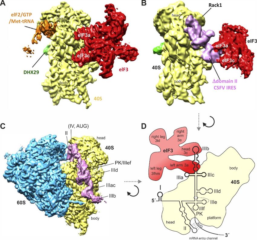
Figure
Figure 4. Binding
4. Binding ofof
thethe
HCVHCVIRES
IREStotothe
theribosome
ribosome andand to
to eIF3.
eIF3. (A)
(A) The
Thesmall
smallribosomal
ribosomal40S40Ssubunit
subunit
(yellow) with bound eIF3 (red). eIF3 makes multiple contacts to the 40S subunit using several several
(yellow) with bound eIF3 (red). eIF3 makes multiple contacts to the 40S subunit using subunits,
subunits,
including including
eIF3a eIF3a and
and eIF3c; (B) eIF3c;
The IRES(B) The IRES of classical
of classical swinevirus
swine fever fever (CSFV)
virus (CSFV)
(pink)(pink) without
without IRES
IRES domain II, binding to the 40S subunit (yellow) and to eIF3 (red). The IRES has displaced eIF3
domain II, binding to the 40S subunit (yellow) and to eIF3 (red). The IRES has displaced eIF3 completely
completely from its binding to the 40S subunit (compare panel A) but keeps it connected to the 40S
from its binding to the 40S subunit (compare panel A) but keeps it connected to the 40S subunit only
subunit only indirectly by contacts between IRES SL IIIabc and eIF3. Figures A and B were reprinted
indirectly by contacts between IRES SL IIIabc and eIF3. Figures A and B were reprinted from [150]
from [150] (Figure 2F) and slightly modified. Reprinted with permission from Elsevier (licence no.
(Figure 2F) and slightly modified. Reprinted with permission from Elsevier (licence no. 4761840166773);
4761840166773); (C) The HCV IRES (pink) binding to the 40S subunit (yellow) in the complete 80S
(C) The HCV IRES (pink) binding to the 40S subunit (yellow) in the complete 80S ribosome (60S subunit
ribosome (60S subunit in blue). The IRES SL IIIdef/PK is in close contact with the 40S subunit, the SLs
in blue). The IRES SL IIIdef/PK is in close contact with the 40S subunit, the SLs II and IV are positioned
II and IV are positioned in the mRNA entry channel, and the SL IIIb is pointing to the solvent side for
in the mRNA entry channel, and the SL IIIb is pointing to the solvent side for binding eIF3. The IRES is
binding eIF3. The IRES is not touching the 60S subunit [121,138]. Figure C was modified from the left
not touching the 60S subunit [121,138]. Figure C was modified from the left panel of Figure 1C in [121].
panel of Figure 1C in [121]. Reproduced with permission from EMBO; (D) Schematic illustration of
Reproduced
the HCV IRESwith binding
permission from
to the 40SEMBO;
subunit(D) Schematic
(yellow) and toillustration
eIF3 (red) of the eIF3
when HCVisIRES binding
largely to the
displaced
40Sfrom
subunit (yellow) and to eIF3 (red) when eIF3 is largely displaced from the 40S subunit
the 40S subunit by the IRES, similar as in (B). The orientation of the 40S subunit is shown by the IRES,
similar as in (B). The orientation of the 40S subunit is shown essentially top-down
essentially top-down as compared with (A–C), viewed approximately from the solvent side. as compared with
(A–C), viewed approximately from the solvent side.
In contrast, the second functional IRES region, the SL II region in its canonical form (see Figures
In contrast, the second functional IRES region, the SL II region in its canonical form (see Figures 2
2 and 3A,E), appears to be rather flexible and fulfills important tasks in reorganizing the IRES
andstructure
3A,E), appears to be rather flexible and fulfills important tasks in reorganizing the IRES structure
and the ribosome in order to unwind the SL IV, place the contained AUG start codon in the
andribosomal
the ribosome mRNA in order to unwind
entry channel, andthe SL IV, place
manipulate thethe
40Scontained
subunit toAUG startinitiation
undergo codon in(seethebelow).
ribosomal
mRNA The entry channel, and manipulate the 40S subunit to undergo initiation (see below).
third region is the SL IIIb. It appears not to bind to the 40S subunit at all [55,119,121,138,149],
butThe
it isthird regionto
connected is the
the rest
SL IIIb.
of theIt IRES
appears notvery
by the to bind to the
flexible SL40S subunit
IIIabc at alljunction
four-way [55,119,121,138,149],
[151] which
but it is connected to the rest of the IRES by the very flexible SL IIIabc four-way junction [151] which isInt. J. Mol. Sci. 2020, 21, 2328 11 of 33
anchored on ribosomal protein eS27 [138]. This SL IIIb is used by the IRES solely to bind eIF3 [126,127]
after the IRES has displaced eIF3 from its binding to the 40S subunit [143]. The reasons for this are not
yet fully understood.
However, it appears likely that the IRES needs to keep eIF3 on hold in close vicinity of cis
for using its contacts to the HCV 30 UTR [97], to avoid premature subunit joining [152,153], and to
use it later in subsequent initiation steps to acquire eIF2, eIF5B, and the 60 subunit to the initiation
complex [141,154,155]. In addition, the AUG start codon of the HCV IRES must be properly positioned
in the 40S mRNA entry channel in order to fully accommodate its contacts on the 40S subunit for proper
initiation [137]. However, it has been reported that eIF3 is not absolutely essential for subsequent
stages in initiation, since 48S complexes formed in the absence of eIF3 on both wild-type and SL IIIb
mutant HCV IRES elements readily underwent subunit joining, forming elongation-competent 80S
ribosomes [143]. Nevertheless, the HCV IRES can still bind eIF3 also in the complete 80S ribosomes [156],
likely in the same way with the IRES SL IIIb.
The IRES core domain with the double pseudoknot, SL IIId, IIIe, and IIIf tightly contacts the
40S subunit on the solvent side [117,129] by binding to the ribosomal 18S rRNA, as well as to
several ribosomal proteins. The HCV IRES SL IIId apical loop GGG sequence contacts a CCC
sequence in the 18S rRNA helix 26 ES (expansion segment) 7 apical loop (close to the 40S subunit
mRNA exit tunnel) [119,138,143,150,157], while also SL IIIe makes a contact to this helix expansion
segment [138,150]. Several specific ribosomal proteins are also known to be contacted by the IRES core
domain and SL III, listed in the following (for the new nomenclature of ribosomal proteins see [158],
where the prefix “u” stands for ribosomal proteins universal to bacterial, archeal, and eukaryotic
ribosomes, and “e” stands for ribosomal proteins unique to eukaryotes). The ribosomal protein eS27
contacts the upper IRES four-way junction with SLs IIIa and IIIc and the SL IIId [138,143,144,159].
Proteins eS1, uS7, uS9, and uS11 contact the lower part of the SL III with SLs IIId and IIIe [138,159,160],
and proteins eS10, eS26, and eS28 (near the ribosomal exit channel) contact the IRES more downstream
at the double pseudoknot and at the beginning of the core coding region [138,143,159,161]. In contrast,
the IRES SL II contacts ribosomal proteins uS7, uS9, uS11, and uS25 [119,138,144,160–162].
The IRES SL II functions in preparing the ribosomal 40S subunit for initiation. Whereas IRES
domain III and pseudoknot contacts to the IRES appear to function primarily in tight binding, the
domain II of the IRES exerts important functions in manipulating the ribosome and to facilitate the
positioning of the AUG start codon region in the 40S entry channel and subsequent 60S subunit
joining [121,141,150]. Therefore, the SL II needs to be flexible, a feature that is mainly conferred by
included bulges to allow flexible changes [112,163]. The apical part of SL II is essential for ribosomal
subunit joining [154]. This part contacts the 40S subunit in the region of the head and the edge of the
platform, near the mRNA entry channel [55,138,149], and causes a slight rotation of the 40S head and
changes in the structure of the platform [55]. Thereby, SL II occupies similar binding sites as the E-site
tRNA and eIF2 [138]. SL II helps to unfold the SL IV with the AUG start codon and to position it in
the mRNA entry channel in the 40S-bound conformation [137,149], perhaps also by base-pairing with
the region with the start codon like tRNA [121,138] (see Figures 2 and 3). Finally, the SL II stimulates
eIF5-mediated hydrolysis of eIF2-bound GTP and joining of a 60S subunit [141,164,165]. Consistently,
a deletion of three nucleotides (GCC) from the apical loop of SL II completely abolishes SL II function
on the 40S subunit and results in an accumulation of 80S ribosomes after 15 min [162], suggesting that
assembled 80S ribosomes are arrested on such defective IRES and are unable to undergo the transition
from initiation to elongation, which normally takes place after about 6 min [66].
Interestingly, the HCV IRES is able to bind to translating ribosomes (which translate regular
cap-dependent cellular mRNAs). Thereby, the IRES binds with its “core” described above (SL IIIacdef
plus PK1 and PK2), but without SL IIIb and SL II, to the solvent side of the 40S subunit in the translating
80S ribosome. In this way, the IRES “hitchhikes” with an actively translating ribosome until regular
termination of the cap-dependent mRNA occurs. After termination, the HCV IRES is already present
on the 40S subunit in cis and efficiently usurps the post-termination 40S subunit [166].Int. J. Mol. Sci. 2020, 21, 2328 12 of 33
5. Steps Involved in HCV Translation Initiation
When
Int. J. we consider
Mol. Sci. 2020, 21, xthe
FOR order of binding events taking place during translation initiation
PEER REVIEW 12 of 33 at the
HCV IRES RNA (Figure 5), it is important to note that the affinity of the HCV IRES to the isolated small
ribosomal 40S When we consider
subunit is much thehigher
order ofthan
binding events taking
its affinity place during
to isolated eIF3. translation
The HCV initiation
IRES canatbindthe to the
HCV IRES RNA (Figure 5), it is important to note that the affinity of the HCV IRES to the isolated
small ribosomal 40S subunit independently of any initiation factors [167]. The dissociation constant
small ribosomal 40S subunit is much higher than its affinity to isolated eIF3. The HCV IRES can bind
(KD ) of IRES binding to isolated 40S subunits is about 2 nM, whereas the K of IRES binding to isolated
to the small ribosomal 40S subunit independently of any initiation factorsD[167]. The dissociation
eIF3 is only
constant about
(KD)35 nM [79,141,154].
of IRES The40S
binding to isolated SLsubunits
II contributes
is about 2only very little
nM, whereas toD of
the K theIRES
overall
bindingIRES-40S
affinity to
[79,141].
isolated eIF3 The ishigh
onlyaffinity
about 35 of
nMthe IRES-40SThe
[79,141,154]. interaction is caused
SL II contributes by multiple
only very and
little to the very close
overall
contactsIRES-40S
of several affinity
IRES [79,141]. Thewith
regions high affinity
the 40S of subunit
the IRES-40S interaction is caused by
[55,119,121,138,149], multiplebinding
whereas and very to eIF3
includes only the apical region of the SL III, in particular the SL IIIb [79,126,127,141,143]. to
close contacts of several IRES regions with the 40S subunit [55,119,121,138,149], whereas binding
eIF3 includes only the apical region of the SL III, in particular the SL IIIb [79,126,127,141,143].
Figure 5. The steps involved in translation initiation at the HCV IRES. (A) The canonical way of
Figure 5. The steps involved in translation initiation at the HCV IRES. (A) The canonical way of
translation initiation with the HCV IRES under conditions of sufficient eIF2 activity. In vivo, the IRES
translation initiation with the HCV IRES under conditions of sufficient eIF2 activity. In vivo, the IRES
most likely binds to 40S-eIF3 post-termination complexes (top right), while in vitro-studies also
most likely binds
suggest thattobinding
40S-eIF3 post-termination
of 40S complexes
and eIF3 can occur (top right),
subsequently while
(top left). Then,inthevitro-studies
ternary 40S-IRES-also suggest
that binding of 40S and
eIF3 complex eIF3eIF2
acquires cancharged
occur subsequently
with tRNAiMet and(topGTP,
left).asThen,
well asthe
eIF1Aternary 40S-IRES-eIF3
and eIF5. After locatingcomplex
acquiresthe
eIF2
HCV charged
AUG start with tRNA
codon, eIF5Met and GTP, asofwell as eIF1A and eIF5. After locating the HCV
i catalyzes release decharged eIF2-GDP from the ribosome. eIF5B
thencodon,
AUG start causes eIF5
subunit joining, and
catalyzes eIF5B-GDP,
release eIF1A, and
of decharged eIF3 leavefrom
eIF2-GDP the complex which is,eIF5B
the ribosome. then, ready
then causes
subunitfor the firstand
joining, translation elongation
eIF5B-GDP, step.and
eIF1A, “Hitchhiking”
eIF3 leaveofthe the complex
IRES on translating
which is,80S ribosomes
then, readyisfornotthe first
shown (see main text); (B and C) Alternative translation initiation pathways for the HCV IRES under
translation elongation step. “Hitchhiking” of the IRES on translating 80S ribosomes is not shown
stress conditions leading to eIF2α phosphorylation, i.e., under limited eIF2 availability. (B) Binary
(see main text); (B and C) Alternative translation initiation pathways for the HCV IRES under stress
IRES-40S complexes, which can or cannot also bind eIF3, bind either eIF2A (left), eIF5B (right) or both
conditions
eIF2Aleading
and eIF5Bto eIF2α phosphorylation,
in combination (middle). In i.e., under
the cases limited
when eIF2AeIF2 availability.
is present, it delivers(B)theBinary
tRNAiMetIRES-40S
.
complexes, which can or cannot also bind eIF3, bind either eIF2A (left), eIF5B (right)
In the absence of eIF2A, eIF5B delivers the initiator tRNA. (C) The function of tRNAi delivery can Met or both eIF2A and
also be taken over by either eIF2D or by two proteins which together are structured
eIF5B in combination (middle). In the cases when eIF2A is present, it delivers the tRNAi . In the similar to Met
eIF2D,
absencenamely
of eIF2A,MCT-1eIF5Banddelivers
DENR [168].
the initiator tRNA. (C) The function of tRNAi Met delivery can also be
taken over by either eIF2D or by two proteins which together are structured similar to eIF2D, namely
These affinities could suggest that the HCV IRES first binds to naked 40S subunits, and only
MCT-1
afterand
that,DENR
the IRES [168].
can additionally acquire eIF3 (as shown in Figure 5A, upper left arrows). This idea
emerges from several studies that analyzed the binding of the HCV IRES to purified 40S subunits
These affinities could suggest that the HCV IRES first binds to naked 40S subunits, and only
[55,79,119,121,141,149,154,169]. However, eIF3 is known to bind to 40S subunits obtained from post-
after that, the IRES can additionally acquire eIF3 (as shown in Figure 5A, upper left arrows). This
idea emerges from several studies that analyzed the binding of the HCV IRES to purified 40S
subunits [55,79,119,121,141,149,154,169]. However, eIF3 is known to bind to 40S subunits obtainedYou can also read