Tick-Borne Encephalitis Virus: A Quest for Better Vaccines against a Virus on the Rise - MDPI
←
→
Page content transcription
If your browser does not render page correctly, please read the page content below
Review
Tick-Borne Encephalitis Virus: A Quest for Better
Vaccines against a Virus on the Rise
Mareike Kubinski 1,† , Jana Beicht 1,† , Thomas Gerlach 1 , Asisa Volz 2 , Gerd Sutter 3
and Guus F. Rimmelzwaan 1, *
 1    Research Center for Emerging Infections and Zoonoses (RIZ), University of Veterinary Medicine Hannover,
      Foundation (TiHo), Buenteweg 17, 30559 Hannover, Germany; mareike.kubinski@tiho-hannover.de (M.K.);
      jana.beicht@tiho-hannover.de (J.B.); thomas.gerlach@tiho-hannover.de (T.G.)
 2    Institute of Virology, University of Veterinary Medicine Hannover, Foundation (TiHo), Buenteweg 17,
      30559 Hannover, Germany; asisa.volz@tiho-hannover.de
 3    Institute for Infectious Diseases and Zoonoses, Ludwig-Maximilians-University (LMU) Munich,
      Veterinaerstr. 13, 80539 Munich, Germany; gerd.sutter@lmu.de
 *    Correspondence: guus.rimmelzwaan@tiho-hannover.de
 †    These authors contributed equally to this work.

 Received: 17 July 2020; Accepted: 10 August 2020; Published: 12 August 2020

 Abstract: Tick-borne encephalitis virus (TBEV), a member of the family Flaviviridae, is one of the
 most important tick-transmitted viruses in Europe and Asia. Being a neurotropic virus, TBEV causes
 infection of the central nervous system, leading to various (permanent) neurological disorders
 summarized as tick-borne encephalitis (TBE). The incidence of TBE cases has increased due to the
 expansion of TBEV and its vectors. Since antiviral treatment is lacking, vaccination against TBEV
 is the most important protective measure. However, vaccination coverage is relatively low and
 immunogenicity of the currently available vaccines is limited, which may account for the vaccine
 failures that are observed. Understanding the TBEV-specific correlates of protection is of pivotal
 importance for developing novel and improved TBEV vaccines. For affording robust protection against
 infection and development of TBE, vaccines should induce both humoral and cellular immunity.
 In this review, the adaptive immunity induced upon TBEV infection and vaccination as well as novel
 approaches to produce improved TBEV vaccines are discussed.

 Keywords: tick-borne encephalitis virus; TBEV; flavivirus; vaccination; vaccine; immunity; antibodies;
 CD4+ T cells; CD8+ T cells

1. Tick-Borne Encephalitis Virus
     Tick-borne encephalitis virus (TBEV), a member of the family Flaviviridae and genus Flavivirus [1],
is one of the most important tick-transmitted pathogens in Europe and Asia, causing annually over
10,000 clinical cases [2]. The genus Flavivirus comprises several human-pathogenic arthropod-borne
viruses such as yellow fever virus (YFV), dengue virus (DENV), Japanese encephalitis virus (JEV),
Zika virus (ZIKV) and West Nile virus (WNV). Since TBEV is neurotropic, it can infect the central
nervous system (CNS) leading to several neurological outcomes summarized as tick-borne encephalitis
(TBE) (reviewed in [3]).
     Mature TBE virions are approximately 50 nm in diameter and have an envelope consisting of
membrane (M) and envelope (E) proteins anchored in a lipid bilayer. The nucleocapsid is composed
of capsid (C) proteins and the RNA genome. The non-segmented, single-stranded RNA in positive
orientation has one open reading frame (ORF) coding for a single polyprotein. This protein is co- and
post-translationally cleaved by viral and host proteases into three structural proteins (C, precursor-M

Vaccines 2020, 8, 451; doi:10.3390/vaccines8030451                              www.mdpi.com/journal/vaccines
Vaccines 2020, 8, 451                                                                               2 of 42

(prM), E) and seven non-structural proteins (NS1, NS2A, NS2B, NS3, NS4A, NS4B, NS5) [1,4,5]. As a
viral surface glycoprotein, the E protein mediates receptor binding and membrane fusion of the viral
and endosomal membrane. Moreover, it is important for inducing protective immunity [4–6]. The prM
protein forms heterodimers with the E protein, thereby protecting the E protein fusion loop from
premature fusion during flavivirus release [7]. In the trans-Golgi network, prM is cleaved by furin
into pr and M [8,9] triggering the rearrangement of E proteins on the viral surface which leads to the
transition from immature to mature virions (reviewed in [10]). The non-structural proteins of TBEV play
an important role in replication, processing of the polyprotein and modulation of host cell functions
(reviewed in [4]). Intracellular NS1 proteins are involved in the viral RNA replication (reviewed in [6]).
However, NS1 is also secreted into the extracellular space as an oligomeric “soluble antigen” [11] and
contributes to a protective immune response. NS3 is the viral serine protease (with NS2B as a co-factor),
RNA helicase and nucleoside triphosphatase, therefore, having a central function in viral replication
and protein processing. The highly conserved NS5 protein functions as the RNA-dependent RNA
polymerase and methyltransferase. NS2A, NS4A and NS4B are presumably part of the replication
complex. In addition, most of the non-structural proteins of TBEV are involved in immunomodulatory
processes (reviewed in [6]).
      TBEV is mainly transmitted to humans and animals via tick bites (reviewed in [12]). Occasionally,
alimentary transmission after consumption of raw milk or dairy products of viremic sheep, cows or
goats is also possible [13–17]. Occurrence of TBEV correlates with the distribution of its vector ticks,
mainly Ixodes ricinus and Ixodes persulcatus, and ranges from Europe to Siberia, Russia and Far-Eastern
countries (reviewed in [18]). Phylogenetic studies based on the E protein revealed three TBEV subtypes:
European (TBEV-Eu), Siberian (TBEV-Sib) and Far-Eastern (TBEV-FE). However, two potential new
subtypes were described: Himalayan (TBEV-Him) and Baikalian (TBEV-Bkl) [19,20]. During the
last decades, the incidence of TBE has been fluctuating annually with a general upward trend in
several European countries (reviewed in [21]). Additionally, TBEV and its vectors have invaded novel
regions and countries, such as the Netherlands and the United Kingdom, as well as higher altitudes
observed in an Austrian alpine region 1500 m above sea level [16,22–26]. Possible reasons are a complex
interplay of abiotic and biotic factors, combined with socio-economic circumstances and anthropogenic
factors [26–28]. Furthermore, migratory birds may contribute to an expanded occurrence [24,29,30].
In general, TBEV has become an increased international health concern (reviewed in [31]). According
to the Centers for Disease Control and Prevention (CDC), several TBE cases in people travelling to
Europe, Russia or China were reported during 2000–2009 in the United States of America [32].
      Most TBEV infections remain asymptomatic in humans. However, when symptoms occur, patients
display a mono- or biphasic course with different degrees of severity depending on the causative TBEV
subtype [3,12,33]. Mortality rates vary among the three main subtypes with an increase in fatal cases
from TBEV-Eu (1–2%) to TBEV-Sib (6–8%) and to TBEV-FE (up to 40%) (reviewed in [34]). In general,
several factors, such as age and immune status of the infected person, infectious dose, TBEV strain
and virulence, may influence the severity of the disease (reviewed in [31,35]). Approximately 75% of
the symptomatic patients infected with TBEV-Eu display a typical biphasic course. The incubation
period after a tick bite, with subsequent transmission of TBEV, ranges from 2–28 days (mainly between
7–14 days) [2,3,12], whereas onset of symptoms after an alimentary transmission is shorter [17].
The disease starts with non-specific symptoms, such as headache, fatigue, nausea or malaise combined
with fever. This initial phase lasts for 1–8 days and reflects the viremic period. After an asymptomatic
phase of 1–20 days, about one third develop a second phase involving the CNS and manifesting in
e.g., meningitis, encephalitis or myelitis. Additionally, the development of long-term or permanent
neurological sequelae in TBE patients has been observed [2,3,12]. During the initial (viremic) phase,
TBEV RNA is present in the blood and can be detected by reverse transcriptase polymerase chain
reaction (RT-PCR). However, patients are often hospitalized after the onset of neurological symptoms
and at this time point, the RNA is already cleared from the blood. Thus, RT-PCR has a minor role in
Vaccines 2020, 8, 451                                                                                                                                                 3 of 42

the routine diagnostics of TBEV cases. Therefore, TBEV infections are mainly confirmed serologically
by the detection of TBEV-specific immunoglobulin (Ig) M and IgG (reviewed in [36]).
     Currently, there are no licensed antiviral drugs against TBEV available and treatment of individual
patients is supportive only (reviewed in [3]). Apart from preventive measures, such as wearing
protective clothes, direct removal of ticks or avoiding consumption of unpasteurized milk (reviewed
in [31]), active immunization is the most important protective measure against infection with TBEV.

2. Currently Available Vaccines
     All licensed vaccines against TBEV are based on inactivated whole viruses, containing various
strains of the European or Far-Eastern TBEV subtype (reviewed in [34,37,38]). In general, they can be
grouped as European, Russian and Chinese vaccines.
     Currently, two European vaccines based on the Austrian isolate Neudoerfl (FSME-IMMUN) and
the German isolate K23 (Encepur), both TBEV-Eu strains, are available in many European countries
and Canada [34,39,40]. Licensed vaccines in Russia and some neighboring countries are based on the
Russian TBEV-FE isolate Sofjin (TBE vaccine Moscow and Tick-E-Vac/Klesch-E-Vac) and Far-Eastern
strain 205 (EnceVir) (reviewed in [3,34]). In China, SenTaiBao based on the Chinese TBEV-FE strain
Sen-Zhang is approved as a TBEV vaccine (reviewed in [37,38]). For the production of these vaccines,
the respective virus isolates are grown in primary chicken embryonic cells (European and Russian
vaccines) or primary hamster kidney cells (Chinese vaccine). After virus purification and inactivation,
the vaccines are supplemented with an adjuvant, stabilizer and buffer/preservative (reviewed in [3,37]).
In general, the vaccines differ in their antigen content and used stabilizer as shown in Table 1. Recently,
an aluminum hydroxide-free, inactivated whole virus TBEV vaccine (Evervac, TBEV-FE strain Sofjin)
produced in a continuous Vero cell line was tested in a phase I/II clinical trial and showed comparable
safety, tolerability and immunogenicity results to the TBE vaccine Moscow. However, this vaccine
is not yet licensed [41]. The vaccination schedules of the TBEV vaccines are very time-consuming.
Besides the need to administer several doses for primary immunization, booster vaccinations are
necessary for maintaining the protective efficacy (Table 1). Apart from the conventional schemes, rapid
vaccination schedules for most of these vaccines are available (reviewed in [3,42]). European vaccines
can be used interchangeably [43,44].

                                    Table 1. Approved TBEV vaccines and immunization schedules.
                                                     Antigen                                             Pediatric Vaccine
   Vaccine [3]        TBEV Strain (Subtype) [3]                      Adjuvant [3]   Stabilizer [3]                                       Immunization Schedule [42]
                                                    Content [3]                                            Available [3]
                                                                                                                                  1st + 2nd dose: 1–3m, 3rd dose: 5–12m,
                                                                                                                                  1st booster dose: after 3y, subsequent
 FSME-IMMUN a           Neudoerfl (TBEV-Eu)            2.4 µg          Al(OH)3          HSA                     Yes
                                                                                                                                  booster doses every 5y (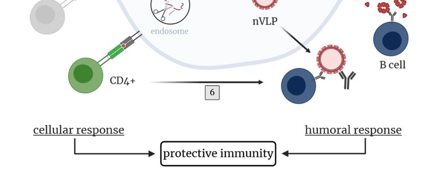
Vaccines 2020, 8, 451                                                                                4 of 42

       All TBEV vaccines are highly immunogenic with high and fast seroconversion rates ranging
from 86–100%, depending on the TBEV vaccine, evaluation method and study design. Various
studies showed seropositivity levels of 99.1–100% for the European vaccines after complete primary
immunization [49], 100% for the Russian vaccines after two immunizations [51,52] and 86–96% for the
Chinese vaccine after two vaccinations (reviewed in [37,53]). In general, waning of the vaccine-induced
immunity over time has been reported in several studies [49,54–57].
       Despite the broad availability of the European vaccines, vaccine coverage in European countries is
relatively low, making TBEV control difficult (reviewed in [3,58]). According to an Austrian field study,
efficacy of European vaccines was calculated to be 96–99% in persons with a complete, and 91.3–92.5%
in persons with an incomplete vaccination schedule. Due to the high vaccination coverage in Austria,
i.e. 85% of the population was vaccinated at least once against TBEV, and the high field effectiveness,
it is assumed that approximately 4000 TBE cases were prevented during 2000–2011 [59]. In addition,
the extensive vaccination of the Austrian population contributed to a strong decrease in TBE incidences
in children, which was 40-times lower compared to the neighboring country Slovenia, although
incidence rates were similar before [60]. Furthermore, the beneficial effect of TBEV vaccination was
also demonstrated in Russia. Vaccination with TBE vaccine Moscow led to decreased incidence rates
and prevention of an estimated annual 1500 novel TBE cases (reviewed in [34]). These facts highlight
the importance of vaccination in preventing TBE cases.
       However, immunization with TBEV vaccines does not provide complete protection. Consequently,
vaccination failures and breakthrough infections can occur in patients with incomplete or even regular
TBEV vaccination history. Although all age groups can be affected, breakthrough infections mostly
occur in elderly persons older than 50 years of age [61–63]. TBE disease severity in patients with
vaccination failure (FSME-IMMUN or Encepur) ranges from mild to severe with sometimes long-term
neurological sequelae or a fatal outcome [61,63–67]. Between 2000–2015, 1.7% of all reported TBE
cases in Slovenia were breakthrough infections [63]. A further study conducted in Stockholm County
(Sweden) identified that 5% of all TBE cases between 2006–2015 occurred in vaccinated subjects [62].
However, it is assumed that the case numbers of TBEV breakthrough infections are higher [61,62].

3. Immune Response to TBEV Infection and Vaccination

3.1. Innate Immunity Against TBEV
     The first defense against pathogens is the innate immune response consisting of anatomical and
chemical barriers as well as innate immune cells such as natural killer cells, macrophages, neutrophils
and dendritic cells. After infection, TBEV-specific pathogen-associated molecular patterns (PAMPs) are
recognized by pattern recognition receptors (PRRs) of nucleated cells. Some important PRRs during
infection of RNA viruses are Toll-like receptors (TLRs) 3, 7, 8 and 9 or retinoic-acid-inducible gene I
(RIG-I)-like receptors. Their activation leads to the production of type I interferon (IFN) (reviewed
in [68]), which was shown to have a protective role in TBEV infection (reviewed in [69]). Besides the
crucial role of innate immune cells to combat TBEV, there is evidence that some of these cells are infected
by TBEV, favor viral spread or contribute to pathogenesis in TBEV infection. Some non-structural
proteins of TBEV, such as NS1, NS2A, NS4A, NS4B or NS5, display antagonistic functions, thus,
interfering with components of the innate immune response (reviewed in [3,69–72]; [73]). In addition,
TBEV infection modulates expression patterns of many antiviral genes which are involved in the
innate immune response such as genes for PRRs, cytokines or chemokines [74]. Besides TBEV itself,
tick-derived saliva was shown to modulate the host innate immune response by influencing pathways,
such as increasing the activation of the Akt pathway in TBEV-infected dendritic cells [75], and innate
immune cells (reviewed in [76]).
     However, vaccination aims for the induction of adaptive immune responses and memory functions.
Hence, the innate immune response will not be further discussed in this review.
Vaccines 2020, 8, 451 5 of 42
3.2. Adaptive Immunity Against TBEV
Virus-specific humoral and cellular immunity, mediated by neutralizing and complement-fixing
antibodies as well as T cells with helper, cytotoxic and memory functions, are essential for protection
against flavivirus infections. However, in the case of TBEV, it may also pose a risk of increased severity
of infection or neuropathology [77].
3.2.1. Antibody Response
Antibodies induced after natural infection and vaccination play an important role as correlates
of protection against TBEV since they can prevent (lethal) disease (reviewed in [3]). Antibodies can
neutralize TBEV in various manners including prevention of viral attachment or fusion and support of
pathogen elimination via the complement system (reviewed in [78]). Therefore, the humoral immune
response is critical for controlling virus dissemination, virus clearance and long-term protective
immunity (reviewed in [79,80]).
Upon TBEV infection, the amount of TBEV-specific antibodies in human serum and cerebrospinal
fluid (CSF) increases and antibodies are usually detectable with the onset of neurological symptoms [2].
The highest IgM levels, which persist for around six weeks, are found in the early stages of TBE, whereas
IgG levels peak in the late convalescence period [81]. IgG antibodies can persist for several years and
protect from reinfection with TBEV (reviewed in [36]). In general, natural infection induces higher
virus-neutralizing (VN) antibody titers than vaccination with an inactivated virus [82]. Low serum
levels of VN antibodies combined with a high number of cells (segmented granulocytes (60–70%) and
lymphocytes (30–40%)) in the CSF at disease onset are thought to be indicative of a more severe clinical
course of TBE [83].
Following primary TBEV vaccination, an increase in antibody titers is observed after the
second dose which subsequently decline, followed by a strong booster response upon the third
immunization [84]. Studies on long-term persistence of vaccine-induced antibodies showed durability
of protective or VN antibodies, respectively, up to 5 years in 99% of children and above 98% in young
individuals [54,56,57]. In addition, other studies reported persistence for up to 8 or even 10 years [55,85].
However, with increasing age, immunosenescence, i.e., alteration of the innate and adapted immunity,
can be observed in the elderly leading to a lower immunogenicity of vaccines (reviewed in [86]).
Noteworthy, avidity and functional activity of TBEV vaccine-induced antibodies are apparently neither
influenced by age, age at primary immunization nor last vaccination, but are rather affected by the
individual [87].
Several human linear B cell epitopes within the structural and non-structural proteins of TBEV
(prM, E, NS1, NS2B, NS3, NS4B and NS5) have been identified. Two of these are located in domain I (DI)
and II (DII) of the E protein, respectively, and one in the C-terminal RNA-dependent RNA polymerase
region of NS5, and showed a positive reaction with almost all positive TBEV sera tested [88]. Intrinsic
factors, such as the conformational flexibility (reviewed in [89]) and the maturation state of flaviviruses
(reviewed in [90]), as well as extrinsic factors, such as the formaldehyde inactivation of virus particles
during the manufacturing process of vaccines (reviewed in [39]; [88,91]), potentially impact sensitivity
to antibody-mediated neutralization by changing the accessibility of epitopes. In addition, antibody
avidity can be influenced by the addition of adjuvants, e.g., aluminum hydroxide, to the vaccines [92].
Upon TBEV infection and vaccination, epitopes in the E protein are the main target for the
induction of VN antibodies (Table 2).inactivation of virus particles during the manufacturing process of vaccines (reviewed in [39];
[88,91]), potentially impact sensitivity to antibody-mediated neutralization by changing the
accessibility of epitopes. In addition, antibody avidity can be influenced by the addition of adjuvants,
e.g., aluminum hydroxide, to the vaccines [92].
Upon TBEV infection and vaccination, epitopes in the E protein are the main target for the
Vaccines 2020, 8, 451 6 of 42
induction of VN antibodies (Table 2).
Table 2. TBEV
Table 2. TBEV proteins
proteins eliciting
eliciting TBEV-specific
TBEV-specific immune
immune responses
responses in
in humans
humans upon
upon TBEV
TBEV infection
infection or
or
TBEV vaccination.
TBEV vaccination.
Infection Vaccination
Infection Vaccination
E [88,93] E [88,93]
Protective antibodies E [88,93]
Protective antibodies NS1 [94–97] NS1E[97]
[88,93]
#†
NS1 [94–97] NS1 [97] #†
C [98–100]
C [98–100]
CD4+ T cells EC[98–100]
[98–100]
C [98–100]
CD4+ T cells E [98–100] E [98–100,102]
NS1 [101] E [98–100,102]
NS1 [103]
NS2A [101]
NS3NS2A [103]
[103,104]
CD8+ T cells * NS3 [103,104] -
CD8+ T cells * NS4B [103] -
NS4B[103]
NS5 [103]
NS5 [103]
†: protective potential was shown in mouse models only [97,101,105–108]. #: presence of NS1-specific
† : protective potential was shown in mouse models only [97,101,105–108]. # : presence of NS1-specific antibodies in
antibodies in vaccinees still under discussion [94–97]. *: CD8+ T cell epitopes were identified for TBE
vaccinees still under discussion [94–97]. *: CD8+ T cell epitopes were identified for TBE patients with HLA-A2 and
patients with HLA-A2
HLA-B7 haplotypes and HLA-B7 haplotypes [103,104].
[103,104].
On
On the
the viral
viral surface,
surface, 180
180 copies
copies of
of the
the EE protein
protein structured
structured into
into 90
90 dimers
dimers can
can be
be found
found inin aa
”herringbone-like” icosahedral arrangement (reviewed
icosahedral arrangement (reviewed in in [109]). The E monomer consists
The E monomer consists of of three
distinct
distinct domains
domains (DI–DIII)
(DI–DIII) connected
connected by
by flexible
flexible linkers
linkers and
and aa membrane
membrane anchor
anchor domain
domain (DIV)
(DIV)
(Figure
(Figure 1).
1).
Figure 1. The structure of the TBEV E protein (PDB-ID 5O6A [5]). (A) Side view of a single TBEV E
protein1.monomer.
Figure Depicted
The structure of theare the four
TBEV domains
E protein (DI: red,
(PDB-ID DII:[5]).
5O6A yellow, DIII: view
(A) Side blue, of
DIV (stem/anchor):
a single TBEV E
gray) and
protein the fusion
monomer. loop (FL:
Depicted areorange).
the four (B) Top view
domains (DI: of a soluble
red, TBEV
DII: yellow, E protein
DIII: dimer.
blue, DIV Color code
(stem/anchor):
same and
gray) as inthe
(A).fusion
Ribbon diagrams
loop were prepared
(FL: orange). with UCSF
(B) Top view Chimera
of a soluble TBEV[110].
E protein dimer. Color code
same as in (A). Ribbon diagrams were prepared with UCSF Chimera [110].
DII has an elongated finger-like structure, which is formed by two loops connecting DI, the central
part of
DIIthe protein,
has in the dimeric
an elongated E protein.
finger-like The hydrophobic
structure, which is formedloop atbythe
twotiploops
of DII,connecting
covered byDI,DI and
the
DIII in mature
central virions,
part of the is responsible
protein, for the
in the dimeric fusion ofThe
E protein. thehydrophobic
viral and endosomal membranes,
loop at the tip of DII,therefore,
covered
termed
by DI andfusion
DIIIloop (FL) [5].
in mature The sequence
virions, of the for
is responsible FL isthehighly
fusionconserved among
of the viral all flaviviruses.
and endosomal DIII has
membranes,
an immunoglobulin-like
therefore, termed fusionstructure
loop (FL)with exposed
[5]. loops protruding
The sequence of the FL from
is the viralconserved
highly surface. The structure
among all
of the E protein is influenced by the pH leading to increased exposure of previous
flaviviruses. DIII has an immunoglobulin-like structure with exposed loops protruding from the viralinaccessible epitopes,
such as the
surface. The FLstructure
[5,111]. Allof three
the Eectodomains are capableby
protein is influenced of inducing a VN antibody
the pH leading response
to increased (reviewed
exposure of
in [109,112]).
previous Epitopesepitopes,
inaccessible can comprise
such asnot only
the individual
FL [5,111]. All domains of the E protein
three ectodomains but also
are capable residues
of inducing
from adjacent dimers and dimers in the quaternary herringbone-like arrangement of E proteins at
the virion surface [109,113]. In addition, the dynamic behavior of flaviviruses, commonly known as
“viral breathing”, can have a great impact on antibody binding (reviewed in [89,109]).
Jarmer et al. [93] provided insights into individual specificity and the variation of the humoral
immune response after TBEV infection or vaccination in humans. Using an immunoassay with
recombinant E protein and combinations of the single E domains, they observed strong individual
variation in antibody titers as well as immunodominance patterns concerning the individual E
domains [93]. The human humoral response was dominated by EDI- and EDII-specific antibodies [88,93],Vaccines 2020, 8, 451 7 of 42
whereas in mice immunized with inactivated, aluminum hydroxide-adjuvanted, purified TBE particles
mostly EDIII-specific VN antibodies were detected [92]. Furthermore, depletion of antibodies with
the dimeric E protein from sera of both naturally infected and vaccinated individuals led to a strong
reduction in VN activity, indicating a minor role of complex quaternary epitopes in the VN antibody
response. Additionally, depletion of EDI- and EDII-specific antibodies also significantly reduced the
VN activity of post-infection or post-vaccination sera, suggesting that antibodies recognizing antigenic
sites independent of the dimeric structure of the E protein display considerable VN activity [93].
The characterization of several flavivirus antibodies identified amino acids of the highly conserved FL
in EDII as a dominant antigenic site for cross-reactive, but not cross-neutralizing, antibodies [114–116].
After solubilization of TBE virions with a mild detergent, EDII-specific antibodies showed a strong
increase in binding avidity, indicating that the EDII-FL epitopes of native viruses may have a limited
accessibility due to partial occlusion. The fact that these broadly cross-reactive, non-neutralizing
antibodies are present in human sera after infection supports the hypothesis that during natural
flavivirus infection, cryptic epitopes become accessible and are presented to the host immune
system [116].
In addition to the E protein, the TBEV NS1 protein plays an important role as a soluble antigen.
In its pentameric or hexameric form, NS1 is secreted into the extracellular space [11], inducing a
NS1-specific antibody response. After natural TBEV infection, NS1-specific IgM and IgG antibodies are
detectable at high levels in human sera (Table 2) [94–97]. Several studies showed that NS1 immunization
or passive transfer of anti-NS1 antibodies afforded protection against infections with flaviviruses,
such as DENV, YFV and WNV in animal models [117–123]. These findings have been confirmed for the
TBEV NS1 protein [97,101,105–108,124]. Mouse studies showed that immunization with a synthetic
peptide corresponding to the structurally conserved α-helix (aa37–55) of NS1 [108] or with various
synthetic fragments of NS1 [101] induced partly protective immunity against lethal challenge infection
with TBEV. In addition, significantly prolonged survival of TBEV-infected mice was observed after
hyperimmunization with a whole recombinant TBEV NS1 protein, although all mice succumbed to
infection [97].
Whether or not an anti-NS1 immune response is induced by TBEV vaccination is matter of debate.
In some studies, anti-NS1 antibodies were almost exclusively observed after natural infection but not
after vaccination [94–96]. However, recent studies on the European vaccines question these findings by
demonstrating the presence of NS1 in Encepur by mass spectrometry and detection of NS1-specific
antibodies by Western blot in sera of vaccinees who repeatedly received Encepur or FSME-IMMUN.
Moreover, vaccination with FSME-IMMUN induced a robust anti-NS1 antibody response in mice after
several immunizations [97]. Further investigations are needed to confirm these results and to gain a
better understanding of the role of vaccine-induced anti-NS1 antibodies. Due to the robust IgM and
IgG response, NS1 might be an interesting target for novel vaccine designs and approaches.
In addition to antibodies against the E and NS1 proteins of TBEV, antibodies against the structural
prM protein have been described. Analysis of sera obtained from TBE patients and vaccinees
showed the presence of prM-specific antibodies, although titers were higher after infection. However,
results from depletion studies indicated that prM-specific antibodies play only a minor role in TBEV
neutralization [93]. In flavivirus infection, the protective potential of prM-specific antibodies has
been shown e.g., for DENV in mice studies [125,126]. In contrast, more recent studies indicated a
role of cross-reacting prM antibodies in antibody-dependent enhancement (ADE) in human DENV
infection [127,128]. Thus, further research is required to identify which role anti-prM antibodies,
induced after infection or vaccination, have in TBEV infection.
Impact of Pre-Existing TBEV-Specific Immunity
Once immunization by natural infection or vaccination has taken place, it remains an open
question which role pre-existing antibodies may play in further immunizations or TBEV infection.
A positive influence of pre-existing TBEV immunity on seroconversion rates following vaccinationVaccines 2020, 8, 451 8 of 42
with Russian TBEV vaccines was observed. However, the effect disappeared 30 days after the first
dose was given and after the administration of a second dose [52]. Furthermore, it has been shown
that pre-existing TBEV-specific antibodies in immune complexes can have immunomodulatory effects.
Immunization of mice with a soluble dimeric E protein only or together with monoclonal antibodies
specific for EDI, EDII and EDIII, respectively, revealed differences in the fine specificity of the antibody
response. The epitope-specific modulations of the immune response were mechanistically related to
the shielding of epitopes in the E protein and the monoclonal antibody-induced dissociation of the E
dimer, thereby revealing cryptic epitopes [129].
Due to cross-reactive epitopes, pre-existing vaccine-induced immunity mediates not only
protection against homologous, but also heterologous TBEV strains. Among different TBEV
subtypes, it was shown that immunization with vaccines based on TBEV-Eu or TBEV-FE elicits
cross-subtype reactive antibodies [130–134]. However, studies with various TBEV vaccines showed
that cross-protection against heterologous TBEV subtypes is not ensured for any strain [134,135].
Interestingly, one mutation in the EDI/EDII hinge region in the K23 vaccine seed virus (used for
manufacturing of Encepur) was identified to be responsible for reduced VN antibody titers against
heterologous Neudoerfl virus (vaccine strain used for manufacturing of FSME-IMMUN) of sera
received from children vaccinated with Encepur Children. In contrast, sera of children immunized
with FSME-IMMUN Junior showed comparable high levels of VN antibodies against both TBEV
strains [136].
Antibodies induced by TBEV vaccination afford not only a certain degree of intra-species
cross-protection but also cross-react within the TBE complex [133] and with other flaviviruses [116].
Vaccination with FSME-IMMUN or TBE vaccine Moscow protected mice against Omsk hemorrhagic
fever virus (OHFV) [130,137]. Furthermore, sera of TBEV vaccinated persons could partially neutralize
louping ill virus (LIV) and WNV [138]. Of interest, pre-existing vaccine-induced immunity to
YFV prior to TBEV vaccination resulted in an impaired TBEV-specific VN antibody response.
Nevertheless, all vaccinated study subjects reached protective TBEV neutralizing antibody titers
after completing the recommended TBEV vaccination schedule [84]. On the other hand, pre-existing
vaccine-induced immunity to TBEV increased the initial JEV-specific neutralizing antibody response
after vaccination with an inactivated JEV vaccine in humans [139]. In general, neutralizing and
non-neutralizing antibodies that cross-react with various flaviviruses can be detected post infection
and vaccination [116,138,140]. Overall, these results indicate the presence of cross-reactive epitopes
among flaviviruses and the effects of pre-existing flavivirus-specific immunity should be taken into
consideration when evaluating flavivirus vaccines.
Antibody-Dependent Enhancement in TBEV Infection?
Besides the potential protective effects of cross-reactive pre-existing antibodies, they may also
have detrimental effects by increasing the severity of disease through ADE of TBEV infection.
Non-neutralizing antibodies could promote virus entry into susceptible host cells, leading to increased
infectivity. The most common mechanism of ADE involves expression of Fc receptors. Complexes of
virus and sub- or non-neutralizing antibodies binding to Fcγ receptors (FcγR) on myeloid cells, such as
monocytes and macrophages, increases the attachment and uptake of the virus. As a consequence,
this opsonization leads to enhanced infectivity (reviewed in [141]). Haslwanter et al. [142] identified
a FcγR-independent mechanism based on interactions of the FL of the TBEV E protein with cellular
membranes. Studies with a monoclonal antibody (A5) recognizing an epitope in the E dimer interface
demonstrated that binding of A5 triggers the dissociation of E dimers, thus exposing the buried FL.
As a consequence, FL-mediated attachment to the plasma membrane increases binding and uptake of
the viral particle into the cell, thereby enhancing TBEV infectivity in vitro [142].
Whereas the phenomenon of ADE was extensively described and studied for DENV infection
in vitro and in vivo (reviewed in [143,144]), a potential role of ADE in TBEV infections is less
well investigated. Early studies showed the ability of polyclonal sera against members of theVaccines 2020, 8, 451 9 of 42
TBE serocomplex to enhance TBEV replication in vitro [145]. These results are in line with the
in vitro observed increased infectivity of TBEV in mouse peritoneal macrophages in the presence of
sub-neutralizing TBEV antibodies [146]. Interestingly, it was shown that murine TBEV hyperimmune
sera as well as TBEV-specific human polyclonal sera enhanced viral replication in mouse macrophages
in vitro, but protected mice from lethal TBEV challenge. Administration of the human or murine
immunoglobulins in different dilutions and combined with sublethal challenging doses revealed no
signs of ADE in vivo [147]. A more recent study investigating TBEV neutralization in vitro and in mice
by treatment with intravenous immunoglobulins containing high amount of TBEV-specific neutralizing
antibodies supports these findings since no ADE was observed by the application of cross-reactive or
virus-specific antibodies at different neutralization levels [148]. In vivo investigations in mice with
sub-neutralizing concentrations of a chimeric antibody against EDIII containing the constant regions
of human IgGκ (chFVN145), proposed as post-exposure treatment, also showed no indications of
ADE for the three main TBEV subtypes [149]. Additionally, in vivo and in vitro studies addressing
the potential effect of TBEV-specific immunity on ZIKV infection suggested that humoral immunity
against TBEV is unlikely to contribute to enhancement of ZIKV-induced pathogenesis in humans [150].
In summary, it is not completely clear if ADE plays a role in TBEV infection but it does not seem to be a
major problem. ADE has been demonstrated for TBEV in vitro but evidence in vivo is lacking.
3.2.2. CD4+ T Cell Response
Virus-specific CD4+ T cell responses are essential for the induction of antiviral immunity. CD4+ T
cells recognize antigenic peptides of 13–25 residues presented by major histocompatibility complex
(MHC) class II molecules, which are expressed on antigen-presenting cells (reviewed in [151]).
Upon stimulation, they produce pro-inflammatory and antiviral cytokines which results in the
recruitment and priming of other lymphoid cells. They may have cytotoxic activity against virus-infected
cells and are essential for promoting virus-specific antibody responses (reviewed in [152]).
Against infections with flaviviruses, CD4+ T cells play an important role as was demonstrated
e.g., for WNV and ZIKV [153,154]. Studies in mice showed that clearance of primary WNV infection
from the CNS is dependent on CD4+ T cells [154]. Likewise, CD4+ T cells contribute to controlling of
ZIKV infection and are pivotal for inducing ZIKV-specific antibodies [153]. For TBEV, adoptive transfer
of CD4+ T cells to TBEV-infected severe combined immunodeficient (SCID) mice, demonstrated a
protective role of these T lymphocytes in limiting the development of TBE and mortality [77].
Analysis of the CD4+ T cell response to TBEV structural proteins demonstrated that the E and C
proteins, but not the prM/M protein, were a major target in infected and vaccinated study subjects
(Table 2) [98,99]. Correlation of protein structure and immunodominance patterns revealed that
peptides corresponding to helices α2 and α4 of the C protein dominated the CD4+ T cell response in
both vaccinated and infected individuals, whereas the E protein-specific CD4+ T cell response was
less focused on selected protein domains. Most E protein-specific CD4+ T cells recognized epitopes in
β-sheets of EDIII as well as exposed loops protruding from the surface of the viral particle. Additionally,
vaccination induced significant EDI- and EDII-specific CD4+ T cell responses, whereas CD4+ T cell
responses against peptides from the stem region of E were predominantly observed in TBE patients [99].
Besides the identified CD4+ T cell epitopes in the structural proteins, several predicted T helper cell
epitopes were located in the NS1 protein of TBEV [101]. In general, individual variation in CD4+ T
cell peptide specificity as well as cytokine profile was seen in TBEV naturally infected and vaccinated
persons [98,99]. Individuals infected with other flaviviruses, like JEV, revealed a comparable CD4+ T
cell response, also against the JEV structural proteins and NS1 [155]. Comparison of immunodominance
patterns from TBEV-, ZIKV-, DENV- and YFV-specific CD4+ T cell epitopes revealed similarities in the
distribution of some epitopes. In summary, all four flaviviruses shared epitopes in the two helices
α2 and α4 of the C protein and in EDI and EDII, respectively, although some amino acid sequence
variation was observed in the respective epitopes [156].Vaccines 2020, 8, 451 10 of 42
Seven days post hospitalization, an elevated number of activated CD4+ T cells with effector
functions (measured by higher human leukocyte antigen- (HLA-) DR, PD-1 and perforin expression
and decreased Bcl-2 expression) was observed in TBE patients compared to control samples [104].
CD4+ T cells responding to DENV, ZIKV, WNV, JEV and YFV are characterized by the production of
interferon-gamma (IFN-γ), interleukin-2 (IL-2) and tumor necrosis factor-alpha (TNF-α), indicating
a T helper (Th) 1 cell response [153,155,157–162]. This Th1 response was also observed after TBEV
infection and vaccination but differed in its specificity and functionality [98–100,102].
Overall, the frequency of TBEV-specific CD4+ T cells was higher after vaccination than after
infection [99]. Antagonistic immunomodulatory properties of viral proteins, such as NS5, during
viral infection [163] and/or an enhanced T cell response after booster vaccinations may explain these
differences. On the other hand, a more polyfunctional phenotype of CD4+ memory T cells was found
in study subjects who had recovered from acute TBEV infection, compared to vaccinees. In recovered
TBE patients, a triple-positive phenotype (IL-2+ TNF-α+ IFN-γ+ ) was observed predominantly, while in
vaccinated subjects a mono- or bifunctional phenotype predominated (TNF-α+ or IL-2+ TNF-α+ ) [100].
Vaccination induced high levels of IL-2, TNF-α and CD40L expression in CD4+ T cells but the IFN-γ
response was considerably lower compared to that observed in TBE patients [98,100]. The CD4+ T
cell response patterns were comparable after all three vaccine doses, with the highest response after
the second immunization [100]. The VN antibody response after vaccination correlated with CD4+ T
cell functions, especially with frequencies of TBEV-specific IL-2+ and TNF-α+ CD4+ T cells [98,99,102].
Whether this is also the case after infection is unclear [98,99].
Studies on cross-reactivity of CD4+ T cells specific for mosquito-borne flaviviruses (reviewed
in [164]) indicate that e.g., JEV-specific CD4+ T cells can recognize other flaviviruses which may result in
stronger CD4+ T cell responses upon secondary infection with a heterologous virus [165,166]. Data on
cross-reactivity of TBEV-specific CD4+ T cells with other flaviviruses are lacking.
Overall, knowledge about CD4+ T cell response after infection or vaccination is limited. CD4+ T
cell subpopulations are important for protection against TBEV, however, their correlation with antibody
responses and their cross-reactive potential require further evaluation.
3.2.3. CD8+ T Cell Response
In addition to virus-specific antibodies and CD4+ T cells, CD8+ T cells are an important immune
correlate of protection against viral infections. CD8+ T cells recognize viral peptides of about 8–10 amino
acid residues that are presented by MHC class I molecules (reviewed in [151]). So far, seven TBEV
CD8+ T cell epitopes have been identified, all located in the non-structural proteins (NS2A, NS3, NS4B
and NS5; Table 2) [103,104]. Almost all of these epitopes are highly conserved among European,
Siberian and Far-Eastern TBEV subtypes [103].
It has been shown that flavivirus-specific CD8+ T cells directed to conserved non-structural
proteins but also to structural proteins, display cross-reactivity across various flaviviruses. It is unclear
if cross-reactive CD8+ T cells can afford protection against a heterologous flavivirus or contribute to a
more severe disease outcome (reviewed in [164]). Especially for DENV infections this is a matter of
debate (reviewed in [167]). However, the potential role of cross-reactive TBEV-specific CD8+ T cells is
largely unknown.
The peak of CD8+ T cell responses in the peripheral blood of acutely infected TBE patients
was about seven days after hospitalization during the second, neuroinvasive stage. TBEV-induced
effector cells were characterized by increased HLA-DR, PD-1, perforin and granzyme B levels in
combination with decreased expression of CD127, Bcl-2 and CD27. Furthermore, most of the CD8+ T
cells (>50%) showed monofunctional properties in virus control (degranulation, cytokine or chemokine
expression) 7, 21 and 90 days after hospitalization. During the course of infection, these cells maintained
their monofunctionality but altered their function. The change in the functional composition was
accompanied by the differentiation of TBEV-specific CD8+ T cells with a dominant Eomes+ Ki67+ T-bet+Vaccines 2020, 8, 451 11 of 42
effector phenotype (peak at day seven) into Eomes− Ki67− T-bet+ memory cells as the infection resolved
(day 21 and 90) [104].
The MHC haplotype dictates the epitope and T cell repertoire as well as magnitude of
the virus-specific CD8+ T cell response (reviewed in [151]). This was also demonstrated for
TBEV by studies investigating CD8+ T cell responses against non-structural TBEV proteins
during acute TBEV infection [103,104]. Depending on HLA alleles, the differentiation of effector
memory CD8+ T cells in two distinct phenotypes was observed. One week after hospitalization,
HLA-A2- and HLA-B7-restricted TBEV epitope-specific effector cells showed a common effector
memory phenotype (CD45RA– CCR7– CD27+ CD57– ). Two weeks later, mainly effector memory
(CD45RA– CCR7– CD27+ CD57+ ; approximately 25% of the cells) and terminally differentiated effector
memory RA (CD45RA+ CCR7– CD27+ CD57+ ; approximately 40% of the cells) phenotypes for HLA-A2-
and HLA-B7-restricted cells, respectively, were found [103].
In the brain of TBE patients with a fatal disease outcome, TBEV and activated CD8+ T cells
have been demonstrated [168,169]. Expression of α4β1 integrin (=VLA-4) on CD8+ T cells during
the neuroinvasive phase of TBEV infection may indicate the migratory capacity of these cells into
the CNS [103]. Since TBEV infects neurons of the CNS [169], the recruitment of CD8+ T cells may
exert, in addition to beneficial effects, detrimental effects and may contribute to the pathogenesis
through the killing of infected neurons and the local secretion of cytokines such as TNF-α. This
hypothesis is supported by the observation that there were no differences in the extent of virus
replication in the brains of mice surviving and in those that succumbed to experimental infection [170].
Since activated TBEV-specific CD8+ T cells show upregulation of perforin and granzyme B [104] and
CD8+ granzyme B+ T cells have been found in the brain tissue of fatal TBE cases associated with
neuronal damage [168], indeed they may contribute to the pathogenesis, although uncontrolled virus
replication may result also in a worse outcome of disease. A correlation between infiltrated CD8+ T
cells and brain damage was also described for other flaviviruses [171–173]. Other evidence for the
detrimental effects of virus-specific CD8+ T cells stems from work with CD8– /– knockout and SCID
mice. Upon infection with a neurovirulent TBEV strain, a significantly prolonged mean survival time
compared to immunocompetent mice was observed. However, all mice succumbed to infection [77].
Comparable findings have been made for WNV. Infection of CD8+ -deficient (β2-m− /− ) mice inoculated
with a high virus dose resulted in a prolonged mean survival time compared to immunocompetent mice.
In contrast, CD8+ -deficient mice inoculated with a low dose of WNV showed, despite an extended
mean survival time, an enhanced mortality rate compared to wild type mice [173]. These findings
indicate that during natural flavivirus infections, which are most likely of low virus dose, CD8+ T cells
may exert beneficial protective effects. Of note, it has been shown that the currently used inactivated
TBEV vaccines induce virus-specific CD8+ T cell responses inefficiently, if at all [100,174,175].
In summary, CD8+ T cells are thought to contribute to protective immunity and recovery
from TBEV infection. However, further research should rule out potential downsides of CD8+ T
cell-mediated immunity.
3.2.4. Vaccine Failures
Understanding the cause of breakthrough infections is very important for improving existing
vaccines and developing new ones. Although a common definition of vaccination breakthrough
infection is missing, TBE patients developing disease despite being vaccinated indicate the inability of
the vaccine to induce a protective immune response in any case [176]. Host-related risk factors, such as
advanced age (beyond 60 years), as well as vaccine-related risk factors, such as incomplete primary
immunization or delayed first booster doses, may favor vaccination breakthrough infections (reviewed
in [177]). Regarding TBEV vaccination, two types of vaccination breakthrough infections have been
suggested: patients with inadequate immune response and non-responders [176].
The humoral immune response upon natural infection of individuals responding to TBEV
vaccination but lacking protective efficacy, differs from those of unvaccinated TBE patients. In vaccinatedVaccines 2020, 8, 451 12 of 42
but unprotected study subjects, IgG and VN antibodies increased rapidly, but the IgM response was
delayed [61,63–67,178,179]. However, this differential serological profile does not explain the lack
of protection.
Comparison of TBEV-specific immune responses post booster vaccination in high and
non-responders revealed significant differences in their antibody response. In the non-responder
group, TBEV-specific geometric mean titers (GMT) of VN antibodies were below the detection limit
pre-booster (6.5 GMT). A slight increase two months after booster vaccination (18.0 GMT) with a decline
six months later (10.7 GMT) was observed. In contrast, high responders showed ten-fold higher VN
antibody titers pre-booster (67.5 GMT) which strongly increased eight weeks after booster vaccination
(139.6 GMT) and decreased slightly afterwards (101.1 GMT) [180]. Investigation of TBEV-specific IgG
avidity in both groups revealed no differences, suggesting alterations in quantity rather than quality of
antibodies [63,67,179,180]. Further studies showed a correlation of humoral and cellular immunity in
both groups: low antibody titers were associated with poor antigen-specific T cell proliferation, whereas
high antibody titers were associated with strong T cell proliferation. Upon TBEV antigen-restimulation,
peripheral blood mononuclear cells from TBEV vaccine non-responders were characterized by low
levels of IL-2 and IFN-γ production prior and post booster vaccination, contrary to high responders
with higher cytokine expression levels. Furthermore, a decrease in regulatory T cells was observed in
the high responder group after the booster vaccination, while non-responders displayed an increase
in IL-10-producing regulatory T cells. Consequently, this might disable T cell proliferation or impair
B cell-induced IgG production. This increase observed in non-responders is thought to be due to
the expansion of natural as well as inducible regulatory T cells. On the other hand, the decrease
in high responders may be explained by the expansion of TBEV-specific effector T cells, changing
the overall distribution of CD4+ subpopulations. In addition, no correlation between HLA class II
haplotype and vaccination failure in TBEV non-responders was found. Interestingly, TBEV vaccine
non-responders were able to develop an efficient immune response upon vaccination with seasonal
influenza vaccines (Inflexal V 2008/2009 and Inflexal V 2009/2010), as observed in high responders.
Hence, the non-responsiveness to TBEV vaccines is supposed to be antigen-dependent at humoral
levels [180].
To overcome impaired vaccine responsiveness, especially in the elderly, further research on the
understanding of TBEV vaccination breakthrough infections is warranted. Optimized or novel vaccine
strategies are needed to ensure sufficient protection for all risk groups.
4. Novel Approaches and TBEV Target Antigens for the Development of Improved
TBEV Vaccines
The currently available TBEV vaccines suffer from some disadvantages as discussed above,
including: (a) the time-consuming vaccination regimen and need of regular booster vaccinations for
maintaining the protective efficacy, (b) reduced immunogenicity in the elderly as well as (c) vaccination
breakthrough infections and non-responders (vaccine failure). Therefore, the development of improved
vaccines against TBEV is desirable. An ideal TBEV vaccine should fulfill several requirements:
• Highly immunogenic in all age and risk groups, rapid and high seroconversion rates.
• Induction of long-lasting immunity without the need for booster vaccinations.
• No vaccine failures.
• Protection against all TBEV subtypes.
• Cost-effective and safe.
To fulfill these requirements, various aspects must be considered. Central to the development
of improved TBEV vaccines is obtaining a better understanding of the correlates of protection
and consequently identifying the targets for humoral and cellular immune responses. In addition,
the vaccine platform chosen for delivery of the antigen(s) of interest can influence the nature of the
induced immune response. Thereby, combining different arms of the immune system is desirableVaccines 2020, 8, 451 13 of 42
to induce a strong/protective and long-lasting immunity. Although various vaccine platforms and
antigens were already utilized for designing novel TBEV vaccine approaches, data on safety, tolerability
and efficacy in men are lacking.
4.1. Novel TBEV Vaccine Strategies Aiming at the Induction of Humoral Immunity
The humoral arm of the adaptive immunity plays a major role in protection against TBEV. Of note,
low VN antibody titers were associated with a more severe TBE outcome [83]. Thus, a vaccine should
induce high affinity VN antibodies which prevent infection, dissemination and support viral clearance.
For TBEV it is known that VN antibodies are mainly directed to epitopes located in the E
protein (Table 2). However, immunization with various soluble forms of dimeric whole or membrane
anchor-free TBEV E protein failed to induce VN antibodies and provide appropriate immunogenicity
in mice [181]. Likewise, DNA vaccines driving intracellular expression of either whole or truncated
TBEV E proteins or secretion of TBEV E dimers, failed to induce VN antibodies and afford robust
protection against TBEV [182]. Thus, the usage of TBEV E protein only was not very successful.
In addition, flavivirus subunit vaccines based on a single protein domain of the E protein, namely
EDIII, were tested. EDIII is a biologically relevant and flavivirus-specific target, inducing antibodies
that are less cross-reactive than those directed against epitopes in EDI/EDII, thus, reducing a possible
risk of ADE [115,144,183]. Immunization of mice with an attenuated recombinant influenza A virus
(rIAV) encoding the WNV EDIII protein domain in frame with the IAV neuraminidase protein, induced
VN antibodies and CD4+ T cells as well as protection against lethal WNV challenge [184]. Contrary,
immunization of mice with EDIII of ZIKV expressed from a plasmid, replication-deficient chimpanzee
adenovirus or given as a recombinant protein confirmed that EDIII of ZIKV induced VN antibody
responses inefficiently [185]. However, results from mouse studies need to be evaluated carefully
since TBEV immunodominant regions were shown to differ in humans (EDI and EDII) [93] and mice
(EDIII) [92]. Since EDIII seems to play a minor role in eliciting VN antibodies upon TBEV infection in
humans [93], it is unclear whether EDIII would be a promising target for novel TBEV vaccines.
In addition to vaccine candidates based on the TBEV E protein only, those based on the E and
prM proteins have been tested extensively. Co-expression of E and prM results in the self-assembly of
protein complexes without a genome, known as virus-like particles (VLPs). Thus, VLPs are unable to
replicate and their use is considered to be safe (reviewed in [186]). In vitro expression of prM and E
in a VLP expression system based on mammalian cell lines [187,188] or the yeast Pichia pastoris [189],
resulted in the production of such recombinant TBEV VLPs. These VLPs were smaller than native whole
virions but showed similar surface protein organization (including antigenic structures) and functional
reactivity (rearrangement and fusion at low pH, hemagglutination activity) when compared to native
virus particles [190]. Due to their authentic morphology, which mimic native virus particles, VLPs are
highly immunogenic and able to provoke humoral as well as cellular immune responses (reviewed
in [186,191]). Immunization of mice with purified VLPs produced in mammalian cells led to a VN
antibody response and complete protection against TBEV challenge. However, immunogenicity and
protective efficacy were similar to formalin-inactivated whole TBE viruses [181]. Thus, improvements
to achieve a stronger immunogenicity, superior to that after vaccination with inactivated whole viruses,
is desirable. One opportunity might be the incorporation of C proteins in the VLPs as demonstrated
for ZIKV VLPs. Direct comparison of recombinant ZIKV VLPs without (prM-E) and with C (C-prM-E),
both produced in a stable cell line, showed that in mice immunized with C-prM-E VLPs neutralizing
antibody titers were enhanced, C-specific antibodies were detectable and viremia upon challenge
infection was prevented [192]. In addition to recombinant VLPs produced in vitro, they can also be
produced in situ after vaccination with nucleic acid-based vaccines or live attenuated viruses encoding
the prM and E proteins (Figure 2).Vaccines 2020, 8, 451 14 of 42
Vaccines 2020, 8, x 14 of 44
Figure 2. TBEV-specific immunity upon immunization with virus-like particles (VLPs). The TBEV
Figure 2. TBEV-specific immunity upon immunization with virus-like particles (VLPs). The TBEV
polyprotein with proteins inducing protective antibody responses (prM, E, NS1) is shown.
polyprotein with proteins inducing protective antibody responses (prM, E, NS1) is shown. Upon
Upon immunization with purified VLPs (1) or vector-based vaccines co-expressing prM and E (in situ
immunization with purified VLPs (1) or vector-based vaccines co-expressing prM and E (in situ
production of VLPs, (2)), mainly humoral immune responses are induced. Inclusion of the NS1 protein
production of VLPs, (2)), mainly humoral immune responses are induced. Inclusion of the NS1
in these vector-based vaccine approaches may improve the protective immunity. (3) Vector-based
protein in these vector-based vaccine approaches may improve the protective immunity. (3) Vector-
immunization leads to the synthesis of proteins of interest (POIs), which are processed on different
based immunization leads to the synthesis of proteins of interest (POIs), which are processed on
pathways leading to humoral and/or cellular immune responses. (4) Humoral immune response: POIs
different pathways leading to humoral and/or cellular immune responses. (4) Humoral immune
are either secreted into the extracellular space (NS1) or assemble into novel VLPs (nVLPs; prM-E)
response:
which are POIs are either
subsequently secreted
released frominto
thethe
cell.extracellular
The secretedspace
proteins(NS1)
andordeassemble into novel
novo produced VLPs
(nVLPs) or
(nVLPs; prM-E) which are subsequently released from the cell. The secreted proteins and
directly administered VLPs, respectively, induce the production of TBEV-specific B cells and antibodies. de novo
produced
(5) Cellular(nVLPs)
immuneor directlyThe
response: administered VLPs, respectively,
POIs or endocytosed induce the
VLPs, respectively, production
are degraded intoofpeptides
TBEV-
specific B cells and antibodies. (5) Cellular immune response: The POIs or endocytosed
mainly by host proteases in endosomes leading to antigen presentation via MHC class II molecules VLPs,
to
respectively,
+ are degraded
+ into peptides mainly by host proteases in endosomes leading
CD4 T cells. (6) CD4 T cells promote the activation and proliferation of TBEV-specific B cells driving to antigen
presentation via MHC
efficient antibody class II
responses molecules
with to CD4of
development
+ T cells. (6) CD4+ T cells promote the activation and
memory responses. Created with BioRender.com.
proliferation of TBEV-specific B cells driving efficient antibody responses with development of
memory responses.
Immunization Created
with with BioRender.com.
a DNA-based vaccine enabling assembly of recombinant TBEV VLPs in vivo,
elicited the production of VN antibodies in mice [182,193] and rhesus macaques [194]. Additionally,
Immunization
immunized with treated
mice or mice a DNA-based vaccine
with serum enablingnon-human
of vaccinated assembly of recombinant
primates TBEV VLPs
were protected in
against
vivo, elicited the production of VN antibodies in mice [182,193] and rhesus macaques
TBEV challenge [182,193,194]. Moreover, in situ generation of TBEV VLPs by vaccination of mice with [194].
Additionally,
a late-defectiveimmunized
recombinantmice or micevirus
Vaccinia treated with serum
(rVACV) of vaccinated
encoding non-human
for the prM primates
and E proteins, were
induced
protected against
VN antibodies andTBEV challenge
a robust [182,193,194].
protection Moreover,
upon challenge in situ
infection withgeneration
TBEV [195].ofAltogether,
TBEV VLPs by
these
vaccination of mice with a late-defective recombinant Vaccinia virus (rVACV) encoding for the prM
and E proteins, induced VN antibodies and a robust protection upon challenge infection with TBEVYou can also read