Retroviral Restriction Factors and Their Viral Targets: Restriction Strategies and Evolutionary Adaptations - MDPI
←
→
Page content transcription
If your browser does not render page correctly, please read the page content below
microorganisms
Review
Retroviral Restriction Factors and Their Viral Targets:
Restriction Strategies and Evolutionary Adaptations
Guney Boso and Christine A. Kozak *
 Laboratory of Molecular Microbiology, National Institute of Allergy and Infectious Diseases,
 Bethesda, MD 20892-0460, USA; guney.boso@nih.gov
 * Correspondence: ckozak@niaid.nih.gov

 Received: 30 October 2020; Accepted: 8 December 2020; Published: 11 December 2020

 Abstract: The evolutionary conflict between retroviruses and their vertebrate hosts over millions
 of years has led to the emergence of cellular innate immune proteins termed restriction factors as
 well as their viral antagonists. Evidence accumulated in the last two decades has substantially
 increased our understanding of the elaborate mechanisms utilized by these restriction factors to
 inhibit retroviral replication, mechanisms that either directly block viral proteins or interfere with the
 cellular pathways hijacked by the viruses. Analyses of these complex interactions describe patterns
 of accelerated evolution for these restriction factors as well as the acquisition and evolution of their
 virus-encoded antagonists. Evidence is also mounting that many restriction factors identified for
 their inhibition of specific retroviruses have broader antiviral activity against additional retroviruses
 as well as against other viruses, and that exposure to these multiple virus challenges has shaped their
 adaptive evolution. In this review, we provide an overview of the restriction factors that interfere with
 different steps of the retroviral life cycle, describing their mechanisms of action, adaptive evolution,
 viral targets and the viral antagonists that evolved to counter these factors.

 Keywords: retroviruses; restriction factors; viral evolution; viral antagonists

1. Introduction
     Retroviruses replicate by converting their single-stranded RNA genome into double-stranded
DNA through a virus-encoded reverse transcriptase. These DNA copies are integrated into host
chromosomes where they persist as a permanent part of the host cell genome. When retroviruses infect
germline cells, integrated copies can be passed on to the next generation. These integrated proviruses
are termed endogenous retroviruses (ERVs). As the retroviral genome encodes only a limited number
of genes, replication of exogenous viruses and expression of ERVs rely on the host cell machinery.
These viruses can be mutagenic and pathogenic, and this puts strong selective evolutionary pressure
on the host genes appropriated for virus replication. The result has been the emergence of defensive
genes that can inhibit viral infection and that have been derived from cellular genes, or more rarely,
from ERVs. These restriction factors comprise the innate immune system that represents the first line
of defense against viral attack.
     While each restriction factor has followed a unique evolutionary path, some common themes have
emerged (Table 1). First, most restriction factors are encoded by interferon-stimulated genes (ISGs) that
are upregulated upon cellular exposure to type I or type II interferons (IFNs). Second, phylogenetic
studies in the last two decades demonstrated that most restriction factors show signatures of positive
or diversifying selection—that is, the rate of nonsynonymous mutations that result in amino acid
substitutions is higher than the rate of synonymous mutations. The rapid evolution observed in the
genes encoding retroviral restriction factors has often been countered by rapidly evolving viral evasive
mechanisms which include virus-encoded antagonists and signatures of positive selection in the viral

Microorganisms 2020, 8, 1965; doi:10.3390/microorganisms8121965           www.mdpi.com/journal/microorganisms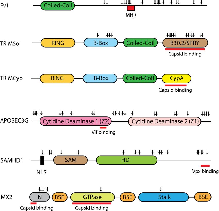
Microorganisms 2020, 8, 1965 2 of 34
target of host restriction factors. This evolutionary conflict represents a genetic arms race described by
the Red Queen hypothesis [1,2].
Table 1. Properties of Retroviral Restriction Factors.
Restricted Viruses
Restriction Factor Viral Antagonist * IFN Induced Positive Selection *
Retroviruses Other *
CAT1 - No Ecotropic MLV - No
XPR1 - No Nonecotropic MLV - Yes
CD4 Vpu, Nef No HIV, SIV - Yes
CCR5 Vpu, Nef No HIV - -
Fv4 - No Ecotropic MLV - -
Rmcf, Rmcf2 - No Nonecotropic MLVs - -
Nef, Vpu
Alphaviruses,
SERINC5 S2 (EIAV) Glycogag No HIV, SIVs, EIAV, MLV No
Filoviruses
(MLV)
MLV, EIAV, Feline
Fv1 - No - Yes
Foamy Virus
TRIM5 - Yes Retroviruses Flaviviruses Yes
huAPOBEC3G Vif HIV, SIVs, MLV HBV
Yes Yes
mApobec3 Glycogag, p50 (MLV) MLV -
HBV, Herpesvirus,
SAMHD1 Vpx, Vpr Yes HIV, SIVs, EIAV, FIV Yes
Vaccinia
Herpesvirus, HBV,
MX2 - Yes HIV, SIVs, EIAV, FIV Yes
Flavivirus
HBV, Alphaviruses,
ZAP - Yes HIV, MLV Yes
Filoviruses
SLFN11 - Yes HIV, EIAV, MLV Flaviviruses Yes
BST2/Tetherin Vpu, Nef Yes All known retroviruses Enveloped Viruses Yes
Influenza, Zika,
GBP5 - Yes HIV, MLV Yes
Measles
Vesicular Stomatitis
MARCH8 - No HIV -
Virus, Ebola Virus
IFITM3 Glycogag (MLV) Yes HIV, MLV Influenza, Measles Yes
*-, none or unknown. HIV: Human Immunodeficiency Virus; MLV: Murine Leukemia Virus; SIV: Simian
Immunodeficiency Virus; EIAV: Equine Infectious Anemia Virus; FIV: Feline Immunodeficiency Virus; HBV:
Hepatitis B Virus.
Here, we describe a well-characterized set of retroviral restriction factors for which there
is substantial information on their restriction mechanisms as well as their evolutionary history.
The majority of these factors were discovered in primates or rodents through studies on HIV-1 which
causes the acquired immune deficiency syndrome (AIDS) in humans, and on mouse leukemia viruses
(MLVs) which induce lymphomas, immunodeficiencies, and neurological diseases. Some of these
factors are restricted to specific taxonomic lineages, while others are carried by many mammalian
species, showing broad antiviral activity against multiple retroviruses and in some cases against other
families of viruses (Table 1). Decades of studies have produced an increased understanding of how
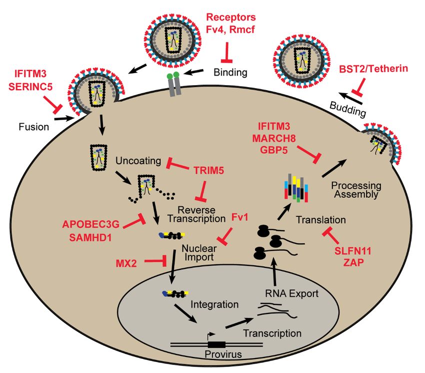
these factors work and what stage of the replication cycle is blocked (Figure 1), and, in many cases,
also describing the coevolutionary adaptations in the interacting host and virus proteins.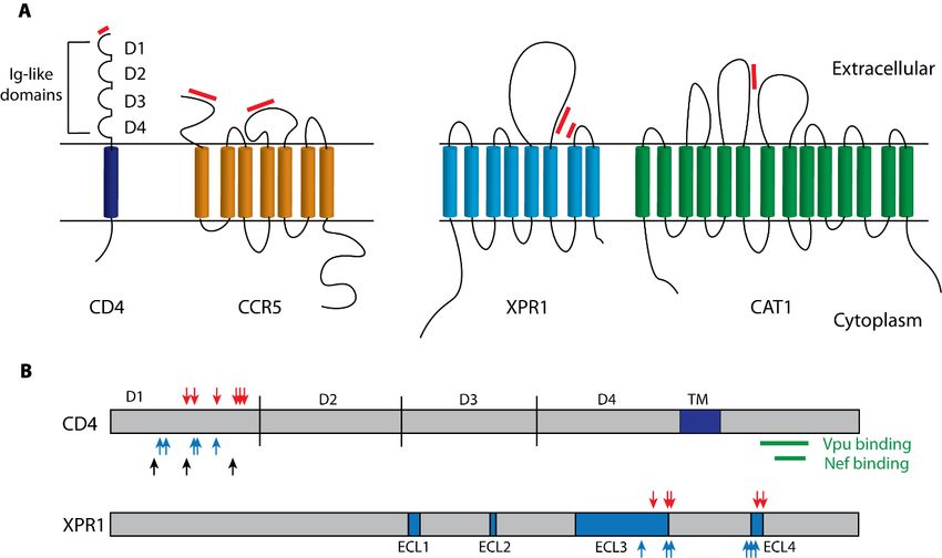
factors are restricted to specific taxonomic lineages, while others are carried by many mammalian
species, showing broad antiviral activity against multiple retroviruses and in some cases against
other families of viruses (Table 1). Decades of studies have produced an increased understanding of
how these factors work and what stage of the replication cycle is blocked (Figure 1), and, in many
Microorganisms 2020, 8, 1965 the coevolutionary adaptations in the interacting host and virus proteins.3 of 34
cases, also describing

                    Figure 1. Restriction factors block specific stages of the retroviral life cycle.
                    Figure 1. Restriction factors block specific stages of the retroviral life cycle.
2. Retroviral Restriction Factors
 2. Retroviral Restriction Factors
2.1. Binding and Entry
 2.1. Binding and Entry
      Retroviruses enter susceptible cells through their interactions with specific cell surface receptors.
TheseRetroviruses
        interactionsenter
                        can besusceptible
                                 inhibited cells    throughpolymorphisms
                                              by receptor     their interactionsaswith
                                                                                    wellspecific    cell surface
                                                                                          as by other             receptors.
                                                                                                          transmembrane
 These   interactions    can be   inhibited   by   receptor
proteins that interfere with binding or membrane fusion.     polymorphisms       as well    as by  other   transmembrane
 proteins that interfere with binding or membrane fusion.
2.1.1. Receptors
 2.1.1. Receptors
      Receptor-mediated restriction of virus entry has been documented for HIV-1 and MLVs. The HIV-1
       Receptor-mediated
envelope      (Env) interactsrestriction
                                  with twoofmembrane
                                                  virus entryproteins
                                                                has beenfordocumented        for HIV-1
                                                                               entry, a receptor,      CD4,andand
                                                                                                                MLVs.oneThe
                                                                                                                          of
 HIV-1
two       envelope (Env)
      co-receptors,          interacts with
                        the chemokine           two membrane
                                            receptors    CCR5 and  proteins
                                                                      CXCR4for(Figure
                                                                                  entry, 2A)
                                                                                           a receptor,
                                                                                                 [3–5]. CD4,
                                                                                                          CD4 is and  one of
                                                                                                                    a type  I
 two co-receptors,
transmembrane            the chemokine
                      protein  that functionsreceptors    CCR5 and for
                                                   as a co-receptor    CXCR4
                                                                          the T (Figure     2A) [3–5].
                                                                                 cell receptor            CD4complex
                                                                                                   signaling     is a typeinI
 transmembrane
response              protein
             to antigen         that functions
                          presentation     by theas MHCa co-receptor   for the T [6].
                                                           Class II molecules     cell These
                                                                                        receptor    signaling
                                                                                                proteins        complex
                                                                                                            mediate   HIV-1in
 responseof
infection     toimmune
                 antigen presentation
                           system cells, andby the   MHC Class II molecules
                                                 receptor-functional     orthologs[6].
                                                                                     areThese
                                                                                          largelyproteins   mediate
                                                                                                    restricted         HIV-1
                                                                                                                to humans
 infection
and   relatedof primates.
                 immune system
                            CD4 bindscells,virus
                                            and receptor-functional      orthologs
                                                  through the D1 Ig domain,       andareCCR5
                                                                                           largely
                                                                                                 hasrestricted
                                                                                                      two virus- to binding
                                                                                                                    humans
 and related
domains          primates.
            in its          CD4and
                   N-terminus      binds   virus
                                       in its     through
                                               second        the D1 Igloop
                                                        extracellular    domain,   and
                                                                             (Figure  2A)CCR5     has two
                                                                                             [7]. CD4        virus- binding
                                                                                                        is downregulated
 domains
in  infected incells
                its N-terminus
                      by the HIV-1 andproteins
                                        in its second    extracellular
                                                   Nef and              loop2B),
                                                              Vpu (Figure     (Figure
                                                                                   and2A)this[7]. CD4 is superinfection,
                                                                                               prevents     downregulated
 in infected
thus  avoiding  cells  by the and
                   apoptosis  HIV-1    proteins of
                                    production      Nef  and Vpu (Figure 2B),virions,
                                                      infection-compromised        and this andprevents     superinfection,
                                                                                                  also reduces   sensitivity
to inhibition by another restriction factor, SERINC5 [8–10]. While there are no known variants reduces
 thus   avoiding      apoptosis    and   production       of  infection-compromised         virions,   and    also   of CD4
in humans that affect the efficiency of HIV-1 infection, CD4 is highly variable in chimpanzees, and this
variation is responsible for restricted susceptibility to SIV, the progenitor of HIV-1 (Figure 2B) [11,12].
Moreover, CD4 has been molded by positive selection in primates with rapidly evolving residues found
in the HIV-1 Env interacting interface of the CD4 protein, but not affecting the sites targeted by Vpu and
Nef (Figure 2B) [13,14]. These findings suggest that co-evolution with SIVs has accelerated the evolution
of CD4 in primates. In some human populations, HIV-1 entry can be blocked by a variant of the CCR5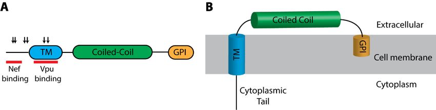
variants of CD4 in humans that affect the efficiency of HIV-1 infection, CD4 is highly variable in
chimpanzees, and this variation is responsible for restricted susceptibility to SIV, the progenitor of
HIV-1 (Figure 2B) [11,12]. Moreover, CD4 has been molded by positive selection in primates with
rapidly evolving residues found in the HIV-1 Env interacting interface of the CD4 protein, but not
affecting the
Microorganisms sites
2020, targeted by Vpu and Nef (Figure 2B) [13,14]. These findings suggest that4 co-
8, 1965 of 34
evolution with SIVs has accelerated the evolution of CD4 in primates. In some human populations,
HIV-1 entry can be blocked by a variant of the CCR5 coreceptor with a 32-base pair (bp) deletion in
coreceptor
the secondwith a 32-baseloop
extracellular pair of
(bp)
thedeletion
proteinin the second
leading to theextracellular
introductionloop
of aofpremature
the protein leading
stop codonto
the introduction of a premature stop codon which
which renders it non-functional as a co-receptor [15]. renders it non-functional as a co-receptor [15].
Figure2.2.Structure
Figure Structure andand functional features of the HIV-1
HIV-1 and
and MLV
MLV cell
cell surface
surfacereceptors.
receptors.(A)
(A)Schematic
Schematic
diagrams of
diagrams of the
the receptors
receptors for
for HIV-1
HIV-1 (CD4
(CD4 and CCR5) and and for
for different
different MLV
MLV subtypes
subtypes (XPR1
(XPR1and and
CAT1). Red
CAT1). Red bars
bars indicate
indicate regions
regions that bind virus envelope
envelope [7,16,17].
[7,16,17]. (B)
(B) XPR1
XPR1and
andCD4CD4receptor
receptor
proteins.Blocks
proteins. Blocksidentify
identifythe
the transmembrane
transmembrane domain
domain of CD4
of CD4 andand the extracellular
the extracellular loopsloops (ECLs)
(ECLs) in
in XPR1.
XPR1. Positively selected residues are marked with red arrows [13,17–19]. Receptor critical
Positively selected residues are marked with red arrows [13,17–19]. Receptor critical sites are marked sites are
marked
with blue with
arrowsblueandarrows and black
black arrows arrows
identify identify polymorphic
polymorphic sites in chimpanzee
sites in chimpanzee CD4 that
CD4 that influence SIV
influenceGreen
binding. SIV binding. GreenCD4
bars identify barssites
identify CD4 sites
susceptible susceptible to downregulation
to downregulation by Nef and Vpuby [8,9,11,12].
Nef and Vpu
[8,9,11,12].
MLVs isolated from laboratory mice have host range subgroups that rely on two receptors, CAT1 for
MLVs isolated
the ecotropic from laboratory
or mouse-tropic MLVs, and mice have
XPR1 forhost
MLVs range
that subgroups
can also infectthatother
rely mammalian
on two receptors,
species
CAT1 for
(Figure 2A)the ecotropic
[20,21]. or host
These mouse-tropic MLVs, and
genes function, XPR1 forasMLVs
respectively, that can
an amino acidalso infect other
transporter and
amammalian species (Figure
phosphate exporter [22–24].2A)
CAT1[20,21]. Theseare
orthologs host genes function,
functional respectively,
as receptors as anbut
only in mice, amino
wild acid
mice
transporter
have and abeen
only recently phosphate
exposed exporter [22–24].
to ecotropic MLVs,CAT1 orthologs
as these ERVs are found
functional as Eurasian
only in receptorsandonly in
some
mice, but wild mice have only recently been exposed to ecotropic MLVs, as
California mice [25]. Only one mouse CAT1 sequence variant has been identified; the Mus dunni CAT1 these ERVs are found
only in infection
restricts Eurasian byand some California
Moloney MLV [26].mice [25]. Only
In contrast, theone
oldermouse CAT1 sequence
XPR1-dependent MLV variant
ERVs has been
are found
identified; the Mus dunni CAT1 restricts infection by Moloney MLV [26]. In contrast,
in all house mouse subspecies [25], and this extended exposure to virus challenge was accompanied the older XPR1-
bydependent MLV of
the evolution ERVs
six are found in
functional all house
XPR1 mouse
variants subspecies
in Mus, five of[25],
whichandrestrict
this extended
differentexposure
subsetstoof
virus challenge
MLVs. was accompanied
These restrictions result fromby the evolution
deletions of six functional
or substitutions in the two XPR1 variants
receptor in Mus, five
determining of
regions
which restrict different subsets of MLVs. These restrictions result from deletions
of XPR1 (Figure 2), all of which were acquired by MLV-infected wild mouse populations (reviewed or substitutions in
inthe twoThis
[17]). receptor determining
suggests that the regions
mutant of XPR1
XPR1 (Figurehave
variants 2), all of whichadvantage
a survival were acquiredwhichbyisMLV-infected
supported by
wild mouse populations (reviewed in [17]). This suggests that the mutant
an observed pattern of positive selection (Figure 2B) and also explains the co-evolution of viral XPR1 variants haveEnv
a
survival advantage which is supported by an observed pattern of positive selection
variants with different receptor usage patterns [19]. Nonpermissive Xpr1 orthologs are rare among (Figure 2B) and
mammals and birds but are found in a few mammalian species like hamsters [27], and in chickens,
which were domesticated in India where their exposure to MLV-infected mice likely selected for
inactivating XPR1 mutations [18]. A third MLV receptor, the Pit2 phosphate transporter [28,29], has no
known functional polymorphisms in mice and is used by wild mouse amphotropic MLVs [30,31],
a virus subtype that has not endogenized, and is found as infectious virus only in isolated mouse
subpopulations in California [32].Microorganisms 2020, 8, 1965 5 of 34
Retrovirus entry can also be blocked by factors that interfere with receptor function (reviewed
in [16,33]). The mouse genome contains several such resistance genes including Fv4, which blocks
ecotropic MLVs, and Rmcf and Rmcf2 which restrict XPR1-dependent MLVs (Figure 1). These genes have
all been identified as ERVs that are defective but have intact env genes capable of producing trimeric
proteins comprised of extracellular surface (SU) subunits that bind virus and the transmembrane
(TM) subunit responsible for fusing host and viral membranes. Fv4, Rcmf, and Rcmf2 are thought to
mask or downregulate the activity of their cognate receptors, and Fv4 additionally has a defect in the
fusion peptide of the transmembrane domain of env, so virions that incorporate this Env have reduced
infectivity [34]. This use of co-opted Env genes to block exogenous infection has also been described in
chickens, sheep, and cats (reviewed in [33]).
2.1.2. SERINC5
SERINC5 belongs to the serine incorporator (SERINC) gene family, a highly conserved group
of genes found in all eukaryotes that encode 9-11 pass transmembrane proteins. SERINC proteins
incorporate the amino acid serine into the lipids of cell membranes [35]. Mammals carry five SERINC
genes while some lower eukaryotes, such as C. elegans and S. cerevisiae have only a single SERINC
gene [35]. SERINC5 is highly expressed in multiple tissues in humans including lymphoid tissues but
is not induced by interferons [36,37].
SERINC3 and SERINC5 restrict replication of HIV-1 variants lacking the viral accessory protein
Nef [36,37]. Analysis of transcriptional profiles or virion proteomes in the presence or absence of
Nef established SERINC3 and SERINC5 as targets of Nef, and identified SERINC5 as having more
impact on infection by Nef-deficient HIV-1 [36,37]. In the absence of Nef, SERINC5 is incorporated into
virions in producer cells [36–38]. The Nef block to SERINC5 incorporation likely involves vesicular
trafficking, endolysosomal degradation, and a role for polyubiquitination in the targeting of SERINC5
to the endolysosomal vesicular bodies [36,37,39–42]. The sensitivity of SERINC5 to Nef-mediated
antagonism maps to the last of the protein’s cytoplasmic loops [43,44].
SERINC5 is also antagonized by the glycogag (g-gag) of MLVs and S2 of EIAV [36,37,40,45–47].
G-gag is a Type II transmembrane protein that is expressed via an alternative start codon upstream
of gag in some MLVs. Both g-gag and S2-mediated downregulation of SERINC5 levels were shown,
like Nef, to function through the endolysosomal system [36,37,40,47]. This convergent evolution of
SERINC5 antagonism in different retroviruses is noteworthy in light of the lack of shared sequence or
structural homology between Nef, S2, and g-gag.
SERINC5-mediated restriction of HIV-1 is virus Env-type dependent, suggesting that SERINC5
restriction could be at the level of viral entry (Figure 1) [36,37,48,49]. In accordance with this finding,
viral reverse transcription in target cells is reduced and viral core delivery to the cytoplasm is blocked
in the presence of SERINC5 [36,37,50]. Moreover, fluorescent microscopy of single viral particles as
well as super-resolution fluorescent imaging showed that the packaging of SERINC5 into viral particles
leads to inhibition of fusion between the virus and the target cell [51,52].
SERINC5 orthologs from multiple mammals as well as amphibians and fish can restrict HIV-1
infection [43,45,53–55]. This is in line with the high degree of conservation in the primary amino acid
sequence of SERINC5 in vertebrate lineages, and this conservation is reflected in the fact that SERINC5
is under purifying rather than positive selection in primates, suggesting that preservation of its cellular
function cannot tolerate alterations [56].
2.2. Post Entry
After traversing the cell membrane, the retroviral capsid begins to uncoat and reverse transcription
(RT) produces a DNA copy that is then transported into the nucleus where it integrates into host
chromosomes. Multiple host factors can interfere with these processes at or after RT (Figure 1).
These factors differ in domain structure and show signatures of positive selection that align with
regions that are either important for restriction or are targeted by viral antagonists (Figure 3).After traversing the cell membrane, the retroviral capsid begins to uncoat and reverse
transcription (RT) produces a DNA copy that is then transported into the nucleus where it integrates
into host chromosomes. Multiple host factors can interfere with these processes at or after RT (Figure
1). These factors
Microorganisms 2020, differ
8, 1965 in domain structure and show signatures of positive selection that align6with
of 34
regions that are either important for restriction or are targeted by viral antagonists (Figure 3).
2.2.1. Fv1
2.2.1. Fv1
Fv1 was the first retroviral restriction factor to be discovered [57]. Pioneering experiments with
Fv1 was the first retroviral restriction factor to be discovered [57]. Pioneering experiments with
different isolates of Friend MLV showed that susceptibility to these isolates varies among inbred
different isolates of Friend MLV showed that susceptibility to these isolates varies among inbred
mouse strains [57,58]. Specifically, NIH Swiss mice with the Fv1n allele are permissive to MLVs classed
mouse strains [57,58]. Specifically, NIH Swiss mice with the Fv1n allele are permissive to MLVs
as N-tropic but not to B-tropic MLVs, while BALB/c mice (Fv1b ) are more permissive to B-tropic
classed as N-tropic but not to B-tropic MLVs, while BALB/c mice (Fv1b) are more permissive to B-
MLVs [58,59]. Other mouse strains and Mus species carry alternative alleles that restrict other MLV
tropic MLVs [58,59]. Other mouse strains and Mus species carry alternative alleles that restrict other
variants such as Fv1nr in 129 mice, Fv1d in DBA, and other restrictive and nonrestrictive variants in
MLV variants such as Fv1nr in 129 mice, Fv1d in DBA, and other restrictive and nonrestrictive variants
wild mouse species [60,61]. Almost three decades after the discovery of Fv1 restriction, the responsible
in wild mouse species [60,61]. Almost three decades after the discovery of Fv1 restriction, the
gene was identified using positional cloning and shown to have homology to the gag gene of an ancient
responsible gene was identified using positional cloning and shown to have homology to the gag
ERV family termed ERV-L [62,63].
gene of an ancient ERV family termed ERV-L [62,63].
Figure 3.
3. Domain organization of retroviral
retroviral post-entry
post-entry restriction
restriction factors.
factors. Schematic representation
identifies positively selected residues (down arrows) found through analyses of Fv1 in rodents and
primate genes forfor TRIM5α,
TRIM5α, APOBEC3G,
APOBEC3G, SAMHD1
SAMHD1 and and MX2
MX2 [64–68].
[64–68]. The
The red bars indicate the
binding regions
regions for
forVif
VifininAPOBEC3G,
APOBEC3G, Vpx
Vpxin in
SAMHDI
SAMHDI andand
capsid in TRIM5α,
capsid TRIMCyp
in TRIM5α, and MX2
TRIMCyp and
[69–74].
MX2 MHR,MHR,
[69–74]. major homology
major region;
homology CypA,
region; cyclophilin
CypA, A; A;
cyclophilin BSE,
BSE,bundle
bundlesignaling
signalingelement;
element; NLS,
NLS,
amino terminal
nuclear localization signal; N, unstructured amino terminal domain.
domain.
The Fv1 block occurs after reverse transcription but before the integration of the viral DNA into
the host
host genome
genome(Figure
(Figure1)1)[75].
[75].Constitutive
Constitutiveexpression
expression Fv1
of of is low
Fv1 andand
is low notnot
IFN-inducible,
IFN-inducible,andand
it can
it
be saturated by high virus titers [76]. Fv1 restriction targets the capsid protein of the virus,
can be saturated by high virus titers [76]. Fv1 restriction targets the capsid protein of the virus, and and specific
residues involvedinvolved
specific residues in restriction have been
in restriction identified
have in the target
been identified region
in the of the
target virus
region of capsid as well
the virus as
capsid
in Fv1 [60,61,77,78]. Fv1 interaction with the capsid requires the assembly of a higher-order capsid
structure suggesting interference with the virus uncoating process [79].
Fv1 is found in all but the most basal species in the Mus phylogenetic tree, and is missing in
the rat genome, so it was initially thought that Fv1 was acquired shortly after the origins of the Mus
genus [62,80,81]. Taking advantage of the availability of whole-genome sequenced species, we showedMicroorganisms 2020, 8, 1965 7 of 34
that Fv1 entered the genome of rodents much earlier than previously thought [64]. These findings
indicate that the ERV ancestor of Fv1 was fixed in the common ancestor of the rodent families Muridae,
Cricetidae, and Spalacidae at least 45 million years ago [64,82,83]. Although Fv1 is lost or substantially
mutated in a variety of rodent lineages, the Fv1 open reading frame (ORF) is present in several branches
of the rodent family Muridae [64,82].
The accelerated evolution of Fv1 led to its loss in several lineages but also established signatures
of strong positive selection (Figure 3) [64,80,82]. We and others have shown that Fv1 is evolving under
positive selection in Mus as well as other lineages in Muridae [64,80,82]. Some of the residues evolving
under positive selection determine Fv1 restriction suggesting that exposure to retroviral pathogens
contributed to this evolution [64,78,80–82]. Fv1 has thus been evolving under positive selection much
longer than the exposure of mice to the MLVs that initially defined Fv1 restriction, and subsequent
analyses showed that some Fv1 variants have demonstrated restriction activity against retroviruses in
other genera including foamy viruses and lentiviruses (Table 1) [25,80,81].
2.2.2. TRIM5
In the late 1990s, Fv1-like early blocks to the replication of HIV-1 and MLV were observed in
various primate and other mammalian cell lines [84,85]. The responsible factor was identified as
TRIM5α [86]. TRIM5 belongs to the tripartite motif (TRIM) family of genes that encode E3 ubiquitin
ligases. The human genome contains more than 80 TRIM family members with roles in various
functions ranging from autophagy and innate immunity to cellular differentiation [87,88]. The TRIM
genes are named after the three main domains they encode; RING, B-Box, and coiled-coil (Figure 3) [89].
A subset of the TRIM family of proteins, including TRIM5, also contains a fourth domain called SPRY
or B30.2 [89]. The restriction factor TRIM5α is encoded by the longest isoform of the primate TRIM5
gene and includes a SPRY domain [86].
TRIM5 contributes to the species-specific post-entry restriction of retroviruses in several
mammalian species [90]. Like Fv1, TRIM5 blocks retroviral replication after entry but before integration
(Figure 1) and targets at least one of the same capsid residues as Fv1 [84,86]. Phylogenetic analysis of
TRIM5 orthologs in primates revealed that this gene has been under positive selection [65]. The rapidly
evolving residues are concentrated in a “patch” in the SPRY domain and at least one of those sites was
subsequently shown to be critical for restriction [65,91]. In fact, TRIM5 binds to the viral capsids of
incoming virus through its SPRY domain [69,70]. This leads to a strong block at reverse transcription
(Figure 1) [86]. TRIM5 binding to viral capsid occurs only when the capsid molecules are assembled
into a higher-ordered structure, and TRIM5 multimers then form a hexagonal net on the viral capsid
core [70,92–97]. In contrast to its rhesus macaque ortholog, human TRIM5α is a weak restriction factor
against HIV-1, but can become a significant contributor to the IFN-mediated inhibition of HIV-1 which
likely involves activation of immunoproteasomes [86,98–102].
Like other TRIM proteins, TRIM5 is an E3 ubiquitin ligase owing to the presence of its RING
domain [89]. This TRIM5 feature suggested that the ubiquitin-proteasome pathway may be involved
in the TRIM5-mediated restriction of retroviruses and in fact, TRIM5 is a short-lived protein that
goes through self-ubiquitination [86,103,104]. Hence one proposed mechanism for TRIM5-mediated
restriction involves TRIM5 multimers binding to the viral capsid lattice and recruiting proteasomes that
degrade the viral core and its components [104–106]. Interestingly, both the addition of proteasome
inhibitors and the introduction of point mutations in the RING domain of TRIM5 that disrupt
self-ubiquitylation restores reverse transcription, but these additions do not restrict virus infection
suggesting that TRIM5 may impose subsequent blocks to viral infection [70,104,106,107].
Due to its ability to recognize retroviral capsid structure, TRIM5 was, until recently, thought to
be a retrovirus-specific restriction factor. However, TRIM5α of humans and rhesus macaques can
inhibit the replication of some flaviviruses by promoting ubiquitination and subsequent degradation
of flavivirus protease [102].Microorganisms 2020, 8, 1965 8 of 34
TRIM5 from multiple mammalian species has antiviral activity and presents a complex evolutionary
history marked by gene duplications, losses, and fusions, some of which have been linked to retroviral
resistance [108–118]. The first such example of fusion associated restriction was identified in owl
monkeys, where HIV-1 restriction is caused by the LINE-1-mediated retrotransposition of an intronless
copy of the cyclophilin A (CypA) gene into the 30 end of the TRIM5 gene to generate a TRIMCyp
fusion protein [108]. This fusion replaces the capsid-binding SPRY domain with the capsid-binding
CypA domain (Figure 3) [108]. Independent CypA insertions creating similar but structurally different
TRIMCyp fusion genes are found in several mammalian lineages, including old world monkeys,
tree shrews and rodents, examples of the remarkable convergent evolution of retrotransposition-driven
gene fusion in disparate lineages, some of which have demonstrated antiviral activity [112,115,116].
In addition to its function as a retroviral restriction factor, TRIM5 can act as a pattern recognition
receptor of the retroviral capsid and activate innate immune signaling pathways [119], although this
function may not be uniform in different species [116,120,121]. TRIM5α binding to the viral capsid
core initiates a cascade that leads to polyubiquitination of TRIM5α and activation of activator protein
1 (AP-1) and nuclear factor kappa B (Nf-κB) pathways as opposed to the monoubiquitination that
happens in the absence of viral infection [122]. This distinct ubiquitination pattern is thought to
act in a way that specifically activates the innate immune response only in the presence of virus
infection [122].
2.2.3. APOBEC3G
APOBEC3G belongs to the apolipoprotein B mRNA editing enzyme catalytic
polypeptide-like/activation induced cytidine deaminase (APOBEC/AID) family of genes [123]. The 11
members of the APOBEC/AID family in humans include AID, APOBEC1, APOBEC2, APOBEC4,
and seven clustered paralogs of APOBEC3A-G. All members of this family except for APOBEC2 and
APOBEC4 can catalyze the deamination of cytosine to uracil in single-stranded DNA/RNA [123].
APOBEC3G (A3G) was originally identified as a restriction factor for its ability to inhibit the
replication of Vif-deficient HIV-1 [124]. Both human A3G and its mammalian orthologs also restrict other
lentiviruses and other retroviruses [125–133]. A3G is packaged into viral particles and, in subsequently
infected cells, it can catalyze the deamination reaction on newly formed viral single-stranded DNA
during reverse transcription [134,135]. This leads to the accumulation of G-to-A mutations on
proviral DNA which can produce defective viral proteins and non-infectious viral particles [134,135].
A3G can also block reverse transcription independently of its deaminase function [136–138]. Moreover,
this deaminase-independent block to reverse transcription of Moloney MLV and mouse mammary
tumor virus is the major factor in virus restriction by mouse APOBEC3 (mA3) in vivo [139–143].
mA3 can also block the proteolytic processing of MLV gag and gag-pol and some mA3 proteins
incorporate an extra exon that decreases translation efficiency [144,145].
In addition to A3G, several other members of the A3 family can restrict HIV-1 to varying degrees
including A3C, A3D, A3F, A3H [125,146–151]. Notably, natural polymorphisms of A3C, A3F and
A3H in human populations can lead to different restriction profiles against HIV-1 [132,133,147,148,152].
In the case of A3C, a single haplotype found in African populations is the only known variant capable
of blocking Vif-deficient HIV-1 [132,148]. Moreover, Both A3F and A3H are highly polymorphic in
humans with several variants showing anti-HIV-1 activity [133,146,147,152–156].
Overexpression of APOBEC/AID proteins in transgenic mice can be mutagenic and oncogenic,
so expression levels must be regulated [157]. APOBEC3 (A3) genes are expressed at higher levels in
hematopoietic cells than the cells of other lineages [158]. Most A3 genes show high expression levels in
T cells, the main target of HIV infection, and the expression of multiple members of the A3 gene family
can be induced by IFNα [158,159]. In addition, mA3 levels are elevated in mice that have an MLV LTR
inserted into this gene, another example of exapted ERVs with an antiviral role [160].
A3G was the first restriction factor shown to be counteracted by a retroviral accessory protein.
The antiviral activity of the A3 proteins is antagonized by the lentiviral Vif protein [124]. Vif degradesMicroorganisms 2020, 8, 1965 9 of 34
A3G by recruiting an E3 ligase complex that is composed of Cullin5, Elongin B, Elongin C, and Rbx1 [161].
This leads to polyubiquitination and eventual proteasomal degradation of A3G [161]. Vif recruitment
and formation of the E3 ligase complex also require the transcription cofactor CBF-β (core-binding factor
subunit beta) which stabilizes Vif binding to A3G and the E3 ligase complex [162–165]. In addition
to Vif as an antagonist of human A3G, the MLV g-gag can counter the mA3 restriction of MLV
replication [140,166,167]. Unlike the degradative impact of Vif on A3G, g-gag antagonizes mA3
indirectly by stabilizing the viral core and preventing A3 access to the viral DNA/RNA by shielding
the viral reverse transcriptase complex [167]. Apart from g-gag, mA3-mediated restriction of MLV
replication is also be counteracted by the viral p50 protein, produced by alternative splicing of gag,
which interacts with mA3 and prevents its packaging into newly produced virions [168,169].
While the APOBEC/AID gene family likely originated in early vertebrates, A3 genes are only
found in placental mammals [170,171]. The A3 paralogs are tightly clustered in a conserved locus,
but the copy number of the A3 genes varies greatly among mammals. For example, while most rodents
only have a single A3 gene, the A3 locus saw an expansion in primates and a recent study described
the acquisition of new A3 copies in primate genomes through retrotransposition, some of which are
active [172,173]. The genomic structure of these A3 paralogs and orthologs also varies as they can
contain one or two zinc-coordinating domains, named according to sequence homology as Z1, Z2,
and Z3, but only one Z domain per gene has deaminase activity [174]. In the case of human A3G,
the C-terminal cytidine deaminase domain (Z1) has catalytic activity [174].
Phylogenetic and computational analyses revealed that A3G has evolved under positive selection
in primates and a subsequent analysis of mammalian A3 genes found signatures of positive selection at
several sites concentrated in the Vif binding region in loop 7 of the N-terminal A3Z2 domain involved
in substrate recognition [66,71,175]. The single copy of mA3 is also under positive selection in Mus,
and the positively selected residues in the catalytically active A3Z2 domain line the substrate groove
that accommodates nucleic acids [160]. The demonstration that A3 proteins can also more broadly
restrict replication of endogenous retroviruses and retrotransposons, indicate that A3 mutators have
been in conflict with retroelements throughout mammalian evolution [175–179]. Furthermore, A3G has
been demonstrated to have broader antiviral activity that can block HBV, replication of which includes
a reverse transcription step [180–182].
2.2.4. SAMHD1
Sterile alpha motif and histidine/aspartic acid (HD) domain containing protein 1 (SAMHD1) is
an ISG that functions as a triphosphohydrolase and regulates the levels of intracellular deoxynucleoside
triphosphates (dNTPs) [183]. SAMHD1 was first identified as a restriction factor that blocks
HIV-1 infection in myeloid and dendritic cells and was later linked to HIV-1 restriction in resting
CD4-positive T cells [184,185]. SAMHD1 blocks HIV-1 early in the replication cycle (Figure 1), as its
dNTPase activity leads to the depletion of the intracellular dNTP pools available for viral reverse
transcription [183,184,186]. When SAMHD1 is depleted from myeloid or dendritic cells, there is
an increase in late reverse transcription products, consistent with SAMHD1 inhibition of HIV-1 prior
to nuclear import [184–186].
The HIV-1 restriction activity of SAMHD1 is limited to non-dividing cells, as these cells have
lower levels of intracellular dNTPs than dividing cells [183–185]. This aligns with the observation
that the phosphorylation status of SAMHD1 at residue T592 is linked to cell cycle regulation [187].
Phosphorylation is accomplished upon S phase entry by the cyclin-dependent kinases (CDKs),
CDK1 and CDK2 together with cyclin A2 [188]. Following M phase exit, SAMHD1 is dephosphorylated
by the phosphatase PP2A-B55α [187]. The phosphorylation status of SAMHD1 is linked to its
anti-retroviral activity, although phosphomimetic mutants of SAMHD1 still decrease dNTP pools but
lack anti-lentiviral activity suggesting that there may be a dNTPase-independent restriction function
by SAMHD1 [189–192].Microorganisms 2020, 8, 1965 10 of 34
SAMHD1 restriction of lentiviruses is counteracted by two related viral accessory proteins found
in different lentiviruses: Vpx in HIV-2 and the primate lentiviruses SIVsm, SIVmac, and SIVrcm,
and the Vpr variants in some SIVs [67,184,186,193,194]. Vpx mediates degradation of SAMHD1
through a proteasome-mediated mechanism [74,184,186]. Vpx interacts with the C-terminal domain
of SAMHD1 (Figure 3) and recruits the Cullin4-DCAF E3 ubiquitin ligase complex [74,184,186,195].
This leads to polyubiquitination and eventual proteasomal degradation of SAMHD1. Some other
lentiviruses, like HIV-1 and its close relative SIVcpz, do not encode Vpx and are thus unable to
counteract SAMHD1-mediated restriction [184]. The in vivo consequences of this antagonism of
SAMHD1 restriction are unclear as the absence of Vpx in HIV-1 and some SIVs may be beneficial to
these viruses by providing a mechanism for immune evasion. This is because unlike HIV-2, HIV-1
cannot efficiently infect dendritic cells and therefore these cells fail to induce IFNβ, so no broad antiviral
response can be activated [184,196,197]. However, while SIVmac, which expresses Vpx, can readily
infect dendritic cells, it causes a pathogenic infection in macaques suggesting that disease induction
involves factors other than immune evasion and Vpx interference [198].
SAMHD1 has evolved under positive selection in primates as well as other mammals, and some
of these positively selected sites overlap with the Vpx interaction sites (Figure 3) [67,193,194,199].
SAMHD1 orthologs from cats, horses, and cows also show dNTPase activity [200,201]. Both feline
and human orthologs inhibit feline immunodeficiency virus (FIV) when overexpressed, and human
SAMHD1 also inhibits equine infectious anemia virus (EIAV) and SIV [200,202]. While alpha-, gamma-,
and betaretroviruses cannot productively infect non-dividing cells, the addition of exogenous Vpx
before infection leads to the increase of late RT products of MLV, Mason Pfizer monkey virus (MPMV)
and Rous sarcoma virus (RSV), but not foamy virus, which largely completes reverse transcription
prior to target cell entry [202]. In addition to its restriction of lentiviral replication, SAMHD1 inhibits
HTLV-1 (human T cell leukemia virus type 1) in non-dividing monocytes [203]. While it remains to be
seen whether SAMHD1 has any role in restricting non-lenti retroviruses in vivo, none of the studies
performed so far has shown antagonism of SAMHD1 by any of these other retroviruses.
SAMHD1 also restricts HBV, a member of the hepadnavirus family as well as multiple
double-stranded DNA viruses, including herpes simplex virus-1 (HSV-1), mouse and human
cytomegalovirus, and vaccinia virus [204–209]. The dNTPase function of SAMHD1 is required
for the restriction of HBV while the inhibition of HSV-1 and vaccinia virus replication was only
observed in non-dividing cells [204,205]. Moreover, as shown for retroviruses, phosphorylation at T592
also relieves the inhibitory impact of SAMHD1 on HBV [205,206,210]. This ability to inhibit multiple
families of viruses suggests that SAMHD1-mediated depletion of dNTP pools has been adapted to
provide a broad innate immune mechanism against viral infection in mammals.
2.2.5. MX2
Myxovirus resistance (MX) proteins are dynamin-like GTPases that are found in most
vertebrates [68]. Humans, like most mammals, encode two MX genes: MX1 and MX2 [68]. Structural
analyses of human MX1 and MX2 proteins reveal a similar domain architecture, despite having only
63% amino acid identity, with a globular GTPase domain connected to the C-terminal stalk domain
via a flexible bundle signaling element (Figure 3) [211,212]. Human MX2 was initially described
as an HIV-1 restriction factor activated by IFNα [213,214]. MX2 expression has no impact on late
reverse transcription products but results in decreased levels of HIV-1 2-LTR circles and integrated
proviral DNA [213–215]. These findings place MX2-mediated restriction of viral replication after
reverse transcription but before integration (Figure 1) [213–215]. While the precise mechanism of MX2
inhibition of HIV-1 infection remains to be determined, the viral capsid is a critical target of MX2
restriction, since capsid-specific replacement mutations can escape MX2-mediated inhibition [213–217].
MX2 interacts with in vitro assembled capsid and capsid-nucleocapsid structures and two binding
domains have been identified in the N-terminus and in the GTPase region (Figure 3) [72,73,218–220].Microorganisms 2020, 8, 1965 11 of 34
The determination that MX2 inhibits viral nuclear import is based on the analysis of the two
transcriptional isoforms in humans that result from alternative use of an internal start codon [221].
The shorter isoform lacks the N-terminal nuclear localization signal and does not show any antiviral
activity, but interferes with the restrictive activity of the longer isoform through competitive capsid
binding [72,213,214]. Moreover, the longer isoform localizes to the nuclear periphery while the
shorter isoform is cytoplasmic as is MX1 [222]. These findings, together with the demonstration
that MX2 inhibition is after reverse transcription, focused attention on nuclear import and led to the
findings that several nuclear import proteins interact with MX2 and are involved in MX2 inhibition of
HIV-1 [223–225].
MX2 has been under accelerated evolution in primates [68]. Both MX genes have a complex
evolutionary history in mammals where they have undergone duplications, losses, and gene conversions
between the two MX genes [68,226,227]. While humans and other primates have MX1 and MX2
orthologs, rodents, except for the squirrel clade, have a duplication of Mx1 and lack an Mx2 ortholog [68].
This observation together with the fact that human MX2 does not affect MLV replication suggests that
MX2 restriction of retroviruses may be specific to lentiviruses [213,214]. Among the species that are
infected by known exogenous lentiviruses, horses carry orthologs of both MX1 and MX2 and equine
MX2 blocks EIAV replication at the same point of the replication cycle as the block to HIV-1 [68,228,229].
In contrast, cats, infectible by FIV, and rabbits that harbor an endogenous lentivirus, only contain
a single MX gene in their genome, an ortholog of MX1 [68].
Like MX1, MX2 inhibits a variety of other viruses including herpesviruses, HBV, and flaviviruses
as well as LINE-1 retrotransposons, suggesting that this restriction factor evolved to play a significant
role in the mammalian defense against viral invasion [201,228,230–234]. Notably, while the GTPase
activity of MX2 is dispensable for its restriction of HIV-1, MX2 mutants that are deficient in GTP
binding or hydrolysis are unable to block the infection of herpesviruses [231]. The fact that sites under
positive selection in primate MX2 do not coincide with the residues that bind HIV-1 capsid structures
suggests that the diversifying evolution that shaped this gene was influenced by its interaction with
a broader set of pathogens (Figure 3) [68,218].
2.3. Post Integration
Restriction factors that operate in late stages of viral replication have been identified in more
recent years. While the two highlighted here, ZAP and SLFN11, do not have well-defined mechanisms
of action or known viral antagonists, both are under positive selection.
2.3.1. ZAP
Zinc finger antiviral protein (ZAP) is a broad restriction factor that is encoded by the human gene
ZC3HAV1 (zinc finger CCCH-type containing, antiviral 1). Originally discovered as an inhibitor of
MLV replication, ZAP is a member of the poly ADP ribose polymerase (PARP) family although both
isoforms of ZAP (ZAP-L and ZAP-S) lack the poly ADP ribosylation activity [235,236]. The four ZAP
zinc fingers form a binding pocket for the CpG dinucleotides in viral mRNAs which leads to either
their degradation or translational repression (Figure 1) [235,237–244]. While the exact mechanism
of the ZAP-mediated viral RNA inhibition is not known, it has been shown that ZAP recruits host
proteins such as TRIM25 and KHNYN known for their antiviral actions [245–247]. ZAP has evolved
under positive selection in primates with rapidly evolving residues concentrated at the PARP-like
domain [248]. ZAP also restricts a variety of other viruses rich in CpG dinucleotides, including
alphaviruses, filoviruses, and HBV as well as retrotransposons (Table 1) [238,249–255].
2.3.2. Schlafen11
Members of the Schlafen family of genes, which includes Schlafen11 (SLFN11), are differentially
expressed during thymocyte development [256]. Schlafens are involved in a variety of processes
including the regulation of cell cycle, immune cell differentiation, and virus replication [257–259].Microorganisms 2020, 8, 1965 12 of 34
These genes are found in mammals as well as a few amphibian and fish species [260]. Humans encode
six Schlafen genes that are clustered on chromosome 17, and all human Schlafen genes are ISGs [258].
The ten mouse Schlafen genes do not include a SLFN11 ortholog.
Human SLFN11 inhibits HIV-1 replication at the level of protein synthesis (Figure 1) [226].
Evidence suggests that SLFN11 interacts with tRNAs and blocks the shift of the composition of the
tRNA pool induced by HIV-1 [226,261]. The impact of SLFN11 on codon usage bias does not seem to
be specific to HIV-1 as SLFN11 can also inhibit translation of EIAV, flaviviruses, and even non-codon
optimized non-viral genes [262–264]. Moreover, SLFN11 has evolved under positive selection in
primates, but positively selected residues do not seem to be responsible for the restriction level
differences observed among primate orthologs suggesting that either an unknown viral antagonist or
a non-viral cellular process may be driving this accelerated evolution [263].
2.4. Envelope Processing and Packaging
2.4.1. GBP5
Guanylate-binding protein-5 (GBP5) is a member of a family of small GTPases that can be induced
by IFNγ [265]. GBP5 was identified as a potential restriction factor of HIV-1 in an evolutionary
screen of human genes under positive selection [266]. GBP5 and, to a lesser extent, its paralog GBP2,
inhibit HIV-1 replication by interfering with the activity of cellular protease furin which leads to
defective envelope processing and incorporation [267,268]. Mutations in the GTPase domain of GBP5
had no impact on its ability to restrict HIV-1 [267]. While there is no evidence of viral antagonism
against the restrictive action of GBP5, it has been suggested that mutations in the Vpu initiation codon
may confer an advantage to the virus against this inhibition [267,269]. Inhibition of furin cleavage by
GBP5 is also responsible for the inhibition of other viruses including MLV, influenza virus and measles
virus, and GBP5 is a major contributing factor to the IFNγ-mediated restriction of respiratory syncytial
virus demonstrating that this restriction factor has broad antiviral activity [268,270].
2.4.2. MARCH8
Membrane-associated RING-CH 8 (MARCH8) is a member of a RING finger E3 ubiquitin ligase
family with 11 members in the human genome [271,272]. MARCH8 blocks HIV-1 Env incorporation
into viral particles through surface downregulation that depends on a tyrosine motif found in the
cytoplasmic tail of the viral Env [272,273]. The antiviral target of MARCH8 is not limited to the
HIV-1 envelope since the Vesicular Stomatitis Virus (VSV) and Ebolavirus glycoproteins were also
downregulated from the cell surface in the presence of MARCH8 [272,274]. Two other members of the
MARCH family, MARCH1 and MARCH2, can also inhibit HIV-1 and VSV envelope incorporation,
and, unlike MARCH8, expression of MARCH1 and MARCH2 can be induced by type I IFNs [275,276].
2.4.3. IFITMs
Interferon-induced transmembrane proteins (IFITMs) are a family of small transmembrane
proteins upregulated by interferon during virus infection that are evolutionarily conserved among
vertebrates [277]. The most well-studied member of this family, IFITM3, restricts the replication of
a variety of viruses including influenza virus, flaviviruses and HIV-1 [278–288]. The mechanism of
this restriction is not fully understood as inhibition targets two different stages of the viral life cycle
(Figure 1); restriction is observed when IFITM3 is expressed in target cells where it interferes with virus
entry as well as in producer cells where it can decrease production of infectious virus. [279,280,289–293].
At the entry level, IFITM3 interferes with fusion between the viral and celluIar membranes and
can also reduce the fusogenic activity of syncytin proteins responsible for trophoblast fusion in
placentation [278,289,294–296]. IFITM3 is embedded in the membranes of endocytic vesicles and after
membrane fusion, IFITM3 traffics infecting viruses to lysosomes [294].Microorganisms 2020, 8, 1965 13 of 34
When IFITM3 is expressed in virus-producing cells, it is incorporated into retroviral particles
which then show substantially decreased ability to infect target cells [280,290–292]. IFITM3 interferes
with the cleavage of the HIV-1 Env protein gp160, leading to a decrease in the amount of mature Env
in viral particles [291,293,297]. This inhibition of envelope packaging was also observed for ecotropic
and xenotropic variants of MLV [293]. That IFITM3 interferes with envelope processing/packaging is
consistent with the observation that CXCR4-tropic strains of HIV-1 are more sensitive to IFITM3 and
IFITM2-mediated restriction than CCR5-tropic strains [298]. While there is no evidence of antagonism
of IFITM3 by any of the HIV-1 proteins, g-gag of Moloney MLV can counteract the IFITM3 restrictive
function [293].
The number of genes in the IFITM cluster varies among vertebrate genomes [299]. For example,
while the human IFITM cluster in chromosome 11 is comprised of five genes; IFITM1, IFITM2, IFITM3,
IFITM5 and IFITM10, the orthologous cluster in the mouse contains six IFITM genes [299]. Signatures of
positive selection were found among the primate IFITM1, 2 and 3 genes located in this cluster, and such
sequence variants were also identified in human populations using haplotype analysis [299,300].
2.4.4. Additional Factors
In addition to the restriction factors described above, other proteins have been shown to inhibit
retroviral envelope processing and packaging including the mannose receptor and endoplasmic
reticulum alpha-mannosidase I (ERManI).
Mannose receptor is a type I transmembrane protein expressed on the surface of macrophages that
acts as a pattern recognition receptor with important roles in pathogen internalization by macrophages
(reviewed in [301]). This protein inhibits virus egress and also reduces HIV-1 envelope levels [302,303].
While the expression of mannose receptor is not controlled by type I IFNs, HIV-1 accessory proteins
Nef and Vpr can counteract the mannose receptor-mediated block to viral release by reducing the
surface expression of mannose receptor [303].
ERManI is a member of the glycoside hydrolase family 47 α-mannosidases and plays a critical
role in the endoplasmic reticulum-associated protein degradation (ERAD) pathway (reviewed in [304]).
ERManI restricts HIV-1 infectivity by initiating the mitochondrial translocator protein (TSPO)-induced
ERAD pathway which leads to the degradation of viral Env [305,306]. While the exact mechanism
of this restriction is unknown, mutations that abrogate the catalytic activity of ERManI reverse
its inhibitory activity on Env suggesting that ERManI may be involved in glycosylation of HIV-1
Env [306]. In addition to its antiviral activity against HIV-1, ERManI is also involved in ERAD-mediated
degradation of HA protein of Influenza virus [307].
2.5. Assembly and Release
BST2/Tetherin
Soon after the discovery of Vpu as an HIV-1 accessory gene, it was demonstrated that HIV-1
without Vpu was not efficiently released from certain cell types [308]. Bone marrow stromal cell
antigen 2 (BST2) was identified as the responsible ISG by comparing microarray results from various
cell types based on their ability to restrict Vpu-deficient HIV-1 [309]. BST2 was originally identified as
a surface antigen that is specifically expressed in differentiated B cells [310]. It was later shown that
BST2 is expressed in a wide variety of tissues with variable levels of expression [311]. BST2 is a type
II single-pass transmembrane protein with a unique domain structure (Figure 4A) [312]. It contains
a short N-terminal cytoplasmic tail followed by an alpha-helical transmembrane domain, a coiled-coil
ectodomain, and a C-terminal glycosylphosphatidylinositol (GPI) anchor (Figure 4B) [312]. It is the
only gene in the human genome that encodes a protein with this structure [313]. This unique topology,
with a transmembrane domain at its N-terminus and a GPI anchor at its C-terminus, allows BST2 to act
as a bridge between the cell membrane and budding virions, and therefore, BST2 has also been referred
to as tetherin [314]. The ability of BST2 to tether budding virions to the cell membrane is not specificMicroorganisms 2020,
Microorganisms 2020, 8,
8, 1965
x FOR PEER REVIEW 14 of
14 of 34
34
membrane is not specific to retroviruses, as BST2 blocks release of several other enveloped viruses,
to retroviruses,
including as BST2 blocks
herpesviruses, release
filoviruses, VSVof several
and SARS other enveloped[315–325].
coronavirus viruses, including herpesviruses,
filoviruses, VSV and SARS coronavirus [315–325].
In addition to cell-free transmission, HIV-1 can also spread between cells via direct contact
In addition
(reviewed to cell-free
in [326]). While thetransmission,
initial studiesHIV-1on BST2 canrestriction
also spread between cells
of retroviruses via direct
clearly contacta
established
(reviewed
strong block in on
[326]). While
the cell freethe initial studies
transmission on BST2the
of viruses, restriction of retroviruses
role of BST2 on cell-to-cellclearly established
transmission of
aHIV-1
strong block on the cell free transmission of viruses, the role of BST2 on
is less clear. BST2 can block cell to cell transmission of Vpu deficient HIV-1 from macrophages cell-to-cell transmission of
HIV-1
to CD4isTless clear.
cells [327],BST2 can block
however cell to cell
conflicting transmission
results have beenofreported
Vpu deficient HIV-1 from macrophages
on BST2-mediated restriction of
to CD4 T cells [327], however conflicting results
cell to cell transmission of HIV-1 between T cells [328,329]. have been reported on BST2-mediated restriction of
cell toThecellHIV-1
transmission of HIV-1 between
Vpu antagonizes BST2 via T cells [328,329].
multiple mechanisms. Initial studies showed that Vpu
The with
interacts HIV-1and Vpu antagonizes
recruits BST2 via repeat-containing
beta-transducin multiple mechanisms. proteinInitial studies
(β-TrCP) showed
to BST2 that Vpu
[330–332]. β-
interacts with and recruits beta-transducin repeat-containing protein
TrCP is an F-box protein that makes up the substrate recognition part of the E3 ligase complex with (β-TrCP) to BST2 [330–332].
β-TrCP is an F-box protein[330,331].
SCF (Skp1-Cullin-F-box) that makes up the
This leadssubstrate recognition part and
to polyubiquitination of theeventual
E3 ligaselysosomal
complex
with SCF (Skp1-Cullin-F-box)
degradation of BST2 [330,331]. There [330,331].
is alsoThis leads that
evidence to polyubiquitination
Vpu may downregulate and eventual lysosomal
BST2 by interfering
degradation of BST2through
with its trafficking [330,331]. theThere is also network
trans-Golgi evidence[333,334].
that Vpu may downregulate
In addition BST2 byby
to antagonism interfering
the HIV-
with
1 Vpu, itsthe
trafficking
Nef proteinthrough the trans-Golgi
of various strains ofnetwork
SIV and[333,334].
the Env of InHIV-2
addition to antagonism
antagonize BST2, byandthe thisHIV-1
Nef
Vpu, the Nef protein of various strains of SIV and the Env of HIV-2
antagonism of BST2 is important during in vivo infection of rhesus macaques [335–340].Unlike their antagonize BST2, and this Nef
antagonism of BST2 is mouse
primate counterparts, important andduring
rat BST2in vivo infection
orthologs of rhesus
cannot macaques by
be counteracted [335–340].Unlike
Vpu, Nef or HIV-2 their
primate
Env [341]. counterparts,
Moreover, mouse mouseBST2 and ratcanBST2
blockorthologs
spread and cannot be counteracted
pathogenesis of MLV by Vpu,when
in vivo Nef or HIV-2
induced
Env [341].
by type Moreover,
I IFNs or polymouse BST2 can block spread and pathogenesis of MLV in vivo when induced
(I:C) [342].
by type I IFNs or poly
Evolutionary (I:C)of
studies [342].
BST2 reveal an ancient origin in early vertebrates [313,343]. BST2 is
Evolutionary
unusual studies of BST2
among retroviral reveal an
restriction ancient
factors inorigin
that in theearly vertebrates
amino [313,343]. of
acid sequence BST2
BST2 is unusual
shows
among retroviral
substantial restriction
sequence factors inbetween
divergence that the amino acidmammalian
different sequence of BST2 showsalthough
lineages, substantial itssequence
unique
divergence between[313].
structure is retained different
Hence, mammalian
the abilitylineages,
of BST2 to although its unique viruses
restrict enveloped structure is retained
relies [313].
on its specific
Hence, the ability of BST2 to restrict enveloped viruses relies on its
domain topology rather than primary amino acid sequence [314]. Within the primate lineage, specific domain topology rather than
primary
however,amino thereacid
is asequence
distinctive [314]. Within
pattern of the primatedivergence.
sequence lineage, however, there isBST2
The primate a distinctive
evolvedpattern
under
of sequence divergence.
diversifying selection with Thetheprimate BST2selected
positively evolvedresidues
under diversifying
concentratedselection with the positively
at the cytoplasmic tail and
selected residues concentrated
the transmembrane domain, where at thethe
cytoplasmic
Vpu and Nef tail and the transmembrane
interacting residues have domain, where the
been mapped Vpu
(Figure
and Nef interacting residues have been mapped (Figure 4A) [344].
4A) [344].
Figure 4.
4. BST2
BST2domain organization
domain andand
organization membrane
membraneassociation. (A) Schematic
association. diagram
(A) Schematic of the domain
diagram of the
organization
domain of BST2. of
organization Positively selected residues
BST2. Positively selected are identified
residues by down arrows
are identified by down [345,346]
arrows .[345,346].
Regions
Regions
of HIV-1 of HIV-1
Vpu Vpuinteraction
and Nef and Nef interaction
are indicatedare indicated
with [347,348]
red barswith red bars
. (B)[347,348]. (B) Structural
Structural representation
representation
of BST2 as a of BST2 as a transmembrane
transmembrane protein withprotein with
a GPI a GPI anchor.
anchor. TM, transmembrane
TM, transmembrane domain;
domain; GPI,
GPI, glycosylphosphatidylinositol
glycosylphosphatidylinositol anchor.
anchor.
3. Concluding Remarks
3. Concluding Remarks
There has been explosive growth in the identification of retroviral restriction factors since the
There has been explosive growth in the identification of retroviral restriction factors since the
discovery of Fv1 in 1976, and this set of genes will continue to expand as new screening methods
discovery of Fv1 in 1976, and this set of genes will continue to expand as new screening methods
reveal additional cellular proteins that have antiviral activity [99,100,349–351]. Most antiretroviral
reveal additional cellular proteins that have antiviral activity [99,100,349–351]. Most antiretroviral
factors are cellular genes that evolved to become part of the innate immune response against viruses,
factors are cellular genes that evolved to become part of the innate immune response against viruses,
although a subset of restriction factors including Fv1, the MLV LTR embedded in mouse Apobec3,
although a subset of restriction factors including Fv1, the MLV LTR embedded in mouse Apobec3, and
and the Env-producing Fv4, Rmcf and Rmcf2 genes are domesticated ERVs that have no known
the Env-producing Fv4, Rmcf and Rmcf2 genes are domesticated ERVs that have no known cellular
cellular purpose.
purpose.You can also read