CANCER AND THE IMMUNE SYSTEM:
       The Vital Connection

            a publication from
←
→
Page content transcription
If your browser does not render page correctly, please read the page content below
CANCER AND THE IMMUNE SYSTEM:
The Vital Connection
a publication from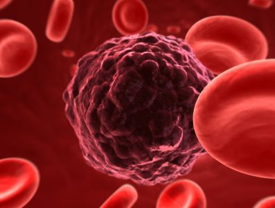
Oki K. Dzivenu, D.Phil., and Jill O’Donnell-Tormey, Ph.D.
Copyright © Cancer Research Institute 2003

TABLE OF CONTENTS

1...............Introduction
2...............An Enigma Called Cancer
9...............The Human Defense System
9...............The Concept of an Innate Defense Network
11.............The Adaptive Defense System
12.............Brian Pricked His Thumb and His Immune System Jumped Into Action
13.............The Humoral Immune Response
14.............The Cellular Immune Response
17.............The Origins of Immunotherapy
19.............Advances in Human Tumor Immunotherapy
19.............Nonspecific Immunotherapy 1: Bacille Calmette-Guerin (BCG) Therapy
20.............Nonspecific Immunotherapy 2: Cytokines
21.............Nonspecific Immunotherapy 3: Cell therapy
22.............Specific Immunotherapy I: Adoptive Transfer
25.............Specific Immunotherapy II: Vaccination
31.............The Advantages of Cancer Immunotherapy
34.............Techniques Behind the Advances in Tumor Immunotherapy
37.............Epilogue and Acknowledgements
38.............Bibliography
41.............Glossary
INTRODUCTION

The Cancer Research Institute was established in 1953 to foster the field of cancer
immunology, which is rooted in the notion that the body’s immune system can be
mobilized against cancer. From our inception, we have championed the development
of new and effective strategies based on the immune system to complement tradi-
tional methods of cancer treatment, such as surgery, radiation, and chemotherapy.
We are a non-profit intermediary organization that provides funding for individual and
collaborative research projects across the country and throughout the world. Our
funding strategy is aimed at providing support to investigators throughout various ca-
reer development stages encompassing a broad spectrum of research such as basic,
preclinical, and clinical sciences.

Today, we are more committed than ever to our long-term goal of fostering cancer im-
munology. We recognize that further advancement in the field depends on increased
public understanding of the enormous power of the immune system and its connec-
tion to cancer. To help build that critical understanding, we have prepared this guide,
which answers a number of commonly asked questions about cancer, the immune
system, and the latest trends in immunotherapy.

In the first chapter, the reader is introduced to the concept of cancer as the defining
term for a panoply of diseases underpinned by two common features. This chapter
ends with an introduction to the human immune system and how its normal function
and cancer prevention are inextricably linked. We move on to chapter 2 for a discus-
sion of the immune system thus setting the stage for the introduction of immunother-
apy in chapter 3. The next three chapters (4—6) constitute a tour de force into almost
every form of currently available immunotherapeutic regimens and how they are
faring in worldwide clinical trials. The seventh chapter is full of hope: reminding us of
how far we have come and how much further we must travel on this exhilarating but
arduous journey of battling cancer. The remainder of the book provides a brief look at
the techniques behind the progress we have made in this field of biomedical science.

We hope that you will find it enlightening.

                                                                                            1
1.0 AN ENIGMA CALLED CANCER
The word “cancer” is an umbrella term that refers to about 200 diseases that share
two common characteristics: first, an uncontrolled growth of cells and second, the
ability to invade and damage normal tissues either locally or at distant sites in the
body. Since a cell is our body’s basic unit of life, this disease could not have chosen
a more effective route to wreak havoc on the entire body. Some human cancers arise
in the epithelium (the layers of cells covering the surface of the body and the lining
of internal organs and various glands); these cancers are called carcinomas. Sar-
comas are cancers of the supporting tissues of the body, such as bone, muscle and
blood vessels. Cancers of the blood and the lymph glands are called leukemias and
lymphomas, respectively. Gliomas are cancers of the nerve tissue. Melanomas arise
from darkly pigmented cells, usually in the skin. There is an imperative to understand
and control cancer because to date, apart from heart disease, more people die from
cancer than any other disease.
1.1 The genesis of cancer
The number of cells in an average human being is about a hundred trillion (1014) or
beyond. Some of them, for example, brain and nerve cells, are not actively dividing
while others like the cells of the skin, gut, bone marrow, and sex organs continually
undergo rapid cell divisions to replace aging and dead cells. It has been estimated
that about one million cells commit suicide every second in the adult human being.
On an average day, the human body produces and concomitantly eradicates about
60 x 109 cells; on an annual basis, this enormous amount of cells is equivalent to an
entire body weight. In order to replace a dead cell, an existing cell must divide and for
each successful cell division, the entire genetic material of the mother cell in the form
of DNA must be faithfully copied by enzymes and handed over to the new daughter
cells. If we compare this to the task of photocopying the entire content of the ency-
clopedia Britannica again and again for over a trillion times a day, one can appreciate
the number of errors (due to exhaustion) that would begin to appear in each copied
volume after only a few hours.
Fortunately, our cells are equipped with enzymes that not only copy but also proof-
2read, edit, and correct errors in the newly manufactured DNA that is destined for
the daughter cells. As with most things in life, this system is not perfect and errors
do get passed these proofreading, editing, and correcting enzymes. Cancer occurs
when the DNA sequence within a gene is altered in such a way that the gene can no
longer instruct the cell in which it resides to produce the normal version of the protein
it encodes. Scientists call such an occurrence a mutation within the gene. Such
alterations can occur more frequently when a gene is exposed to ionizing radiation
or certain drugs or chemicals or when some, as yet, unexplained internal switch is
flipped on or off. These factors can cause the DNA sequence within the gene to break
and recombine incorrectly or to mutate.
The Oncogene: A key factor in development of cancer
3Once one of these changes has taken place, certain genes (proto-oncogenes) may
be transformed into oncogenes (cancer-causing genes), while other genes (cancer-
suppressing ones called anti-oncogenes or tumor-suppressors) may be rendered
useless by inactivation. A cell containing mutated genes that result in the loss of
its growth control is referred to as a transformed cell. Those who have peered into
such a cell report that it contains a veritable gallery of cellular horrors like inactivated
genes, extra or missing chromosomes, and a host of other genetic abnormalities that
cause cancer.
Say, for example the alteration of the DNA is like the ignition of an unoccupied car
being turned on. The transformation of a normal gene into an oncogene is like moving
the gear from P (Park) to D (Drive) while the handbrake is still on. Inactivation of anti-
oncogenes is akin to the release of the handbrake and the car is now free to wander
onto a highway and cause mayhem. That is the ultimate fate of a transformed cell. It
is important to note that it normally takes multiple mutations before cancer occurs.
Mutations of our DNA are occurring constantly due to environmental insult. However,
a single uncorrected mutation event will not guarantee cancer, rather multiple muta-
tions are required. This is why, for instance, cancer occurs only after years of expo-
sure to a carcinogen (smoking, sun, asbestos, etc). If a gene has become an onco-
gene, the cell in which it is located may begin to produce unusually large amounts of
one of its normal proteins or to manufacture an altered form of that protein. If an anti-
oncogene has been rendered inactive, the cell containing it can no longer produce
a normal protein whose function is to suppress cancer. On some rare occasions, a
normal cell becomes cancerous when a particular virus enters the cell and introduces
an oncogene into the genome of the host cell. Once any of these deviations in normal
protein production and/or function has occurred, the size, shape, surface character-
istics, or morphology and behavior of the cell becomes altered. Thus, it becomes a
cancer cell that is distinguishable from a normal cell.
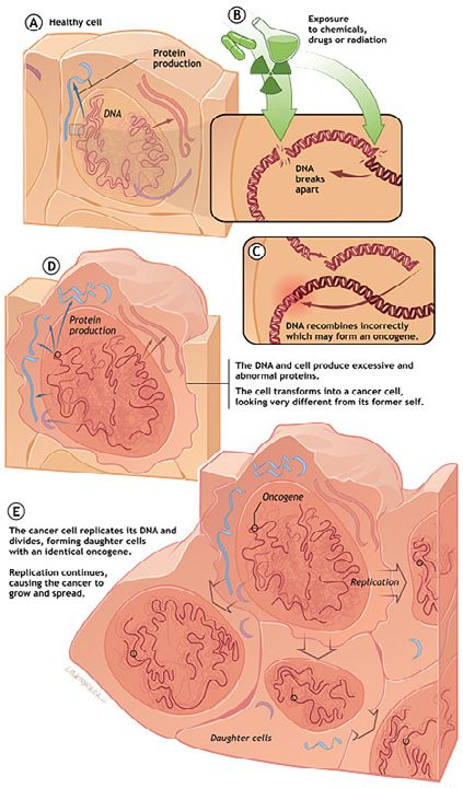
1.2 The progress of cancer within the body
Every cancer starts with a single cell that has been unleashed from the growth
restraints placed on all normal cells. Because the changes that took place within the
cancer cell were directed by the cell’s DNA (the molecular basis of heredity), they are
passed on to each of the daughter cells arising from the original cancer cell. As these
cells continue to divide, collections of abnormal cells accumulate. Except in the case
4of leukemia, these cells form a mass or tumor.
The cells of the tumor then push outward from their boundaries, infiltrating surround-
ing normal tissues. Small clumps of cells may then dislodge from the tumor (primary
site) and migrate to distant (secondary) sites, often by piggybacking on the circulatory
system of the blood or lymph. After traveling to a new organ, the cancer cells burrow
out of the blood or lymph vessels and invade the surrounding tissues, where they
continue to multiply and form secondary tumors. This process of spreading to a dis-
tant site is called metastasis. Eventually, either local invasion or metastasis disrupts
the body’s normal function and often leads to death.
In the 1980s, the American government declared war on cancer. That war is still
raging and the latest report from the frontline is a mixed bag of good and bad news.
According to the latest figures from the American Cancer Society, more than half a
million Americans will die at the hands of this scourge in this year alone. This makes
cancer second only to heart disease as the leading cause of death among Ameri-
cans. The cost of cancer to the American economy was estimated at almost $200
billion for the year 2000 alone.
According to the latest projections, one out of every four Americans alive today will
eventually die of cancer in the absence of major breakthroughs in prevention and
control. In terms of mortality rate, lung cancer is by far the most frequent killer among
all Americans, followed distantly by cancer of the colon and rectum, the breast, and
the prostate. Even when men and women are considered separately, lung cancer is
still the biggest killer, although the mortality rate is much higher for men than for wom-
en. After lung cancer, prostate and colorectal cancers kill men the most often. Among
women, breast cancer is by far the most prevalent form, followed by colorectal and
uterine cancers. The next most common killer for men is cancer of the pancreas and
for women is ovarian cancer.
A recent and disturbing development is the increasing evidence that suggests that
obesity increases the likelihood of colon and prostate cancers in men and breast,
ovarian, and gall-bladder cancers in women. With an estimated 97 million American
adults classified as obese, this new link does not bode well for the battle against
cancer.
1.3 Cancer incidence and mortality rates in the United States
The good news is that the rate of new cancer cases and deaths for all cancers com-
5bined, as well as for most of the top 10 cancers in the United States has been declin-
ing. The report shows that the incidence rate—the number of new cancer cases per
100,000 persons per year—for all cancers combined declined by an average of 0.8
percent per year between 1990 and 1997. The greatest decline in cancer incidence
rates has been among men, who overall have higher rates of cancer than women.
6The reduction in deaths from cancer has been attributed to better screening and
advances in treatment. These uplifting findings reflect the considerable progress that
Americans have made against cancer. This decline in cancer deaths is all the more
remarkable if we consider the fact that the size of the American population has been
increasing while deaths from cancer have been declining.
1.4 The connection between cancer and the immune system
In 1909, a scientist by the name of Paul Ehrlich proposed that the incidence of cancer
would be much greater were it not for the vigilance of our immune defense system
in identifying and eliminating nascent tumor cells. This suggestion gave birth to the
generally accepted concept that the immune system plays a vital role in the iden-
tification and elimination of transformed cells. About 50 years later, two scientists,
Lewis Thomas and Frank MacFarlane Burnet, took Paul Ehrlich’s original idea a step
further and proposed that a special type of immune cell called a T cell was the pivotal
sentinel in the immune system’s response against cancer. This elaboration led to
the coinage of the term “immune surveillance or immunosurveillance” to describe the
concept whereby the immune system is on perpetual alert against transformed cells.
As dictated by the scientific method, theories must in the course of time either
withstand rigorous experimental testing, crumble and be discarded or be improved
upon. This basic requirement brought the theory of immunosurveillance under severe
attack and great controversy when scientists like Osías Stutman showed in the 1970s
that mice supposedly lacking an intact immune system (so-called nude mice) did not
become more susceptible to tumor growth as predicted by the theory.
Thus, the theory of immunosurveillance remained controversial until an important
scientific article entitled “IFN-gamma and lymphocytes prevent primary tumor devel-
opment and shape tumor immunogenicity” was published in the journal Nature on
April 26, 2001. This breakthrough article was authored by Robert D. Schreiber, Ph.D.,
and his colleagues at Washington University School of Medicine, St. Louis, MO, in
collaboration with Lloyd J. Old, M.D., of the Ludwig Institute for Cancer Research and
Memorial Sloan-Kettering Cancer Center, New York, NY. The experimental evidence
presented in their paper unambiguously showed that the immune system can and
often does prevent tumors from developing, and thus plays a strong protective role
against cancer. These researchers also uncovered important new insights regarding
the immune system and tumor development that they dubbed “immunoediting.”
Utilizing genetically engineered mice that lacked a functional immune system, the
7authors showed that lymphocytes and the immune stimulator, IFN-gamma, cooper-
ate to inhibit the development of both spontaneous and carcinogen-induced tumors.
Unfortunately, this natural body defense is imperfect, and some tumor cells escape
identification and go on to cause cancer. These renegade tumors are less immuno-
genic, having undergone a process of immunoselection triggered by the actions of
the immune system. Conceptually, in much the same way as bacteria can become
resistant to antibiotic treatment and lead to more potent and harmful strains, so too
can the body’s own tumor defense system lead to tumors that escape elimination.
Importantly, these researchers went on to demonstrate that there are ways to over-
come the “camouflage” of such renegade tumors by increasing their antigen expres-
sion and making them visible to the immune system. This suggests that even tumors
that have escaped recognition can be turned into targets for an immune response.
Further experimentation is underway to test how these results can be used to develop
cancer immunotherapies. The Cancer Research Institute (CRI) sponsored this
research with a grant to Dr. Schreiber’s lab and provided further funding through its
pre- and post-doctoral training programs to two graduate students, one of whom was
the first author of the paper, and a postdoctoral fellow.
Despite the tremendous scientific progress (such as that described above) that has
been made over the years, a complete and precise understanding of the immune
system’s response to cancer remains elusive. The continued exploration of these
questions is the province of cancer immunology; the scientific discipline to which CRI
is dedicated to supporting and nourishing.
82.0 THE HUMAN IMMUNE DEFENSE SYSTEM
The environment in which we live contains a wide range of organisms called patho-
gens that view the human body as a rather juicy target to invade and live off till
death do us apart. The job of the mammalian immune system is to defend the body
against these pathogens, which include bacteria, viruses, fungi and parasitic worms.
This task is so complex that mammals have evolved a very sophisticated network of
defense units that recognize and attack such a diverse array of potential enemies.
The immune system is characterized by three universal features, which are namely
specificity, diversity, and memory. We say the immune system is specific because it
only reacts against certain specific molecular targets called antigens. It is very impor-
tant that the immune system is able to select what it reacts to because this prevents
it from attacking components of our own body: a phenomenon called autoimmunity.
The immune system is described as diverse because it has the remarkable ability to
react specifically to any molecule in the universe. At the end of a successful immune
reaction, the immune system assigns a unique group of immune cells, called memory
cells, the task of remembering the particular enemy encountered (virus, bacterium,
tumor cell, etc). In this way, there will be immune combat units ready to attack and kill
off the fresh invaders very swiftly if they are encountered in the future.
The defense system of the human body consists of surface barriers such as skin, in-
ternal barriers such as mucus, and special groups of cells, chemicals, and hormones
that act in concert to keep the body free of pathogenic invaders. In broad terms, the
immune system is divided into two branches called the innate and adaptive defense
systems. In evolutionary terms, the innate branch predates the adaptive branch by
about 500 million years.
In the next three sections, the reader will be introduced to both the innate and adap-
tive immune systems. These sections will also examine how the two systems com-
municate effectively to bring about a coordinated defense of their host organisms.
2.1 The concept of an innate defense network
The innate immune system evolved to protect host organisms about 900 million
years ago. It consists of mechanical, chemical, microbiological, and cellular defense
9networks. The function of the innate immune defense system is akin to “turning back
the barbarians at the gates.” We can view the skin as a huge missile defense shield
that prevents the entry of pathogens and foreign substances into the body. In addi-
tion, the skin produces acidic substances that make it difficult for bacteria to grow
on it. Nevertheless, there is a class of harmless bacteria and fungi that thrive on our
skin. These also constitute a defensive mechanism, as they tend to compete with and
crowd out pathogenic organisms. Some membranes produce mucus that block the
entry of pathogens into the body. Another function of mucus is to trap pathogens that
choose the digestive and respiratory routes to invade the body. The hairs in our nose
for example, filter out bacteria entering the nasal passage.
Further down the nasal cavity, en route to the respiratory tract are structures called
cilia whose job it is to move trapped pathogens away from the respiratory tract. If the
cilia fail to do their job properly, coughing and sneezing is induced to expel pathogens
from the upper portion of the respiratory tract. Any pathogen that makes it passed
all these road blocks into the stomach is assailed by a deadly potion called gastric
juice: this is a mixture of concentrated acid and enzymes that chew up the invading
pathogens into harmless bits of protein.
Apart from their normal function of lubrication and cleansing, our tears and saliva
contain an enzyme called lysozyme that cuts and destroys the bacterial cell wall.
Finally, the body produces mild acids into the vagina in order to prevent the growth
of pathogens in the female reproductive tract. All these security barriers are part of
the innate defense system because they are general protection mechanisms that are
designed to keep pathogens out of the body. But as it is often the case in life, security
breaches do occur and some pathogens succeed in lodging themselves within the
cells and tissues of the body. What happens then?
In the case of the innate defense network, cellular combat units called myeloid cells
are deployed to fend off the invading pathogens. The cells that mediate the innate
immune response include macrophages, dendritic cells, neutrophils, eosinophils,
mast cells, natural killer cells, some B lymphocytes (like B1 B cells and marginal zone
B cells) and some T lymphocytes (like TCR-gamma/delta T cells and natural killer
T cells). How does recognition occur by the innate immune system? In other words,
how do all these cells of the innate defense system tell apart a normal cell such as a
red blood cell from an invading bacterial cell such as streptococcus?
The answer to this important question lies in two key evolutionary developments
called Pathogen Associated Molecular Patterns (PAMPs) found in all microorganisms
10and Pattern Recognition Receptors (PRRs) found in all the cells of the innate defense
system. A PAMP is a molecular pattern that is unique to microorganisms. The PRR
is like a molecular velcro patch that is capable of recognizing and latching onto each
unique PAMP. So, for each PAMP in a pathogen, there exists a corresponding PRR
in one or more of the cells within the innate defense network. Examples of PAMPs
are LPS (endotoxin), peptidoglycan (cell walls), lipoproteins (bacterial capsules),
hypomethylated DNA (such as CpG found in bacteria and other parasites), double-
stranded DNA as found in viruses, and a molecule called flagellin that is found in
bacterial flagella.
It is estimated that several hundred PRRs exist in the mammalian innate defense
system and that they are so vital to the immune defense system that their genes are
encoded in germline cells to ensure limited variability in their molecular structures.
PRRs are classified as membrane proteins because they are associated with the cell
membrane. Examples include mannose binding lectin, pulmonary surfactant protein,
C-reactive protein, toll-like receptors (TLRs), C-Type lectin, NOD and MX proteins.
In summary, the immune system recognizes and rapidly responds to microbial patho-
gens via pattern recognition. A complex example of pattern recognition can be found
in our extraordinary ability as human beings to recognize patterns in the environment
using cognitive processes to distinguish visual images such as models of cars or
species of birds. In the innate immune system, cell surface receptors (like PRRs) that
recognize distinct biochemical patterns (like PAMPs) displayed by microbial invaders
constitute a receptor-ligand interaction that forms the bedrock of the innate immune
system.
2.2 The adaptive defense system
In her infinite wisdom, Mother Nature evolved far more sophisticated and specific
responses to pathogenic invasions eons before humans developed complex military
systems. The specific immune response involves a repertoire of specialized cells,
chemicals, and hormones that work in a highly coordinated fashion to rid the body of
these invaders before they get a chance to multiply and cause harm to the body.
The mammalian immune system possesses the ability to recognize every mol-
ecule—known and unknown—in the universe! This awe-inspiring feat is even more
remarkable when we see that in a healthy human being, the extraordinary recognition
capabilities of the immune system does not normally lead to an attack on the body
itself. That is why in normal circumstances, my immune cells will tend to guard my
11kidney against pathogenic invasions but they will ferociously attack a kidney from
you (a donor) that was being transplanted into me unless, of course, you were my
identical twin. The only way around this transplantation hurdle is the administration of
strong drugs to suppress my immune system. Immunologists refer to the second part
of our defense system as the adaptive immune system because it has the potential to
modify itself and adapt to any weapon that the enemy (pathogens) throws at it.
The vast majority of immune cells are created in the bone marrow as stem cells. As
they mature into specialized cells, they exit from the bone marrow and circulate in
the blood. Some of the immune cells are deployed to most of the tissues in the body.
In order to maximize the efficiency of this defense unit, the immune cells produce
chemicals and hormones that enable them to communicate with each other, for
example, to alert another group of immune cells that there is an ongoing invasion in
some distant part of the body. The innate and adaptive arms of the immune system
work in synergy to defend the body against pathogenic onslaughts. How do they do
that?
2.3 Brian pricked his thumb and his immune system jumped into action
Perhaps, we can answer the above question by describing what happens when
five-year old Brian pricks his thumb during an apple-picking trip to an orchard. The
pricking of Brian’s thumb constitutes a breach of the physical barrier that is part
of the innate or natural immune system. In addition, the thorn that pricked Brain’s
thumb would have transferred several hundred or so bacterial cells into the wound
on Brian’s thumb. The initial response of Brian’s immune system will most certainly
involve a type of white blood cell called a macrophage. These cells usually roam the
body like sentinels looking for foreign invaders.
The macrophage is able to recognize the invaders as foreign and harmful because
the invaders come bristling with PAMPs on their surfaces. Each PAMP can be
recognized by the appropriate PRR on the macrophage. In simple terms, it is a bit like
recognizing enemy troops by the uniform they wear. In addition, the invading bacterial
cells actually produce chemical messengers that can be detected by macrophages:
just like invading foreign troops communicating in a coded language on a particular
radio frequency that can be picked up and decoded by United States soldiers as for-
eign. The net result is an aggressive onslaught on the colony of bacterial cells by an
army of macrophages that actually eat up the invading cells. A few minutes after sus-
taining that wound, a casual observer would have noticed that the wound on Brian’s
thumb has become red and swollen: a sure indication that an immune response is
12in progress! In addition to killing off some of the invading bacteria, the macrophages
alert other cells of the immune system that there is an invasion in progress. After
the macrophage has swallowed the bacteria, it chews it into tiny bits of protein and
then deposits the pieces into the groove of a protein called major histocompatibility
complex (MHC). Imagine the MHC molecule as the bun of a hotdog; the antigen to
be displayed is placed in the groove where the sausage sits. There are two types of
MHC proteins subdivided into class I and class II. Macrophages call on MHC class II
molecules to shuttle the fragments of antigens to the surface of the macrophage so
that they can be presented to the immune system. In this role, immunologists refer to
the macrophage as a professional antigen-presenting cell (APC).
2.4 The humoral immune response
B-cells (so-called because they mature in the bone marrow) are white blood cells
that work chiefly by producing soluble substances known as antibodies. Each B cell
is programmed to make one specific antibody. When a B cell encounters its specific
or eliciting antigen (along with various accessory cells), it differentiates into a plasma
cell. The latter is essentially a factory for producing that one specific antibody. Anti-
bodies play a crucial role in a cascade of events called the humoral immune response
that ultimately leads to the destruction of some of the invading bacteria. Like all
immune responses, the humoral immune response can be subdivided into activation
and effector phases.
The activation phase begins when invading bacteria are phagocitized by an antigen-
presenting cell (APC), such as a dendritic cell or macrophage. The bacterium is di-
gested and its antigens processed and presented in combination with the MHC Class
II complex on the surface of the APC. The antigen-MHC complex is recognized by a
type of immune cell called a CD4+ or helper T cell (Th). The helper T cell begins the
attack by docking its antigen receptor to the displayed antigen. The docking process
requires the presence of co-stimulatory molecules like B7 and CD28.
After successful docking, the helper T cell releases a class of chemical messenger
called cytokines. This achieves the following: it causes the helper T cells to multiply,
and stimulates both the APC and the helper T cell to exchange chemical messages
between themselves and with other cells of the immune system. The helper T cell
also releases a cytokine called interleukin-2 (IL-2): this cytokine has a panoply of
immune functions, one of which is the proliferation of lymphocytes following activa-
tion by a specific antigen. The APC releases cytokines called interleukin-1 (IL-1) and
tumor necrosis factor (TNF). The latter steps up the production of IL-1 and performs
13many of the same functions as IL-1, including the induction of fever in Brian so that
his body can assist in fighting off the bacterial infection more effectively. The prolifer-
ating helper T cells release substances that signal another type of lymphocyte, the B
cell (that also specifically recognizes the antigen), to begin multiplying and differen-
tiating into antibody-producing cells. This initiates the effector phase of the humoral
immune response.
The antibodies released by the B cells bind in a lock-and-key fashion to antigens on
the surfaces of bacterial invaders that survived the initial attack by macrophages and
bacterial products. The binding of the antibody to the bacterial antigens achieves
two things: first, it makes it easier for “killer” cells to attack and destroy the invading
bacteria by both phagocytosis and the release of other factors that can directly lyse
the bacteria. Second, it activates another immune military unit called complement—
a group of proteins that act like the special forces of the immune defense system
because their duty is to begin the lethal process of punching holes in the cell walls of
the residual invading bacterial army.
The humoral immune response
2.5 The cellular immune response
As the fruit picking was going on, Brian heard someone shout, “Bless you!” after a
fellow fruit-picker let out a loud sneeze. Unbeknownst to poor little Brian, some of
14the air he was breathing in the orchard was now laden with particles of the influenza
virus. While the humoral immune response is under way, some of the influenza viral
particles will have been consumed by phagocytes and neutrophils while others would
have began infecting other cells such as Brian’s epithelial cells. So, how does the
immune system deal with these infected cells? By a battle plan called the cellular
immune response.
The celluar immune response
Like the humoral immune response, this is also divided into activation and effector
phases. The activation phase begins when an antigen-presenting cell (APC) of the
host organism encounters and attacks an invading virus. Meanwhile, other viruses
look for nearby epithelial cells to infect. A lysosome containing digestive enzymes
combines with the phagosome to process the antigens. The processed antigens com-
bine with MHC class II proteins and are presented on the surface of the APC. The
virus also infects Brian’s epithelial cells. Within the infected epithelial cells the virus is
processed, attached to an MHC class I protein and is presented on the cell surface.
A helper T cell (CD4+) recognizes the displayed antigen on the APC and binds to the
MHC class II protein-antigen complex. The activated helper T cell releases chemical
messengers such as the cytokine IL-2 and gamma interferon (IFN-g).
The effector phase begins when activated cytotoxic T cells (CD8+) which were stimu-
lated to proliferate by the cytokine IL-2, recognize the MHC class I protein-antigen
15complex on the infected epithelial cells. Cytokines also attract other killer T cells to
the site of infection. The activated cytotoxic T cell binds to the MHC class I protein-
antigen complex on the surface of the infected epithelial cell. The binding causes
the cytotoxic T cell to release a potent chemical called perforin. Perforin perforates
the cell membrane of the infected cells causing the cells to lyse (burst) and die. As
the viral infection is brought under control, regulatory T cells turn off the activated
cytotoxic T cells. Memory T cells remain behind to respond quickly if the same virus
attacks again.
Finally, as the infection in Brian’s thumb is brought under control, yet another type of
T cell, the regulatory T cell, instructs the activated combat units consisting of B cells,
helper T cells, and killer T cells to switch from battle mode to stand-by mode. Most of
these immune cells will die, but a few will live to fight another day. These cells, called
memory cells, will be able to respond more quickly the next time Brian is unfortunate
enough to be invaded by the same strain of bacteria. The above account highlights
the overwhelming reliance that our body places on the T cell to fight off microbial in-
fections. But accidents do happen (as it is in life in general) within the immune system
and sometimes these cells mistake part of our body (self) for a microbe (non-self) and
the resulting “friendly fire” can lead to autoimmune diseases such as multiple scle-
rosis and juvenile diabetes. To avoid this kind of “collateral damage” nascent T cells
are subjected to a strict training program in the thymus. As part of their education, the
developing T cells are exposed to as many self-proteins as possible and any T cell
that displays any reactivity is eliminated. This rigorous training regime ensures that
the remaining T cells will react only to non-self molecules.
Although the foregoing description of the immune response applies mainly to viruses
and bacteria, it is important to note that the immune system reacts in a similar man-
ner when it encounters cancer cells, which it also recognizes as foreign or “non-self”
and therefore, must destroy. Scientists have observed in the laboratory that the cells
and other components of the immune system are capable of destroying malignant
tumor cells. They have found that certain antibodies that recognize tumor cells help
the macrophages and the natural killer cells to accomplish their mission. Over the
years, further study of the immune system has demonstrated that the body defends
itself against cancer in much the same way that it seeks to eliminate other intruders
such as bacteria and viruses. Further study of the immune system is expected to
reveal ways to bolster it, allowing the body to become a more active partner in the
fight against cancer.
163.0 THE ORIGINS OF IMMUNOTHERAPY
As it applies to cancer, immunotherapy might be considered a revolutionary form of
medicine but its roots actually go back as far as 1778, when Edward Jenner, an Eng-
lish physician, administered the first vaccine, which was targeted against smallpox.
Jenner observed that milkmaids who contracted cowpox, a relatively mild disease,
seemed protected from the deadly smallpox infection. To test this hypothesis, he
injected some material from a pustule on the body of a milkmaid infected with cowpox
into the arm of a small boy. After the boy had recovered from the cowpox infection,
Jenner inoculated him with smallpox. As Jenner had expected, the boy never devel-
oped the disease. Jenner named his technique “vaccination,” a term derived from the
Latin word “vacca” for cow. Even without a scientific understanding of why his method
worked, Edward Jenner had discovered an effective way to prevent people from
developing a serious disease.
It was not until the late 19th century that medical science disclosed the reason: Jen-
ner had created a condition of acquired immunity in the boy. When the child contract-
ed the less serious disease of cowpox, his immune system had mounted an attack
against the invading virus. Later, when the boy was inoculated with the smallpox virus
he did not contract the disease because the memory lymphocyte (T and B cells) of
his immune system “remembered” the cowpox infection and were able to stimulate
the immediate production of the specific antibodies needed to kill the related but more
deadly smallpox virus. This pioneering immunological work eventually gave rise to a
number of other vaccines against such diseases as rabies, diphtheria, yellow fever,
polio, mumps, hepatitis B, measles, rubella, influenza, whooping cough, and teta-
nus. These days, this type of immunotherapy is in widespread use to protect against
microbial infection.
The connection between cancer and the immune system was first uncovered nearly
100 years ago; long before an in-depth knowledge of the intricate workings of the
immune system existed. In the early 1890s, Dr. William B. Coley, a New York physi-
cian, became intrigued by the dramatic disappearance of malignant tumors that
he observed in cancer patients who had contracted acute streptococcal infections.
Suspecting that the onset of bacterial infection was in some way responsible for the
regression of the tumor, he decided to try an experiment in which he injected live
streptococci into a patient with inoperable cancer to see whether the patient’s tumor
17would regress. After he had tried administering three different bacterial cultures to the
patient, he finally injected a fourth that resulted in the complete disappearance of the
tumor.
Dr. Coley continued to pursue his approach and ultimately developed a mixture of
killed bacteria that became known as Coley’s mixed bacterial toxin. He and other phy-
sicians treated over 1,000 cancer patients with this substance, with varied success.
His results were unpredictable however, and neither he nor the medical community at
large could explain precisely why his mixture worked in some patients. This was due
to the fact that the science of immunology was in its rudimentary stages at that time.
Thus, his results were disregarded and virtually forgotten for years.
Scientific interest in Coley’s work has been accumulating since his daughter, Helen
Coley Nauts, started compiling and disseminating information on his remarkable
observations. Gradually, scientists began to understand why Dr. Coley’s preparation
worked—the bacterial products of which it was composed had acted as immune po-
tentiators. In other words, they had stimulated certain immune cells to kill the cancer
cells directly or through cancer-killing factors. With the founding of the Cancer Re-
search Institute by Mrs. Nauts in 1953, resources were provided to pursue research
into the link between cancer and the immune system. Today, cancer immunology
is a rapidly advancing field and Dr. Coley has come to be regarded as the “father of
cancer immunotherapy.”
184.0 ADVANCES IN HUMAN TUMOR IMMUNOTHERAPY
Tumor immunotherapy is an anticancer approach in which the patient’s immune
system is either prodded or cajoled to fight tumors. Over time, our understanding of
the immune system and tumor immunology has increased and this has enabled sci-
entists to develop the ability to apply specific immunotherapies designed to enhance
the immune response of a particular patient against unique targets. The prevailing
techniques of tumor immunotherapy can be divided into two broad groups called non-
specific and antigen-specific therapies. The latter can be attained by either adoptive
transfer or vaccination.
Adoptive transfer means the physician directly transfers into the patient, the actual
components of the immune system that are already capable of producing a specific
immune response. Vaccination on the other hand involves the administration of a
particular antigen to induce a specific immune response. Nonspecific immunotherapy
refers to therapies that can stimulate the immune system by using a substance that
activates or enhances immune cell function regardless of their antigen specificity. In
the early days of immunotherapy, many non-specific immunostimulants were tested
as antitumor reagents in their own right, but today their use in this way has declined.
The majority of these substances are now recognized for the supporting roles they
play such as enhancing cellular communication between immune cells and therefore
are being tested for use in combination with antigen-specific immune stimulation. In
the next section, we will consider three examples of nonspecific immunotherapeutic
reagents.
4.1 Nonspecific immunotherapy 1: Bacille Calmette-Guerin (BCG) therapy
In 1975, the Cancer Research Institute presented its first Award for Distinguished
Research in Immunology to a group of 16 scientists it called the “Founders of Cancer
Immunology.” The award, which was renamed the William B. Coley Award for Distin-
guished Research in Basic and Tumor Immunology in 1993, is now presented annu-
ally to scientists who have made outstanding achievements in the fields of basic and
cancer immunology. Their work has deepened our understanding of the response
of the immune system to disease, including cancer, and holds forth the promise of
further progress in the development of novel and effective immunotherapies. In 1993,
one of the recipients of this award was Dr. Alvaro Morales.
19In the early 1970s, it was found that the administration of weakened forms of a myco-
bacterial strain called Bacille Calmette-Guerin (BCG) had anticancer effects. During
this period, the CRI awarded one of its grants to Dr. Alvaro Morales for his BCG
research. His work led to the publication of a seminal paper in the Journal of Urology
on the use of intravesical BCG for the prophylaxis of transitional cell carcinoma of the
bladder.
The work of Dr. Morales paved the way for the use of this agent as standard therapy
worldwide. Prior to its use as an anticancer agent, the live and potentially infectious
form of BCG was used as an effective vaccine against tuberculosis. After extensive
clinical testing, the efficacy of BCG against a few cancers such as metastatic mela-
noma and certain types of early bladder cancer were established: so much so, that
today BCG is the treatment of choice for early forms of bladder cancer.
Like other nonspecific immunotherapeutic agents, scientists do not know the ex-
act mechanism by which BCG generates anticancer immune responses in certain
patients but they speculate that BCG probably activates both macrophages and
lymphocytes. With modern techniques, scientists have been able to separate BCG
into separate components, which on their own have been shown to have anticancer
effects. Despite this, the most useful application of BCG (either used whole or frac-
tionated) is as an adjuvant or supplement to other forms of therapy. Now, let us turn
our attention to the cytokines.
4.2 Nonspecific immunotherapy 2: Cytokines
Laboratory studies have shown that the use of cytokines in immunotherapy can lead
to the destruction of tumors by one of two general mechanisms: first, a direct antitu-
mor effect or second, an indirect enhancement of the antitumor immune response. In
the first, cytokines, such as tumor necrosis factor (TNF) alpha, interferon (IFN) alpha,
IFN-beta, interleukin-4 (IL-4), and IL-6, interact directly with tumor cells, inducing the
latter to either commit suicide or stop further growth. Although these cytokines are
effective when given as single agents, the administration of a cytokine cocktail can
be even more potent as an anticancer agent due to the synergistic effect accruing
from all the different cytokines. A note of caution: some cytokines can have danger-
ous side effects. For instance, TNF-alpha and IL-6 are able to suppress the growth of
some tumors while actually promoting the growth of others so the immunotherapeutic
use of cytokines demands great care.
20The cytokines that fight tumors via the indirect mechanism do so by stimulating
immune cells to fight tumors through a variety of different pathways. For example, a
cytokine such as IL-2 promotes T-cell and natural killer (NK) cell growth. Other cyto-
kines such as the interferons and granulocyte–macrophage colony-stimulating factor
(GM-CSF) can act on professional antigen-presenting cells (APCs) and increase the
production of important immune molecules such as MHC molecules and immune
co-stimulators such as B7 that have important roles in facilitating the activation of
lymphocytes. Although more clinical trials are necessary to determine the right dos-
age and to predict immunological responses in the more complex environment within
humans, currently, a number of cytokines have proven effective in cancer therapy.
For example, in patients with metastatic melanoma or renal cell carcinoma, the ad-
ministration of intravenous IL-2 can induce objective tumor regression in 17 percent
and 20 percent of cases, respectively. Also, clinical studies have shown that combin-
ing IL-2 with other cytokines, such as IFN-alpha, can lead to a synergistic response.
IL-2 has been approved by the US Food and Drug Administration (FDA) for the treat-
ment of both of these cancers. IFN-alpha is also FDA approved for the treatment of
malignant melanoma, chronic myelogenous leukemia (CML), hairy cell leukemia, and
Kaposi’s sarcoma. The FDA has approved both GM-CSF and G-CSF because they
have been shown to promote the revival of the immune system (following chemo/
radiation therapies) and improve patient survival. Recently, advances in biotechnol-
ogy have allowed scientists to clone the genes of cytokines leading to the large-scale
production and administration of cytokines to cancer patients. Due to this advance
in biotechnology, a large number of cytokines, including IL-1, IL-4, IL-6, IL-7, IL-11,
IL-12, macrophage inflammatory protein (MIP)-1-alpha, IFN-beta, and IFN-gamma,
are currently being tested in humans for anticancer therapy. For the next topic on
nonspecific tumor immunotherapy, let us turn our attention to cell therapy.
4.3 Nonspecific immunotherapy 3: Cell therapy
The transfer of live, whole cells into patients can also be used to achieve non-specific
immunotherapy against cancer. For example, in patients with metastatic melanoma,
human peripheral blood mononuclear cells (PBMCs) can be isolated and fed with
IL-2 to generate a class of cells called lymphokine-activated killer (LAK) cells. When a
combination of LAK cells and IL-2 are given to patients with either advanced meta-
static melanoma or renal cell carcinoma, complete tumor regression can be achieved
in about 10 percent of cases. Many of the above therapies now provide the basis for
specific immunotherapeutical approaches that are currently undergoing clinical trials.
215.0 SPECIFIC IMMUNOTHERAPY I: ADOPTIVE TRANSFER
As mentioned earlier, the term adoptive transfer applies to all the therapies that con-
sist of the transfer of components of the immune system that are already capable of
mounting a specific immune response. This takes us straight into the effector phase
of the immune response. Examples of adoptive transfer include both the transfer of
antibodies and also, specific types of cells that are capable of mediating antigen-
specific tumor regression such as LAK and T cells. Now, let us take a closer look at
antibodies therapy and how they can act as anticancer agents.
5.1 Antibody therapy
Monoclonal antibodies (MAbs) constitute highly pure populations of immune system
proteins that attack specific molecular targets. Their role in anticancer therapy can be
likened to that of heat seeking or guided missiles. In this sense, monoclonal antibod-
ies tipped with poisons or radioactive isotopes can home in on tumor cells and deliver
their deadly payloads; thus, selectively wiping out cancer cells. By the late 1970s, sci-
entists had shown that monoclonal antibodies could be targeted to tumor cells. Since
then, numerous animal and human studies have shown that antibody administration
can mediate tumor regression in some patients.
Although scientists are not quite sure about the exact manner by which antibody
therapy works, they think the antitumor effect may be achieved via two different
mechanisms: first, the activation of the complement system and second, by the use
of antibodies to mark these tumor cells for destruction. These two mechanisms need
not be mutually exclusive. It has been speculated that the binding of antibodies to
the target tumor cells may compel the latter to either stop growing (antiproliferative
effect) or commit suicide (apoptosis). The interest in antibody therapy has been such
that by the 1970s, a revolutionary technique called hybridoma technology had been
developed to mass produce antibodies (further discussed in chapter 8, section 8.2).
Since this breakthrough, there has been considerable progress in our ability to exploit
antibodies therapeutically against cancer.
To date, two antibodies have been approved by the FDA for use in cancer treatment.
Rituxan is specific for an antigen called CD20, which can be found on the surface of
both normal and malignant B-lymphocytes. In a phase III clinical study, 50 percent
of patients with non-Hodgkin’s lymphoma (NHL) responded to Rituxan. Herceptin is
22the other FDA-approved antibody and it is specific for the human epidermal growth
factor receptor 2 (HER2) protein. HER2 is over-expressed in 25-30 percent of primary
breast cancers and it has been shown to be effective against this disease in phase III
clinical studies.
Despite these promising advances, antibody therapy has certain disadvantages that
have limited its use as a tool against cancer. The main limitation is that since bulky
tumors tend to be inaccessible to antibodies, the use of this technique has been lim-
ited to tumors that are relatively small in size. On the plus side, antibodies are almost
never toxic so this bodes well for their continuous use in the fight against cancer.
5.2 Adoptive transfer of T cells
After decades of experiments conducted on mice and other animals, scientists have
shown that they can isolate antigen-specific T cells from a cancer patient, expand
them to large numbers in a test-tube, and re-infuse them back into the patient to kill
off the remaining tumor cells. When this technique was used in a clinical study to treat
patients with metastic melanoma, it was observed that the tumor regressed in about
34 percent of the patients. The drawback of this therapy is that the killer instinct of
the transferred killer or cytotoxic T cells (CTLs) is rather short-lived after infusion into
patients. This is because the patient cannot provide all the accessory immune mol-
ecules that the killer T cells need to be maintained and to finish the job. If the original
immune response to the tumor cells had been effective, immune cells responsible for
producing these supplementary molecules would have been called into action and
this problem would not exist.
In the immune system, these accessory molecules are sometimes called adjuvants
or co-stimulatory molecules. Examples include cytokines like IL-2 and co-stimulators
like CD28. These days scientists are using genetic-engineering techniques to boost
the production of co-stimulators for infused T cells within cancer patients. As we saw
in the earlier chapters of this booklet, IL-2 is a vital molecule that ensures the growth
and multiplication of antigen-specific killer T cells. The CD4-helper T cell is respon-
sible for the production of IL-2. Some researchers have demonstrated that they could
ensure the continuous supply of IL-2 to infused CD8 killer T cells by transferring the
gene for CD28 into CD4 helper T cells. This enabled the CD8 killer T cells to grow,
multiply and seek out tumor cells for eradication. The main advantage of this modern
approach is that it is not limited to a particular patient or type of cancer. In the future,
we can hope that the broad applicability of this technique will produce T cells at a
lower cost in a shorter time for widespread use in adoptive T-cell therapy.
23A great deal of basic and clinical research is being conducted on how to use vac-
cination to combat cancer. Instead of transferring T cells that have been stimulated
in vitro (outside the patient), as in the case with adoptive T-cell transfer therapy, vac-
cination relies on the injection of known cancer antigens into the patient with the hope
of provoking an immune response against that particular cancer. In this case, the T
cells are stimulated in vivo (within the patient).
246.0 SPECIFIC IMMUNOTHERAPY II: VACCINATION
The aim of cancer vaccination is to place an antigen within the body of a cancer
patient so that the immune system can be provoked to unleash the wrath of the killer
T cells on the patient’s tumor cells. Armed with the knowledge of how T cells interact
with antigens at the atomic level, scientists are able to design antigens that can se-
lectively activate specific T cells to eradicate cancer cells. In general, the success of
vaccine strategies depends on the mode of antigen delivery, the choice of adjuvant,
and the particular antigen being used. For the rest of this section, let us examine a
few examples of anticancer vaccination therapies including tumor-based, virus-based,
peptide-based, and professional APC-based vaccinations.
6.1 Tumor-based vaccines
A relatively ancient but still useful form of anticancer vaccination strategy is to extract
whole tumor cells, mash them up, and inject the crude extract back into the patient.
These days, this method has been refined somewhat: whole tumor cells are extracted
from the patient, blasted with radiation to weaken them substantially before they are
transferred into the patient in the presence of an adjuvant such as BCG. Although the
exact mechanism of action remains unknown, scientists think that the BCG supple-
ment creates a suitable environment within the patient so that the antigens associ-
ated with the tumor cells can be properly presented to the immune system for the
subsequent generation of T cells that can seek and destroy tumor cells.
In a study conducted in 1999, patients with stage II cancer of the colon who had
undergone surgery to remove part of the tumor were given a tumor-BCG mixture to
fight against the reappearance of the tumor and it was observed that this approach
reduced the risk of recurrence by about 61 percent. Other scientists have shown via
studies in mice that the use of cytokines, such as GM-CSF, or co-stimulatory mole-
cules, such as B7, can dramatically improve the efficacy of tumor-based vaccinations.
Adjuvants, cytokines and co-stimulators are all believed to improve this anticancer
therapy by creating an optimal in vivo environment for the presentation of tumor-
associated antigens to the immune system.
The main advantage of this method is that scientists do not have to isolate a specific
antigen: the use of a crude cancer cell extract is good enough. A disadvantage is that
it is difficult to measure specific immune responses without knowing the stimulating
25antigen within the crude mixture. This limitation, therefore, makes it more difficult for
researchers to learn from these tests if the vaccine fails to generate a tumor regres-
sion. Also, isolating and persuading cancer cells to grow in a test-tube is a tedious
and costly job whose application is limited to the single patient from which the tumor
cells were originally isolated. As we shall see in section 6.3, modern techniques have
rendered the use of crude cell extracts almost obsolete in the fight against cancer.
6.2 Virus-based vaccines
In 1910, physicians observed that the tumor cells of a woman suffering from a cancer
of the cervix went into remission while she was receiving a rabies vaccine. This has
become one of those serendipitous discoveries in medical science that provided the
impetus for a form of anticancer strategy called in vivo viral oncosylate vaccination
that involves the direct injection of viruses into tumor sites. Scientists rationalized the
success of this technique as follows: the viral proteins are foreign to the body and as
such, they are highly immunogenic whereas the tumor proteins that arose from the
body’s own cells are weakly immunogenic. The association of the two types of protein
makes the tumor proteins immunogenic enough for them to elicit a tumor-specific
immune response.
Although earlier clinical trials of this technique were encouraging, the results were
deemed too inconsistent. This led to a change in the strategy whereby the tumor
cells were infected with the virus in vitro. After the virus had successfully infected and
broken up the tumor cells, scientists isolate parts of the mixture that lack nuclei and
use that to vaccinate cancer patients. This approach has led to results that are more
consistent and its efficacy has been demonstrated in patients suffering from mela-
noma, colon and ovarian cancers.
These days, scientists are attempting to further improve this technique with the help
of genetic engineering techniques. This involves the isolation of the human genes
that code for tumor antigens and then genetically engineering them into viral vec-
tors. The latter is a gene courier that delivers the gene to a particular address within
the body. Infecting a patient with such an engineered virus will hopefully, lead to an
immune response against both the virus and the tumor antigen. In one example of
this approach, scientists equipped the vaccinia virus with the gene of a human cancer
antigen called carcinoembryonic antigen (CEA). After injecting this into patients with
CEA-expressing tumors, the scientists observed that an effective immune response
was generated and this led to the production of CEA-specific T cells that fought off
26You can also read