Fetal Awareness Review of Research and Recommendations for Practice - Royal College of Obstetricians and Gynaecologists
←
→
Page content transcription
If your browser does not render page correctly, please read the page content below
Royal College of Obstetricians and Gynaecologists

      Fetal
    Awareness
       Review of Research
      and Recommendations
           for Practice

                  March 2010
Fetal Awareness
 Review of Research
and Recommendations
     for Practice

REPORT OF A WORKING PARTY

          March 2010

      Royal College of Obstetricians
          and Gynaecologists
© 2010 Royal College of Obstetricians and Gynaecologists First published 2010 All rights reserved. No part of this publication may be reproduced, stored or transmitted in any form or by any means, without the prior written permission of the publisher or, in the case of reprographic reproduction, in accordance with the terms of licences issued by the Copyright Licensing Agency in the UK [www.cla.co.uk]. Enquiries concerning reproduction outside the terms stated here should be sent to the publisher at the UK address printed on this page. Registered names: The use of registered names, trademarks, etc. in this publication does not imply, even in the absence of a specific statement, that such names are exempt from the relevant laws and regulations and therefore free for general use. Product liability: Drugs and their doses are mentioned in this text. While every effort has been made to ensure the accuracy of the information contained within this publication, neither the authors nor the publishers can accept liability for errors or omissions. The final responsibility for delivery of the correct dose remains with the physician prescribing and administering the drug. In every individual case the respective user must check current indications and accuracy by consulting other pharmaceutical literature and following the guidelines laid down by the manufacturers of specific products and the relevant authorities in the country in which they are practising. Published by Royal College of Obstetricians and Gynaecologists 27 Sussex Place, Regent’s Park London NW1 4RG Registered Charity No. 213280 RCOG Press Editor: Jane Moody Design & typesetting: Karl Harrington, FiSH Books, London
v

Contents

    Glossary                                         vi

    Summary                                         viii

    Background                                       ix

1   Introduction                                      1

2   Neurobiological developments relevant to pain     3

3   Current practice                                14

4   Information for women and parents               20

5   Conclusions                                     23

    Additional reading                              25
vi
Glossary
4-D (four-dimensional) images Three-dimensional images that move in real time (time
being the fourth dimension)
anencephalic fetus A fetus with the major part of the brain missing
anoxic stress Physiological stress through lack of sufficient oxygen
anterior cingulate A higher cortical (brain) structure responsible for
processing the unpleasantness of pain
arborisation Branching – in this case of nerve fibres growing into a
brain region; this is required before all the correct
connections can be formed
auditory cortex The part of the brain responsible for processing sound
axons ‘cables’ or nerve fibres connecting different parts of the
brain
brainstem A lower brain structure, lying between the spinal cord
and the thalamus which is responsible for many reflex
actions such as breathing
catecholamines A chemical typically released during stress
cerebral cortex A sheet of densely packed neuronal cells which form the
outer, folded part of the brain associated with higher
functions
cognition/cognitive Thinking, knowing, sensing and perceiving
cortical plate Develops before the cerebral cortex proper
EEG (electroencephalogram) Measures electrical discharges in the brain. Electrodes
are placed on the scalp of a subject and the activity of
the neurons in the underlying cortex is recorded
electrophysiological Techniques used to directly record the electrical activity
of the peripheral or central nervous system in the body
endocrine Hormone circulating in the body
endorphins A neurochemical released naturally in the body that, in
adults, suppresses pain
endoscopic laser ablation A technique for destroying tissues directed by a small
telescope inserted into the body
ex utero intrapartum treatment Delivery of the head and shoulders at caesarean section
so that surgery can be performed while the baby is still
receiving oxygen from the placenta
fetal magnetoencephalography A technique to measure brain activity in fetus
haemodynamic The movement of blood
hypoxaemia Decreased blood oxygenhysterotomy Surgical incision in the uterus, usually to remove the vii
fetus
insular cortex Part of the cerebral cortex believed to be responsible for
integrating sensory information
Fetal Awareness
fMRI (functional magnetic A technique for measuring blood flow in the brain,
resonance imaging) which is indirectly related to neuronal activity
neurobiological A generic term relating to the biological functions of the
central nervous system
neuronal connection A communicative contact between two neurons
neuropsychological A psychological function associated with a part of the
brain
nociceptor activity Passage of electrical signals through a nerve fibre that
detects noxious stimuli
noxious stimuli Stimuli that do or could cause damage to the body
opiate/opioid A neurochemical that suppresses pain, of which
endorphins are an example
sensory cortex Part of the cortex responsible for processing sensory
stimuli from the body, such as touch
sentience The ability to detect and experience a sensory stimulus
somatosensory The senses that are detected on the surface or deep
within the body, such as touch, temperature, pressure
spinothalamic pathways Major pathway transmitting noxious information
through the spinal cord
stress/stress response Typically the release of catecholamines following an
adverse event but may also include other chemical and
behavioural responses
subcortical sensory nucleus A part of the brain between the spinal cord and cortex
that processes sensory information, such as the thalamus
subplate zone A developmental structure that holds and guides
neurons to their correct place in the cortex
synapse A communication juncture between two neurons
thalamic Pertaining to the thalamus
thalamus afferents Fibres carrying information into the thalamus
transient tachypnoea Rapid breathing observed shortly after birth indicating a
temporary difficulty with respiration
venepuncture Penetrating a vein for injection or for withdrawal of
blood
viability Ability to survive
visual cortex Part of the cortex responsible for processing vision
Attention is also drawn to the glossary entitled Medical Terms Explained
available on the RCOG website:
www.rcog.org.uk/womens-health/patient-information/medical-terms-explained.viii
Summary
The need to review the 1997 RCOG Working Party Report on Fetal Awareness arose following
discussion during the House of Commons Science and Technology Committee Report on
Scientific Developments relating to the Abortion Act 1967. In accepting the findings and
conclusions of the House of Commons report, the Minister of State for Public Health
recommended that ‘the College review their 1997 report into fetal pain’. Accordingly, this
Working Party was established with the remit and membership described. The intention was
to review the relevant science and clinical practice relevant to the issue of fetal awareness and,
in particular, evidence published since 1997. In so doing, the report was completely rewritten,
not only to take account of recent literature but also the evidence presented to the House of
Commons Committee.
In reviewing the neuroanatomical and physiological evidence in the fetus, it was apparent that
connections from the periphery to the cortex are not intact before 24 weeks of gestation and,
as most neuroscientists believe that the cortex is necessary for pain perception, it can be
concluded that the fetus cannot experience pain in any sense prior to this gestation. After 24
weeks there is continuing development and elaboration of intracortical networks such that
noxious stimuli in newborn preterm infants produce cortical responses. Such connections to
the cortex are necessary for pain experience but not sufficient, as experience of external stimuli
requires consciousness. Furthermore, there is increasing evidence that the fetus never
experiences a state of true wakefulness in utero and is kept, by the presence of its chemical
environment, in a continuous sleep-like unconsciousness or sedation. This state can suppress
higher cortical activation in the presence of intrusive external stimuli. This observation
highlights the important differences between fetal and neonatal life and the difficulties of
extrapolating from observations made in newborn preterm infants to the fetus.
The implications of these scientific observations for clinical practice are such that the need for
analgesia prior to intrauterine intervention, for diagnostic or therapeutic reasons, becomes
much less compelling. Indeed, in the light of current evidence, the Working Party concluded
that the use of analgesia provided no clear benefit to the fetus. Furthermore, because of possible
risks and difficulties in administration, fetal analgesia should not be employed where the only
consideration is concern about fetal awareness or pain. Similarly, there appeared to be no clear
benefit in considering the need for fetal analgesia prior to termination of pregnancy, even after
24 weeks, in cases of fetal abnormality. However, this did not obviate the need to consider
feticide in these circumstances and, in this respect, further recommendations of relevance are
included in the parallel report on Termination of Pregnancy for Fetal Abnormality.ix
Background
Remit
The Working Party was established in May 2008 with the following remit:
1. To review the RCOG Working Party Report Fetal Awareness, published in October
1997.
2. To review all evidence submitted to the Science and Technology Committee relating to
the Abortion Act 1967.
3. To review all other evidence of relevance to fetal awareness and pain.
4. To publish a report based on the Working Party’s findings.
The Working Party met on four occasions between July 2008 and July 2009 and reported to
Council in November.
Membership
The Membership of the Working Party was:
Professor Allan Templeton FRCOG (Chair)
Professor Richard Anderson FRCOG, Reproductive Medicine Specialist,
University of Edinburgh
Ms Toni Belfield, Member of the RCOG Consumers’ Forum
Dr Stuart Derbyshire, Senior Lecturer, School of Psychology, University of Birmingham
Mrs Kay Ellis, Department of Health Observer
Ms Jane Fisher, Director, Antenatal Results and Choices (ARC)
Professor Maria Fitzgerald, Professor of Developmental Neurobiology, UCL London
Dr Tahir Mahmood, RCOG Vice President (Standards)
Professor Neil Marlow, Neonatologist, UCL London
Professor Vivienne Nathanson, Director of Professional Activities,
British Medical Association
Professor Donald Peebles FRCOG, Obstetrician, UCL, London
Ms Stephanie Michaelides, Royal College of Midwives
Supported by Mrs Charnjit Dhillon, RCOG Director of Standards, and Miss Maria Finnerty,
Secretary to the Working Partyx This report was peer reviewed by the following individuals, to whom the Working Group
wishes to express gratitude:
Professor David Archard, Professor of Philosophy and Public Policy, Lancaster University
Royal College of Obstetricians and Gynaecologists
Mrs Gillian Baker, Chair Consumers’ Forum, Royal College of Obstetricians and
Gynaecologists, London
Professor Linda S Franck, Professor and Chair of Children’s Nursing Research, UCL
Institute of Child Health, London
Professor Ruth E Grunau, Department of Pediatrics, University of British Columbia,
Vancouver, Canada
Dr Kate Guthrie, Consultant Gynaecologist, Hull and East Yorkshire
Professor James Trussell, Director, Office of Population Research, Princeton University,
Princeton, New Jersey, USA
Dr Suellen Walker, Consultant in Paediatric Anaesthesia and Pain Medicine, London
Professor John Wyatt, Professor of Ethics and Perinatology, UCL, London1 1. Introduction Following concerns generated by the debate on fetal awareness and, particularly, the contro- versy around whether the fetus could feel pain, the RCOG published, in October 1997, a working party report.1 A guiding principle in that report was concern that the fetus should be protected from any potentially harmful or painful procedure but, at the same time, the as- sessment of the capacity to be harmed should be based on established scientific evidence. A major and important conclusion of the report was that the human fetus did not have the nec- essary structural integration of the nervous system to experience awareness or pain before 26 weeks of gestation. In addition, the report recommended that those carrying out diagnostic or therapeutic procedures on the fetus in utero at or after 24 weeks should consider the need for fetal analgesia. This guidance was welcomed by the clinical and scientific communities, although, in recent years, the report has from time to time come under criticism in some quarters for being out of date and perhaps not having assessed all the known scientific evidence. This criticism has been most evident in discussing the age of viability (at present taken as 24 weeks of gestation in the UK) and the upper gestational limit in the context of induced abortion. The House of Com- mons Science and Technology Committee, in its report on Scientific Developments Relating to the Abortion Act 1967 (published in October 2007),2 made a number of important conclusions and recommendations, including some of direct relevance to this issue: ‘We conclude that, while the evidence suggests that foetuses have physiological reactions to noxious stimuli, it does not indicate that pain is consciously felt, especially not below the current upper gestational limit of abortion. We further conclude that these factors may be relevant to clinical practice but do not appear to be relevant to the question of abortion’.2 A minority report, however, recorded in the minutes of the Committee on 29 October 2007 said, ‘We are deeply concerned that the RCOG failed to give full information to the House of Commons Select Committee…since 1997 the RCOG has consistently denied that foetuses can feel pain earlier than 26 weeks, without acknowledging that amongst experts in this field there is no consensus. Professor Anand is a world authority in the management of neonatal pain and has put forward a cogent argument suggesting that the RCOG position is based on a num- ber of false or uncertain presuppositions’.1 In the Government response to the House of Commons report (released November 2007) the Minister of State for Health welcomed the report and its conclusions and recommendations but importantly also indicated that ‘we note the Committee’s findings and are in agreement that the consensus of scientific evidence with regard to fetal pain at gestations below 26 weeks and we will be commissioning the College to review their 1997 working party report into fetal pain which will re-examine the latest evidence, much of which has been considered by the Committee, and any new research currently underway’.3 Accordingly, a Working Party was formed to review the 1997 report. At its first meeting it de- cided to review not only the evidence in the original report but also, more importantly, any relevant evidence published since, including particularly the literature referred to in the mi- nority report. As with the original report, it was decided not to reconsider the ethical situation
2 surrounding viability and abortion, not least because many of the relevant issues had been ad-
dressed in the Nuffield Council publication Critical Care Decisions in Fetal and Neonatal
Medicine: Ethical Issues (2006).4 Their terms of reference centred on the ethical, social, eco-
nomic and legal issues arising from recent developments in fetal and neonatal medicine relating
Royal College of Obstetricians and Gynaecologists
to prolonging life, as well as issues raised by advances in research and practice. This discus-
sion very much revolved around 24 weeks as the age at which survival without impairment
becomes more likely and, with the acceptance that survival without serious impairment or dis-
ability is highly unusual at 22 weeks of gestation, this led to the conclusion that there was no
obligation to attempt resuscitation at gestational age of 23 weeks or lower. Importantly, the
report recommended that a group of specialists and interested parties should develop a defi-
nition of ‘born alive’, with consideration to incorporating such a definition in statute. The
RCOG has now considered this issue and intends to pursue further discussion with the De-
partment of Health in relation to the clinical and legal consequences.
Furthermore, the Working Party agreed that, in reviewing past and current evidence, the re-
port would need to be completely rewritten and that, while it should retain its relevance for
practitioners and those with a professional interest in the area, it should also contain advice
of relevance to women and parents. At the same time, the Working Party was aware of a par-
allel piece of work, also arising from the Government response to the House of Commons
Science and Technology Report on termination of pregnancy for fetal abnormality.5 Much of
that Working Party’s report and, in particular, the conclusions and recommendations are of rel-
evance to the issue of fetal awareness and, in this respect, the reports complement each other.
Particular acknowledgement is paid to those who took the lead in drafting the various chap-
ters but responded constructively to discussion and modification, such that the report is one
in which all of the participants contributed significantly. It is hoped that most will find the re-
port helpful and that it goes some way to answering some of the criticisms of recent times, as
well as offering sound advice to practitioners and consumers.
References
1. Royal College of Obstetricians and Gynaecologists. Fetal Awareness: Report of a Working Party. London:
RCOG Press; 1997.
2. House of Commons Science and Technology Committee. Scientific Developments Relating to the Abortion
Act 1967: Twelfth Report of Session 2006–07. Volume I: Report, Together with Formal Minutes. HC
1045-I. London: The Stationery Office; 2007
[www.publications.parliament.uk/pa/cm200607/cmselect/cmsctech/1045/1045i.pdf].
3. HM Government. Government Response to the Report from the House of Commons Science and
Technology Committee on the Scientific Developments Relating to the Abortion Act 1967. Presented to
Parliament by the Secretary of State for Health by Command of Her Majesty November 2007. Cm 7278.
London: The Stationery Office; 2007 [www.official-documents.gov.uk/document/cm72/7278/7278.pdf].
4. Nuffield Council on Bioethics. Critical Care Decisions in Fetal and Neonatal Medicine: Ethical Issues.
London: Nuffield Council on Bioethics; 2006
[www.nuffieldbioethics.org/fileLibrary/pdf/CCD_web_version_22_June_07_%28updated%29.pdf].
5. Royal College of Obstetricians and Gynaecologists. Termination of Pregnancy for Fetal Abnormality:
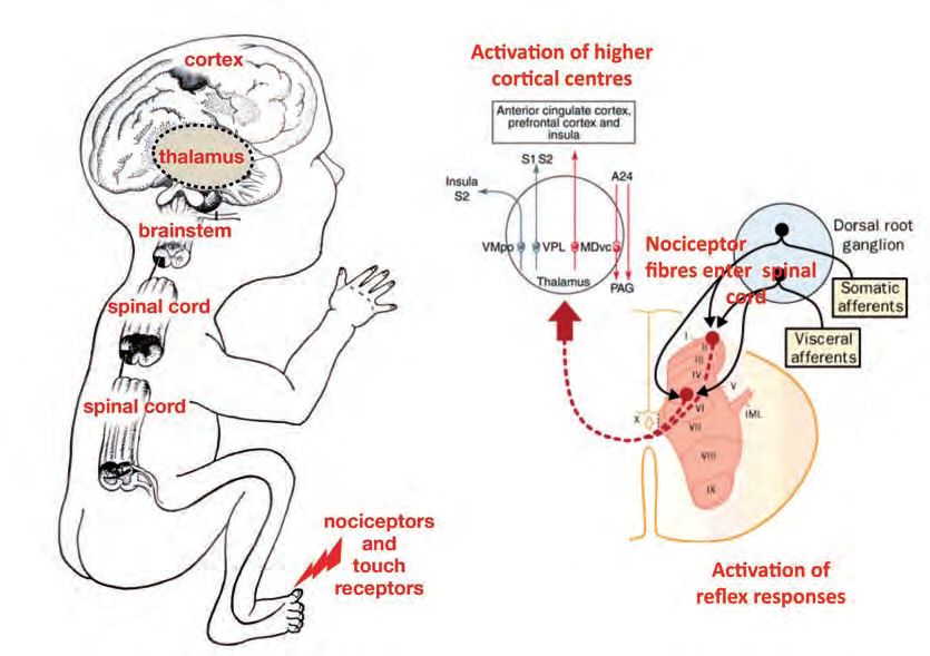
Report of a Working Party. London: RCOG; 2010 [in press].3 2. Neurobiological developments relevant to pain This section examines current knowledge of central nervous system function during fetal and neonatal periods of human development. The aim is to provide a description of key events and changes to inform whether the fetus can reasonably be said to experience pain. To do this, we reviewed all new evidence related to the neurobiology of fetal pain that has been published in peer-reviewed journals listed on PubMed. We begin by considering the scientific evidence for the presence of specific anatomical and physiological connections in the brain that are responsible for signalling noxious events to the central nervous system. Noxious stimuli are those that damage the tissues of the body or threaten to do so, such as surgical incision or physical trauma of the skin. In this context, we define pain as ‘the unpleasant sensory or emotional response to such tissue damage’ and trace the development of those responses through fetal development. We follow the path of the sig- nals produced by tissue damage at sensory detectors in the skin and other organs, through to sensory circuits in the spinal cord, brainstem and thalamus and finally to the cerebral cortex, the site of higher level sensory processing. At each stage, we consider the scientific evidence for functional development and how this evidence may be interpreted. This section includes de- tails derived from over 50 papers identified as relevant. Most were published since the last Working Party report1 but this current report also considers the older material included in the previous report. In addition to understanding the anatomical and physiological connections, it is also impor- tant to consider the psychological aspects of pain. Broadly accepted definitions of pain refer to pain as a subjective experience involving cognition, sensation and affective processes.2 These psy- chological concepts are inevitably harder to address in a fetus but should not be ignored. A discussion of the importance of psychological processes in pain can be found in Box 1. Development of neural pathways related to pain The neural regions and pathways that are responsible for pain experience remain under debate but it is generally accepted that pain from physical trauma requires an intact pathway from the periphery, through the spinal cord, into the thalamus and on to regions of the cerebral cortex including the primary sensory cortex (S1), the insular cortex and the anterior cingulated cor- tex.3,4 Fetal pain is not possible before these necessary neural pathways and structures (figure 1) have developed. The generation of nerve signals from damaged tissue For the fetus to respond to surgical damage, receptors in the affected tissue, such as skin and muscle, must signal the noxious stimulus or damage to the central nervous system. Nocicep- tors are sensory nerve terminals found in the skin and internal organs that convert tissue
4                                       damage into electrical signals. The pattern and strength of these nociceptor signals is the first
                                                    determining step in generating pain. If nociceptor activity is prevented, such as following local
                                                    anaesthesia, then pain is blocked. Deep tissue damage, for example, that cuts through nerve
                                                    bundles causes a brief burst of electrical activity in some of the cut nerve endings known as an
Royal College of Obstetricians and Gynaecologists

                                                    injury discharge.5 The injured tissue, however, is now isolated from the central nervous system
                                                    and, within a few minutes, the isolated tissue becomes ‘numb’ and pain free. Similarly, rare ge-
                                                    netic defects that prevent all nociceptive signals result in a complete inability to sense pain.6

                                                    Figure 1. Pathways from the periphery through the spinal cord and into the thalamus and to the cortex. Nociceptor activity evoked by
                                                    tissue damage reaches the spinal cord and can activate reflex responses through spinal cord connections. Pathways projecting to the
                                                    thalamus and cortex may also be activated. Higher-level pain processing is thought to occur through a medial system (red lines) which
                                                    has both ascending and descending components and a lateral system (blue lines) from the ventroposterior lateral (VPL) and
                                                    ventromedial posterior (VMpo) nuclei. MDvc = mediodorsal ventral caudal nuclei; PAG = periaquaductal gray; S2 = secondary
                                                    somatosensory cortex; S1 = primary somatosensory cortex; A24 = area 24, anterior cingulate cortex (adapted from Cervero and
                                                    Laird,55 Derbyshire56 and Fitzgerald & Walker57)

                                                    Anatomical studies of human fetal skin shows the presence of nerve terminals and fibres deep
                                                    in the skin from 6 weeks of gestational age. These terminals are not nociceptors and are spe-
                                                    cialised for the processing of non-damaging sensations such as touch, vibration and
                                                    temperature, rather than pain. From 10 weeks, nerve terminals become more numerous and
                                                    extend towards the outer surface of the skin.7,8 The terminals closer to the surface are likely
                                                    to be immature nociceptors, necessary for pain experience following tissue damage, but they
                                                    are not unequivocally present until 17 weeks.8 In other mammals, newly formed fetal noci-
                                                    ceptors are able to signal tissue damage but the intensity of their signals is weaker than in
                                                    adults.9 The internal organs develop nerve terminals later than the skin, beginning to appear
                                                    from 13 weeks and then increasing and spreading with age, so that the pancreas, for example,
                                                    is innervated by 20 weeks.10
Interpreting these data 5
Specialised nerve terminals, nociceptors, are likely to detect surgical tissue damage from early
in fetal life (around 10 weeks for the skin and 13 weeks for the internal organs). These noci-
Fetal Awareness
ceptors gradually mature over the next 6–8 weeks and the strength of their signals increases
over fetal life. The presence of nociceptors is necessary for perception of acute surgical pain and
so pain is clearly not possible before the nociceptors first appear at 10 weeks. The presence of
nociceptors alone, however, is not a sufficient condition for pain experience. The electrical ac-
tivity that is generated at nociceptor terminals by tissue damage must also be conducted along
nerve fibres from the skin and into the spinal cord and brain. It is only when the brain receives
information about the damage that the fetus can have any potential of awareness of it.
The transmission of signals from damaged tissue to the
lower levels of the central nervous system
Before any information about a noxious or tissue damaging stimulus can reach the brain, it has
to be transmitted through the spinal cord (for the body) or the brainstem (for the head and
neck). This transmission requires the growth of nerve fibres from the skin to the spinal cord
or brainstem and then further growth of nerve fibres along the spinal cord or brainstem and
into the brain. Staining of postmortem tissue reveals that nerve fibres grow into the fetal spinal
cord from 8 weeks. These fibres, however, are specialised for the control of movement and
some aspects of touching or prodding the body or positioning a limb.
The growth of nerve fibres connecting nociceptive terminals to the spinal cord lags behind that
of other sensory inputs in non-human mammals. Similar connections in the human are also likely
to lag but the specific timings remain unknown. Preliminary studies have failed to demonstrate
nerve fibres from nociceptive terminals in the fetal post-mortem spinal cord before 19 weeks.11
The growth of sensory nerve fibres into the spinal cord is required for the fetus to display re-
flex movements in response to external stimuli. Sensory reflex responses are relatively simple,
central nervous reactions to external events, some of which provide simple protection against
damage. Examples of these reflexes include blinking in response to an air puff to the eye or the
withdrawal of a limb in response to prodding the skin. The presence or absence of these reflexes
at various stages of fetal life can provide information about the first functional sensory con-
nections. In mammals these reflexes are mediated by the spinal cord and brainstem (Figure 1).
During the first 8 weeks of pregnancy, the human fetus displays a range of spontaneous move-
ments, which are not actually reflexes, as they arise from random muscle actions rather than
as reactions to a sensory stimulus. However, when sensory nerves have reached the skin, me-
chanical stimulation of the body can produce reflex movements. This confirms that these nerve
fibres are carrying information about touch and have connected to the spinal cord and acti-
vated nerve fibres controlling motor actions. The fetal spinal cord and brainstem develop well
before the cerebral cortex. This means that these reflex movements occur without any possi-
bility of fetal awareness.
The exact timing of the first nociceptive reflex responses to more traumatic mechanical stim-
ulation is not known but they are unlikely to occur before the second trimester, somewhat
later than responses to touch. It is known that the fetus withdraws from a needle from about
18 weeks and also launches a stress response following needle puncture.12 This stress response
includes the release of hormones and neurotransmitters dependent on activity in areas of the
midbrain. These findings confirm that signals about tissue damage are transmitted from the
spinal cord and brainstem to the midbrain from at least 18 weeks.6
Box 1. A discussion of the nature of pain
Royal College of Obstetricians and Gynaecologists
The word ‘pain’ is used in different ways. The most frequent use, especially with respect to
subjects that cannot communicate verbally, is in describing the behavioural response to nox-
ious stimulation. However, if we accept this use, we are presented with the difficulty of
distinguishing between the responses of simple versus complex organisms. Fruit fly larvae,
for example, have been demonstrated to bend and roll away when approached with a naked
flame but most people would agree that larvae do not feel pain in the way that we do.
Ruling out the responses of larvae and similarly simple organisms as indicating pain is
possible if we suggest that responses must include more than mere reflex responses to be
labelled as a pain response. When someone reaches out and accidentally touches something
very hot, there is an immediate tendency to drop the object. That reaction is entirely reg-
ulated by a simple loop of sensory neurons speaking to motor neurons in the spinal cord.
Typically, the person will drop the object before there is any conscious appreciation of
pain. The action of dropping the object indicates the presence of something noxious but
does not necessarily indicate the presence of pain.
Most pain researchers adopt a definition of pain that emphasises the sensory, cognitive and
affective response to a noxious event. This understanding of pain is supported by the In-
ternational Association of Pain (IASP) which defines pain as ‘an unpleasant sensory and
emotional experience associated with actual or potential tissue damage, or described in
terms of such damage…pain is always subjective. Each individual learns the application
of the word through experiences related to injury in early life’.1 By this definition, pain does
not have primacy over subjectivity, existing before and in addition to subjectivity, but is
experienced through subjectivity. It suggests that pain is a part of knowledge and requires
the existence of a conceptual apparatus that can marshal all its dimensions into a coher-
ent experience.
Although there is considerable merit in the IASP definition of pain, it does tend towards a
view of pain as being a constituent part of higher cognitive function. There is disquiet in
denying a rawer, more primitive, form of pain or suffering that the fetus, neonate and many
animals might experience.2–4 One possible solution is to recognise that the newborn infant
might be said to feel pain, whereas only the older infant can experience that they are in
pain and explicitly share their condition with others as an acknowledged fact of being.5
Currently there is no immediately obvious way of resolving these arguments empirically.
It is possible, however, to argue that even a raw sense of pain involves more than reflex
activity and will, therefore, require the higher regions of the cortex to be connected and
functional. The age when this minimum requirement is fulfilled is explored in the rest of
this chapter.
References
1. Merskey H. The definition of pain. Eur Psychiatry 1991:6;153–9.
2. Anand KJS, Craig KD. New perspectives on the definition of pain. Pain 1996;67:3–6.
3. Lowery CL, Hardman MP, Manning N, Hall RW, Anand KJS. Neurodevelopmental changes of fetal
pain. Semin Perinatol 2007;31:275–82.
4. Anand KJS. Consciousness, cortical function, and pain perception in non-verbal humans. Behav Brain
Sci 2007;30:82–3.
5. Tallis R. The Knowing Animal: A Philosophical Inquiry into Knowledge and Truth. Edinburgh:
Edinburgh University Press; 2005.Interpreting these data 7
Observations of fetal movements in response to sensory stimulation show us that information
about tissue stimulation has reached the spinal cord from 8 weeks. The demonstration of a hor-
Fetal Awareness
monal stress response at 18 weeks following needle puncture shows us that information about
tissue damage has reached the midbrain. A connection from the skin to the spinal cord and
brain is a basic requirement for the fetus to feel or be aware of pain. Again, it is important to
emphasise that, while such input to the spinal cord and brain is necessary for perception of
acute surgical pain, it is not sufficient. Activity in the spinal cord, brainstem and subcortical
midbrain structures are sufficient to generate reflexive behaviours and hormonal responses
but are not sufficient to support pain awareness. At 18 weeks of gestational age, local spinal
cord or brainstem reflexes control movement and, even as movement becomes more coordi-
nated from 24 weeks, it does not require the involvement of higher brain centres. Extremely
preterm infants of 24–30 weeks of gestation show the same motor responses to a noxious heel
lance (required for clinical blood sampling) even when there is severe damage of the pathways
connecting the spinal cord and brainstem to higher brain centres.13 Also, such reactions to
noxious stimuli, even those involving changes in facial expression, do not always correlate
with cortical activity14 when the nervous system is intact, showing that they cannot be assumed
to reflect higher brain function.
Hormonal responses to needling show that there are functional brainstem and midbrain me-
diated reactions to noxious events but they, too, do not require higher brain processing to take
place and can occur independently of sensory awareness. The specific relationship between
pain and the release of hormones and neurotransmitters is unclear. In a prospective crossover
study on 50 extremely low gestational age infants (less than 28 weeks of gestation), no differ-
ence in hormonal response was observed after heel lance15 and, in adult mice, it is difficult to
distinguish changes in levels of naturally occurring opioids due to stressful handling from those
due to tissue damage.16
The transmission of signals from damaged tissue to
cortical regions of the brain
Reflex movements and hormonal stress responses provide information about sensory connec-
tions at lower levels of the nervous system and cannot be assumed to indicate perception or
awareness. For perception or awareness, the sensory information needs to be transmitted to
the thalamus, the major subcortical sensory nucleus and then to the cortex, the highest region
of the brain.
Anatomical evidence
At 8 weeks, the fetal brain is profoundly immature and its surface layer, the cerebral cortex,
is smooth, with no indication of the folds (sulci and gyri) that are so prominent later.17 There
is also no internal cellular organisation in either the thalamus, which is the main source of
sensory input to the cortex, or the cortex itself.18,19 The limbic system, an evolutionary older
part of the brain, consisting of interconnected deep brain structures involved in various fun-
damental drives and regulatory functions, is already discernable and has began to form
interconnections.20 The external surface of the brain is about 1 mm thick and consists of an
inner and outer layer with no cortical plate, the structure that will gradually develop into the
layers of the cortex proper.21 At 13 weeks, a furrow or groove appears on each side of the
brain,17 which becomes part of the insular cortex around 15 weeks, a key region involved in
the experience of external stimuli, including pain.22 In spite of this, the fetal brain is still largely8 smooth at 26 weeks. Massive growth of the brain after 34 weeks rapidly results in the char-
acteristic folds and surface features of the more mature brain.
An important stage of cortical development is the formation of the subplate zone, a prominent,
Royal College of Obstetricians and Gynaecologists
transient layer of the human fetal cerebral wall which develops around 13 weeks and gradu-
ally disappears after 32–34 weeks. The subplate is composed of newly arrived neurons and
their connections together with other brain cells and cellular components and a large amount
of extracellular material. All this makes the subplate very clearly distinguishable in fetal and
neonatal brain scans (magnetic resonance images) and in postmortem brains. The subplate is
thought to be the main synaptic or neuronal connection zone in the human fetal cortex where
incoming fibres from the thalamus, the main sensory (and pain) relay centre, and other re-
gions of the cortex gather during the crucial phase of cortical target area selection. Recent
neurobiological evidence from other mammals shows that subplate is a site of spontaneous
electrical activity and that this activity is required to build a framework for the precise organ-
isation of cortical connections. The subplate is a focus of interest of paediatric neurology
because damage to this area may lead to cognitive impairment in later life.23
The first projections to the subplate from the thalamus arrive between 12 and 18 weeks21,24,25
and wait for the overlying cortical plate to mature and facilitate the invasion of neurons from
the subplate.26 Electrical activity arising from synaptic connections has been recorded in sub-
plate neurons in isolated slices of mammalian brain but it is not known whether that activity
can be selectively produced by thalamic connections or by noxious stimulation of body tissues
in intact animals. It is known that this synaptic activity in the subplate performs a maturational
function. In non-human mammals, synaptic activity in the subplate facilitates connections be-
tween thalamus and cortex and refines the early, initially crude, connections between the
thalamus and cortex.27
By 24 weeks, substantial thalamocortical fibres have accumulated at the superficial edge of the
subplate, which is the stepping-off point for axons growing towards their final cortical tar-
gets.21 Between 24 and 32 weeks, there is substantial ingrowth of thalamocortical axons in
the cortical plate of the frontal, somatosensory, visual and auditory cortex, and formation of
the first synapses in the deep cortical plate. This is consistent with observations in neonates
with rare brain malformations, such a lyssencephaly, where the brain resembles that of a fetus
before 23–24 weeks of gestation, and which shows a lack of connections between the cortex
and subcortical nuclei and an abnormal limbic system.28
At the same time, the relocation of neurons from the subplate to the cortical plate also begins
around 24 weeks, thus coinciding with the invasion of thalamic afferents. This relocation is ex-
tremely rapid from about 34 weeks, leading to the dissolution of the subplate as the
extracellular matrix and other growth-related and guidance molecules disappear.21 The sub-
plate has been observed to thin in the insula and in areas where cortical folding occurs rather
earlier than the rest of the cortex, from at least 20 weeks.29 It is currently uncertain whether
this thinning is due to earlier maturation and potentially earlier synaptic activity in these re-
gions, some of which are key areas in the experience of pain in adults,3 or attributable to
incidental morphological changes.
The arrival of thalamic fibres and formation of thalamocortical synapses in the newly formed
cortex from 24 weeks onwards provides the minimum connection required for cortical pro-
cessing of sensory events in the body. However, completion of the major pathways from the
periphery to the cortex, at around 24 weeks, does not signal the end of cortical development
but the beginning of a further maturational process. As spinothalamic pathways complete their
connections with the cortex, they increasingly stimulate the development of intracortical path-
ways, which is the next major phase of neuronal maturation. Furthermore, the cortex sendsconnections down to the brainstem and spinal cord; the motor centres of the brain have begun 9
to form connections with the spinal cord and brainstem by 26–28 weeks.30 This phase involves
elaboration and refinement of neuron processes and connections, including selective elimina-
tion of some cell populations and corresponds to the cortical maturation described by
Fetal Awareness
Goldman-Rakic31 in primates and by Chugani32 in humans. McKinstry et al.33 illustrated the
effects of this development using diffusion tensor imaging in neonates born at 26 and 35 weeks.
The proliferation of cortical neurons and the overgrowth of arborisation and synaptic contacts
begins prenatally32 but continues postnatally, together with synaptic elimination, pruning and
programmed cell death.31,32,34,35
Physiological evidence
While the study of anatomical connections between brain regions provides important infor-
mation about developing pain processes, the existence of a connection is not evidence of its
function. Connections viewed under the microscope between the thalamus and the cortical
plate at 24 weeks, for example, may or may not transmit information from nociceptors upon
tissue damage. Fetal magnetoencephalography has been used to effectively record fetal audi-
tory and visual evoked responses and spontaneous brain activity of cortical origin from 28
weeks and fetal brain activation to sound has been demonstrated using functional magnetic res-
onance imaging (fMRI) from 33 weeks. It has not been possible to record directly from human
fetal cortex to establish when cortical neurons first begin to respond to tissue damaging inputs.
Near infrared spectroscopy with preterm infants in intensive care, however, has demonstrated
localised somatosensory cortical responses in premature newborn infants (from 24 weeks) fol-
lowing noxious heel lance36 and venepuncture.37 More recently, EEG has demonstrated a clear,
time-locked, nociceptive-evoked potential in preterm infants following heel lance.38 Thus, there
is direct evidence of neural activity in primary sensory cortex following tissue damage in very
premature infants equivalent to 24 weeks of gestational age.
Behavioural evidence
Fetal behavioural responses have also been used as indicators of stress or pain.22,39 Shortly
after the development of skin sensitivity, around 10 weeks, repeated stimulation results in hy-
perexcitability and a generalised movement of all limbs. After 26 weeks, this generalised
movement gradually gives way to more coordinated behavioural responses that indicate im-
proved organisation within the nervous system. Infants delivered at 26–31 weeks, for example,
show coordinated facial expressions in response to heel prick,23 although these are immature
compared to older infants.40 Four-D images of the fetus have also been reported to show fe-
tuses ‘scratching’, ‘smiling’, ‘crying’ and ‘sucking’ at 26 weeks of gestational age.
Although these later behavioural responses are not spinal cord reflexes, the responses are still
unlikely to involve higher cortical centres. An anencephalic fetus withdraws from noxious
stimulation, demonstrating that this response is mediated at a subcortical level.41 Similarly, in-
fants with significant neonatal neurological injury due to a parenchymal brain injury respond
to noxious stimulation with a pattern of behavioural reactions similar to infants without brain
injury.13
Interpreting these data
The cortex is required for both the discriminative and emotional aspects of the processing of
noxious stimuli and both anatomical and functional studies show that cortical neurons begin
to receive input about sensory events in the body and the external environment from 24 weeks.10 Long axonal tracts now course through the brain to the cortex and evoked responses in the pri-
mary sensory cortex indicate the presence of a spinothalamic connection and the ability of
somatosensory cortical neurons to generate specific activity in response to tissue damaging
stimulation. The primary sensory cortex is an important area in pain processing but it is only
Royal College of Obstetricians and Gynaecologists
one of many areas that are active during pain experience.4 Other important areas include the
secondary somatosensory, the anterior cingulate and the insular cortices. Although we may
speculate that these regions will also be functionally active from 24 weeks, similar to primary
sensory cortex, there is no evidence for this at the moment.
It has been suggested that subcortical regions, including the brainstem, and transient brain
structures, including the subplate, organise responses to noxious information at each stage of
development and provide for a pain experience complete within itself at each stage.42–44 There
is, however, no evidence or rationale for subcortical and transient brain regions supporting
mature function. Although developing brain circuits often display spontaneous neuronal ac-
tivity this activity is a fundamental developmental process and not evidence of mature function.
The fact that the cortex can receive and process sensory inputs from 24 weeks is only the be-
ginning of the story and does not necessarily mean that the fetus is aware of pain or knows that
it is in pain. It is only after birth, when the development, organisation and reorganisation of
the cortex occurs in relation to the action and reaction of the neonate and infant to a world
of meaning and symbols, that the cortex can be assumed to have mature features. The cortex
is an important step beyond the spinal cord and brainstem because it facilitates pain experi-
ence by enabling the higher functions of cognition, emotion and self-awareness that are realised
in the postnatal environment. Thus, there is good evidence for claiming that the cortex is nec-
essary for pain experience but not sufficient.
The interpretation of 4-D ultrasound images as evidence for emotional or sentient experience
in the fetus is similarly problematic. While 4-D ultrasound provides better-quality images that
can be useful to diagnose problems in fetal growth or structure, they provide no new evidence
relevant to fetal sentience. As noted above, behavioural reactions can be mediated at a very low
level in the brain and are not, therefore, evidence for experienced emotion or sentience. It is
also important to recognise that ‘labelling’ a set of movements with a functional or emotional
purpose can import too much certainty. Yawning, for example, is most likely a protective lung
reflex that maintains proper lung inflation and prevents the developing alveoli (a kind of
sponge-like material) from collapsing. While this protective reflex is unnecessary in the womb
where oxygen is delivered by the umbilicus, it will be necessary soon after birth and therefore
the neural connections that mediate it need to be fully functional well in advance of birth.
Sleep and wakefulness in the womb
It has been proposed that arguments around fetal pain can be resolved by the fact that the
fetus never enters a state of wakefulness in utero.45 This evidence is derived largely from ob-
servations of fetal lambs. Rigatto et al.,46 for example, directly observed an unanaesthetised
sheep fetus, in utero, through a Plexiglas window, for 5000 hours without observing signs of
wakefulness such as eyes opening or coordinated movement of the head. Several factors explain
this lack of wakefulness, including the environment of the womb, which is warm, buoyant
and cushioned, and the presence of a chemical environment (most notably adenosine) that pre-
serves a continuous sleep-like unconsciousness or sedation and suppresses higher cortical
activation in the presence of intrusive external stimulation. Mellor et al.45 also propose that the
fetus is unconscious based on the presence of sleep-like EEG patterns observed in the lamb
fetus, which enter a more quiescent state together with lack of movement, during hypoxic
stress,46,47 although it should be emphasised that this is quite different from the kind of nox-ious stress generated by surgery discussed here. Mellor et al.45 report that the general pattern 11
of EEG during gestation is equivalent to a sleep-like state analogous to non-rapid eye move-
ment and rapid eye movement sleep.
Fetal Awareness
Interpreting these data
Although these data are derived from sheep, this species has been a useful investigative model
of human pregnancy and the extrapolation of these data to the human fetus is plausible. Being
asleep or awake is not as easy to distinguish in the fetus and newborn as it is in adults48 but
the broad categories can still be classified on the basis of EEG recordings. On this basis, sleep
state differentiation appears in humans as early as 25 weeks in preterm infants and is complete
at 30 weeks.49 EEG recordings in late fetal baboons support these observations and define only
two physiological states from EEG analysis, quiet sleep and active sleep.50
While the lack of fetal movement during anoxic stress in sheep may not be the same as the re-
sponse to acute surgical tissue damage in humans, this work does highlight the important
differences between fetal and neonatal life and the potential pitfalls of extrapolating from ob-
servations of newborn preterm infants to observations of the fetus. Sedation of the fetus and
suppression of cortical arousal in times of stress imply that the cortex in utero responds dif-
ferently from the neonatal cortex and that it is only after birth, with the separation of the baby
from the uterus and the umbilical cord, that wakefulness truly begins. This conclusion is not
inconsistent with reports of fetal conditioning and habituation to repeated exposure of sounds
and smells in late pregnancy which are often referred to as fetal learning. Such responses do
not require a cortex in a state of wakefulness and can be induced in simple circuits in lower
organisms.51
Summary
Connections from the periphery to the cortex are not intact before 24 weeks of gestation. Most
pain neuroscientists believe that the cortex is necessary for pain perception; cortical activation
correlates strongly with pain experience and an absence of cortical activity generally indicates
an absence of pain experience.52–54 The lack of cortical connections before 24 weeks, therefore,
implies that pain is not possible until after 24 weeks. Even after 24 weeks, there is continuing
development and elaboration of intracortical networks. Furthermore, there is good evidence
that the fetus is sedated by the physical environment of the womb and usually does not awaken
before birth.
References
1. Royal College of Obstetricians and Gynaecologists. Fetal Awareness: Report of a Working Party. London:
RCOG Press; 1997.
2. Merskey H. The definition of pain. Eur Psychiatry 1991:6;153–9.
3. Apkarian AV, Bushnell MC, Treede RD, Zubieta JK. Human brain mechanisms of pain perception and
regulation in health and disease. Eur J Pain 2005;9:463–84.
4. Tracey I, Mantyh PW. The cerebral signature for pain perception and its modulation. Neuron
2007;55:377–91.
5. Blenk KH, Jänig W, Michaelis M, Vogel C. Prolonged injury discharge in unmyelinated nerve fibres
following transection of the sural nerve in rats. Neurosci Lett 1996;215:185–8.
6. Cox JJ, Reimann F, Nicholas AK, Thornton G, Roberts E, Springell K, et al. An SCN9A channelopathy
causes congenital inability to experience pain. Nature 2006;444:894–8.
7. Narisawa Y, Hashimoto K, Nihei Y, Pietruk T. Biological significance of dermal Merkel cells in
development of cutaneous nerves in human fetal skin. J Histochem Cytochem 1992;40;65–71.8. Terenghi G, Sundaresan M, Moscoso G, Polak JM. Neuropeptides and a neuronal marker in cutaneous
12 innervation during human foetal development. J Comp Neurol 1993;328:595–603.
9. Koltzenburg M. The changing sensitivity in the life of the nociceptor. Pain 1999;Suppl 6:S93–102.
10. Amella C, Cappello F, Kahl P, Fritsch H, Lozanoff S, Sergi C. Spatial and temporal dynamics of innervation
Royal College of Obstetricians and Gynaecologists
during the development of fetal human pancreas. Neuroscience 2008;154:1477–87.
11. Konstantinidou AD, Silos-Santiago I, Flaris N, Snider WD. Development of the primary afferent projection
in human spinal cord. J Comp Neurol 1995;354:11–12.
12. Gitau R, Fisk NM, Glover V. Human fetal and maternal corticotrophin releasing hormone responses to
acute stress. Arch Dis Child Fetal Neonatal Ed 2004;89:F29–32.
13. Oberlander TF, Grunau RE, Fitzgerald C, Whitfield MF. Does parenchymal brain injury affect
biobehavioral pain responses in very low birth weight infants at 32 weeks’ postconceptional age? Pediatrics
2002;110:570–6.
14. Slater R, Cantarella A, Franck L, Meek J, Fitzgerald M. How well do clinical pain assessment tools reflect
pain in infants? PLoS Med 2008;5(6):e129.
15. Gibbins S, Stevens B, Beyene J, Chan PC, Bagg M, Asztalos E. Pain behaviours in extremely low gestational
age infants. Early Hum Dev 2008;84:451–8.
16. Rasmussen NA, Farr LA. Beta-endorphin response to an acute pain stimulus. J Neurosci Methods
2009;177:285–8.
17. Afif A, Bouvier R, Buenerd A, Trouillas J, Mertens P. Development of the human fetal insular cortex: study
of the gyration from 13 to 28 gestational weeks. Brain Struct Funct 2007;212:335–46.
18. Larroche JC. The marginal layer in the neocortex of a 7 week-old human embryo: a light and electron
microscopic study. Anat Embryol (Berl) 1981;162:301–12.
19. Hevner RF. Development of connections in the human visual system during fetal mid-gestation: a
DiI-tracing study. J Neuropathol Exp Neurol 2000;59:385–92.
20. Müller F, O’Rahilly R. The amygdaloid complex and the medial and lateral ventricular eminences in staged
human embryos J Anat 2006;208:547–64.
21. Kostovi I, Jovanov-Milosevi N. The development of cerebral connections during the first 20–45 weeks’
gestation. Semin Fetal Neonat Med 2006;11:415–22.
22. Craig AD. A new view of pain as a homeostatic emotion. Trends Neurosci 2003;26:303–7.
23. Kostovi I, Jovanov-Milosevi N. Subplate zone of the human brain: historical perspective and new
concepts. Coll Antropol 2008;32 Suppl 1:3–8.
24. Kostovic I, Judas M. Correlation between the sequential ingrowth of afferents and transient patterns of
cortical lamination in preterm infants. Anat Rec 2002;267:1–6.
25. Bystron I, Blakemore C, Rakic P. Development of the human cerebral cortex: Boulder Committee revisited.
Nat Rev Neurosci 2008;9:110–22.
26. Molnar Z, Blakemore C. How do thalamic axons find their way to the cortex? Trends Neurosci
1995;18:389–97.
27. Kanold PO, Kara P, Reid RC, Shatz CJ. Role of subplate neurons in functional maturation of visual
cortical columns. Science 2003;301:521–5.
28. Rollins N, Reyes T, Chia J. Diffusion tensor imaging in lissencephaly. Am J Neuroradiol 2005;26:1583–6.
29. Huang H, Zhang J, Wakana S, Zhang W, Ren T, Richards LJ, et al. White and gray matter development in
human fetal, newborn and pediatric brains. NeuroImage 2006;33:27–38.
30. Eyre JA, Miller S, Clowry GJ, Conway EA, Watts C. Functional corticospinal projections are established
prenatally in the human foetus permitting involvement in the development of spinal motor centres. Brain
2000;123:51–64.
31. Goldman-Rakic PS. Development of cortical circuitry and cognitive function. Child Development,
1987;58:601–22.
32. Chugani HT. Biological basis of emotions: brain systems and brain development. Pediatrics
1998;102:S1225–9.
33. McKinstry RC, Mathur A, Miller JH, Ozcan A, Snyder AZ, Schefft GL, et al. Radial organization of
developing preterm human cerebral cortex revealed by non-invasive water diffusion anisotropy MRI.
Cereb Cortex 2002;12:1237–43.
34. Huttenlocher PR, Dabholkar AS. Regional differences in synaptogenesis in human cerebral cortex. J Comp
Neurol 1997;387:167–78.
35. Fitzgerald M. The development of nociceptive circuits. Nat Rev Neurosci 2005;6:507–20.
36. Slater R, Cantarella A, Gallella S, Worley A, Boyd S, Meek J, et al. Cortical pain responses in human
infants. J Neurosci 2006;26:3662–6.37. Bartocci M, Bergqvist LL, Lagercrantz H, Anand KJ. Pain activates cortical areas in the preterm newborn
brain. Pain 2006;122:109–17.
13
38. Slater R, Worley A, Fabrizi L, Roberts S, Meek J, Boyd S, et al. Evoked potentials generated by noxious
stimulation in the human infant brain. Eur J Pain 2009;May 28 [Epub ahead of print].
Fetal Awareness
39. Anand KJS, Hickey PR. Pain and its effects in the human neonate and fetus. N Engl J Med
1987;317:1321–9.
40. Johnston CC, Stevens BJ, Franck LS, Jack A, Stremler R, Platt R. Factors explaining lack of response to
heel stick in preterm newborns. J Obstet Gynecol Neonat Nurs 1999;28:587–94.
41. Visser GH, Laurini RN, de Vries JIP, Bekedam DJ, Prechtl HFR. Abnormal motor behaviour in
anencephalic fetuses. Early Hum Dev 1985; 12:173–82.
42. Anand KJS, Craig KD. New perspectives on the definition of pain. Pain 1996;67:3–6.
43. Glover V, Fisk NM. Fetal pain: implications for research and practice. Br J Obstet Gynaecol
1999;106:881–6.
44. Lowery CL, Hardman MP, Manning N, Hall RW, Anand KJS. Neurodevelopmental changes of fetal pain.
Semin Perinatol 2007;31:275–82.
45. Mellor DJ, Diesch TJ, Gunn AJ, Bennet L. The importance of ‘awareness’ for understanding fetal pain.
Brain Research Reviews, 2005;49:455–71.
46. Rigatto H, Moore M, Cates D. Fetal breathing and behavior measured through a double-wall Plexiglas
window in sheep. J Appl Physiol 1986;61:160–4.
47. Gunn AJ, Cook CJ, Williams CE, Johnston BM, Gluckman PD. Electrophysiological responses of the fetus
to hypoxia and asphyxia. J Dev Physiol 1991;16:147–53.
48. Vecchierini MF, André M, d’Allest AM. Normal EEG of premature infants born between 24 and 30 weeks
gestational age: terminology, definitions and maturation aspects. Neurophysiol Clin 2007;37:311–23.
49. Hunter CJ, Bennet L, Power GG, Roelfsema V, Blood AB, Quaedackers JS, et al. Key neuroprotective role
for endogenous adenosine A1 receptor activation during asphyxia in the fetal sheep. Stroke
2003;34:2240–5.
50. Stark RI, Garland M, Daniel S, Myers MM. Diurnal rhythm of fetal behavioral state. Sleep
1998;21:167–76.
51. Hawkins RD, Kandel ER, Bailey CH. Molecular mechanisms of memory storage in Aplysia. Biol Bull
2006;210:174–91.
52. Fransson P, Skiöld B, Horsch S, Nordell A, Blennow M, Lagercrantz H, et al. Resting-state networks in the
infant brain. Proc Natl Acad Sci U S A 104:15531–6.
53. Rosen SD, Camici PG. The brain-heart axis in the perception of cardiac pain: the elusive link between
ischaemia and pain. Ann Med 2000;32:350–64.
54. Coghill RC, McHaffie JG, Yen YF. Neural correlates of interindividual differences in the subjective
experience of pain. Proc Natl Acad Sci U S A 2003;100:8538–42.
55. Cervero F, Laird J. Visceral pain. Lancet 1999;353:2145–8
56. Derbyshire SWG. Measuring our natural painkiller. Trends Neurosci 2002;25:65–6.
57. Fitzgerald M, Walker SM. Infant pain management: a developmental neurobiological approach. Nat Clin
Pract Neurol 2009;5:35–50.You can also read