Nerve Growth Factor Biodelivery: A Limiting Step in Moving Toward Extensive Clinical Application?
←
→
Page content transcription
If your browser does not render page correctly, please read the page content below
REVIEW
                                                                                                                                                published: 15 July 2021
                                                                                                                                       doi: 10.3389/fnins.2021.695592

                                            Nerve Growth Factor Biodelivery: A
                                            Limiting Step in Moving Toward
                                            Extensive Clinical Application?
                                            Giuseppe Alastra 1† , Luigi Aloe 2† , Vito Antonio Baldassarro 1† , Laura Calzà 1,2,3† ,
                                            Maura Cescatti 2† , Jason Thomas Duskey 4† , Maria Letizia Focarete 1,5† ,
                                            Daria Giacomini 1,5† , Luciana Giardino 2,6* † , Valentina Giraldi 1,5† , Luca Lorenzini 6† ,
                                            Marzia Moretti 2† , Irene Parmeggiani 4† , Michele Sannia 1† and Giovanni Tosi 4†
                                            1
                                              Interdepartmental Centre for Industrial Research in Health Sciences and Technologies, University of Bologna, Bologna,
                                            Italy, 2 IRET Foundation, Bologna, Italy, 3 Department of Pharmacy and Biotechnology, University of Bologna, Bologna, Italy,
                                            4
                                              Nanotech Laboratory, TeFarTI Center, Department of Life Sciences, University of Modena and Reggio Emilia, Modena, Italy,
                                            5
                                              Department of Chemistry “Giacomo Ciamician”, University of Bologna, Bologna, Italy, 6 Department of Veterinary Medical
                                            Sciences, University of Bologna, Bologna, Italy

                                            Nerve growth factor (NGF) was the first-discovered member of the neurotrophin family,
                                            a class of bioactive molecules which exerts powerful biological effects on the CNS
                            Edited by:
                        Robert Nistico,
                                            and other peripheral tissues, not only during development, but also during adulthood.
  Tor Vergata University of Rome, Italy     While these molecules have long been regarded as potential drugs to combat acute
                       Reviewed by:         and chronic neurodegenerative processes, as evidenced by the extensive data on
                Hubert Hondermarck,         their neuroprotective properties, their clinical application has been hindered by their
The University of Newcastle, Australia
                          Ilias Kazanis,    unexpected side effects, as well as by difficulties in defining appropriate dosing
             University of Cambridge,       and administration strategies. This paper reviews aspects related to the endogenous
                      United Kingdom
                                            production of NGF in healthy and pathological conditions, along with conventional and
                    *Correspondence:
                       Luciana Giardino
                                            biomaterial-assisted delivery strategies, in an attempt to clarify the impediments to the
               luciana.giardino@unibo.it    clinical application of this powerful molecule.
     † These   authors have contributed
                                            Keywords: nerve growth factor, nanomedicine, drug delivery, electrospinning, hydrogels
                    equally to this work

                   Specialty section:
         This article was submitted to
                                            INTRODUCTION
                  Neuropharmacology,
               a section of the journal
                                            Since its discovery in the 1950s (Levi-Montalcini, 1987) and the award of the Nobel Prize
            Frontiers in Neuroscience       to Rita Levi-Montalcini and Stanley Cohen for their discoveries of growth factors in 1986,
                                            the number of basic science discoveries and preclinical studies supporting the use of nerve
             Received: 15 April 2021
            Accepted: 21 June 2021
                                            growth factor (NGF) for therapeutic purposes has constantly increased over the years, principally
            Published: 15 July 2021         for neurodegenerative diseases (Alzheimer’s disease, AD; and Parkinson’s disease, PD) and
                                            brain injuries (perinatal hypoxia/ischemia, traumatic brain, and spinal cord injury), but also
                               Citation:
    Alastra G, Aloe L, Baldassarro VA,      retinopathies, optic nerve degeneration, and peripheral neuropathies associated with diabetes and
       Calzà L, Cescatti M, Duskey JT,      HIV. The potential applications have also extended from the nervous system as the primary target
            Focarete ML, Giacomini D,       to an increasing number of tissues and organs, including epithelial tissue, parenchymal organs, and
      Giardino L, Giraldi V, Lorenzini L,   the osteoarticular system (Manni et al., 2013; Rocco et al., 2018), as well as inflammation (Skaper,
  Moretti M, Parmeggiani I, Sannia M        2017) and cancer (Griffin et al., 2018).
      and Tosi G (2021) Nerve Growth
                                               The road toward the clinical translation of NGF, however, has encountered numerous obstacles.
    Factor Biodelivery: A Limiting Step
  in Moving Toward Extensive Clinical
                                            In spite of the success of Genentech in translating NGF production from male mouse salivary gland
                            Application?    extract (Bocchini and Angeletti, 1969) to the recombinant technology of the human form (Altar
           Front. Neurosci. 15:695592.      et al., 1991; Rogers, 1996), results from early clinical trials led Genentech to terminate the rhNGF
      doi: 10.3389/fnins.2021.695592        (recombinant human NGF) project. This was based on side effects observed in two sets of phase

Frontiers in Neuroscience | www.frontiersin.org                                    1                                             July 2021 | Volume 15 | Article 695592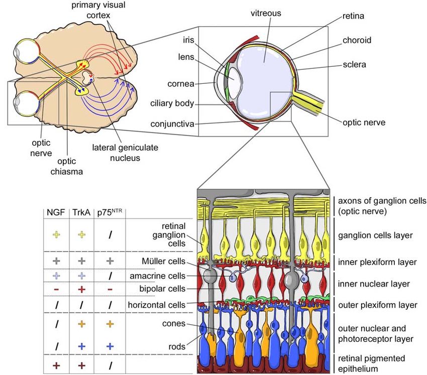
Alastra et al.                                                                                                                               NGF Biodelivery

II clinical trials, suggesting that despite the efficacy of rhNGF               There is little data available on the biodistribution of
administration at ameliorating the symptoms associated with                 exogenously administered NGF. In the initial human applications
both diabetic polyneuropathy and HIV-related neuropathy, side               and clinical trials, mouse NGF or recombinant human NGF
effects were dose limiting for NGF (Apfel, 2002). Moreover, a               (rhNGF) was SC (Petty et al., 1994; Rogers, 1996; Apfel
large-scale phase III clinical trial of 1019 patients randomized            et al., 1998, 2000; McArthur et al., 2000; Schifitto et al., 2001)
to receive either rhNGF or placebo for 48 weeks failed to                   (clinical trial NCT00000842) or ICV administered (Olson et al.,
confirm these earlier indications of efficacy (Apfel, 2002).                1992; Jönhagen et al., 1998; Chiaretti et al., 2005, 2008), but
Intravenous (IV), subcutaneous (SC), and intradermal (ID) NGF               basic information on absorption, distribution and excretion
injection induces myalgia, mechanic and thermal hyperalgesia,               following parenteral administration derives from animal studies,
emerging rapidly after injection and lasting for weeks (Mizumura            in particular in adult rats and in cynomolgus monkeys. In
and Murase, 2015). As concerns human studies, SC or                         this few biodistribution studies, mouse NGF was administrated
intracerebroventricular (ICV) administration of rhNGF to                    intravenously (IV) and SC in rats as a single injection (35 µg/kg,
healthy subjects or patients with diabetic polyneuropathy, HIV-             single dose) or by continuous infusion via osmotic mini-pump
associated peripheral neuropathy (SC, 0.03–1 µg/kg), AD (ICV,               (50–450 µg/pump) (Tria et al., 1994). In monkeys, rhNGF
75 µg/day for 3 months), PD (ICV, 3.3 mg infused over 23 days)              was administered SC (2 mg/kg), and pharmacokinetic analysis
or hypoxic-ischemic perinatal brain injury (0.1 mg/day for                  was conducted after single and multiple doses (for 15 days,
10 days) (reviewed by Tria et al., 1994; Mizumura and Murase,               every other day) (Nguyen et al., 2000). In both studies, the
2015) always produced hyperalgesia at the injection site, and in            maximum plasma concentrations (Cmax ) confirmed that the drug
some cases also mild to moderate-severe transient muscle pain               is absorbed after SC administration. In rats, the maximum blood
(Petty et al., 1994; Rogers, 1996) (clinical trial NCT00000842).            concentration of NGF after SC administration was 65-fold lower
    Actually, the current evidence regarding the painful side               than after IV injection. The calculated time to reach maximum
effects of NGF administration is taking pharmacological research            plasma concentrations (T max ) are very similar in two studies,
in two new directions: development of humanized anti-                       despite the different doses employed. Multiple dosing in monkeys
NGF monoclonal antibodies (anti-NGF mAbs) for conditions                    shifts the T max from a mean value of 2.5 to 3.3 h (Nguyen et al.,
as osteoarthritis, lower back pain, and interstitial cystitis               2000; Tria et al., 1994).
(Wise et al., 2021), and synthesis of TrkA ligands in an                        Subcutaneous administration via osmotic mini-pump
attempt to overcome this severe and limiting side effect                    (450 µg/pump, corresponding to 37.5 µg/day) in rats resulted
(Carleton et al., 2018; Bagal et al., 2019). The rhNGF has                  in detectable NGF plasma levels after 6 h, reaching peak values
finally received FDA approval as Cenegermin eye drops R
                                                                            during day 1, confirming the T max delay after multiple dosing
by Dompé, first-in-class with the potential to completely                   (Tria et al., 1994). IV injection allows calculation of the half-life
heal rare neurotrophic keratitis (clinical trials NCT04293549,              distribution phase (t 1/2α ), reached in 5–6 min, indicating a rapid
NCT03836859, NCT02101281, NCT03019627).                                     disappearance from plasma, probably due to the binding of NGF
    However, the major obstacles to clinical translation of NGF             to the α2-macroglobulin cleared from blood by hepatocytes (Tria
are also due to other factors, such as the biodistribution of this          et al., 1994). With regard to NGF metabolism, no degradation
large molecule, including its crossing of blood-tissue barriers, and        products were observed in plasma after immunoprecipitation
to issues of dosage, since NGF is produced by many different                and SDS-PAGE, suggesting a long-term stability of the protein
cell types (Gostynska et al., 2020), and because endogenous NGF             (Tria et al., 1994; Nguyen et al., 2000). Table 1 summarizes
production is altered in many of the pathologies included in a              the basic pharmacokinetic parameters following comparable
tentative list of potential targets for the NGF drug.                       administration routes.
    This review addresses some of these major issues affecting the              Data regarding tissue distribution were obtained following the
development of innovative NGF delivery solutions, discussing                administration of radiolabeled rhNGF (125 I-rhNGF) in primates
possible reasons for their success, and in many cases their                 (multiple dosing, for 15 days, every other day). The large central
failures. Sections “Parenteral Administration” and “Topical                 volume of distribution (V2 /F, 827 ml/kg) indicated distribution
Application” refer to both preclinical and clinical studies, these
latter also indicated by the respective clinicaltrials.gov code;
section “Biomaterial-Assisted Delivery” refers to in vitro and              TABLE 1 | Main PK parameters evaluated in animal studies, following comparable
preclinical studies.                                                        single subcutaneous injection (data from Tria et al., 1994; Nguyen et al., 2000).

                                                                                                               Monkeys                              Rats
PARENTERAL ADMINISTRATION
                                                                                                                 rhNGF                             mNGF

NGF Endogenous Levels, Biodistribution                                      Dose                                2 mg/kg                           35 µg/kg
and Metabolism                                                              Cmax (ng/ml)                      1300 ± 120                         3.57 ± 0.33
In the body, NGF is produced according to a delicate balance                T max (h)                           2.5 ± 2.7                        3.20 ± 0.49
that varies from tissue to tissue also according to specific diseases       t1/2β (h)                           4.1 ± 1.0                        4.47 ± 0.15
and pathological states (Lorenzini et al., 2021), and that can be           Cmax , maximum plasma concentration; Tmax , time to reach maximum plasma
reflected by NGF blood levels                                               concentration; t1/2β , elimination phase half-life.

Frontiers in Neuroscience | www.frontiersin.org                         2                                              July 2021 | Volume 15 | Article 695592
Alastra et al. NGF Biodelivery
in extravascular tissues. The organs were then collected at 8 While the restrictive nature of the BBB allows for proper
and 24 h following dose 1 and dose 15. In non-neuronal tissues neuronal function and protection of the neural tissue, and
125 I-rhNGF was detected at both time points and doses in all maintains CNS homeostasis, it also constitutes an obstacle for
studied tissues, particularly in the thyroid, adrenals, kidneys, liver, drug delivery. Whether NGF can penetrate the BBB and be
spleen, peripheral and axillary tissues, and at the injection site. As absorbed by the brain tissue, and under what conditions, is still
expected, the radiolabeled drug was observed in the peripheral controversial, but the poor permeability of NGF through the
nervous system, whereas it was minimal in the spinal cord, and BBB under physiological conditions has been widely described
absent in the brain (Nguyen et al., 2000). (Pan et al., 1998). However, the BBB is not a fixed structure,
The plasma elimination half-life (t 1/2β ) is very close both undergoing pathophysiological adaptations which are not yet
in rats and in monkeys (Table 1) and can be extended by fully understood. Although BBB formation starts during the
varying the administration route, e.g., 4.47 h in SC versus 2.30 h embryonic stage, soon after vessel formation in the developing
in IV injection in rats. The administration schedule does not CNS (E11, in rats), the system continues to mature following
appear to affect this parameter, e.g., 4.1 h versus 4.8 h following birth, increasing the strength of the paracellular barrier and
single and multiple dosing. The clearance values (Cl) are not expression of the efflux transporter (Blanchette and Daneman,
affected by administration route, e.g., 6.38 ml/min/kg in SC 2015), while pathological conditions, particularly inflammation,
versus 6.93 ml/min/kg in IV injection in rats, but decrease are known to modify its structure and dynamics. In general,
following multiple administration, e.g., 140 ml/kg/hr versus different diseases (e.g., stroke, multiple sclerosis, epilepsy) are
63 ml/kg/hr following single and multiple (Tria et al., 1994; characterized by the internalization and down-regulation of tight
Nguyen et al., 2000). junctions, increased rates of transcytosis, increased expression
With regard to NGF metabolism, analysis of radiolabeled of adhesion molecules for leukocytes leading to increased
NGF in monkeys shows that urinary excretion represents the leukocyte extravasation, degradation of the basal membrane,
main route of elimination, although traces of radioactivity were and reduced microvessel coverage by pericytes and astrocytes
found in the feces. No difference in elimination pattern was (Profaci et al., 2020). Specific pathologies may therefore offer time
observed between 24 and 120 h following single and multiple window opportunities for exploiting altered BBB permeability
dosing (Tria et al., 1994; Nguyen et al., 2000). There is also and increasing NGF transportation to the CNS.
very little data available on specific NGF metabolic products, Strategies to overcome the BBB for the CNS delivery of large
derived from immunoprecipitation and SDS-PAGE in monkey molecules as NGF represent a major goal, driving alternative
tissue lysates. In non-neuronal tissues, low molecular mass bands routes of administration [see section “Intranasal (IN)” and “Eye
were observed in the kidney, liver and spleen, indicating that Drops”] and pharmaceutical technologies as nanocarrier (see
intensive metabolism occurs in these organs. SDS-PAGE from section “Nanomedicines for NGF Delivery”).
lysate of sympathetic ganglia and dorsal root ganglia also show
intense NGF metabolism, while material present in the peripheral
nerves (radial, sciatic, and tibial) was mostly negative (Tria et al., TOPICAL APPLICATION
1994; Nguyen et al., 2000).
Intranasal (IN)
Due to its large surface area, the high degree of vascularization,
NGF and the Blood Brain Barrier and the “nose-to-brain” pathways, the nasal cavity is an
Peripheral administration of NGF to target the CNS is limited interesting portal for systemic delivery and to by-pass the BBB.
by the poor ability of this molecule to cross the blood-brain The advantages of nose-to-brain drug delivery include safety
barrier (BBB), and by peripheral enzymatic degradation. The and avoidance of the hepatic first pass metabolism, as well as
unique properties of the BBB stem from CNS capillary histology, its non-invasive nature and high patient compliance (Colombo
where endothelial cells are held together by tight junctions which et al., 2011). Limitations include the possible dosing volume
limit the paracellular flux of solutes, and the presence of specific through the nasal cavity and the consequent total amount of
transporters which regulate the passage of molecules to the drug delivered systemically or into the brain (Dong, 2018), active
CNSs. Endothelial cells are covered by mural cells (pericytes mucociliary clearance of the mucosa, short retention time for
and smooth muscle cells) which contribute to the dynamics of drug absorption, low permeability for hydrophilic drugs, and low
BBB control, and the microvascular tube is also surrounded central nervous system (CNS) delivery for proteins (Erdõ et al.,
by the inner vascular and the outer parenchymal basement 2018). For these reasons, many strategies are currently being
membrane, providing an anchor for many signaling processes tested to enhance drug transport and distribution through the
and an additional barrier for cells and molecules accessing the “nose-to-brain” pathways.
CNS. The blood vessels also interact with different immune To give a brief anatomical overview, the nasal cavity consists
cells, mainly perivascular macrophages, and microglial cells, of three anatomical areas, the nasal vestibule, respiratory region,
representing the first line of innate immunity. Lastly, the direct and olfactory region, each characterized by different mucosal
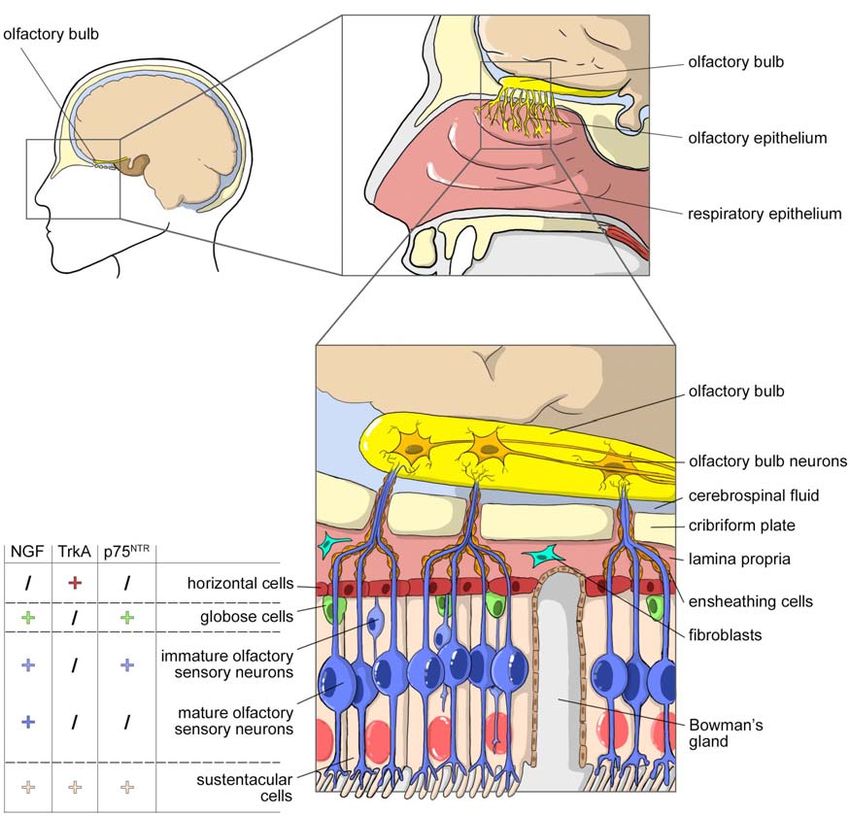
bridge from the microvessel to the neurons is the astrocyte, epithelia. Figure 1 shows the cell composition of the olfactory
a glial cell with extending processes which completely envelop part of nasal cavity, mucosa, olfactory epithelium and lamina
the vascular tube, connecting the microvessels to the neurons propria, including the detail of the NGF, TrkA, and p75NTR
(Daneman and Prat, 2015). expression on different cell types.
Frontiers in Neuroscience | www.frontiersin.org 3 July 2021 | Volume 15 | Article 695592Alastra et al. NGF Biodelivery
FIGURE 1 | NGF and olfactory system. (A) Sagittal view of the head with highlighted the area occupied by the nasal cavity. (B) Magnification of the nasal cavity with,
in the square, the area occupied by the olfactory epithelium. (C) Cell composition of the olfactory part of the nasal cavity, mucosa, olfactory epithelium, and lamina
propria. The olfactory epithelium is surrounded by layer globose and horizontal basal cells. It consists in many cellular types: sustentacular cells, Bowman’s glands
producing mucus, immature olfactory neurons and mature olfactory sensory neurons that project their axons toward the olfactory bulb through the cribriform plate.
Axons are enclosed by olfactory ensheating cells and olfactory nerve fibroblasts. The picture includes data on the expression of NGF, TrkA and p75NTR in the
different cellular populations of the olfactory epithelium (from Feron et al., 2008).
The respiratory epithelium is a ciliated pseudostratified brain. The olfactory mucosa consists of a ciliated chemosensory
columnar epithelium composed by four main cell types – ciliated pseudostratified columnar epithelium that contains three types
and non-ciliated columnar cells, basal or horizontal cells and of cells – olfactory sensory neurons, immature olfactory sensory
goblet cells – with high vascularization, supplied by the arterial neurons and supporting (or sustentacular) cells – all connected
branch of the maxillary artery. A mucus gel layer covers the by tight junctions (Figures 1A,B). The cilia are non-motile, and
epithelium, that, together with ciliary tip movements, constitutes the overlying mucus gel has a very slow turnover (several days).
the first protective barrier against inhaled particulates and The olfactory mucosa presents the main inter-species
irritants. The respiratory mucus layer is renewed every 10–20 min anatomical differences, an aspect which must be considered when
(Pardeshi and Belgamwar, 2013). translating animal data to humans. In humans, this mucosa
While the respiratory region is mainly involved in systemic covers 10% of the total surface area, while in rodents, the most
drug absorption, the olfactory area is important not only for the widely used species for intranasal administration studies, it can
ability of its neurons to provide the sense of smell, but also for constitute up to 50% of the total area. This is an important aspect,
the “nose-to-brain path,” which delivers drugs directly into the because results obtained from animal models do not always
Frontiers in Neuroscience | www.frontiersin.org 4 July 2021 | Volume 15 | Article 695592Alastra et al. NGF Biodelivery
correlate with those of humans, a discrepancy which probably Although interest in this delivery route for preclinical
comes from an insufficient consideration of the anatomical and and clinical studies is increasing, very few studies of NGF
physiological differences between the respective nasal cavities pharmacokinetics or biodistribution are available. In a study
(Cho et al., 2010). Rodents are more widely used for preliminary on the Sprague Dawley rat hippocampus, for example, the
nose-to-brain drug absorption studies, while rabbits and dogs are bioavailability of intranasally administered NGF with or without
used for pharmacokinetic studies. chitosan was ∼14 fold greater than the group treated with NGF
Drugs transferred from the olfactory mucosa to the CNS without chitosan (Vaka et al., 2009). In a preclinical model of AD,
bypassing the BBB follow two pathways, olfactory and trigeminal, polymeric nanoparticles appear to be promising carriers for the
with molecular transfer taking place outside or within the nerve nose-to-brain delivery of drugs (Rabiee et al., 2021). Following
axon. The olfactory path includes the neuronal cells of the IN administration, rhNGF reached the brain within an hour,
olfactory epithelium, and the lamina propria and the olfactory achieving a concentration of 3400 pM in the olfactory bulb, 660–
bulb in the CNS. The olfactory bulb then projects to the 2200 pM in other brain regions and, 240 and 180 pM in the
cortex, amygdala and hypothalamus, providing an anatomical hippocampus and the amygdala, respectively, while, little or no
link between nasal administration and the brain structures (Khan rhNGF was found in the brain following IV administration (Chen
et al., 2017). The trigeminal path consists of the trigeminal et al., 1998). The therapeutic efficacy of IN NGF administration
nerve with its three major branches, ophthalmic, maxillary, and has also been evaluated in many other brain diseases, and in
mandibular, thus promoting the entrance of drugs to the caudal clinical trials of traumatic brain injury, acute ischemic stroke
and rostral parts of the brain. The olfactory path delivers drugs to and frontotemporal dementia (Eftimiadi et al., 2021). Notably,
the rostral areas of the brain only, whereas the trigeminal pathway no systemic or local side-effects have been described in clinical
delivers to both the rostral and caudal areas. trials using IN NGF administration in both adult (10 µl of NGF
Following drug administration into the nasal cavity, the at 200 µg/ml concentration, daily, for a 1-year period) (de Bellis
first step of absorption is the passage through the mucus et al., 2018) and pediatric patients (0.1 mg/kg, three times daily
layer and ciliary movement. After crossing this barrier, several for 7 consecutive days) (Chiaretti et al., 2020).
mechanisms are involved in the transmucosal transfer, such as the
paracellular pathway (the passive transport of molecules between Eye Drops
cells), or the transcellular pathway (active transport of the drug The eye is regarded as one of the main therapeutic targets
across the cells). Carrier-mediated transport, transcytosis, and for NGF topical treatments. Local application of NGF exerts
transport through the intercellular tight junctions are other a healing action on corneal and cutaneous ulcers associated
possible pathways. with pathological conditions such as inflammation, diabetes and
The entry of a wide range of molecules into olfactory sensory rheumatoid arthritis (Aloe et al., 2008), and the use of NGF as a
neurons via an intracellular mechanism such as pinocytosis drug in ophthalmology is the best characterized and developed
or receptor-mediated endocytosis was first demonstrated for clinical use of this neurotrophin (Eftimiadi et al., 2021). Since the
BDNF (Deckner et al., 1993), and more recently for other initial discovery that goldfish retinal cells are receptive to NGF
drugs such as ribavirin, an antiviral drug potentially useful action (Turner and Delaney, 1979), many studies have shown
for the treatment of viral infections in both humans and the potential therapeutic use of NGF to treat ophthalmic diseases
animals (Colombo et al., 2011; Giuliani et al., 2018). This (Aloe et al., 2012), leading to a number of pre-clinical research
is the mechanism used by many viruses such as poliovirus studies and clinical trials on different eye-related pathologies
or herpesvirus, as well as by the latest example, the SARS- (Aloe et al., 2012; Manni et al., 2013).
CoV-2 virus. Following internalization in olfactory neurons, The most recent research into NGF treatments has focused
the molecules (or viruses) run down the soma via retrograde on neurotrophic keratitis, dry eye disease, optic neuropathy
axonal transport. Neuronal transport is considered a slow and optic pathway glioma (Eftimiadi et al., 2021), and the
process. For example, intranasal delivery of 70 µg radiolabeled treatment of corneal ulcers of different etiologies, treated by
BDNF, CNTF, NT-4, or erythropoietin (EPO) resulted in 0.1– topical NGF application in more than 200 patients, is of major
1.0 nM neurotrophin concentrations within 25 min in brain interest (Lambiase et al., 2012). However, it was only in 2018,
parenchyma (Alcalá-Barraza et al., 2010). Intranasal studies following 30 years of clinical trials (Bonini et al., 2018), that
using labeled IGF-1 suggest that the rapid distribution toward research finally led to the approval of a rhNGF produced in
the CNS (∼30 min) is due to extracellular convection or bacteria, named Cenegermin (OxervateTM ; Dompè Farmaceutici
intracellular transport rather than to diffusion (Thorne et al., SpA, Milan, Italy) for the treatment of neurotrophic keratitis. The
2004). Other studies report 45 min for the axonal transport phase topical administration of NGF leads to complete corneal healing
(Crowe et al., 2018). (Bonini et al., 2018; Deeks and Lamb, 2020; Pflugfelder et al.,
Despite the presence of tight junctions (TJs), the use of 2020), without inducing the development of pain and circulating
intercellular spaces has been hypothesized. These spaces are anti-NGF antibodies (Lambiase et al., 2007a).
generated by channels through which proteins, peptides (such But the retinal cells in the eye are part of the CNS and
as insulin, IGF-1, albumin) and even stem cells can reach the constitutes the visual system together with the brain areas
CNS, as demonstrated in the nasal mucosa, and by a transient receiving retinal input. The retina, which is part of the posterior
loosening of the BBB by decreasing expression of TJ proteins such segment, is composed of different layers of nerve cell bodies
as claudin-1, occludin, and tricellulin (Jackson et al., 2017). organized in nuclear and synaptic layers, transforming light into
Frontiers in Neuroscience | www.frontiersin.org 5 July 2021 | Volume 15 | Article 695592Alastra et al. NGF Biodelivery
nerve signals. From the retina, the retinal ganglion cell (RGCs) detected as early as 2 h after administration, reaching maximum
axons form the nerve fibers which converge in the optic disk and level at 6 h and disappearing from the eye tissues after 48 h
form the optic nerve. (Lambiase et al., 2005).
Thanks to this neural connection to the brain, the topical Ocular and intranasal application, with their ease of delivery,
application of NGF on eye is also regarded as a delivery route to offer attractive alternatives to the systemic delivery of NGF,
the brain. In fact, and in addition to innervating primary visual bypassing the BBB (Frey et al., 1997; Thorne and Frey, 2001; Aloe
areas, RGCs also extend their projections to the hypothalamus et al., 2014). A drawback is the low delivery efficiency. Moreover,
and direct/indirect projections to different limbic structures the specificity of the treatment is uncertain and highly variable,
including the hippocampus and the septum (Tirassa et al., 2018; with unpredictable, albeit minimal systemic effects.
Murcia-Belmonte and Erskine, 2019; Eftimiadi et al., 2021).
Figure 2 shows the structures of the visual system and in
particular of the eye (Figures 2A,B), including the detail of the Skin
cell composition of the retina and the expression of NGF, TrkA Topical NGF applications also include the skin, where it acts
and p75NTR in the different cell populations (Figure 2C). locally and is highly effective in wound healing promotion. The
Although the eye has a number of anatomical and cellular actors involved in epithelial tissue repair (keratinocytes,
physiological barriers which limit the absorption and transport dermal fibroblasts, and myofibroblasts) are cells which produce
of molecules, topical application is nevertheless highly appealing. or respond to NGF, expressing the TrkA high-affinity receptor
Although bioavailability and efficacy after this route are lower (Palazzo et al., 2012; Matsumura et al., 2015; Samarasena et al.,
than a number of injection routes in different eye compartments 2015). In this context, NGF also exerts an angiogenic action on
(intravitreal, subconjunctival, and retrobulbar), different drugs endothelial cells (Calzà et al., 2001; Nico et al., 2008), a direct
are still capable of reaching the posterior segment of the eye. action on inflammatory and immune cells (Minnone et al., 2017),
Topical application also reduces the chance of systemic side and a direct effect on the thinly myelinated Aδ- or unmyelinated
effects, and the drug can even be self-administered as eye drops. C-fibers that innervate the dermis and epidermis (Indo, 2010).
The administration of NGF to the target areas of the brain via This is also demonstrated by the role of endogenous NGF in
the ophthalmic route is theoretically hindered by the molecular skin and mucosal wound healing in various animal models and
weight of the active form of NGF (14.5 KDa) which does human pathologies (Levi-Montalcini, 1987; Chéret et al., 2013).
not permit its passage through the cornea. However, NGF is Taking this evidence as a starting point, several reports have
unexpectedly absorbed, albeit at a low concentration, reaching described the positive effect of NGF in epithelial wound healing,
the retina, optic nerve and finally the brain (Lambiase et al., 2005; including chronic non-healing cutaneous ulcers in diabetic
Di Fausto et al., 2007; Lambiase et al., 2007b) via different paths. rodent models, where a defect of endogenous NGF is supposed
From the optical surface, several routes direct the transport of (Tiaka et al., 2011). Our group demonstrated in vitro that NGF
the molecule to the posterior segment (Maurice, 2002; Koevary, action is directed at the main cell types involved in wound
2005). NGF receptors are highly expressed throughout the visual healing (keratinocytes, fibroblasts, and endothelial cells), as well
system (Wang et al., 2014), and its voyage starts by binding as at hyperglycemic conditions which mimic the pathological
the high affinity receptor TrkA in the anterior part of the eye microenvironment of diabetes (Gostynska et al., 2019). We
(Roberti et al., 2014). also tested the efficacy of a non-algogenic NGF derivative
Nerve growth factor appears to be transported mainly by (hNGFP61S/R100E), named CHF6467 (Chiesi Farmaceutici).
the trans-conjunctive/trans-sclera pathways, although systemic This molecule is a rhNGF containing an amino acid substitution,
absorption and passage through the retrobulbar space have also which removed the NGF-related hyperalgesic effect, while
been hypothesized (Maurice, 2002; Koevary et al., 2003). maintaining its ability to induce wound healing. CHF6467
Following the passage from the anterior to the posterior part treatments of pressure ulcers in diabetic mice accelerated
of the eye, the cells in the retina and the RGCs transport NGF skin repair, increasing re-epithelization, re-innervation, and re-
along their axons via anterograde or retrograde mechanisms vascularization (Giuliani et al., 2020). Our results confirmed
(Carmignoto et al., 1991), indeed anterograde transport and other studies (Muangman et al., 2004), with the remarkable
systemic absorption may explain the increased levels of the difference that we used a non-algogenic rhNGF, thus potentially
molecule in the contralateral eye. NGF eye drops also induce overcoming the main limitation to the clinical application of NGF
c-Fos in the neurons of the primary visual areas of the (Giuliani et al., 2020).
CNS, supraoptic and paraventricular nuclei, hippocampus, Besides its role in angiogenesis (Calzà et al., 2001; Ahluwalia
frontal cortex and amygdala, indicating that all the retinal et al., 2017; Li X. et al., 2018) and its action on skin cells
pathways are activated and that NGF also acts through post- (Gostynska et al., 2020), NGF may act by improving local re-
synaptic modulation of cells localized in different brain areas innervation, fundamental to the wound healing process (Kiya and
which receive the retinal signals, either directly or indirectly Kubo, 2019). Our transcriptomic study on the CHF6467 molecule
(Tirassa et al., 2018). also points to the modulation of Akt/mTOR signaling as the main
An animal study on rats using radiolabeled NGF driver of NGF action (Giuliani et al., 2020). This pathway is
demonstrated that following eye drop administration, the in fact in involved in the wound healing process (Huang et al.,
molecule is present in the conjunctiva, sclera, choroid, retina 2015; Jere et al., 2019) and is regarded as a therapeutic target
and optic nerve. In the retina and the optic nerve, NGF was (Squarize et al., 2010).
Frontiers in Neuroscience | www.frontiersin.org 6 July 2021 | Volume 15 | Article 695592Alastra et al. NGF Biodelivery
FIGURE 2 | NGF and the visual system. (A) Horizontal cross section of the brain, showing the optic nerves originating from retina and crossing at the optic chiasm.
Each optic tract travels to its corresponding cerebral hemisphere to reach the lateral geniculate nucleus in the thalamus and to the contralateral hemisphere to reach
the primary visual cortex. (B) Horizontal cross section of the eye showing the anterior (cornea, conjunctiva, iris, ciliary body, and lens) and a posterior (sclera, choroid,
retina, and optic nerve) ocular segment, filled with the vitreous fluid. From the retina, the retinal ganglion cells axons form the nerve fibers converging in the optic disk
and forming the optic nerve. (C) NGF, TrkA, and p75NTR expression in the different cellular populations of the retina (data from Garcia et al., 2017).
BIOMATERIAL-ASSISTED DELIVERY as scaling up reproducible manufacturing processes, the low
stability of encapsulated proteins and their rapid inactivation by
Biomaterial-based systems (nanomedicine, hydrogels and enzymes under physiological conditions.
scaffolds) are a common strategy to ameliorate drug delivery,
and enormous advances have been made over the last decades Hydrogels for NGF Delivery
to assist tissue repair and regeneration by biomaterial loading Hydrogels are polymers with a 3D network and a hydrophilic
different growth factors (GF) (Lee et al., 2011). The use of structure with the potential to absorb up to thousands of
biomaterials has been proposed to support macromolecule times their dry weight in water (Hoffman, 2002). Their unique
topical application and to facilitate the body’s barriers crossing. properties, including gelation time and gelation temperature,
For example, nanotherapeutics and nanomaterials improve the mechanical strength, degradability together with their good
biodistribution of drugs throughout the brain for more effective affinity and compatibility with biological tissues, make hydrogels
treatments, not only via convection-enhanced delivery, but also versatile materials for drug delivery and scaffolding for tissue
via IN delivery (Keller et al., 2021). engineering applications (Naahidi et al., 2017). The use of
But in spite of this progresses at the material side in controlling hydrogels as carrier materials for NGF is an important
hydrophobicity/hydrophilicity, micro/nano-architectures, strategy to protect this protein from inactivation, ensure its
porosity, stiffness, and degradation rate, translation of materials sustained delivery over time, and improve its regenerative effects.
to clinical applications is still limited due to difficulties such Conventional hydrogels may be unsuited to wrapping NGF
Frontiers in Neuroscience | www.frontiersin.org 7 July 2021 | Volume 15 | Article 695592Alastra et al. NGF Biodelivery
due to a poor affinity to NGF, or to the lack of particular gelatin solution, followed by physical absorption of NGF on the
requirements such as a certain mechanical strength or shape dried scaffold. When used in grafting a 15 mm gap in a rat sciatic
at normal body temperature, and thermo-sensitive hydrogels nerve, these NCs showed significantly better results compared
may offer a valid alternative. These polymers are liquid at to the random scaffold, and even matched the performance
room temperature, changing into a 3D-network structure at of the autograft.
normal body temperature, thus rapidly transforming from a To treat chronically compressed nerves, a chitosan and sericin
solution to a viscoelastic gel making them particularly suitable for (CS-SS) scaffold cross-linked with genipin was developed for
in vivo application. NGF delivery (Zhang et al., 2017). The round flake-like scaffolds
were folded and adhered to the injured nerve after decompression
Polysaccharide-Based-Hydrogels in the in vivo rat model. The number and thickness of myelinated
Heparin poloxamer (HP) is a thermo-sensitive hydrogel with nerve fibers and axons increased, and atrophy and function
good affinity to NGF (Zhao et al., 2016). NGF-HP hydrogel impairment of the gastrocnemius muscle was suppressed.
maintains its thermosensitive nature and has a porous sponge- Another scaffold-based strategy using hydrogels is aimed to
like structure which is ideal for carrying NGF and controlling obtain NGF concentration gradients, thus supporting axonal
its release. In an in vivo study on spinal cord injury (SCI) regeneration by adapting NGF release to the stage of the repair
rat model, the NGF-HP hydrogel by in situ injection reduced process. The use of such gradients in vivo to repair a challenging
the formation of a glial scar by inhibiting the generation of 20 mm gap in rat sciatic nerve was recently reported (Dodla and
reactive astrocytes following SCI, promoting axon regeneration Bellamkonda, 2008). A polysulphone nerve guidance channel was
and inhibiting the formation of proteoglycans and collagen fibers, filled with agarose hydrogel containing gradients of NGF and/or
as well as promoting the formation of the new blood capillaries laminin, and nerve regeneration was evaluated in comparison
required for regeneration process. Moreover, and improvement with an autograft implant and an isotropic scaffold, containing
in the locomotion performance was also observed. a homogenous distribution of NGF and laminin. The anisotropic
Controlled delivery of multiple GFs to lesion areas is becoming hydrogel with a concentration gradient in both NGF and laminin
an attractive strategy to achieve successful axonal regrowth was the only one leading to an improved axonal regeneration,
following SCI. The HP hydrogel was therefore used for the suggesting a synergistic effect, although the nerve autograft gave
delivery of both NGF and fibroblast growth factors (bFGF) (Hu again the best results.
et al., 2020). The release of these GFs from the hydrogel exhibited The ability of NGF to trigger the survival and neuronal
an initial rapid phase during the first week, and a slow sustained differentiation of human adipose-derived stem cells (hADSCs)
release. The GF-HP hydrogel was also used in a diabetic rat model was exploited in the treatment of erectile disfunction in
with sciatic nerve crush injury to enhance the peripheral nerve a rat model caused by an injury of the cavernous nerve.
regeneration with a single injection of GF-HP hydrogel. After A biocompatible and biodegradable hydrogel composed of
30 days, the GFs attenuated gastrocnemius muscle atrophy, and hyaluronic acid and polyethylene oxide was used as a delivery
promoted the formation of myelinated axons, the proliferation of vehicle for both NGF and hADSCs by a single injection at the
Schwann cells, and motor function recovery. However, the study injury site. The hydrogel guaranteed a continuous release of NGF
lacks electrophysiology data and control experiments by single in vitro and led to an improved regeneration of the cavernous
growth factor administration (Li R. et al., 2018). nerve, leading to a recovery of erectile function (Kim et al., 2013).
Hydrogels carrying bioactive molecules can be used as Other approaches have been used to exploit the biological
cavity fillers in nerve conduits (NCs) for nerve reconstruction, effect of NGF without using the isolated protein itself, such as
in order to provide an ideal microenvironment for axonal the use of NGF-overexpressing genetically modified hADSCs,
regeneration. To promote the regeneration of a 5 mm gap which has been for example incorporated into a thermosensitive
in a rat facial nerve, an autologous vein was filled in situ chitosan β-glycerophosphate/hydroxyethyl cellulose hydrogel to
with a thermosensitive Chitosan/β-glycerophosphate hydrogel treat a spinal cord contusion in rats (Alizadeh et al., 2020).
loading NGF. While good functional recovery was achieved, the
performance of the hydrogel was inferior to autologous nerve Protein- and Peptide-Based-Hydrogels
grafting (Cao et al., 2012). Alternatively, an electrospun conduit The thermo-responsive hydrogel consisting of methoxy-poly
composed of aligned poly-L-Lactide-co-caprolactone (PCLC) (ethylene glycol)-b-poly(γ-ethyl-L-glutamate) (mPEG-PELG)
nanofibers was filled with an NGF-loaded collagen/hyaluronan was also successfully used to load NGF and obtain a controlled
hydrogel (Jin et al., 2013). This NGF/PCLC/Hydrogel system release (Liu et al., 2019). In a rat model, a 10 mm segment of
enhanced neurite outgrowth from cultured dorsal root ganglia sciatic nerve was dissected and removed, and the gap bridged
explants, compared to the plain PCLC/hydrogel. This result was using a chitosan conduit with the lumen filled of NGF/mPEG-
not replicated in vivo to repair 10 mm gap in rat sciatic nerve, PELG. The morphological, electrophysiological and functional
where no statistical difference in motor functional recovery and analyses revealed that the chitosan scaffold with NGF/mPEG-
histomorphology were observed. PELG achieved superior regenerative outcomes compared to
The combination of NGF with scaffolds presenting an ordered plain scaffolds or to a daily intramuscular injection of NGF.
microstructure has also been employed (Singh et al., 2018). For Microporous hydrogels are another useful material. GelMA
example, an aligned open pore structure was generated inside a is a photo-crosslinking hydrogel composed of modified collagen
3D printed conduit by directional cryogelation of a chitosan and components which retains cell adhesive peptide (arginyl-glycyl
Frontiers in Neuroscience | www.frontiersin.org 8 July 2021 | Volume 15 | Article 695592Alastra et al. NGF Biodelivery aspartic acid, RGD) as well as matrix metalloprotease peptides tested to treat stress urinal incontinence (SUI). The combined (MMP). The GelMA hydrogel was used to create an adaptable action of NGF and bFGF, which were released at different microporous hydrogel (AMH), facilitating the formation of a rates, led to a significant improvement in regeneration and stable 3D porous scaffold (Hsu et al., 2019). The adaptable reinnervation of the damaged smooth muscle around the urethra microporous scaffold has cell-penetrable pore sizes and was in a rat model of SUI (Oh et al., 2015). integrated with a propagating gradient of NGF in a NC. Finally, NGF and BDNF with mimicking peptides were used The GelMA hydrogel loaded with NGF (NGF-G-AMH@) was to functionalize RADA16-1, a self-assembling peptide capable implanted into the 5 mm transected sciatic nerve in SD mice. of forming nanofibrous hydrogels under certain conditions (Lu NGF-G-AMH@ directed axon outgrowth of up to 4.7 mm et al., 2018). The hydrogel was used to fill a chitosan NC to graft in 4 days in vivo, with well aligned axons and functional a 10 mm gap in rat sciatic nerve. recovery within 30 days post-surgery. A gel material composed of collagen, nanohydroxyapatite and carrageenan (Col/nHA/Carr) closely mimics natural bone composition and microstructure, Nanofibrous Electrospun Scaffolds for and provides a sustained release of human NGF-A upon NGF Delivery loading (Wang et al., 2009). In a rabbit model of mandible Among the more useful processing strategies to fabricate distraction osteogenesis (DO), a single injection of NGF-A in a nanofibers, electrospinning is one of the best known methods Col/nHA/Carr gel at the end of a distraction period enhanced (Greiner and Wendorff, 2007). Electrospun nanofibers with a histological and morphometric nerve parameters. A more rapid defined micro/nanoarchitecture in terms of fiber size (fiber recovery from the inferior alveolar nerve injury was observed diameters range from a few hundreds of nanometers to tens of due to a sustained release of NGF from the gel, which continued micrometers) and fiber orientation, have been used as a scaffold to exert its biological activity for a prolonged period. However, for a wide range of tissue engineering applications including neurophysiological and behavioral studies are needed to test neural, cardiovascular, bone and skin tissue engineering. the effects of the locally applied NGF/Col/nHA/Carr gel on Nanofibrous electrospun scaffolds offer a promising alternative neurosensory functions (Wang et al., 2010). to autologous grafting in peripheral nerve injuries, and have The Col/nHA/NGF construct also accelerated bone formation been extensively studied for neural tissue repair and regeneration in the same model. Although in vitro release studies were (Ghane et al., 2021), due to their ability to act both as matrices not conducted, the authors hypothesized that the hydrogel for cells and as a delivery vehicle for various biomolecules such system prevents biodegradation of the NGF and guarantees a as NGF and glial cell line-derived neurotrophic factor (GDNF) sustained release in vivo, which, combined with the intrinsic (Liu et al., 2018; Bighinati et al., 2020). There are several reasons osteoconductive action of COL/nHA, led to an improvement in for the great interest in electrospun constructs in neural tissue bone regeneration (Chao et al., 2016). engineering: ease of manufacture, production using a variety of Nerve growth factor concentration gradients have been natural and synthetic polymers, structural similarity with the recently achieved using a modified 3D printer apparatus to get a extracellular matrix, and tunable morphology and mechanical continuous NGF concentration gradient in a silk fibroin/collagen properties. Of their various advantages, the ease of nanofiber hydrogel then subjected to directional freezing to finally functionalization is perhaps the most relevant, since biomolecules obtain a 3D scaffold displaying both biochemical gradient and and drugs can easily be incorporated into electrospun scaffolds longitudinally oriented microchannels. It was demonstrated that by means of several methods, including physical adsorption, both the NGF gradient and the oriented structure synergistically blend electrospinning, coaxial electrospinning, and covalent promoted nerve regeneration on a 15 mm gap in rat sciatic nerve immobilization. The nanometer scale of the fibers provides in vivo, accelerating functional recovery, but these results were an extremely high surface-to-volume ratio, and contributes to not compared to an autograft nerve repair (Huang et al., 2020). improving biological functionality and biomolecule delivery (Ji Amphiphilic diblock co-polypeptide hydrogels (DCH) using et al., 2011). To tackle the problems related to the possible poly-leucine and poly-glutamate or poly-lysine can be deformed destabilization and denaturation of biomolecules such as growth and thinnered by stress, thus injected through small-bore factors when exposed to organic solvents in a traditional cannula, after which they self-assemble into rigid gel networks electrospinning process, variations in the technique, such as that degrades in about 56 days. NGF could be loaded in DCH coaxial or emulsion electrospinning, have been employed to which mediate its sustained release in vivo inside the BBB of the preserve the bioactivity of the incorporated biomolecules, thus CNS (Song et al., 2012). When injected in the basal forebrain, enhancing the efficiency of incorporation, while controlling the depots of DCH-NGF provided a more prolonged delivery of release kinetics of the biomolecules at the same time. NGF compared with NGF injected in buffer, which induced A variety of natural and synthetic materials have been used to and maintained the hypertrophy of local forebrain cholinergic manufacture aligned structures for nerve regeneration, however neurons for at least 28 days. This hypertrophic reaction of only a few studies report significant results on the biomaterial- neurons seems to follow a gradient effect from the depot, and assisted delivery of NGF for in vivo applications. being more evident close and attenuate far from the depot. In a detailed study recently published by Zhu et al. (2020) Nerve growth factor loaded in a gelatin-polyethylene glycol- highly aligned poly(ε-caprolactone) (PCL) fibers with NGF tyramine hydrogel together with bFGF loaded in heparin- gradients were developed for peripheral nerve regeneration. NGF pluronic nanogels and PCL beads as a passive bulking agent was was incorporated into the conduit following its manufacture, Frontiers in Neuroscience | www.frontiersin.org 9 July 2021 | Volume 15 | Article 695592
Alastra et al.                                                                                                                     NGF Biodelivery

preventing the biomolecule from being negatively affected by the          acid, PLLA) with natural polymer and biomolecules (spider
organic solvents used during the electrospinning process. In vitro        silk protein, Lysine and NGF). In vitro tests revealed that the
studies demonstrated that the conduits enhanced and attracted             scaffold was able maintain a stable structure for at least 4 months
the longitudinal neurite growth of the dorsal root ganglion               in buffered solution, with a degradation rate comparable to
(DRG) neurons toward their high-concentration gradient side.              the nerve growth rate. Good biocompatibility and good cell
In vivo, the conduits directed a stronger longitudinal attraction         adhesion with PC 12 cells were demonstrated. In vivo evaluation
of axons and migration of Schwann cells in 15 mm rat sciatic              also showed that the composite fibrous conduit was effective
nerve defects. At 12 weeks, rats transplanted with the conduits           at bridging a 20 mm sciatic nerve gap in adult rats within
showed satisfactory morphological and functional improvements             10 months, and electrical stimulation through the conduit
in g-ratio and total number and area of myelinated nerve                  promoted Schwann cell migration and axonal regrowth.
fibers, as well as sciatic function index, compound muscle action             In addition to coaxial electrospinning, emulsion
potentials, and muscle wet weight ratio, as compared to aligned           electrospinning can be also used to incorporate biomolecules
conduits with uniform NGF distribution. mRNA-seq and RT-                  while preserving their bioactivity, a method used to load
PCR results also revealed that Rap1, MAPK, and cell adhesion              recombinant human NGF into the core of emulsion electrospun
molecule signaling pathways were closely associated with axon             PLLA nanofibers (Xia and Lv, 2018). The resulting nanofibrous
chemotactic response and attraction. The performance of the               scaffold was then additionally loaded with recombinant human
NGF-gradient aligned conduits was similar to that of autografts,          vascular endothelial growth factor (VEGF) on the surface to
demonstrating the great potential of the proposed scaffolds in            achieve a controlled dual-delivery of the biomolecules. In vitro
repairing peripheral nerve defects.                                       studies showed a sequential release pattern of VEGF and NGF,
   More commonly, NGF is incorporated homogeneously into                  with most of the VEFG released in the first few days, whereas
the nanofibers by means of coaxial or emulsion electrospinning.           the NGF loaded in the fiber core was continuously released
In the study by Kuihua et al. (2014), an artificial nerve                 for more than 1 month. After demonstrating that the scaffold
guidance conduit for nerve gap regeneration was designed                  enhanced neural differentiation of iPSC-NCSC cells in vitro,
and manufactured via coaxial electrospinning. Aligned core-               it was implanted into a critical-size defect in a rat sciatic
shell nanofibers were obtained, with the shell made of a silk             nerve model. Footprint analysis, electrophysiological tests, and
fibroin/poly(lactic-acid-co-caprolactone) blend [SF/P(LLACL)],            histological analysis revealed a significant improvement in
and the core consisting of SF encapsulating NGF. This approach            neovascularization and nerve healing 3 months after surgery.
permitted stabilization of the NGF during the electrospinning                 The potential of electrospinning to prepare an aligned
process, and contributed to a controlled sustained release of NGF.        fiber matrix able to influence the directionality and growth
A sustained release of biologically active NGF was observed,              of axons in the CNS was investigated in the study by
using ELISA and a PC12 cell-based bioassay, over a 60-day                 Colello et al. (2016). A composite material was prepared
time period, although the number of neurons was lower than                by electrospinning polydioxanone (PDO) in the presence of
the positive control. The core-shell fibrous conduits were then           alginate beads incorporating NGF and chondroitinase ABC
used as a bridge implanted across a 15-mm defect in the sciatic           (ChABC). Upon implantation in a completely transected rat
nerve of rats. The outcome in terms of regenerated nerve at               spinal cord, the composite matrices supplemented with NGF and
12 weeks was evaluated by a combination of electrophysiological           (ChABC) promoted significant functional recovery. Examination
assessment, histochemistry, and electron microscopy, and the              of the conduits post-implantation revealed that electrospun
results, taken together, demonstrated that the NGF-aligned fibers         aligned fibers induced a more robust cellular infiltration than
promoted peripheral nerve regeneration significantly better than          random fibers. A vascular network was also generated in these
the same conduit without NGF, suggesting that the released NGF            matrices, since electrospun fibers acted as a growth substrate for
may effectively promote the regeneration of peripheral nerves. In         endothelial cells. The presence of axons within the implanted
an analogous study, very similar random core-shell nanofibers             electrospun matrix demonstrated that the aligned composite
were prepared by coaxial electrospinning, consisting of a shell           fibers containing NGF are able to provide trophic support
of P(LLA-CL) and a core of BSA/NGF (Liu et al., 2011), and                and directional guidance cues to regenerating axons following
the conduits used for sciatic nerve regeneration in rats. The             spinal cord injury.
functional and histological analyses revealed that the parameters             In a very recent and exhaustive study, emulsion
related to the number and arrangement of regenerated nerve                electrospinning      was     used     to    develop      innovative
fibers, myelination, and nerve function reconstruction for the            microenvironment-responsive                       (pH-responsive)
P(LLA-CL)/NGF group were similar to those obtained for the                immunoregulatory electrospun fibers to promote nerve function
group where the autograph nerve was implanted, and were                   (Xi et al., 2020). PLLA-based scaffolds were manufactured,
significantly better than for the group in which plain P(LLA-CL)          containing Rat-β-NGF microsol particles wrapped in the core of
electrospun fibers were implanted, even in the presence of an             the fiber during the electrospinning process from a homogeneous
injection of NGF solution.                                                and stable water-in-oil emulsion. IL-4 plasmid-loaded liposomes
   In the study by Zhang et al. (2015), a composite micro/nano-           (pDNA) were then grafted onto the surface of the electrospun
fibrous scaffold with core–shell structure was manufactured               fiber scaffolds. The resulting biomimetic scaffold responded
by coaxial electrospinning, combining synthetic polymers                  directly to the acidic microenvironment at focal areas, followed
(polypyrrole, PPy) as a conductive polymer and poly(L-lactic              by triggered release of the IL-4 plasmid-loaded liposomes within

Frontiers in Neuroscience | www.frontiersin.org                      10                                      July 2021 | Volume 15 | Article 695592
Alastra et al.                                                                                                                         NGF Biodelivery

a few hours to suppress the release of inflammatory cytokines and            poly(alkyl-cyanoacrylate) polymer coated with polysorbate 80 to
promote the neural differentiation of mesenchymal stem cells                 promote BBB crossing (Kurakhmaeva et al., 2008, 2009). This
in vitro. A Sprague Dawley (SD) rat spinal hemisection model                 coating promoted the adsorption of apolipoproteins onto the
was used to investigate the in vivo performance on inflammation              nanoparticle (NPs) surface, and the contact of the NPs with the
suppression, nerve regeneration and functional recovery.                     brain capillary endothelial cells which promoted endocytosis and
Once implanted into the rats with acute spinal cord injury,                  the intracellular release of the drug.
the scaffold showed sustained NGF release, achieved by the                       In the same study, NGF was adsorbed on the surface of
core-shell structure, and brought a significantly shifted immune             polybutylcyanoacrylate (PBCA) NPs coated with polysorbate-80
subtype to down-regulate the acute inflammation response,                    (PS-80) surfactant for antiparkinsonian effects (Kurakhmaeva
reduce scar tissue formation, promote angiogenesis and neural                et al., 2008). Pre-treatment of the mice with NGF-loaded NPs
differentiation at the injury site, and enhance functional recovery          coated with PS-80 15 min before MPTP (used to provoke
in vivo.                                                                     parkinsonian syndrome) showed a considerable decrease in
    Overall, electrospinning-based technologies allow an                     parkinsonian symptoms such as a 37% decrease in latero- and
extraordinary range of manufacturing opportunities for finely                retropulsion and a 34% decrease in catalepsy as early as day 1 of
tuned design suitable for topical application. Moreover, several             observation when compared to control groups.
studies have also demonstrated that NGF bioactivity is not                       It is noteworthy that the total index of vertical and horizontal
compromised by the electrospinning processing, making this                   motor activity in the group receiving NGF-loaded NPs coated
technology suitable for applications in dermatology, but also                with PS-80 after MPTP was 1.78-fold higher compare to control,
neurosurgery and orthopedics.                                                while treatment before MPTP induction was 2.86 times higher
                                                                             suggesting a potential protective effect. The effects of NGF-loaded
Nanomedicines for NGF Delivery                                               NPs persisted for 7 and 21 days following a single injection
While biomacromolecules offer promising and possibly                         of the neurotoxin proving to be one of the most promising
fundamental pharmaceutical treatments for controlling and                    NMed carriers by preventing the scavenging of the NGF by the
tacking diseases, their action is hampered by severe limitations             cells of the reticuloendothelial system, prolonging circulation of
in delivery. This is due to chemical and physical instabilities, as          these particles in the blood and increasing their concentration in
well as difficulties in crossing physiological barriers, and to being        cerebral vessels.
accumulated and released over time at the correct site of action                 Similar experiments were conducted to explore the effect of
(Duskey et al., 2017; Tosi et al., 2019).                                    NGF adsorbed on PBCA NPs coated with polysorbate-80 in
    Conventional drug delivery strategies cannot address these               Alzheimer’s disease. Acute amnesia in mice was induced by
limitations leading to the increase in the number of polymeric or            subcutaneous injection of scopolamine before training in the
lipidic nanomedicine (NMed) applications which have incredible               step-through passive avoidance reflex (PAR) test to determine
potential for the medical field (Germain et al., 2020) to: (i)               effects on memory (Kurakhmaeva et al., 2009). The NGF-loaded
stabilization of the biomacromolecules by encapsulation within               PBCA NP formulation produced significantly increased latent
a polymeric or lipidic matrix, therefore assuring the required               periods in the passive-avoidance reflex (PAR) test, compared
level of protection of biological activity, and (ii) a controlled            to the control animals who only received scopolamine. In
release of pharmacologically relevant amounts of therapeutics at             contrast, systemic administration of the NGF in solution did
the site of action.                                                          not induce any significant changes in the mental or cognitive
    Depending on the material used, NMeds can be tuned                       activity of the animals after induction of these changes by
in terms of size, shape, charge, binding capacity and                        scopolamine pretreatment.
hydrophobicity/hydrophilicity, and are easily scaled-up in                       Nerve growth factor was also encapsulated into a chemically
view of future production on an industrial scale. This allows                crosslinked albumin nanocarrier matrix (HSA) with ultrasmall
for a quality by design approach of an NMed with tunable                     particles of iron oxide surface-modified with apolipoprotein E
characteristics to be compatible with (i) the drug characteristics;          to facilitate active transport into the brain and allow it to be
(ii) the required drug release profiles, and (iii) the characteristic        used as a theranostic agent (Feczkó et al., 2019). The HSA
or biological/pathological environment in order to control the               NPs exhibited a size of 212 ± 1 nm, a polydispersity index
pharmacokinetic half-life, biodistribution, stability, and overall           (PDI) of 0.075 ± 0.022 and a zeta potential of -48.3 mV. The
therapeutic activity of the loaded macromolecule to be managed               biocompatibility of these nanocarriers and the bioactivity of
and regulated ad hoc.                                                        NGF were confirmed in rat pheochromocytoma (PC12) cells.
    One example, NGF is the most potent growth stimulating                   Following modification of the particle surface with Apo E, the
factor for cholinergic neurons and has been shown to prevent the             particles were able to cross the BBB and remained bioactive in
degeneration of dopaminergic neurons, making it a promising                  terms of neurite outgrowth regulation.
candidate for the treatment of neurodegenerative diseases such as                In addition to Apo E, Apolipoprotein A-I was used to coat
Alzheimer’s and Parkinson’s disease (Kurakhmaeva et al., 2008,               NGF lipoprotein (HDL)-mimicking NPs (Zhu and Dong, 2017).
2009). Regarding NGF delivery by means of nanomedicines,                     High-density lipoprotein (HDL)-mimicking NPs is a natural
several attempts have been made to improve loading and                       NP consisting of a lipid core coated with apolipoproteins,
delivery across the BBB by engineering various polymers with                 and a phospholipid monolayer which plays a critical role in
different BBB targeting ligands. One example was the use of a                the transport of lipids, proteins, and nucleic acids via its

Frontiers in Neuroscience | www.frontiersin.org                         11                                       July 2021 | Volume 15 | Article 695592
You can also read